Abstracts
PURPOSE: To compare experimentally, the healing of cervical oesophageal anastomoses performed either with stapler or 2-layer hand-sewn sutures. METHODS: Thirty six dogs were randomised to two groups: stapled anastomoses (n = 18); hand-sewn anastomoses (n = 18). Each group was divided into three subgroups, corresponding to the day of sacrifice (3rd, 7th and 14th postoperative day). Healing was assessed by: a) anatopathology b) anastomotic resistance to bursting pressure test; c) quantification of hidroxyproline RESULTS: Group 1 heal by second intention, group 2 showed a healing by first intention. Bursting pressure was similar between groups at day 3, though group 1 animals showed it significantly higher at day 7and day 14 compared with group 2. Statiscally, there were no interaction between the day of sacrifice and groups, as well as there was no difference among the dates of observation regarding the results of hidroxyproline CONCLUSIONS: a) mechanical suture is more resistant than hand-sewn; b) In stapler anastomoses, healing was as secondary union, whereas in hand-sewn anastomoses, healing was by first intention; c) no correlation was found in the results of bursting pressure and hidroxyproline quantification.
Anastomosis, surgical; Suture techniques; Esophagus; Dogs
OBJETIVO: Comparar o processo de cicatrização do esôfago cervical com sutura mecânica ou manual em dois planos. MÉTODOS: Foram utilizados 36 cães, distribuídos em dois grupos: com sutura mecânica (n = 18), e manual (n = 18). Cada grupo foi dividido em três subgrupos, que correspondiam ao dia do sacrifício (3º p o; 7º p o e 14º p o). Avaliou-se: a) o estudo anatomopatológico; b) a resistência da anastomose à pressão interna; e c) a dosagem da hidroxiprolina na anastomose. No estudo anatomopatológico, o grupo 1 apresentou cicatrização por segunda intenção, o grupo 2 apresentou cicatrização em primeira intenção. RESULTADOS: Os resultados da pressão de ruptura foram semelhantes em ambos os grupos no 3º p o; porém, o grupo 1 apresentou valores significantemente maiores no 7º p o e no 14º p o em relação ao grupo 2. Não houve diferença significante para os resultados de hidroxiprolina. CONCLUSÕES: a) a anastomose mecânica foi mais resistente que a manual; b) na anastomose mecânica o processo de cicatrização foi em segunda intenção, enquanto que na manual foi em primeira intenção; c) não houve correlação entre a resistência da anastomose e dosagem da hidroxiprolina tecidual.
Anastomose cirúrgica; Técnicas de sutura; Esôfago; Cães
ORIGINAL ARTICLE
Hand-sewn and stapled esophageal anastomosis. Experimental study in dogs1 1 . Discipline of Digestive Surgery, Department of Gastroenterology, São Paulo University Medical School (FMUSP), Brazil.
Anastomose esofágica manual e mecânica. Estudo experimental em cães
Cervantes CaporossiI; Ivan CecconelloII; José Eduardo Aguilar-NascimentoIII; Filadelfio VençoIV; Joaquim José Gama-RodriguesV
IPost-Graduate student FMUSP. Associate Professor of Federal University of Mato Grosso (UFMT), Brazil
IIAssociate Professor Discipline of Digestive Surgery, Department of Gastroenterology FMUSP, Brazil
IIIAssociate Professor of UFMT, Brazil
IVMD, Pathologist FMUSP, Brazil
VFull Professor Discipline of Digestive Surgery, Department of Gastroenterology FMUSP, Brazil
Correspondence Correspondence to José Eduardo Aguilar Nascimento R. Marechal Deodoro, 582 78005-101 Cuiabá - MT aguilar@terra.com.br
ABSTRACT
PURPOSE: To compare experimentally, the healing of cervical oesophageal anastomoses performed either with stapler or 2-layer hand-sewn sutures.
METHODS: Thirty six dogs were randomised to two groups: stapled anastomoses (n = 18); hand-sewn anastomoses (n = 18). Each group was divided into three subgroups, corresponding to the day of sacrifice (3rd, 7th and 14th postoperative day). Healing was assessed by: a) anatopathology b) anastomotic resistance to bursting pressure test; c) quantification of hidroxyproline
RESULTS: Group 1 heal by second intention, group 2 showed a healing by first intention. Bursting pressure was similar between groups at day 3, though group 1 animals showed it significantly higher at day 7and day 14 compared with group 2. Statiscally, there were no interaction between the day of sacrifice and groups, as well as there was no difference among the dates of observation regarding the results of hidroxyproline
CONCLUSIONS: a) mechanical suture is more resistant than hand-sewn; b) In stapler anastomoses, healing was as secondary union, whereas in hand-sewn anastomoses, healing was by first intention; c) no correlation was found in the results of bursting pressure and hidroxyproline quantification.
Key words: Anastomosis, surgical. Suture techniques. Esophagus. Dogs.
RESUMO
OBJETIVO: Comparar o processo de cicatrização do esôfago cervical com sutura mecânica ou manual em dois planos.
MÉTODOS: Foram utilizados 36 cães, distribuídos em dois grupos: com sutura mecânica (n = 18), e manual (n = 18). Cada grupo foi dividido em três subgrupos, que correspondiam ao dia do sacrifício (3º p o; 7º p o e 14º p o). Avaliou-se: a) o estudo anatomopatológico; b) a resistência da anastomose à pressão interna; e c) a dosagem da hidroxiprolina na anastomose. No estudo anatomopatológico, o grupo 1 apresentou cicatrização por segunda intenção, o grupo 2 apresentou cicatrização em primeira intenção.
RESULTADOS: Os resultados da pressão de ruptura foram semelhantes em ambos os grupos no 3º p o; porém, o grupo 1 apresentou valores significantemente maiores no 7º p o e no 14º p o em relação ao grupo 2. Não houve diferença significante para os resultados de hidroxiprolina.
CONCLUSÕES: a) a anastomose mecânica foi mais resistente que a manual; b) na anastomose mecânica o processo de cicatrização foi em segunda intenção, enquanto que na manual foi em primeira intenção; c) não houve correlação entre a resistência da anastomose e dosagem da hidroxiprolina tecidual.
Descritores: Anastomose cirúrgica. Técnicas de sutura. Esôfago. Cães.
Introduction
Anastomoses involving the esophagus present higher risk of complications, since some of the anatomic and physiological characteristics of this organ negatively interfere with healing (1). Several techniques are used in the performance of anastomosis to reduce negative outcomes, such as hand-sewn single-layer or double-layer sutures or use of modern suture lines and stapled anastomosis (2,3,4)
From the advent of mechanical suturing in the l960s, reported by Ravitch & Steichen (1979) (5), both circular and linear automatic stapling devices have been developed. Presently staplers are known to be safe and easy to use, performing anastomoses quickly and with technical standards.
By comparing mechanical and hand-sewn suturing techniques of the esophagus, with the use of a perfect stratification of the population (site of anastomosis, underlying disease, nutritional status), results are similar in what refers to leak rates, although a discreetly higher incidence of stenosis is reported in patients mechanically sutured(5,6,7,8,9). However, in certain cases such as anastomoses in higher segments of the esophagus, mechanical suture allows much easier performance (3,10,17). Comparative data on both techniques are generally obtained in clinical studies. Further data from experimental studies are required for better understanding in depth the healing process with either method.
Therefore, the present study was designed to comparatively investigate the healing process in hand-sewn and stapled-assisted esophageal anastomoses, taking into consideration: a) studies of macroscopic and microscopic aspects of anastomoses; b) resistance of the anastomosis to intraluminal strength (bursting pressure); c) the hydroxyproline content in the anastomosis.
Methods
Half-breed adult dogs (N=36), mean weight 19kg, of both genders were studied. Subjects were randomly assigned to one of two groups of study.
After anesthesized and trough a left cervical incision, dogs underwent section and reconstruction of the cervical esophagus (at the level of the third or fourth vertebra). In Group 1 (N=18), esophageal anastomosis was performed by mechanical suturing, with a 25mm outer diameter circular stapler, introduced through the animals mouth. These animals were re-grouped into three subgroups, according to the number of days from surgery to sacrifice, as follows: the third, the seventh and the fourteenth postoperative day (3rd p.o., 7th p.o., 14th p.o.), with six animals in each subgroup. In Group 2 (N=18) the animals underwent 2-layer manual esophageal anastomosis (mucosa-to-mucosa with 4.0 chromium catgut running suturing, and adventitia muscularis with interrupted 4-0 polygalactyne suturing). These animals were also re-grouped into three subgroups similar to those in Group 1. Both groups underwent pharyngotomy with the introduction of a tube down to the stomach, so that the animals could be fed during the postoperative period, maintaining the anastomosis inactive.
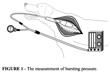
Subjects were assorted to be sacrificed on days determined before. After sacrificed, they had the esophagus dissected 3cm above and below the anastomosis, avoiding lysis of adhesions eventually existing on the anastomosis. A plastic tube was placed through the mouth and its distal extremity was positioned at the level of the suture. The other extremity was linked to a two-way system, where one was connected to a continues air flux equipment whereas other to a pressure meter. Next, the esophagus was tied distally and proximally of the anastomosis, being the second suture around the catheter (Figure 1).
Introduction of continuous flux of air into the esophagus determines a distention of the organ until any portion of the wound disrupt (bursting pressure). A camera recorded this pressure.
Next, the esophagus was resected and longitudinally opened for macroscopic inspection. Finally, the material was placed in formol for fixation, to be histologically studied. The macroscopic study aimed to examine in detail the presence of leaks; the microscopic study assessed: a) ischemic aspects; b) acute inflammation process (a.i.p.), and c) regenerative connective tissue, after stained with hematoxylin and eosin and Masson's trichrome. To study the collagen, the hydroxyproline content in tissue samples of the anastomotic area was also measured.
Fischer's exact test was used to analyse the hypothesis of equal proportions between the two groups. The hypothesis of similar mean values was tested by the analysis of variance of two factors, and multiple comparisons by Tukeys analysis. Significance level was established at 5%.
Results
There was no mortality in the intra- and postoperative periods during the experiment.
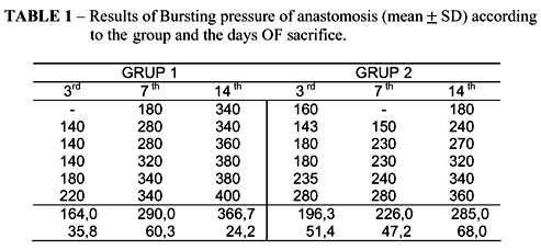
Bursting pressure The results are listed in Table 1. Evaluation of the results in Group 1 showed that a significant difference was found from the 7th p.o. to the 3rd p.o. (p=0.003), and also from the 14th p.o. to the 7th p.o. (p=0.001) and to the 3rd p.o. (p=0.001). In Group 2 that difference only had statistical significance when the 14th p.o. was compared with the 3rd p.o. (p=0.005). Thus, in the group manually sutured the bursting intraluminal resistance of the anastomosis took longer to develop. (Figure 2)
In the comparison between the two groups, the group of mechanical suturing evidenced statistically significant difference from the group of manual anastomosis at the 7th p.o. (p=0.0457) and at the 14th p.o. (p=0.0092). No significant difference was found at the 3rd p.o. (p=0.2997). Therefore, results of bursting pressure were similar in both groups at the 3rd p.o., however Group 1 evidenced higher resistance of anastomosis at the 7th p.o. and at the 14th p.o. (Figure 3).
Anatomopathological study The macroscopic study in Group 1 revealed: -3rd p.o.: suturing with impaired apposition of mucosae and evidence of ulcerative process along the full extent of the suture, characterizing healing by second intention; -7th p.o.: over-abundant granulation tissue, but without complete coaptation of mucosae; -14th p.o.: progress in the granulation process with closing of mucosae. Group 2 showed: -3rd p.o.: suture with apposition of layers - three animals (50%) with mucosal integrity; the other three (50%) showed shallow leaks over the suturing line; - 7th p.o.: leaks of various sizes in the mucosa, which at that time were present in all of the animals; -14th p.o.: progress of the regenerative tissue, with three dogs (50,0%) showing healing of the mucosa, although leakages over the suturing line could still be found in the other three animals (50,0%) (Figure 4).
The histological study showed the following results: ischemic aspects, mainly in the 3rd p.o. subgroup, apparently more intense in Group 1, which decreased in both groups thereafter, along the days of observation. The acute inflammatory process was most intense in Group 2 already at the 3rd p.o.; it remained equally intense at the 7th p.o., and in two animals at the 14th p.o. Buildup of reparative connective tissue was seen in similar ways in both groups, as assessed by either staining methodologies. It increased at the 14th p.o., when compared with the 3rd p.o. and the 7th p.o.
With reference to the hydroxyproline content, no difference was found between the days to sacrifice in Group 1 or in Group 2, and also no difference was found between those groups in any of the days of study. However, Group 2 average content was higher(p=0.0007). Therefore, the hand-sewn sutured group showed higher hydroxyproline concentration in the anastomosis, when compared with the stapled sutured group (Table 2).
Discussion
Hermann et al.(11), on studying the morphological evolution of anastomoses of the digestive tract, described three phases. Phase 1 (from day 0 to day 4), or delayed phase, is basically characterized by edema and inflammation, and by important neutrophylic reaction. In parallel, two other researchers reported intense lythic activity of the collagen and decrease of bursting pressure of the anastomosis during this phase. Phase 2 (day 3 to day 10), or lag phase, is characterized by fibroblasts regeneration. During this period edema and inflammation subside and an intense proliferation of fibroblasts is observed. Some investigators(12,13,14) reported, concomitantly to this phase, the occurrence of a balance between lysis and synthesis of the collagen. Phase 3 (from day 10 to day 180), or stable phase, is characterized by reorganization and progressively there is complete recovery of the intestinal wall layers.
According the above, the third, seventh and fourteenth days postoperative were chosen, considering that those are the days most used in studies of the healing process of the gastrointestinal tract, corresponding to those three healing phases(12).
In the present study data on bursting strength corroborated the sequence of facts described above, considering that in the study groups there was progressive gain in resistance of the anastomosis from the third to the seventh day postoperative, and from this to the fourteenth day. However, the statistic analysis showed significant increasing difference (of the 7th p.o. values compared with those of the 3rd p.o., and the 14th p.o. compared with the days prior to sacrifice) only in the stapled sutured group. In the hand-sewn sutured group, a significant difference was only shown by comparing the 14th p.o. with the 3rd p.o. Comparison of the two groups showed similar bursting pressure in the 3rd p.o. However, the group of mechanical suturing presented a significantly greater resistance in the 7th and 14th p.o., compared with the hand-sewn group.
These results indicate that, through the viewpoint of the bursting pressure, the dynamics of the healing process yield better outcome with mechanical suture.
In relation to the macroscopic study, the group mechanically sutured evidenced impaired mucosal apposition at the time of the suturing, which then progressed to a important proliferation of granulating tissue around the seventh day postoperative and after that to abundant local healing. At the microscope, ischemic aspects and little inflammatory reaction in the initial phase were seen, which then rapidly regressed. Next, there was progressive appearance of granulation tissue, which characterized this evolution as healing by second intention.
In Group 2 the mucosae were well apposed to construct the anastomosis, however leaks of various sizes appeared on the seventh day, which remained in great proportion until the fourteenth day postoperative, showing that the apparent mucosae union in the early stages had been illusory. Microscopically, a progressive inflammatory reaction called the attention, which was maximally expressed at the seventh day and still maintained at the 14th p.o. This healing evolution is characteristic of healing by first intention, and we believe that the prolonged inflammatory reaction can be credited to the suturing line used(2,15).
Therefore, both methods of anastomosis progressed with classical, although different ways of healing.
This observation has clinical correlation with those of other studies, e.g. by Fok, Wong(2), and by Bardini et al.(1), who provided evidence of healing by second intention with stapled sutures and detected higher incidence of delayed stenoses in mechanically sutured anastomoses probably as consequence of the over-abundant granulation tissue.
The analysis of the hydroxyproline content was employed to quantify the collagen content in the anastomosis, believing that its resistance is due to the concentration of that material in the submucosal layer. However, this study evidenced lack of that correlation, since its concentration is not significantly higher in the group with greater bursting strength. The same is reported by other studies in the literature.
Perhaps this could be reflecting the importance of studying the degreee of collagen maturation instead of its concentration in the anastomosis. The strength of collagenous tissue may depend on the types of collagen present. For instance, the major collagen types in soft tissues, type I and type III, form fibrils with large and small diameters, respectively. In addition to the fact that fibril diameter plays a major role in determining its mechanical properties, it has also been suggested that collagen in small fibrils shows a higher turnover rate than collagen in large fibrils. Thus, changing type distributions will directly influences upon the resistance capacity of anastomoses (1,11,13,17).
Conclusions
Analysis of the healing process in stapled and hand-sewn anastomoses in dogs, with samples collected at the third, seventh and fourteenth days postoperative, and having as parameters of study the anatomical pathology, the bursting pressure of rupture and the hydroxyproline content, led to the following conclusions: the healing process was different in both types of anastomoses, the mechanical suturing being healed by second intention and the manual suturing by first intention; mechanical anastomoses provided greater bursting strenght than the manual ones; and there was no correlation between bursting strength of the anastomosis and collagen concentration, as assessed by measurement of the hydroxyproline content in the tissues.
Received: February 10, 2004
Review: March 9, 2004
Accepted: April 12, 2004-07-21
Conflict of interest: none
Financial source: none
How to cite this article:
Caporossi C, Cecconello I, Aguilar-Nascimento JE, Venço F, Gama-Rodrigues JJ. Hand-sewn and stapled esophageal anastomosis: experimental study in dogs. Acta Cir Bras [serial online] 2004 Jul-Aug;19(4). Available from URL: http://www.scielo.br/acb [also in CD-ROM].
- 1. Bardini, R.; Asolati, M.; Ruol, A.; Bonavina, L.; Baseggio, S.; Peracchia, A. Anastomosis. Word J. Surg. 1994;18:373-8.
- 2. Fok, M.; Ah-Chong, A. K.; Cheng, S. W. K.; Wong, J. Comparison of single layer continuous hand sewn method and circular stapling in 580 oesophageal anastomoses. Br. J. Surg. 1991; 78:.342-5.
- 3. Celis J. Ruiz E. Berrospi F. Payet E. Mechanical versus manual suture in the jejunal esophageal anastomosis after total gastrectomy in gastric cancer. Rev Gastroenterol Peru 2001; 21:271-5.
- 4. Brundage SI. Maier RV. Jurkovich GJ. Re; Witzke JD. Stapled versus hand sewn anastomes in patients with small bowel injury: a changing perspective. J Trauma. 2000;49:660-65.
- 5. Steichen, F.M.; Ravitch, M.M. Mechanical sutures in esophageal surgery. Ann. Surg. 1979; 191:373-81.
- 6. Sallum, R.A.; Cecconello, I.; Zilberstein, B.; Felix, V.N.; Pinotti, H.W. Hand-sewn versus stapled cervical esophagogastrostomy-analisys of 129 cases. [Apresentado no 6th World Congress of the ISDE, Milăo, 1995].
- 7. Voros A. Ender F. Jakkel T. Cserepes E. Tota J. Szanto I. Ereifej S. Seli A. Farsang Z. Kesseru B. Laszlo S. Polanyi C. Esophageal anastomosis--based on the experience with 1460 operations. Magyar Sebeszet. 2001; 54:132-7.
- 8. Parker J. Sell H Jr. Stahlfeld K. A new technique for esophagojejunostomy after total gastrectomy for gastric cancer. Am J Surg 2001; 182:174-6.
- 9. Picardi N. History of mechanical sutures in digestive system surgery. Ann Ital Chir 2002;73:1-10.
- 10. Cochrane JP. Modifications to the double-staple technique for oesophago-jejunal anastomosis. Eur J Surg Oncol 2002; 28:186-8.
- 11. Hermann, J.B.; Woodward, S.C.; Pulaski, E.J. Healing of colonic anastomosis in the rat. Surg Gynecol Obstet 1964; 119:169-75.
- 12. Ballantyne, G.H. Intestinal suturing. Review of the experimental foundations for traditional doctrines. Dis. Col. Rectum 1983; 26: 836-43.
- 13. Hawley, P.R.; Faulk, W.P.; Hunt, T.K.; Dunphy, J.E. Collagenase activity in the gastro intestinal tract. Br J Surg 1970; 57:896-900.
- 14. Koruda, M.J.; Rolandelli, R.H. Experimental studies on the healing of colonic anastomoses. J Surg Res 1990; 48:504-15.
- 15. Wong, J. Esophagogastric anastomosis performed with a stapler: the occurrence of leakage and stricture. Surgery 1987; 101:408-15.
- 16. Cronin, K.; Jachson, D.S.; Dunphy, J.E. Changing bursting strength and collagen content of the healing colon. Surg. Gynecol. Obstet 1968;126:747-53.
- 17. Singh D. Maley RH. Santucci T. Macherey RS. Bartley S. Weyant RJ. Landreneau RJ. Experience and technique of stapled mechanical cervical esophagogastric anastomosis. Ann Thor Surg 2001; 71:419-24.
Publication Dates
-
Publication in this collection
12 Aug 2004 -
Date of issue
Aug 2004
History
-
Accepted
12 Apr 2004 -
Received
10 Feb 2004 -
Reviewed
09 Mar 2004