Abstracts
PURPOSE: Analyse the histologic changes of rat kidneys perfused with isotonic saline solution (ISS), Euro-Collins solution (ECS) and Euro-Collins solution with diltiazem (ECSD). METHODS: Thirty-six Wistar rats were used divided equally, as follow: group A (ISS), group B (ECS) and group C (ECSD). Through a catheter placed into the abdominal aorta, a renal perfusion was performed using a solution according to the group to which the animal belonged. After the complete perfusion, bilateral nephrectomy was performed and the organs were preserved under hypothermia for five distinct periods of time. Glomerulus and tubule were evaluated through optical microscopy. RESULTS: Renal perfusion with ECS and ECSD proved effectiveness in the preservation of the organs up to 36 hours and an increase in the percentage of injured glomeruli was noticed only in the period of 48 hours. CONCLUSIONS: The results showed that exists an association between the tubular injury and the glomeruli lesion degree; kidneys with a higher degree of tubular damage were related to severe glomerular lesion. Also, the addition of a calcium channel blocker, diltiazem, to the ECS for the renal perfusion does not decrease the percentage of glomerular lesion.
Diltiazem; Perfusion; Kidney; Rats
OBJETIVO: Analisar as alterações histológicas nos rins de ratos perfundidos com solução salina isotônica (ISS), solução Euro-Collins (ECS) e solução Euro-Collins com diltiazem (ECSD). MÉTODOS: Foram divididos, de forma igual, 36 ratos Wistar, como se segue: grupo A (ISS), grupo B (ECS), grupo C (ECSD). Através de um cateter localizado na aorta abdominal, foi realizada a perfusão renal com a solução de acordo com o grupo ao qual o animal pertencia. Após a perfusão total, realizou-se nefrectomia bilateral com a preservação dos órgãos sob hipotermia por cinco períodos distintos de tempo. Glomérulos e túbulos foram avaliados por microscopia óptica. RESULTADOS: Tanto a perfusão renal com ECS quanto a com ECSD provaram sua efetividade na preservação dos órgãos em até 36 horas e aumento da porcentagem de glomérulos injuriados foi notada apenas no período de 48 horas. CONCLUSÕES: Os resultados mostraram haver uma correlação entre a injúria tubular e o grau de lesão glomerular; rins com um maior grau de dano tubular foram relacionados com lesão glomerular severa. Além disso, a adição de um bloqueador de canal de cálcio, diltiazem, à ECS para a perfusão renal não diminui a porcentagem de lesão glomerular.
Diltiazem; Perfusão; Rim; Ratos
7 ORIGINAL ARTICLE
EXPERIMENTAL UROLOGY
Histologic alterations of rat kidneys perfused with a Euro-Collins diltiazem solution1 1 Research performed at Department of Urology, Pontifical Catholic University of Parana (PUC-PR), Curitiba - PR, Brazil.
Alterações histológicas ocorridas em rins de ratos perfundidos com solução de Euro-Collins associada à diltiazem
Fernando MeyerI; Juliana Navarro LizanaII; Luiz Felipe DziedrickiII; Luiz Fernando Bleggi-TorresIII
IMSc, Associate Professor, Department of Urology, Cajuru University Hospital, PUC-PR, Curitiba, Brazil
IIGraduate student, PUC-PR, Curitiba, Brazil
IIIPhD, Full Professor, Department of Medical Pathology, Clinics Hospital, Federal University of Parana (UFPR), Curitiba, Brazil
Correspondence Correspondence: Fernando Meyer Pontifícia Universidade Católica do Paraná (PUCPR) Departamento de Urologia Rua Portugal, 307 80510-280 Curitiba - PR Brasil Phone: (55 41)3074-7478 Fax: (55 41)3015-0303 fmeyer@onda.com.br
ABSTRACT
PURPOSE: Analyse the histologic changes of rat kidneys perfused with isotonic saline solution (ISS), Euro-Collins solution (ECS) and Euro-Collins solution with diltiazem (ECSD).
METHODS: Thirty-six Wistar rats were used divided equally, as follow: group A (ISS), group B (ECS) and group C (ECSD). Through a catheter placed into the abdominal aorta, a renal perfusion was performed using a solution according to the group to which the animal belonged. After the complete perfusion, bilateral nephrectomy was performed and the organs were preserved under hypothermia for five distinct periods of time. Glomerulus and tubule were evaluated through optical microscopy.
RESULTS: Renal perfusion with ECS and ECSD proved effectiveness in the preservation of the organs up to 36 hours and an increase in the percentage of injured glomeruli was noticed only in the period of 48 hours.
CONCLUSIONS: The results showed that exists an association between the tubular injury and the glomeruli lesion degree; kidneys with a higher degree of tubular damage were related to severe glomerular lesion. Also, the addition of a calcium channel blocker, diltiazem, to the ECS for the renal perfusion does not decrease the percentage of glomerular lesion.
Key words: Diltiazem. Perfusion. Kidney. Rats.
RESUMO
OBJETIVO: Analisar as alterações histológicas nos rins de ratos perfundidos com solução salina isotônica (ISS), solução Euro-Collins (ECS) e solução Euro-Collins com diltiazem (ECSD).
MÉTODOS: Foram divididos, de forma igual, 36 ratos Wistar, como se segue: grupo A (ISS), grupo B (ECS), grupo C (ECSD). Através de um cateter localizado na aorta abdominal, foi realizada a perfusão renal com a solução de acordo com o grupo ao qual o animal pertencia. Após a perfusão total, realizou-se nefrectomia bilateral com a preservação dos órgãos sob hipotermia por cinco períodos distintos de tempo. Glomérulos e túbulos foram avaliados por microscopia óptica.
RESULTADOS: Tanto a perfusão renal com ECS quanto a com ECSD provaram sua efetividade na preservação dos órgãos em até 36 horas e aumento da porcentagem de glomérulos injuriados foi notada apenas no período de 48 horas.
CONCLUSÕES: Os resultados mostraram haver uma correlação entre a injúria tubular e o grau de lesão glomerular; rins com um maior grau de dano tubular foram relacionados com lesão glomerular severa. Além disso, a adição de um bloqueador de canal de cálcio, diltiazem, à ECS para a perfusão renal não diminui a porcentagem de lesão glomerular.
Descritores: Diltiazem. Perfusão. Rim. Ratos.
Introduction
The goal of ideal renal preservation is to promote the necessary time for cross matching, receiver selection, transportation of graft and preparing the receiver for surgery as well as leading to early reestablishment of renal function as the delay would incur into considerable costs due to longer hospital stay and need for repeated dialysis1.
Prolonged organ preservation has only become possible after the development of intracellular electrolyte composition solutions2. Since then many changes in the composition of solutions have been performed. Aiming at prolonging renal graft preservation time without, however, altering the organ's viability and function, studies have been performed by adding different substances to the perfusion solution such as oxygen3, steroids4, free radical scavengers5,4 and calcium channel blockers such as trifluoperazine6, verapamil7 and diltiazem8,9.
Considerable interest has been shown recently in the calcium function as possible irreversible cell damage mediator after ischemia period. Cell membrane ischemic injury is associated to the influx of calcium from the extracellular compartment into the intracellular compartment and as a consequence with the activation of calcium-dependent catabolic process10.
Based on previously mentioned data this study has been proposed in order to assess histological changes occurred in rat kidneys submitted to perfusion with isotonic saline solution (ISS), Euro-Collins solution (ECS) and Euro-Collins solution associated to diltiazem (ECSD), calcium channel blocker in different periods of preservation.
Methods
Thirty six Wistar rats have been used (Rattus norvegicus albinus, Rodentia mammalia) divided into three groups with twelve animals each. Group A: perfusion with ISS, group B: perfusion with ECS and group C: perfusion with ECSD (25mg/l). The rats have been submitted to inhalation anesthesia with commercial ether followed by xyphopubic median laparotomy with the exposure of the abdominal cavity. A dissection of the aorta above and below the renal vessels has been conducted after laparotomy. The aorta has been isolated above the celiac trunk being repaired at this level with 5-0 silk yarn. In the distal portion a ligation of the aorta and the vena cava has been performed right above its bifurcation to the iliac vessels. Aorta has been catheterized with catheter type Abocath® 24 introduced right above the iliac bifurcation11. After checking of blood reflux in the catheter, meaning it was correctly positioned; renal blood flow has been interrupted by the traction of superior aortic repair. Immediately after a renal perfusion has been initiated using the solution according to the group the animal belonged to. Temperature of used solutions were always at 4ºC12,13 and perfusion pressure was of 100 cm H2O14 for every group (A, B and C). During perfusion a change in color of the bowel and renal parenchyma has been observed. Such changes have been used as criteria for effective perfusion, being interrupted at the moment kidneys showed a homogeneous color or at least five minutes after beginning of perfusion. After bilateral nephrectomy kidneys have been stored in plastic package containing 30ml of perfusion solution (ISS, ECS, ECSD) at 4ºC and placed inside another plastic package containing 10ml of ISS and ice15. After being properly identified, the package has been stored in thermal container with ice. The kidneys have been preserved and fixed for histological analysis in five different periods: 0 (right after perfusion was finished), 12, 24, 36 and 48 hours. The choosing of period of kidney has been done through draw.
Histological study
From 72 kidneys used in this experiment 12 have been analyzed from the histological point of view at period 0 and 15 kidneys in each one of the other periods. After draw, kidney was sectioned longitudinally on its anti-hilar edge for adequate fixation. The blades have been made with three cutting levels, with intervals of 5 micrometers between them stained with hematoxylin and eosin (HE)16. Two renal structures have been evaluated: glomeruli and tubules. Total number of glomeruli as well as damaged glomeruli have been accounted for. Glomeruli were considered normal (Figure 1) whenever capillary loops were open, with thin walls and whenever there was no content inside the Bowman's capsule. On the other hand, glomeruli were considered as damaged (Figure 2) whenever they presented a contraction of glomerular tuft with approximation of structures, contents inside the Bowman's capsule or vacuolization of endothelial cells. The degree of glomerular lesion has been characterized as: light (up to 25% of damaged glomeruli), moderate (26% to 50% of damaged glomeruli), sharp (51% to 75% of damaged glomeruli) and severe (above 75% of damaged glomeruli).
Evaluation of tubular damage has been carried out according to kidney's compromising extension. Renal tubules have been considered normal whenever coated by a single layer of cuboid cells of dense eosinophilic cytoplasm and tubular lights were optically empty. Injured tubules presented cytoplasmic vacuolization in different degrees. Four different categories were used, according to Chart 1.
Statistical analysis
Thirteen rats were considered for the statistical analysis, randomly placed in 3 groups (10 rats each group), being both kidneys from each rat used. In total, 60 kidneys have been considered. Result evaluations have been done in 4 periods: 12 hours, 24 hours, 36 hours and 48 hours. For each one of the variables (weight of rats, time and volume of perfusion) null hypothesis of equal averages has been tested for the three groups considered (A, B and C), versus an alternative hypothesis in which at least one of the groups presented an average different from the others. For that matter Cochran test has been applied. In case there was significant difference between the groups "t as in student" tests was adopted for independent samples. For variable analysis (total number of glomeruli, percentual of damaged glomeruli and tubular damage) the Mann-Whitney non-parametrical test was adopted. For research of association between damaged glomeruli and tubular damage the Spearman's rank correlation coefficient for evaluation of this association was estimated. In every test the adopted significance level was of 5%.
Results
From 36 rats used in the experiment 6 of them were used in period 0 (4 kidneys in each group: A, B and C). In every kidney histological analysis showed normal renal architecture, presenting normal glomeruli and absence of tubular damage in period 0. Therefore, such kidneys were not part of statistical analysis; however, they have been used as pattern for histological study in the remaining periods (Table 1).
As for rats' weight (p=0.1786) and perfused solution volume (p=0.9288) in each rat, results showed that there was no significant difference between the three groups. As for perfusion time, kidneys perfused with ECS presented a bigger perfusion time than kidneys perfused with ISS (p=0.0000001) and ECSD (p=0.0014).
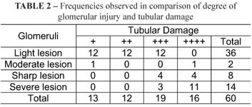
There was no significant difference in total number of glomeruli in the different periods (p<0.05). There was a significant difference, however, in the percentual of damaged glomeruli in each group between the periods 12x24 (p=0.0088), 12x36 (p=0.0088) and 12x48 (p=0.0088) hours in group A, between periods 12x48 (p=0.0465) hours in group B and between periods 12x48 (p=0.0232) and 24x48 (p=0.0439) hours in group C (Table 2).
Discussion
The choice of rat as an experimental animal in this study was due to ease in obtaining and handling such animal and the accordance of literature which states rats' isolated kidney as a doable and reliable method for evaluation of organ preservation4,17.
The choice for diltiazem in this work was based on the action mechanism of this blocker in the calcium channel. Diltiazem enhances blood renal flow through direct vasodilation of gromeruli afferent arterioles. It also prevents calcium ions from entering intracellular space, consequentely interrupting vicious cycle initiated by ischemia which leads to cellular damage. Besides, this substance enhances urinary volume and it is also natriuretic, as it inhibits sodium reabsorption by the distal tubules. That fact itself enhances glomerular filtering index due to the bigger renal perfusion produced by the increase of cardiac debit.
Perfused kidneys with ECS presented bigger perfusion time than kidneys perfused with ISS (p=0.000001) and ECSD (p=0.0014). On the Iriarte and collaborators study, performed in 1981, there has been a difference in perfusion time between kidneys perfused with the same solution (Collins and Sacks-Kaufman solutions). Difference in perfusion time found on this work may be due to the different compositions of solutions used in renal perfusion, when a given solution demands a bigger time to perfuse the same volume.
Normal and damaged glomeruli counting has been carried out in a comparative way and aiming at evaluating kidney preservation. This method is little regarded in literature. In the present study and average of 95.85 glomeruli were counted in each kidney, varying from 66 to 126 glomeruli. In order to keep an average of counted glomeruli the same level of histological cut has been chosen, making sure the same renal area was assessed in every case. Results showed statistically significant difference in the total number of counted glomeruli between groups ISS x ECS and ISS x ECSD in the 24-hour period and between groups ISS x ECS in the 36-hour period (p<0.05). There was no significant difference, however, between the different periods in each group (p<0.05), confirming that glomeruli counting was similar in the evaluated groups.
Evaluation of glomerular damage is another factor used for analysis under the histological point of view, changes and preservation occurred in graft after perfusion18. In the present study, glomerular damage present in the different groups and periods have been evaluated. When groups were compared there was no significant difference in the percentual of damaged glomeruli between groups A, B and C in the period of 12 hours, showing that the three solutions obtained similar results in the renal preservation during this period. Such data is according to the Fisher and collaborators work, in 1967, which demonstrated little glomerular lesion in kidneys perfused with Ringer lactate solution and preserved under simple hypothermia for up to 12 hours. Results, however, showed that there has been significant difference in the percentual of damaged glomeruli between ISS X ECS groups in the periods of 24, 36 and 48 hours and between ISS x ECSD groups in the period of 24 hours (p<0.05). Data showed that ECS, being in intracellular solution, has been more effective in the preservation of glomerular damage than ISS, which is extracellular.
As for comparisons between ECS x ECSD groups there hasn't been significant differences between the percentual of damaged glomeruli in any of the evaluated periods (p<0.05), showing that the addition of calcium channel blocker to the Euro-Collins solution has not been effective in the preservation of glomerular damage. As for the percentual of damaged glomeruli the present study evaluated different periods for each group. Perfused kidneys with ECS showed significant difference when compared to periods of 12x48 and 24x48 hours. Data suggests that ECS, associated to diltiazem, provides protection against glomerular damage for a period up to 36 hours.
Tubular damage is another important and observed factor in histological changes occurred in preserved kidneys19. A tubular damage evaluation has been carried out during this study as well as the association to the degree of glomerular damage. Obtained results showed direct correlation between tubular damage and the degree of glomerular damage. Eleven out of fourteen kidneys which presented severe glomerular damage have also presented tubular damage. However, none of the kidneys presenting light glomerular damage showed severe tubular damage. The result was similar to the one obtained by Hill and collaborators in 1976, which performed renal biopsy one hour after transplantation and observed that kidneys with a higher degree of tubular alteration correlated to a more intense glomerular damage, which proves the direct correlation between the degree of tubular alteration and glomerular damage.
Literature on the subject is controversial when it comes to the results obtained by calcium channel blockers in renal preservation. Blank et al.20 evaluated the effects of hypothermia and verapamil in rats' kidneys submitted to sixty minutes of ischemia, showing that isolated hypothermia provides a better protection and that verapamil has not obtained protective effects against hot and cold ischemia. Such results have been confirmed by findings of electronic microscopy.
Nakamoto et al.17 has evaluated the effects of verapamil in renal preservation after 24 hours of hypothermic perfusion compared to simple perfusion using Euro-Collins solution. Authors showed that verapamil has not only enhanced kidney preservation but also improved their function after reperfusion.
The fact that difference in the percentual of glomerular damage has not been found between groups ECS x ECSD in this study may be explained by the renal damage caused by tissue reperfusion. Lesions due to ischemia/tissue reperfusion have been described in the myocardium, brain, intestine, liver and kidney. Besides, tissue reperfusion may lead to cellular damage when tissue reoxygenating.
Cellular injury derived from ischemia/renal reperfusion may lead to a series of events: depletion of ATP reserves, free radicals formation derived from oxygen, cellular acidosis, increase in calcium cellular ions influx, lytic actions of some enzymes (proteases, phospholipases) and cellular apoptosis. The ischemic/reperfusional phenomena also involves the production of inflammatory mediators, activation of endothelial cells and leukocitary activation.
Therefore, protective action of calcium channel blockers may happen when beginning tissue reperfusion. Anaise et al.6 has used calcium channel blocker (verapamil) during kidney perfusion in dogs and submitted to reperfusion after preservation for 48 and 72 hours. This substance showed favorable effect in the renal blood flow, decreasing the effects caused by tissue reperfusion.
Conclusions
Based on the results in this work it was concluded that the addition of diltiazem to the Euro-Collins solution has not decreased the percentual of damaged glomeruli when compared to the Euro-Collins solution itself in the periods of 12, 24, 36 and 48 hours. Is has also been observed that perfusion with Euro-Collins solution and Euro-Collins solution with addition of diltiazem are effective in the preservation of renal histology for a period up to 36 hours.
Received: March 18, 2010
Review: May 20, 2010
Accepted: June 23, 2010
Conflict of interest: none
Financial source: none
How to cite this article
Meyer F, Bleggi-Torres LF, Lizana JN, Dziedricki LF. Histologic alterations of rat kidneys perfused with a Euro-Collins diltiazem solution. Acta Cir Bras. [serial on the Internet] 2010 Nov-Dec;25(6). Available from URL: http://www.scielo.br/acb
*Color figures available from www.scielo.br/acb
- 1. Halasz NA, Collins GM. Forty-eight-hour kidney preservation: a comparison of flushing and ice storage with perfusion. Arch Surg. 1976;111:175-7.
- 2. Collins GM, Halasz NA. Letter: composition of intracellular flush solutions for hypothermic kidney storage. Lancet. 1975;1(7900):220.
- 3. Pegg DE, Foreman J, Hunt CJ, Diaper MP. The mechanism of action of retrograde oxygen persufflation in renal preservation. Transplantation. 1989;48:210-7.
- 4. Biguzas M, Jablonski P, Howden BO, Thomas AC, Walls K, Scott DF, Marshall VC. Evaluation of UW solution in rat kidney preservation: the effect of pharmacological additives. Transplantation. 1990;49:1051-5.
- 5. Green CJ, Healing G, Lunec J, Fuller BJ, Simpkin S. Evidence of free-radical-induced damage in rabbit kidneys after simple hypotermic preservation and autotransplantation. Transplantation. 1986;41:161-5.
- 6. Anaise D, Lane B, Waltzer WC, Rapafort FT. The protective effect of calcium inhibitors and of Captopril on the renal microcirculation during reperfusion. Transplantation. 1987;43:128-33.
- 7. Hertle L, Garthoff B. Calcium channel blocker nisoldipine limits ischemic damage in rat kidney. J Urol. 1985;134:1251-4.
- 8. Burke TJ, Arnold PE, Gordon JA, Bulger RE, Dobyan DC, Schrier RW. Protective effect of intrarenal calcium membrane blockers before or after renal ischemia: functional, morphological, and mitochondrial studies. J Clin Invest. 1984;74:1830-41.
- 9. Puig JM, Lloveras J, Oliveras A, Costa A, Aubia J, Masramon J. Usefulness of Diltiazem in reducing the incidence of acute tubular necrosis in Euro-Collins-Preserved cadaveric renal grafts. Transplant Proc. 1991;23:2368-9.
- 10. Changnac A, Zevin D, Ori Y, Korzets A, Hirsh J, Levi J. The effect of high-dose nifedipine on renal hemodynamics of ciclosporine-treated renal allograft recipients. Transplantation. 1992;53:766-9.
- 11. Lee S. An improved technique of renal transplantation in the rat. Surgery. 1967;61:771-3.
- 12. Williams G, Peet TND, Hamshere RJ. Assessment of a test of renal viability. Br Med J. 1976;2(6027):75-7.
- 13. Bittard H, Benoit G, Moukarzel M, Bensadoun H, Charpentier B, Fries D. Prediction of renal fuction by intraoperative renal blood flow and renal vascular resistence during kidney transplantation. Transplant Proc. 1991;23:2396-9.
- 14. Kemp E, Larsen S, Jorgensen KA, Dieperink H, Starklint H. Flush perfusion of rabbit kidneys with autogeneic, allogeneic and xenogeneic blood. Scand J Urol Nephrol. 1991;25:45-9.
- 15. Howden B, Rae D, Jablonski P, Marshall VC, Tange J. Studies of renal preservation using a rat kidney transplant model: evoluation of citrate flushing. Transplantation. 1983;35:311-4.
- 16. Bancroft JD, Stevens A. Theory and practice of histological techniques. 2ed. New York: Churchill Livingstone; 1982.
- 17. Nakamoto M, Shapiro JI, Mills SD, Schrier RW, Chan L. Improvement of renal preservation by verapamil with 24-hour cold perfusion in the isolated rat kidney. Transplantation. 1988;45:313-5.
- 18. Grenier JF, Dauchel J, Jaeck D, Kachelhoffer J, Stock C. Comparison of two electrolyte solutions for short perfusion and hypothermic storage in experimental renal preservation. Br J Surg. 1973;60:964-8.
- 19. Dahlager JI, Bilde T. The integrity of tubular cell function after preservation in Collins' or Sacks' solution. Transplantation. 1976;21:365-9.
- 20. Blank W, Mooppan MM, Chhajwani B, Chou SY, Kim H. Effects of verapamil on preservation of renal function after ischemia: functional and ultrastructural study. J Urol. 1984;131:992-4.
Publication Dates
-
Publication in this collection
19 Nov 2010 -
Date of issue
Dec 2010
History
-
Received
18 Mar 2010 -
Reviewed
20 May 2010 -
Accepted
23 June 2010