Abstract
Purpose:
To investigate the long non-coding RNAs (lncRNAs) profile on renal ischemia reperfusion in a mouse model.
Methods:
Microarray analysis was used to study the expression of misregulated lncRNA in a mouse model of renal ischemia reperfusion(I/R) with long ischemia time. Quantitative real-time PCR (qPCR) was used to verify the expression of selected lncRNAs and mRNAs.The potential functions of the lncRNA was analyzed by bioinformatics tools and databases.
Results:
Kidney function was impaired in I/R group compared to the normal group. Analysis showed that a total of 2267 lncRNAs and 2341 messenger RNAs (mRNAs) were significantly expressed in I/R group (≥2.0-fold, p < 0.05).The qPCR result showed that lncRNAs and mRNAs expression were consistent with the microarray analysis. The co-expression network profile analysis based on five validated lncRNAs and 203 interacted mRNAs showed it existed a total of 208 nodes and 333 connections. The GO and KEEG pathway analysis results showed that multiple lncRNAs are involved the mechanism of I/R.
Conclusion:
Multiple lncRNAs are involved in the mechanism of I/R.These analysis results will help us to further understand the mechanism of I/R and promote the new methods targeted at lncRNA to improve I/R injury.
Key words:
Kidney Transplantation; Ischemia; Reperfusion; RNA, Messenger; Mice
Introduction
Occurrence of renal ischemia-reperfusion (I/R) injury affects the outcome of kidney transplantation, which has a direct relationship with the survival of the recipient. The delayed graft function (DGF) rate of kidney transplantation from donation after cardiac death (DCD) reached up to 40-70% and was significantly higher than that from brain death donors and living donors11 Elgharably H, Shafii AE, Mason DP. Expanding the donor pool: donation after cardiac death. Thorac Surg Clin. 2015;25(1):35-46. doi: 10.1016/j.thorsurg.2014.09.011.
https://doi.org/10.1016/j.thorsurg.2014....
. The main factor responsible for this difference was the prolonged warm ischemic time22 Morrissey PE, Monaco AP. Donation after circulatory death: current practices, ongoing challenges, and potential improvements. Transplantation. 2014;97(3):258-64. doi: 10.1097/01.TP.0000437178.48174.db.
https://doi.org/10.1097/01.TP.0000437178...
. The I/R injury mechanisms in DCD kidney transplantation are multifactorial and may include oxidative stress, mitochondrial Ca2+ overload, inflammation, cell apoptosis, necrosis, loss of cell polarity, dedifferentiation and proliferation of viable cells, and disruption of the generation of free radicals. Long non-coding RNAs (lncRNAs) are typically longer than 200 nucleotides. It has been demonstrated that lncRNAs exert comprehensive effects on biological processes, such as transcription, translation, splicing, and intracellular and extracellular trafficking, and are associated with numerous diseases33 Yu SY, Tang L, Zhou SH. Long noncoding RNAs: new players in ischaemia-reperfusion injury. Heart Lung Circ. 2018;27(3):322-32. doi: 10.1016/j.hlc.2017.09.011.
https://doi.org/10.1016/j.hlc.2017.09.01...
. lncRNAs can interact with proteins, DNAs, and RNAs and regulate gene expression at various levels, including epigenetic, transcriptional, and post-transcriptional regulation44 Wu X, Zhu H, Zhu S, Hao M, Li Q. lncRNA expression character associated with ischemic reperfusion injury. Mol Med Rep. 2017;16(4):3745-52. doi: 10.3892/mmr.2017.7051.
https://doi.org/10.3892/mmr.2017.7051...
. However, the possible role of lncRNAs in I/R injury has not received much attention.
Therefore, in this study, we used microarray analysis to analyze the lncRNA and mRNA expression differences in I/R model.The selected lncRNA and mRNA were verified by q-PCR. Gene Ontology(GO) analysis and Kyoto Encyclopedia of Genes and Genomes (KEGG) analysis were performed to predict possible biological processes and potential signal pathways. In addition, co-expression network of lncRNA-mRNA was clarified by coding/non-coding gene co-expression (CNC) analysis.
Methods
Animal preparation and experimental design
The research project was approved by the research ethics committee (NSCF 81400753).
Male C57BL/6 mice (body weight 250-300g) were obtained from the Experimental Animal Center of the Medical College of Wuhan University (Wuhan, China). The animals were maintained at the Central Animal Facility of Affiliated Renmin Hospital of Wuhan University according to standard guidelines, and experiments were conducted according to the guidelines of the Chinese Council for Animal Care. The mice were kept in an air-filtered, homoiothermal (20-22°C), and light-controlled room (light from 7 a.m. to 7 p.m.) and allowed free access to a standard diet.
Sample collection
Mice were separated into two groups.Group 1: control group (n = 5), in which mice were subjected to a right nephrectomy but without the induction of a left renal ischemia, and Group 2: I/R group (n = 5), in which mice were subjected to a right nephrectomy and left renal ischemia for 45 min followed by a reperfusion period of 24 h. All mice were anesthetized with intraperitoneal pentobarbital (50 mg/kg) and were killed via decapitation. Mice were placed on an electric heating pad to maintain their body temperature at 37°C. Their kidney tissues were removed and frozen in liquid nitrogen followed by storage at −80°C prior to analysis.
Kidney function test and hematoxylin and eosin staining
After the 24-h reperfusion period, blood samples were collected from the inferior vena cava and centrifuged to determine the concentration of creatinine (Cr). The Cr was measured in the blood using standard techniques with an Olympus AU 2700 Analyzer (Olympus Optical Co., Ltd., Tokyo, Japan).
After a 24 h reperfusion, the left kidney was excised. Then, the kidney tissue was fixed with 10% phosphate-buffered formalin, paraffin embedded, and sectioned to a thickness of 4 mm according to a standard procedure. Sections were deparaffinized and gradually hydrated before they were examined by hematoxylin and eosin (HE) staining. Morphological assessments were performed by an experienced renal pathologist who had not been informed of the experimental protocol.
Microarray analysis
Gene microarray analysis was performed on 5 pairs of kidney tissues from I/R group and normal group to detect differentially expressed lncRNA and mRNA. Approximately 35,923 lncRNAs and 24,881 coding transcripts were detected by the Arraystar Mouse LncRNA Microarray V3. The tissue preparations and microarray hybridization were performed using the Agilent Gene Expression Hybridization Kit (Agilent Technology Inc., USA). Then, the arrays were scanned using the Agilent Microarray Scanner and were finally analyzed using the Agilent Feature Extraction software. Differentially expressed transcripts were identified by fold-change screening at a threshold of ≥2-fold and a p-value of <0.0555 Hanjin C, Tao L, Pengfei L, Ali Y, Huajun Z, Jiekun L, Yang W, Tao T. Altered long noncoding RNA and messenger RNA expression in experimental intracerebral hemorrhage - a preliminary study. Cell Physiol Biochem. 2018;45(3):1284-301. doi: 10.1159/000487464.
https://doi.org/10.1159/000487464...
. Pathway analysis was used to study the significant signaling pathways of the differentially expressed genes. GO analysis was used to explore the biological roles of the aberrantly expressed mRNAs, which included to three domains-molecular function, biological process, and cellular component.
qRT-PCR validation
The kidney tissue collected for lncRNA microarray analysis were used for quantitative real-time polymerase chain reaction (qRT-PCR) validation.According to the instructions of the product, SuperScript III reverse transcriptase(Invitrogen, Grand Island, NY, USA) is used to reverse transcribe the total RNA into cDNA. The qRT-PCR was performed with the Applied Biosystems ViiA 7 RT PCR System and 2× PCR Master Mix. The PCR conditions consisted of: incubation at 95°C for 10 min, followed by 40 cycles of 95°C for 10 s and 60°C for 1 min. The relative expression levels of lncRNAs were calculated using the 2−ΔΔCt method and were normalized against β-actin. The primers for each gene are listed in Table 1. The data represent the means of three experiments.
GO annotations and KEGG analysis
We conducted a GO analysis to construct a meaningful annotation of genes and gene products. The ontology covers the domains of biological processes, cellular components, and molecular functions. The −log10 (p-value) denotes enrichment score representing the significance of GO term enrichment among differentially expressed genes. Pathway analysis was performed to explore the significant pathways in differentially regulated gene profiles according to KEGG. Also, the −log10 (p-value) denotes an enrichment score showing the significance of the pathway correlations.
Construction of a co-expression network with GO and KEGG analysis
In order to identify the interaction between differentially expressed lncRNA and mRNA, we constructed the co-expression network with the verified lncRNA and related mRNA on the basis of correlation analysis. Cytoscape software was applied into construct the network between lncRNA and mRNA, while the pearson’s correlation coefficients should not less that 0.992. In the figure of CNC, mRNAs were represented by a red node, while lncRNAs were represented by a green node.
Statistical analyses
Statistical analyses were performed using Graphpad Prism 5.0 (San Diego, CA, USA). Results were expressed as mean ± standard error of the mean. The t-test was used to analyze the differences between the normal control and I/R group data in this study. Spearman’s correlation analysis was used to detect the relationship between lncRNAs and mRNAs.A p-value of <0.05 and a fractional disappearance rate of <0.05 were used as thresholds to define markedly enriched GO terms/pathways.
Results
Kidney function test and HE stain
Compared with the normal control group, we observed increased levels of serum creatine in I/R group. HE staining of the kidney tissues showed that renal tubular cells in I/R group presented I/R injury features, including tubular necrosis, renal tubular expansion, and renal tubular epithelial cellular microvilli disappearance (Fig. 1).
Renal function was impaired in the I/R group when compared with the normal control group. a. The level of serum Cr is higher in the I/R group; “*” represents p<0.05. b, c. HE staining in kidney tissue of the I/R (b) and control groups (c), showing I/R features in the tubular cells of the I/R group, including tubular necrosis, renal tubular expansion, and renal tubular epithelial cellular microvilli disappearance (magnification ×200).
lncRNA and mRNA expression analysis in renal I/R injury
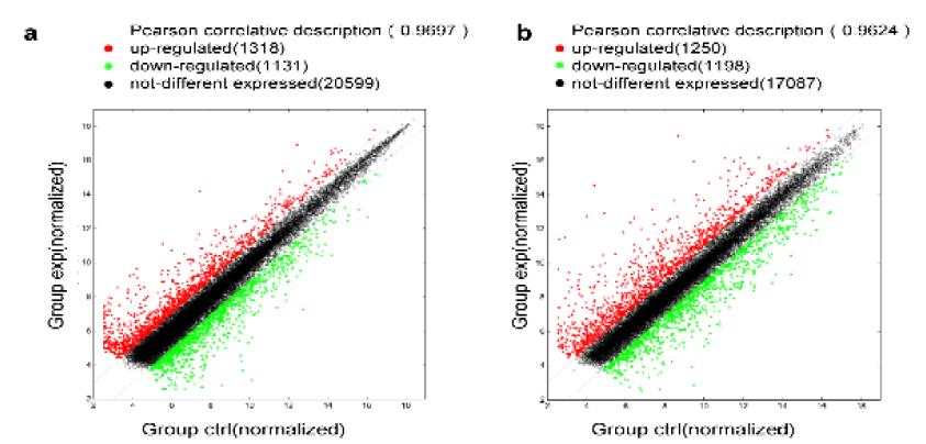
To explore the difference in the expression of lncRNA and mRNA in I/R model, microarray analysis was used to assess the expression levels. We identified 2267 significantly dysregulated lncRNAs in the I/R mice, while 1180 were upregulated, while 1087 were downregulated (≥2.0-fold, p < 0.05). Table 2 presents the 50 most significantly differentially expressed lncRNAs.
Heat map and hierarchical clustering of the 50 most significantly differentially expressed lncRNAs expression patterns in different samples (Fig. 2a). All the variation in lncRNA expression between the I/R and normal control groups is shown using a scatter plot (Fig. 3a). At the same time, 2341 significantly dysregulated mRNAs were identified in the I/R group, while1166 were upregulated, while 1175 were downregulated (≥2.0-fold, p<0.05). Table 3 presents the 50 most significantly differentially expressed mRNAs. Heat map and hierarchical clustering of the 50 most significantly differentially expressed mRNA expression patterns in different samples (Fig. 2b). All the variation in mRNA expression between the I/R and control groups is shown using a scatter plot (Fig. 3b).
Heat map and hierarchical clustering of the 50 most significantly differentially expressed lncRNAs (a) and mRNAs (b) between the I/R and control groups. The data are depicted as a data matrix, in which each row represents one lncRNA (mRNA) and each column represents one sample. The relative lncRNA (mRNA) expression follows the color scale at the top. Red represents high relative expression, and green represents low relative expression; −3.0, 0, and 3.0 are fold changes in the corresponding spectrum. The magnitude of deviation from the median is represented by the color saturation.
Scatter plot of lncRNA (a) and mRNA (b) expression variation between the I/R and control kidney samples. The values shown on the X-axis and Y-axis are normalized signal values for each sample (log2 scale). The dark lines are fold-change lines (the default fold-change value given is 2.0). The green dot and red plots showed an expression fold-change of >2.0 between the two samples compared.
Validation of the microarray data using qRT-PCR
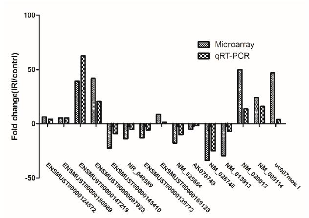
To validate the result of mRNA and lncRNA from microarray analysis, we performed the qRT-PCR assay.Ten mRNA and lncRNA were randomly selected to perform the qRT-PCR.The results showed that the expressions of lncRNA ENSMUST00000124572, ENSMUST00000180989, ENSMUST00000147219, ENSMUST00000097928, uc007mos1 and ENSMUST00000169128 were upregulated, whereas those of ENSMUST00000145410, NR_040589, ENSMUST00000139773, and AK078749 were downregulated (Fig. 4). Meanwhile, when compared with the control group, three target mRNAs (NM_028746, NM_013913, and NM_025684)were found to be downregulated in the I/R group, whereas NM_020013 and NM_009114 were upregulated (Fig. 4). These results are consistent with those of the microarray analysis (Fig. 4), thereby confirming the validity of the microarray results. The finding provides compelling evidence that these lncRNAs and mRNAs could be implicated in the pathogenesis of renal I/R injury.
The differential expression of lncRNAs and mRNAs was validated by quantitative real-time PCR (qRT-PCR). The data show that expression levels of lncRNAs ENSMUST00000145410, NR_040589, ENSMUST00000139773, NM_025684, and AK078749, along with mRNAs NM_028746 and NM_013913 were downregulated, while expression levels of lncRNAs ENSMUST00000124572, ENSMUST00000180989, ENSMUST00000147219, ENSMUST00000097928, ENSMUST00000169128, uc007 mos.1, and mRNAs NM_020013 and NM_009114 were upregulated in kidney tissue samples from I/R mouses when compared with the control mouses. The heights of the columns in the chart represent fold changes. The qRT-PCR results were consistent with the microarray data.
GO analysis and KEGG pathway analysis
The GO defines concepts/classes used to describe gene function, and relationships including three aspects molecular function, biological process and cellular component. In this study the GO term analysis indicates that the most enriched GO terms targeted by mRNAs co-expressed with lncRNAs were organic acid metabolic process (ontology: biological process, GO:006082), catalytic activity (ontology: molecular function, GO:0003824), and extracellular vesicular exosome (ontology: cellular component, GO:0070062) (Fig. 5).
The 40 most significant GO terms for differences in co-expressed lncRNA genes in I/R animals and controls. GO enrichment analysis provided a controlled vocabulary to describe co-expressed genes of differentially expressed lncRNAs. The ontology covered three domains: biological process (blue), cellular component (red), and molecular function (green).
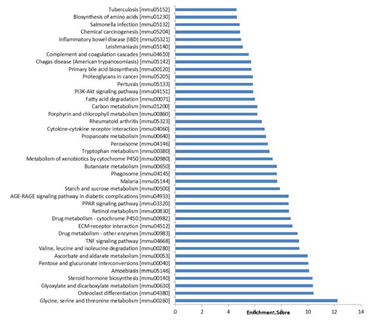
Furthermore, we applied the KEGG tool to explore pathway maps on the molecular interaction, reaction and relation networks .the results indicated that mRNAs co-expressed with lncRNAs were involved in the regulation of glycine, serine, and threonine metabolism; TNF signaling pathway; AGE-RAGE signaling pathway in diabetic complications; cytokine-cytokine receptor interaction; ECM-receptor interaction; PI3K-Akt signaling pathway; and others. The 40 most significant KEGG pathways are listed in Figure 6.
The 40 most significant pathways for differences in lncRNA genes co-expressed in I/R injury animals and controls.
lncRNA-mRNA CNC network analysis
LncRNA is involved in the occurrence and development of I/R, but lncRNA mainly play functions by regulating the expression of mRNA, so we further analyzed the CNC network of lncRNA and mRNA. The 5 differentially expressed lncRNAs that were validated by qRT-PCR (uc007 mos.1, ENSMUST00000147219, ENSMUST00000124572, ENSMUST00000145410, and NR_040589) with 203 interacting mRNAs were used to construct the co-expression network. This co-expression network was consisted of 208 nodes and 333 connections, of which 89 were negative and 244 were positive interactions. The resulting network revealed that uc007 mos.1 is correlated with 119 mRNAs, ENSMUST00000147219 is correlated with 54 mRNAs, ENSMUST00000124572 is correlated with 22 mRNAs, ENSMUST00000145410 is correlated with 81 mRNAs, and NR_040589 is correlated with 57 mRNAs (Fig. 7) .
lncRNA-mRNA network analysis. Green nodes represent dysregulated lncRNAs, whereas red nodes represent dysregulated mRNAs. The dotted lines between lncRNAs and mRNAs indicate a negative correlation, whereas solid lines indicate a positive correlation.
GO and KEGG analyses based on the results of the co-expression network were performed. According to the enrichment score level, the result showed that the targeted mRNA was focused on the organic acid metabolic process (ontology: biological process, GO:0004930), oxidoreductase activity (ontology: molecular function, GO:0016491), and extracellular exosome (ontology: cellular component, GO:0070062) (Fig. 8a).
GO analysis and pathway analysis with which targeted genes are correlated. a. According to the enrichment score levels, the results showed that mRNAs were enriched for organic acid metabolic process (ontology: biological process, GO: 0004930), oxidoreductase activity (ontology: molecular function, GO:0016491), and extracellular exosome (ontology: cellular component, GO:0070062). b. The result of KEGG pathway analysis with the targeted gene. Most of genes were predicted to interplay with glycine, serine, and threonine metabolism.
In addition, most genes predicted by KEGG analysis were involved with glycine, serine, and threonine metabolism (pathway ID: mmu00260) (Fig. 8b). These results show that randomly selected lncRNA samples are very representative.
Discussion
lncRNAs encompass a large and diverse class of transcribed RNA molecules that are longer than 200 nucleotides and do not encode proteins66 Li SY, Susztak K. The long noncoding RNA Tug1 connects metabolic changes with kidney disease in podocytes. J Clin Invest. 2016;126(11):4072-5. doi: 10.1172/JCI90828.
https://doi.org/10.1172/JCI90828...
. In recent years, more and more studies have shown that 1ncRNA participate in the occurrence and development of many diseases through a variety of mechanisms. lncRNAs contain complementary binding sites to micro RNAs (miRNAs) and serve as endogenous miRNA sponges, forming an lncRNA-miRNA axis to regulate cell processes, such as apoptosis, necrosis, and autophagy. lncRNAs can be categorized as sense, antisense, bidirectional, intronic, or intergenic, depending on their position with regard to protein coding genes77 Batista PJ, Chang HY. Long noncoding RNAs: cellular address codes in development and disease. Cell. 2013;152(6):1298-307. doi: 10.1016/j.cell.2013.02.012.
https://doi.org/10.1016/j.cell.2013.02.0...
,88 Yang B, Xia ZA, Zhong B, Xiong X, Sheng C, Wang Y, Gong W, Cao Y, Wang Z, Peng W. Distinct hippocampal expression profiles of long non-coding RNAs in an Alzheimer's disease model. Mol Neurobiol. 2017;54(7):4833-46. doi: 10.1007/s12035-016-0038-5.
https://doi.org/10.1007/s12035-016-0038-...
. LncRNAs interact with components of the cellular machinery, including protein, DNA, RNA and chromatin remodelling complexes, to regulate the expression of target genes.
Recent discoveries suggest that lncRNAs participate in renal processes and play pathogenic roles in kidney diseases99 Jiang X, Lei R, Ning Q. Circulating long noncoding RNAs as novel biomarkers of human diseases. Biomark Med. 2016;10(7):757-69. doi: 10.2217/bmm-2016-0039.
https://doi.org/10.2217/bmm-2016-0039...
10 Wilflingseder J, Reindl-Schwaighofer R, Sunzenauer J, Kainz A, Heinzel A, Mayer B, Oberbauer R. MicroRNAs in kidney transplantation. Nephrol Dial Transplant. 2015;30(6):910-7. doi: 10.1093/ndt/gfu280.
https://doi.org/10.1093/ndt/gfu280...
-1111 Zhou P, Chen Z, Zou Y, Wan X. Roles of non-coding RNAs in acute kidney injury. Kidney Blood Press Res. 2016;41(6):757-69. doi: 10.1159/000450566.
https://doi.org/10.1159/000450566...
. Animal studies have linked lncRNAs to diabetic nephropathy, glomerular disease, acute renal allograft rejection, renal cell carcinoma, acute kidney injury, and hypertension. The regulation of lncRNAs expression will become novel targets for the treatment of kidney diseases44 Wu X, Zhu H, Zhu S, Hao M, Li Q. lncRNA expression character associated with ischemic reperfusion injury. Mol Med Rep. 2017;16(4):3745-52. doi: 10.3892/mmr.2017.7051.
https://doi.org/10.3892/mmr.2017.7051...
,66 Li SY, Susztak K. The long noncoding RNA Tug1 connects metabolic changes with kidney disease in podocytes. J Clin Invest. 2016;126(11):4072-5. doi: 10.1172/JCI90828.
https://doi.org/10.1172/JCI90828...
,1212 Richards EJ, Zhang G, Li ZP, Permuth-Wey J, Challa S, Li Y, Kong W, Dan S, Bui MM, Coppola D, Mao WM, Sellers TA, Cheng JQ. Long non-coding RNAs (LncRNA) regulated by transforming growth factor (TGF) ß. LncRNA-HIT-mediated TGF-induced epithelial to mesenchymal transition in mammary epithelia. J Biol Chem. 2016;291(43):22860. doi: 10.1074/jbc.A114.610915.
https://doi.org/10.1074/jbc.A114.610915...
.
Although the role of non-coding RNA in renal ischemia reperfusion has been reported1313 Jun Zhou, Hongtao Chen and Youling Fan. Systematic analysis of the expression profile of non-coding RNAs involved in ischemia/reperfusion-induced acute kidney injury in mice using RNA sequencing. Oncotarget. 2017;8,(59):100196-215. doi: 10.18632/oncotarget.22130.
https://doi.org/10.18632/oncotarget.2213...
, we tried to use a longer time model of thermal ischemia reperfusion (ischemia time 45 min), which may lead to cell necrosis and inflammation and get the different resuls. We explored whole transcriptome profiles in a mouse model of I/R using microarray analysis and bioinformatics analysis. The results of serum creatine levels and HE pathological staining verified the reliability of the I/R model (Fig. 1). By microarray analysis, it has been identified 2267 significantly dysregulated lncRNAs in the I/R group, as well as 2341 significantly deregulated mRNAs in the I/R mouse (Figs. 2 and 3). These data provide the groundwork for a comprehensive analysis of potential lncRNAs involved in I/R.
Many studies have demonstrated that lncRNAs play an important role in AKI and other kidney diseases33 Yu SY, Tang L, Zhou SH. Long noncoding RNAs: new players in ischaemia-reperfusion injury. Heart Lung Circ. 2018;27(3):322-32. doi: 10.1016/j.hlc.2017.09.011.
https://doi.org/10.1016/j.hlc.2017.09.01...
,77 Batista PJ, Chang HY. Long noncoding RNAs: cellular address codes in development and disease. Cell. 2013;152(6):1298-307. doi: 10.1016/j.cell.2013.02.012.
https://doi.org/10.1016/j.cell.2013.02.0...
,1414 Lin J, Zhang X, Xue C, Zhang H, Shashaty MG, Gosai SJ, Meyer N, Grazioli A, Hinkle C, Caughey J, Li W, Susztak K, Gregory BD, Li M, Reilly MP. The long noncoding RNA landscape in hypoxic and inflammatory renal epithelial injury. Am J Physiol Renal Physiol. 2015;309(11):F901-13. doi: 10.1152/ajprenal.00290.2015.
https://doi.org/10.1152/ajprenal.00290.2...
. In the mouse model of renal I/R injury model, significant RANTES expression was observed in the renal tubular cells of wild type mice. RANTES-deficient mice showed improved renal function with reduced acute tubular necrosis, serum Cr levels, infiltration of inflammatory cells, and cytokine expression compared with the wild type mice1515 Yu TM, Palanisamy K, Sun KT, Day YJ, Shu KH, Wang IK, Shyu WC, Chen P, Chen YL, Li CY. RANTES mediates kidney ischemia reperfusion injury through a possible role of HIF-1a and LncRNA PRINS. Sci Rep. 2016;6:18424. doi: 10.1038/srep18424.
https://doi.org/10.1038/srep18424...
. The four specific target genes-ankyrin repeat and SOCS box 3, cation transport regulator homolog 2, peroxisomal membrane protein 11B, and trans-acting transcription factor 5 had been identified as being similarly associated with differentially expressed lncRNAs to regulate blood pressure and kidney disease1616 Lorenzen JM, Thum T. Long noncoding RNAs in kidney and cardiovascular diseases. Nat Rev Nephrol. 2016;12(6):360-73. doi: 10.1038/nrneph.2016.51.
https://doi.org/10.1038/nrneph.2016.51...
.In addition, we identified several of the dysregulated lncRNAs and mRNAs, based on qRT-PCR validation, and the results confirmed the microarray analysis findings to some extent (Fig. 4).
Recent studies have implicated lncRNAs can become potential biomarkers for related diseases in remodeling and dysfunction after I/R1717 Lorenzen JM, Schauerte C, Kölling M, Hübner A, Knapp M, Haller H, Thum T. Long noncoding RNAs in urine are detectable and may enable early detection of acute T cell-mediated rejection of renal allografts. Clin Chem. 2015;61(12):1505-14. doi: 10.1373/clinchem.2015.243600.
https://doi.org/10.1373/clinchem.2015.24...
. Thus, circulating or urinary lncRNAs may be fascinating novel biomarkers, which noninvasively reflect intra nuclear processes. And the lncRNA (e.g.AK139328 and lncRNA-PRINS) in the pathogenesis of I/R may therefore be markers of intracellular processes than currently established conventional biomarkers1111 Zhou P, Chen Z, Zou Y, Wan X. Roles of non-coding RNAs in acute kidney injury. Kidney Blood Press Res. 2016;41(6):757-69. doi: 10.1159/000450566.
https://doi.org/10.1159/000450566...
,1818 Huang YS, Hsieh HY, Shih HM, Sytwu HK, Wu CC. Urinary Xist is a potential biomarker for membranous nephropathy. Biochem Biophys Res Commun. 2014;452(3):415-21. doi: 10.1016/j.bbrc.2014.08.077.
https://doi.org/10.1016/j.bbrc.2014.08.0...
. lncRNAs are strongly altered in the urine of patients with acute rejection, and urinary RP11-354P17.15-001 may serve as a novel biomarker of acute kidney rejection and predict loss of kidney function1919 Chen W, Peng W, Huang J, Yu X, Tan K, Chen Y, Lin X, Chen D, Dai Y. Microarray analysis of long non-coding RNA expression in human acute rejection biopsy samples following renal transplantation. Mol Med Rep. 2014;10(4):2210-6. doi: 10.3892/mmr.2014.2420.
https://doi.org/10.3892/mmr.2014.2420...
. Plasma levels of circulating lncRNAs (TapSAKI, also known as MGAT3-AS1), could predict survival in patients with dialysis-dependent AKI. Arid2-IR is a novel lncRNA that functions to promote NF-κB-dependent renal inflammation2020 Lorenzen JM, Schauerte C, Kielstein JT, Hübner A, Martino F, Fiedler J, Gupta SK, Faulhaber-Walter R, Kumarswamy R, Hafer C, Haller H, Fliser D, Thum T. Circulating long noncoding RNATapSaki is a predictor of mortality in critically ill patients with acute kidney injury. Clin Chem. 2015;61(1):191-201. doi: 10.1373/clinchem.2014.230359.
https://doi.org/10.1373/clinchem.2014.23...
,2121 Zhou Q, Huang XR, Yu J, Lan HY. Long noncoding RNA Arid2-IR is a novel therapeutic target for renal inflammation. Mol Ther. 2015;23(6):1034-43. doi: 10.1038/mt.2015.31.
https://doi.org/10.1038/mt.2015.31...
. lncRNA-H19 expression was significantly upregulated in TGF-β2-induced HK-2 cell fibrosis and unilateral ureteral obstruction-induced renal fibrosis in vivo2222 Xie H, Xue JD, Chao F, Jin YF, Fu Q. Long non-coding RNA-H19 antagonism protects against renal fibrosis. Oncotarget. 2016;7(32):51473-81. doi: 10.18632/oncotarget.10444.
https://doi.org/10.18632/oncotarget.1044...
.
Furthermore, to explore the potential functions of the differentially expressed lncRNAs identified in this study, GO and KEGG pathway analysis were performed using coding genes associated with significantly differentially expressed lncRNAs. GO analysis revealed that these lncRNAs are involved in biological processes such as organic acid metabolic process, catalytic activity, and extracellular vesicular exosome. KEGG pathway analysis indicated the enrichment of multiple pathways including glycine, serine, and threonine metabolism; cytokine-cytokine receptor interaction and PI3K/Akt signaling pathway (Figs. 5 and 6) . These results are consistent with previous studies of renal I/R.lncRNAs could serve as sponges to bind to miRNAs to regulate gene expression. The interactive networks of lncRNAs that regulate mRNAs reveal the important role of lncRNA function, which has biological significance2323 Mercer TR, Dinger ME, Mattick JS. Long non-coding RNAs: insights into functions. Nat Rev Genet. 2009;10(3):155-9. doi: 10.1038/nrg2521.
https://doi.org/10.1038/nrg2521...
,2424 Shi X, Sun M, Liu H, Yao Y, Song Y. Long non-coding RNAs: a new frontier in the study of human diseases. Cancer Lett. 2013;339(2):159-66. doi: 10.1016/j.canlet.2013.06.013.
https://doi.org/10.1016/j.canlet.2013.06...
. The CNC network was performed to analyze the indicated lncRNA in this study, the result also showed these lncRNAs intersect multiple mRNAs and play multiple function in different pathways (Figs. 7 and 8). lncRNAs play multiple functions in different types of kidney cells, including renal tubular cells, endothelial cell, and podocytes. Long et al.66 Li SY, Susztak K. The long noncoding RNA Tug1 connects metabolic changes with kidney disease in podocytes. J Clin Invest. 2016;126(11):4072-5. doi: 10.1172/JCI90828.
https://doi.org/10.1172/JCI90828...
reported that the lncRNA Tug1 contributes to CKD development. lncRNAs also exert the biological functions in many cellular components, such as the extracellular exosome, extracellular vesicle, and extracellular organelle77 Batista PJ, Chang HY. Long noncoding RNAs: cellular address codes in development and disease. Cell. 2013;152(6):1298-307. doi: 10.1016/j.cell.2013.02.012.
https://doi.org/10.1016/j.cell.2013.02.0...
,2525 Carpenter S, Aiello D, Atianand MK, Ricci EP, Gandhi P, Hall LL, Byron M, Monks B, Henry-Bezy M, Lawrence JB, O'Neill LA, Moore MJ, Caffrey DR, Fitzgerald KA. A long noncoding RNA mediates both activation and repression of immune response genes. Science. 2013;341(6147):789-92. doi: 10.1126/science.1240925.
https://doi.org/10.1126/science.1240925...
. All these suggest the great potential of lncRNA in the occurrence and development of renal diseases.
Conclusions
lncRNA-mRNA expression was detected in the mouse model of kidney I/R by high-throughput microarray analysis. The expression profile showed that lncRNAs participate in several biological processes in renal I/R injury. Further research is needed to explore the potential role of lncRNAs in renal I/R injury.
References
-
1Elgharably H, Shafii AE, Mason DP. Expanding the donor pool: donation after cardiac death. Thorac Surg Clin. 2015;25(1):35-46. doi: 10.1016/j.thorsurg.2014.09.011.
» https://doi.org/10.1016/j.thorsurg.2014.09.011 -
2Morrissey PE, Monaco AP. Donation after circulatory death: current practices, ongoing challenges, and potential improvements. Transplantation. 2014;97(3):258-64. doi: 10.1097/01.TP.0000437178.48174.db.
» https://doi.org/10.1097/01.TP.0000437178.48174.db -
3Yu SY, Tang L, Zhou SH. Long noncoding RNAs: new players in ischaemia-reperfusion injury. Heart Lung Circ. 2018;27(3):322-32. doi: 10.1016/j.hlc.2017.09.011.
» https://doi.org/10.1016/j.hlc.2017.09.011 -
4Wu X, Zhu H, Zhu S, Hao M, Li Q. lncRNA expression character associated with ischemic reperfusion injury. Mol Med Rep. 2017;16(4):3745-52. doi: 10.3892/mmr.2017.7051.
» https://doi.org/10.3892/mmr.2017.7051 -
5Hanjin C, Tao L, Pengfei L, Ali Y, Huajun Z, Jiekun L, Yang W, Tao T. Altered long noncoding RNA and messenger RNA expression in experimental intracerebral hemorrhage - a preliminary study. Cell Physiol Biochem. 2018;45(3):1284-301. doi: 10.1159/000487464.
» https://doi.org/10.1159/000487464 -
6Li SY, Susztak K. The long noncoding RNA Tug1 connects metabolic changes with kidney disease in podocytes. J Clin Invest. 2016;126(11):4072-5. doi: 10.1172/JCI90828.
» https://doi.org/10.1172/JCI90828 -
7Batista PJ, Chang HY. Long noncoding RNAs: cellular address codes in development and disease. Cell. 2013;152(6):1298-307. doi: 10.1016/j.cell.2013.02.012.
» https://doi.org/10.1016/j.cell.2013.02.012 -
8Yang B, Xia ZA, Zhong B, Xiong X, Sheng C, Wang Y, Gong W, Cao Y, Wang Z, Peng W. Distinct hippocampal expression profiles of long non-coding RNAs in an Alzheimer's disease model. Mol Neurobiol. 2017;54(7):4833-46. doi: 10.1007/s12035-016-0038-5.
» https://doi.org/10.1007/s12035-016-0038-5 -
9Jiang X, Lei R, Ning Q. Circulating long noncoding RNAs as novel biomarkers of human diseases. Biomark Med. 2016;10(7):757-69. doi: 10.2217/bmm-2016-0039.
» https://doi.org/10.2217/bmm-2016-0039 -
10Wilflingseder J, Reindl-Schwaighofer R, Sunzenauer J, Kainz A, Heinzel A, Mayer B, Oberbauer R. MicroRNAs in kidney transplantation. Nephrol Dial Transplant. 2015;30(6):910-7. doi: 10.1093/ndt/gfu280.
» https://doi.org/10.1093/ndt/gfu280 -
11Zhou P, Chen Z, Zou Y, Wan X. Roles of non-coding RNAs in acute kidney injury. Kidney Blood Press Res. 2016;41(6):757-69. doi: 10.1159/000450566.
» https://doi.org/10.1159/000450566 -
12Richards EJ, Zhang G, Li ZP, Permuth-Wey J, Challa S, Li Y, Kong W, Dan S, Bui MM, Coppola D, Mao WM, Sellers TA, Cheng JQ. Long non-coding RNAs (LncRNA) regulated by transforming growth factor (TGF) ß. LncRNA-HIT-mediated TGF-induced epithelial to mesenchymal transition in mammary epithelia. J Biol Chem. 2016;291(43):22860. doi: 10.1074/jbc.A114.610915.
» https://doi.org/10.1074/jbc.A114.610915 -
13Jun Zhou, Hongtao Chen and Youling Fan. Systematic analysis of the expression profile of non-coding RNAs involved in ischemia/reperfusion-induced acute kidney injury in mice using RNA sequencing. Oncotarget. 2017;8,(59):100196-215. doi: 10.18632/oncotarget.22130.
» https://doi.org/10.18632/oncotarget.22130. -
14Lin J, Zhang X, Xue C, Zhang H, Shashaty MG, Gosai SJ, Meyer N, Grazioli A, Hinkle C, Caughey J, Li W, Susztak K, Gregory BD, Li M, Reilly MP. The long noncoding RNA landscape in hypoxic and inflammatory renal epithelial injury. Am J Physiol Renal Physiol. 2015;309(11):F901-13. doi: 10.1152/ajprenal.00290.2015.
» https://doi.org/10.1152/ajprenal.00290.2015 -
15Yu TM, Palanisamy K, Sun KT, Day YJ, Shu KH, Wang IK, Shyu WC, Chen P, Chen YL, Li CY. RANTES mediates kidney ischemia reperfusion injury through a possible role of HIF-1a and LncRNA PRINS. Sci Rep. 2016;6:18424. doi: 10.1038/srep18424.
» https://doi.org/10.1038/srep18424 -
16Lorenzen JM, Thum T. Long noncoding RNAs in kidney and cardiovascular diseases. Nat Rev Nephrol. 2016;12(6):360-73. doi: 10.1038/nrneph.2016.51.
» https://doi.org/10.1038/nrneph.2016.51 -
17Lorenzen JM, Schauerte C, Kölling M, Hübner A, Knapp M, Haller H, Thum T. Long noncoding RNAs in urine are detectable and may enable early detection of acute T cell-mediated rejection of renal allografts. Clin Chem. 2015;61(12):1505-14. doi: 10.1373/clinchem.2015.243600.
» https://doi.org/10.1373/clinchem.2015.243600 -
18Huang YS, Hsieh HY, Shih HM, Sytwu HK, Wu CC. Urinary Xist is a potential biomarker for membranous nephropathy. Biochem Biophys Res Commun. 2014;452(3):415-21. doi: 10.1016/j.bbrc.2014.08.077.
» https://doi.org/10.1016/j.bbrc.2014.08.077 -
19Chen W, Peng W, Huang J, Yu X, Tan K, Chen Y, Lin X, Chen D, Dai Y. Microarray analysis of long non-coding RNA expression in human acute rejection biopsy samples following renal transplantation. Mol Med Rep. 2014;10(4):2210-6. doi: 10.3892/mmr.2014.2420.
» https://doi.org/10.3892/mmr.2014.2420 -
20Lorenzen JM, Schauerte C, Kielstein JT, Hübner A, Martino F, Fiedler J, Gupta SK, Faulhaber-Walter R, Kumarswamy R, Hafer C, Haller H, Fliser D, Thum T. Circulating long noncoding RNATapSaki is a predictor of mortality in critically ill patients with acute kidney injury. Clin Chem. 2015;61(1):191-201. doi: 10.1373/clinchem.2014.230359.
» https://doi.org/10.1373/clinchem.2014.230359 -
21Zhou Q, Huang XR, Yu J, Lan HY. Long noncoding RNA Arid2-IR is a novel therapeutic target for renal inflammation. Mol Ther. 2015;23(6):1034-43. doi: 10.1038/mt.2015.31.
» https://doi.org/10.1038/mt.2015.31 -
22Xie H, Xue JD, Chao F, Jin YF, Fu Q. Long non-coding RNA-H19 antagonism protects against renal fibrosis. Oncotarget. 2016;7(32):51473-81. doi: 10.18632/oncotarget.10444.
» https://doi.org/10.18632/oncotarget.10444 -
23Mercer TR, Dinger ME, Mattick JS. Long non-coding RNAs: insights into functions. Nat Rev Genet. 2009;10(3):155-9. doi: 10.1038/nrg2521.
» https://doi.org/10.1038/nrg2521 -
24Shi X, Sun M, Liu H, Yao Y, Song Y. Long non-coding RNAs: a new frontier in the study of human diseases. Cancer Lett. 2013;339(2):159-66. doi: 10.1016/j.canlet.2013.06.013.
» https://doi.org/10.1016/j.canlet.2013.06.013 -
25Carpenter S, Aiello D, Atianand MK, Ricci EP, Gandhi P, Hall LL, Byron M, Monks B, Henry-Bezy M, Lawrence JB, O'Neill LA, Moore MJ, Caffrey DR, Fitzgerald KA. A long noncoding RNA mediates both activation and repression of immune response genes. Science. 2013;341(6147):789-92. doi: 10.1126/science.1240925.
» https://doi.org/10.1126/science.1240925
-
Financial source:
National Natural Science Foundation of China (No. 81400753) -
1
Research performed at Department of Organ Transplantation, Renmin Hospital, Wuhan University, Hubei, China.
Publication Dates
-
Publication in this collection
29 Apr 2019 -
Date of issue
2019
History
-
Received
12 Dec 2018 -
Reviewed
08 Feb 2019 -
Accepted
13 Mar 2019