Abstract
PURPOSE:
To evaluate the level of cytokines and keratinocyte growth factor (KGF) or Fibroblast Growth Factor 7 (FGF-7) in the culture medium of cultured human dermal fibroblasts from patients with large burn in comparison to small burn.
METHODS:
Fibroblasts of 10 patients (four large burns, four small burns and two controls) were initiated by the enzymatic method using collagenase. Cytokines and KGF in the supernatant of the culture medium was measured by, respectively, flow cytometry using Cytometric Bead Array Human Inflammation kit (CBA, BD Biosciences, USA) and the enzyme immunoassay method using the Quantikine (r) Human KGF. The experiments were performed in triplicate.
RESULTS:
The expression of IL-12 protein in patients with large burns showed a tendency to increase. IL- 6, IL- 10, and IL- 1beta were observed no difference. For IL - 8, TNF - alpha and KGF was observed a significant difference between the expression in large and small burned patient.
CONCLUSION:
That IL-8, TNF-alpha and KGF showed higher expression in cultured fibroblasts of large burned patients.
Burns; Fibroblasts; Flow Cytometry; Immunoenzyme Technique; Fibroblast Growth Factor 7; Interleukins; Tumor Necrosis Factor Alpha
Introduction
Burn is a major public health problem for the world. Occur per year, more than 300,000 deaths caused by accidents with fire, without considering the scald burns, electrical burns, chemical burns, and others forms that do not have their statistics available on global levels. In addition, millions cases occur resulting in permanent disabilities and aesthetic problems that often cause social rejection and depressive disorders11. Mock C, Peck M, Krug E, Haberal M. Confronting the global burden of burns: a WHO plan and a challenge. Burns. 2009 Aug;35(5):615-7..
Agay et al.22. Agay D, Andriollo-Sanchez M, Claeyssen R, Touvard L, Denis J, Roussel A, Chancerelle Y. Interleukin-6, TNF-alpha and interleukin-1 beta levels in blood and tissue in severely burned rats. Eur Cytokine Netw. 2008;19:1-7. evaluated the interleukin 6 (IL- 6), tumor necrosis factor alpha (TNF- alpha), and interleukin 1 beta (IL- 1beta) in the blood, lung, liver, and brain of rats subjected to full thickness burns on the dorsal region between 20 and 40% bovine serum albumin (BSA). They found no changes in plasma levels of IL- 1beta and TNF -alpha in groups 20 and 40% TBSA22. Agay D, Andriollo-Sanchez M, Claeyssen R, Touvard L, Denis J, Roussel A, Chancerelle Y. Interleukin-6, TNF-alpha and interleukin-1 beta levels in blood and tissue in severely burned rats. Eur Cytokine Netw. 2008;19:1-7.. IL-6 showed no detectable levels in tissues within the first five days. In the lung, an increase was detected in the synthesis of IL- 1beta in the first six hours after injury, regardless of the size of the burn and the presence of lung infection; this level remained high in the first five days, probably indicating that this cytokine may be involved in the pathophysiology of systemic inflammatory response syndrome (SIRS). Mikhal'chik et al.33. Mikhal'chik EV, Piterskaya JA, Budkevich LY, Pen'kov LY, Facchiano A, De Luca C, Ibragimova GA, Korkina LG. Comparative study of cytokine content in the plasma and wound exudates from children with severe burns. Bull Exp Biol Med. 2009;148:771-5. compared the cytokine profile in plasma and wound exudates in children with severe burns during treatment surgical. The results indicate that during the acute phase treatment, the wound serves as a source of inflammatory cytokines33. Mikhal'chik EV, Piterskaya JA, Budkevich LY, Pen'kov LY, Facchiano A, De Luca C, Ibragimova GA, Korkina LG. Comparative study of cytokine content in the plasma and wound exudates from children with severe burns. Bull Exp Biol Med. 2009;148:771-5..
A variety of growth factors and cytokines can influence the production of keratinocyte growth factor (KGF) or Fibroblast Growth Factor 7 (FGF-7)44. Chedid M, Rubin JS, Csaky KG, Aaronson SA. Regulation of keratinocyte growth factor gene expression by interleukin 1. J Biol Chem. 1994;269:10753-7.. When analyzed cytokines and growth factors that influence the production of KGF by fibroblasts, the results suggested that other growth factors such as PDGF -beta, IL-6 and TGF -alpha moderately stimulate KGF RNA levels.
The aim of the present study as to evaluate the level of KGF and IL- 1beta, IL - 6, IL - 8, IL - 10, IL -12, and TNF -alpha produced in the culture medium of dermal fibroblasts from patients with burns.
Methods
The project was approved by the Ethics Committee of Federal University of Sao Paulo (UNIFESP) (0842/11).
The present study design is experimental, in vitro, using donated tissues from burned patients. It is observational, analytic, controlled, and conduced in a single center. All the patients included in this study read and signed the Free and Clarified Consentment Term.
The patients recruited to this study were burn victims (Table 1) admitted in the Burns Treatment Unit, Plastic Surgery Division, Federal University of Sao Paulo, University Hospital.
Samples were taken from four patients with large burns and four patients with small burns; two healthy patients in the control group were included in this study. The control group comprised two healthy, non-smoking female patients with no previous disease or medication use and undergoing aesthetic surgery. The first patient was 38 years old and underwent breast lift surgery; the second patient was 32 years old and underwent abdominoplasty.
Inclusion, exclusion and non-inclusion criteria
Inclusion criteria for the study were patients of both genders, over 18 years old, who agreed to participate and signed a consent form, being hospitalized in Burns Unit, and requiring surgery. A criterion was added to the group with large burns: having deep partial thickness or full thickness burns affecting between 25% and 50% of total body area surface (TBSA) or which require partial skin graft in 10% TBSA. For the small burns group, the criterion added was that the affected TBSA should be 5% or less, for deep partial thickness or full thickness burns and the need of partial skin graft. To the control group was included the criterion of not having previous diseases, not smoking, and performing aesthetic surgery.
Patients who had previous skin diseases, such as psoriasis and similar, superficial skin lesions or illnesses that might interfere directly in the inflammatory process, as rheumatologic diseases in general were not included.
Exclusion criteria were contamination of the culture flasks, low proliferation rate without achieving confluence of 80% of the cells in the culture flasks, insufficient quantity of extracted RNA that prevents the evaluation of patient data or non-viability of the extracted material.
Surgery
The debridement of burned tissue with deep partial thickness or full thickness burns occurred four days after injury. Normal skin around the lesion is removed because of the surgical procedure itself.
Fibroblasts culture
The sterile tissue was then immediately immersed in Dulbecco's modified Eagle's medium (DMEM) (Gibco, Grand Island, NY, EUA), supplemented with 100UI/ml of penicilin and 100µg/ml streptomycin (Gibco, Grand Island, NY, EUA) for the transportation to the Translational Surgery Laboratory. It was kept in 4°C temperature and manipulated immediately or in the six subsequent hours.
The culture was initiated by the enzymatic method using collagenase55. Hirsch T, Peter SV, Dubin G, Mittler D, Jacobsen F, Lehnhardt M,1 Elof Eriksson, Steinau HU, Steinstraesser L. Adenoviral gene delivery to primary human cutaneous cells and burn wounds. Mol Med. 2006;12(9-10):199-207.. The dermis was placed in sterile Petri dishes with collagenase solution sterile type-2 (Gibco, Grand Island, NY, USA), which was diluted in phosphate buffered saline (3000 units/ml PBS: 137 mM NaCl, 2.7 mM KCl, 9.1 mM Na2HPO4.2H2O, 1.8 mM KH2PO4, pH 7.4), being 3.0 ml of solution for every gram of tissue placed on the plate. This procedure was made overnight at 37°C. After digestion of the tissue, the suspension was filtered through a 100micron filter and centrifuged at 400g for 10 min. The cells were placed in a culture bottle, sub cultivated and used in the second passage. The centrifugation precipitate was ressuspended in with fibroblast culture medium (DMEM) with fetal bovine serum (FBS; Hyclone, Logan, Utah, EUA) 20%, 1% of penicillin/streptomycin solution (100UI/ml penicillin and 100µg/ml streptomycin [Gibco, Grand Island, NY, EUA]) and buffered with 1N sodium bicarbonate. The solution's pH was adjusted for 7.2.
Dosage of KGF by enzyme immunoassay
The dosage of KGF was obtained in the supernatant of the culture medium of primary human fibroblasts by the method of enzyme-linked immunosorbent assay (ELISA) using the Quantikine(r) human KGF (R & D Systems cat DKG00, Minneapolis, USA), following the manufacturer›s guidelines. All doses of KGF were performed in triplicate. The result of the first reading was subtracted from the second reading and calculated the average of the sample results in triplicate.
Flow cytometry
Concentrations of IL- 1beta, IL- 6, IL- 8, IL- 10, IL- 12p70, and TNF -alpha were analyzed in the culture medium of cells of patients by Cytometric Bead Array Human Inflammation kit (CBA, BD Biosciences, USA) according to the manufacturer's instructions. Samples were analyzed by FACSCalibur cytometer (Becton Dickison Immunodiagnostic System [BDIs], San Jose, CA, USA). The acquisition of the events and analysis of results were performed with the help of Cell Quest software and FCAP Array (BDIs, San Jose, CA, USA).
Statistical analysis
Was used for comparisons between patients with burn and control Student's t unpaired test and p values less than 0.05 was considered significant difference between patients and controls.
Results
Interleukins 12, 10, 8, 6, 1-beta, and TNF -alpha expression
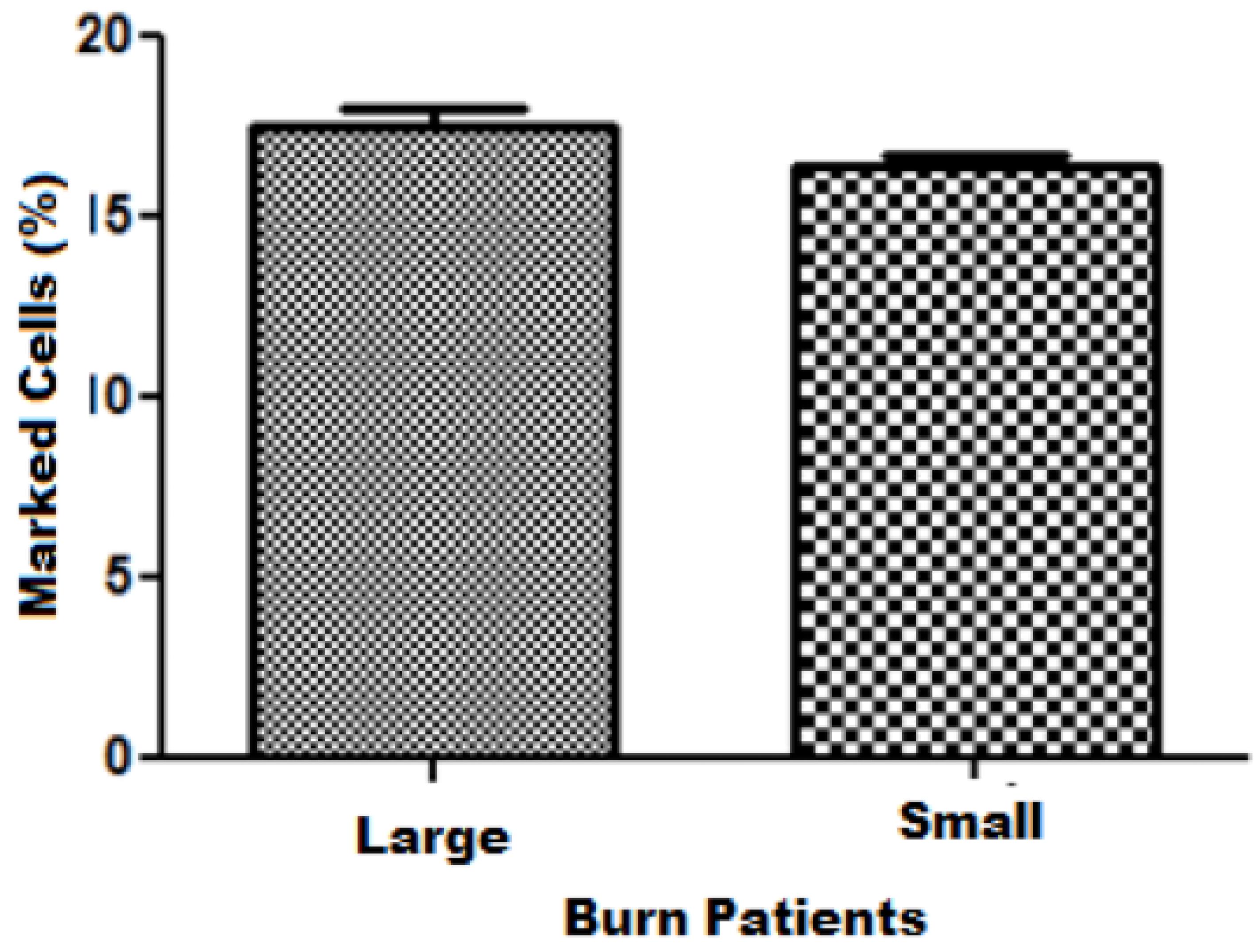
Analyzing the concentrations of IL-12 found in the culture medium of cells from the patients it was noted that there is a tendency to significance (p= 0.077) for patients with major burns shows a tendency to increase the IL-12 protein expression compared to patients with small burn. The mean expression and standard deviation, respectively, for major and small burn was 17.82 ± 0.43 and 16.99 ±0.09 (Figure 1).
Regarding to IL - 10 no difference was observed between large and small burned (p = 0.48), and the mean expression and standard deviation observed was, respectively, 15.27 ± 1.19 and 16.27 ± 0.56 (Figure 2).
For IL - 8 was observed that there was significant difference between the expression between large and small burned patient (p = 0.018*). There was a large increase in burn patients and the mean expression and standard deviation was, respectively, 18.18 ± 0.40 and 15.85 ± 0.45 to large and small burn patients (Figure 3).
Analyzing IL - 6 was noted that there is no difference between large and small burn (p = 0.14) and the mean expression and standard deviation was, respectively, 17.47 ± 0.56 and 16.43 ± 0.26 (Figure 4).
As for IL - 1 beta, it was not observed difference between groups (p= 0.28) and the mean and standard deviation was 17.14 ± 0.93 and 15.85 ± 0.45 to large and small burn patients, respectively (Figure 5).
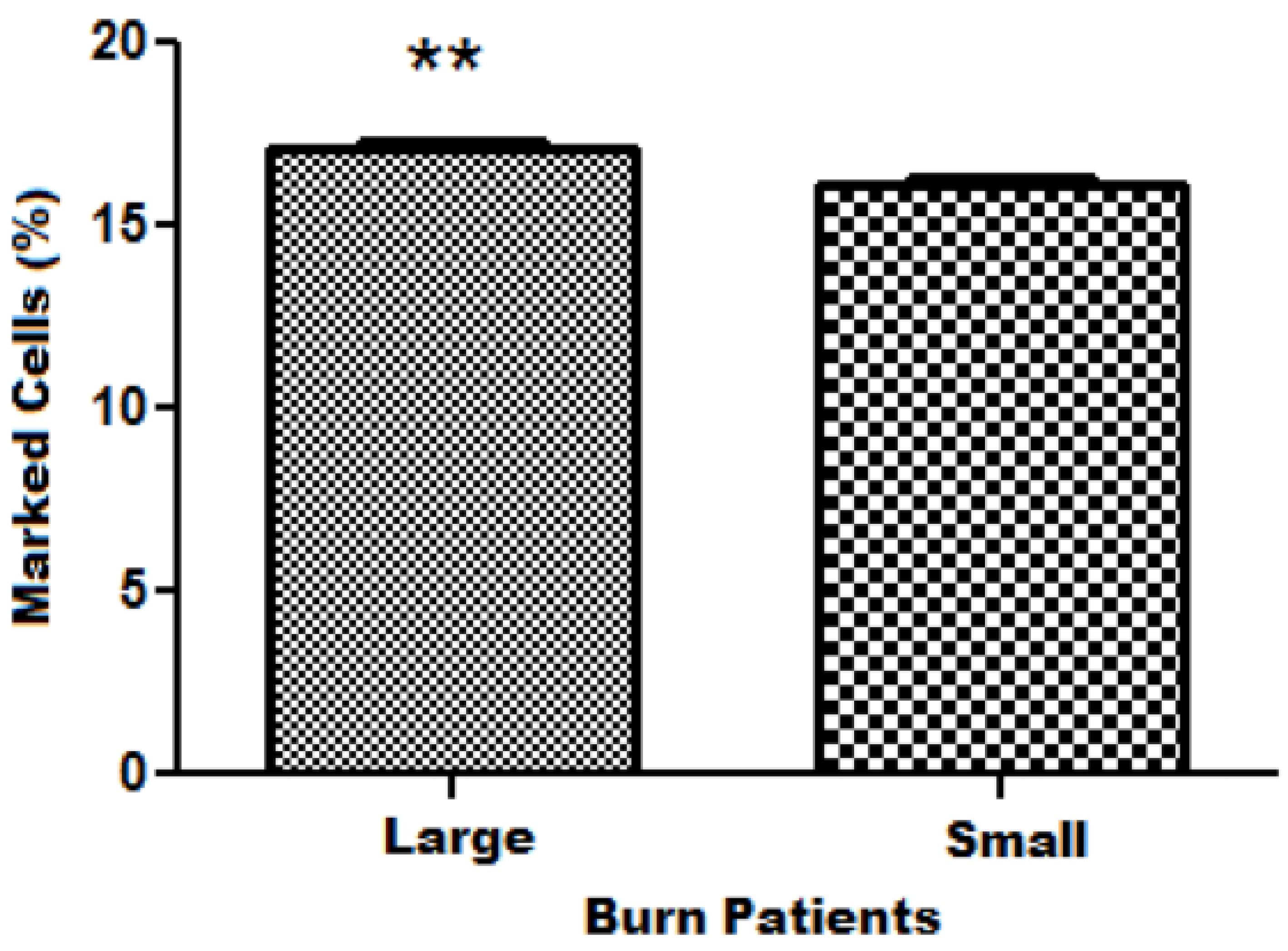
TNF -alpha protein expression presented difference between large and small burn patients (p = 0.0054*). We observed a pattern of 17.13 ± 0.19 for large increase in patients with large burns comparing those who had small burn and mean and standard deviation and 16.16 ± 0.12 for patients with small burn (Figure 6).
Average expression of TNF-alpha obtained by flow cytometry of the experimental groups protein.
KGF expression
Analyzing the concentrations of KGF, there is difference in protein expression to large compared to small burn (p = 0.0378 *). Comparing and interpolating the data between the three groups came to the following result seen in Figures 7 and 8.
KGF concentration observed by ELISA in patients, P1, P2, P3 and P5 large group of burned patients, P4, P6, P7 and P8 burned small group and the control group (CTRL).
Average KGF concentration obtained by the ELISA experiments between large and small burn groups.
Discussion
Studies have shown that the stimulatory effects of cytokines promote wound healing by
promoting the recruitment and maturation of neutrophils, increase in collagen synthesis,
proliferation of keratinocytes and fibroblasts, increased chemotaxis of neutrophils and
monocytes, increased neutrophil degranulation, and expression of cell adhesion
molecules66. Gosain A, Gamelli RL. A primer in cytokines. J Burn Care Rehabil.
2005; 26:7-12.
7. Rumalla VK, Borah GL. Cytokines, growth factors and plastic surgery.
Plast Reconstr Surg. 2001;108:719-33.
8. Efron PA, Moldawer LL. Cytokines and wound healing: The role of
cytokine and anticytokine therapy in repair response. J Burn Care Rehabil.
2004;25:149-60.
-
99. Ono I, Gunji H, Suda K, Iwatsuki K, Kaneko F. Evaluation of citokynes
in donor site wound fluids. Scand J Reconstr Hand Surg.
1994;28:269-73..
Although Singer and Clark1010. Singer AJ, Clark RA. Cutaneous wound healing. N Engl J Med. 1999;341:738-46. show that pro-inflammatory cytokines, particularly IL- 1, IL -6 and TNF -alpha are increased during the inflammatory phase of healing, the present study showed no significant difference in the concentration of IL -6 between the groups of large and small burn, whose cells were obtained immediately following an injury suffered in the skin by burning phase, unlike a single healing process analyzed by the authors.
Rumalla and Borah77. Rumalla VK, Borah GL. Cytokines, growth factors and plastic surgery. Plast Reconstr Surg. 2001;108:719-33. showed that the concentration of IL - 10 was taken as detectable on the first day and may remain so until ten days after the trauma. In the present study the difference between large and small burn did not show small differences, although present in both groups as the aforementioned study.
This may suggest standard concentrations of IL-1, IL-6, IL-10, and IL-12 independent of the extent of injury increase. As for IL - 8 significant changes was observed.
It is known that IL -12 is produced by activated macrophages and antigen presenting cells and induces differentiation of helper T lymphocytes and thereby of cellular immunity. It is considered a pro-inflammatory cytokine, together with TNF-alpha. The increased expression in body fluids is detected in the first twelve hours after injury, and is primarily released by local macrophages, which induces the recruitment and maturation of Neutrophils66. Gosain A, Gamelli RL. A primer in cytokines. J Burn Care Rehabil. 2005; 26:7-12. , 77. Rumalla VK, Borah GL. Cytokines, growth factors and plastic surgery. Plast Reconstr Surg. 2001;108:719-33. , 1111. Elenkov IJ, Chrousos GP, Wilder RL. Neuroendocrine regulation of IL-12 and TNF-alpha/IL-10 balance - Clinical Implications. Ann N Y Acad Sci. 2000;917:94-105..
In the present study a trend was observed the difference in the concentration of IL -12 between the two groups, large and small burned, showing that there may be a direct relationship. Therefore, the greater the extent of burning, increased production of IL -12, a proinflammatory cytokine, and that big difference could maintain a state of burned patient much more intense inflammation, and can determine systemic changes.
IL -8 family of chemokines component is secreted primarily by macrophages activated by pathogens or acute inflammation response, but may also be secreted by fibroblasts in the acute phase of wound healing. These components increase neutrophils, monocytes chemotaxis, and increased expression of cellular adhesion molecules. And in the first 24 hours after the assault is found in its highest concentration and promotes maturation and migration of keratinocytes77. Rumalla VK, Borah GL. Cytokines, growth factors and plastic surgery. Plast Reconstr Surg. 2001;108:719-33..
The IL - 8, IL - 6, and IL - 1 are elevated in the first hours after the burn, after trauma, severe burns, and elective surgeries remained, and were associated with increased complications and increased mortality66. Gosain A, Gamelli RL. A primer in cytokines. J Burn Care Rehabil. 2005; 26:7-12..
One involved in wound healing is the most well known cytokine tumor necrosis factor alpha (TNF -alpha). The increased expression occurs in body fluids in the first twelve hours after the trauma. The peak level of TNF- alpha in the wound fluid is affected three days after the attack in the dermis and is responsible for the increased proliferation and vascular permeability77. Rumalla VK, Borah GL. Cytokines, growth factors and plastic surgery. Plast Reconstr Surg. 2001;108:719-33.. The effects of TNF -alpha are dependent on the concentration and duration of exposure, highlighting the importance of the balance of inflammatory signals in the control of wound healing.
The IL -8 and TNF -alpha showed a significant difference when compared in both groups. In the group of severe burn them perform at higher concentration when compared to the small burned. This shows a direct relationship between the extent of the burn and the concentration of IL - 8 and TNF -alpha in exaggerated inflammatory responses as seen in major burns.
Chedid et al.44. Chedid M, Rubin JS, Csaky KG, Aaronson SA. Regulation of keratinocyte growth factor gene expression by interleukin 1. J Biol Chem. 1994;269:10753-7. studied cytokines and growth factors that influenced the production of KGF by fibroblasts where the results suggested that other growth factors such as IL - 1, PDGF -beta, IL - 6 and TGF -alpha stimulate moderately KGF RNA levels. These data suggest that a variety of growth factors and cytokines can influence the production of KGF in vivo.
KGF showed a significant difference when compared between the two study groups. In the group of large burns the expression of this protein was in greater concentration than in the group of small burned.
Gauglitz et al.1212. Gauglitz GG, Zedler S, von Spiegel F, Fuhr J, von Donnersmarck GH, Faist E. Functional characterization of cultured keratinocytes after acute cutaneous burn injury. PLoS One. 2012;7(2):e29942. conducted a study in which they compared the morphology and cytokine expression profile of keratinocytes in the skin following acute burn and unburned skin. It was observed that skin allograft in the concentration of unburned keratinocytes was higher when compared with the proliferation of these substances on the edges of burn injuries.
In the present study we noted that the control group had the KGF concentration increased when compared with the other two groups. This discrepancy resulted from the control group shows no support in the literature and the studies on gene expression of KGF in inflammatory processes of different tissues show increased expression in the presence of acute inflammation. So this new finding should be one of the prospects of future projects within the research group that during cosmetic surgery with general anesthesia there is a possible stimulus for the production of KGF by dermal fibroblasts at the site of the surgical procedure.
Conclusion
The IL-8, TNF-alpha, and KGF showed higher expression in large burned patients.
Acknowledgement
To Sao Paulo Research Foundation (FAPESP) for Research Grants number 2011/12945-4 and 2011/23.985-7.
References
-
1Mock C, Peck M, Krug E, Haberal M. Confronting the global burden of burns: a WHO plan and a challenge. Burns. 2009 Aug;35(5):615-7.
-
2Agay D, Andriollo-Sanchez M, Claeyssen R, Touvard L, Denis J, Roussel A, Chancerelle Y. Interleukin-6, TNF-alpha and interleukin-1 beta levels in blood and tissue in severely burned rats. Eur Cytokine Netw. 2008;19:1-7.
-
3Mikhal'chik EV, Piterskaya JA, Budkevich LY, Pen'kov LY, Facchiano A, De Luca C, Ibragimova GA, Korkina LG. Comparative study of cytokine content in the plasma and wound exudates from children with severe burns. Bull Exp Biol Med. 2009;148:771-5.
-
4Chedid M, Rubin JS, Csaky KG, Aaronson SA. Regulation of keratinocyte growth factor gene expression by interleukin 1. J Biol Chem. 1994;269:10753-7.
-
5Hirsch T, Peter SV, Dubin G, Mittler D, Jacobsen F, Lehnhardt M,1 Elof Eriksson, Steinau HU, Steinstraesser L. Adenoviral gene delivery to primary human cutaneous cells and burn wounds. Mol Med. 2006;12(9-10):199-207.
-
6Gosain A, Gamelli RL. A primer in cytokines. J Burn Care Rehabil. 2005; 26:7-12.
-
7Rumalla VK, Borah GL. Cytokines, growth factors and plastic surgery. Plast Reconstr Surg. 2001;108:719-33.
-
8Efron PA, Moldawer LL. Cytokines and wound healing: The role of cytokine and anticytokine therapy in repair response. J Burn Care Rehabil. 2004;25:149-60.
-
9Ono I, Gunji H, Suda K, Iwatsuki K, Kaneko F. Evaluation of citokynes in donor site wound fluids. Scand J Reconstr Hand Surg. 1994;28:269-73.
-
10Singer AJ, Clark RA. Cutaneous wound healing. N Engl J Med. 1999;341:738-46.
-
11Elenkov IJ, Chrousos GP, Wilder RL. Neuroendocrine regulation of IL-12 and TNF-alpha/IL-10 balance - Clinical Implications. Ann N Y Acad Sci. 2000;917:94-105.
-
12Gauglitz GG, Zedler S, von Spiegel F, Fuhr J, von Donnersmarck GH, Faist E. Functional characterization of cultured keratinocytes after acute cutaneous burn injury. PLoS One. 2012;7(2):e29942.
-
1
Research performed at Division of Plastic Surgery, Department of Surgery, Paulista School of Medicine (EPM), Federal University of Sao Paulo (UNIFESP), Brazil.
Publication Dates
-
Publication in this collection
2014


