ABSTRACT
Objective:
To verify which component of body composition (BC) has greater influence on postmenopausal women bone mineral density (BMD).
Subjects and methods:
Four hundred and thirty women undergoing treatment for osteoporosis and 513 untreated women, except for calcium and vitamin D. Multiple linear regression analysis was performed in order to correlated BMD at lumbar spine (LS), total femur (FT), femoral neck (FN) with body mass (BM), total lean mass (LM) and total fat mass (FM), all determined by DXA.
Results:
BM significantly correlated with all bone sites in untreated and treated women (r = 0.420 vs 0.277 at LS; r = 0.490 vs 0.418 at FN, r = 0.496 vs 0.414 at FT, respectively). In untreated women, the LM correlated better than FM with all sites, explaining 179% of LS; 32.3% of FN and 30.2% of FT; whereas FM explained 13.2% of LS; 277% of FN, 23.4% of FT In treated women, correlations with BC were less relevant, with the LM explaining 6.7% of BMD at LS; 15.2% of FN, 16% of FT, whereas the FM explained 8.1% of LS; 179% of FN and 176% of FT.
Conclusion:
LM in untreated women was better predictor of BMD than FM, especialy for distal femur, where it explained more than 30% of the BMD, suggesting that maintaining a healthy muscle mass may contribute to decrease osteoporosis risk. Treatment with anti-osteoporotic drugs seems to mask these relationships. Arch Endocrinol Metab. 2018;62(4):431-7
Keywords
Osteoporosis; treatment; body weight; body composition
INTRODUCTION
Osteoporosis is intimaly associated to the aging process and represents a social problem nowadays. Populational statistics shows that the situation can get even worse in the future due to the increase in longevity. Low bone mass is one of the main determinants of osteoporosis, which associates with bone microstructural changes resulting in a higher fracture risk. By comprehending what positively influences bone mass of individual health professionals will be able to create strategies to control bone loss throughout aging (11. Marin RV, Pedrosa MA, Moreira-Pfrimer LD, Matsudo SM, Lazaretti-Castro M. Association between lean mass and handgrip strength with bone mineral density in physically active postmenopausal women. J Clin Densitom. 2010;13(1):96-101.,22. Radominski SC, Bernardo W, Paula AP, Albergaria B, Moreira C, Fernandes CE, et al. Diretrizes brasileiras para o diagnóstico e tratamento da osteoporose em mulheres na pós-menopausa. Rev Bras Reumatol. 2017;57(2):452-66.).
Total body mass is one of the biological variables that best correlates with bone mass. However, it remains unclear what would be the influence of the different body mass components on bone metabolism (11. Marin RV, Pedrosa MA, Moreira-Pfrimer LD, Matsudo SM, Lazaretti-Castro M. Association between lean mass and handgrip strength with bone mineral density in physically active postmenopausal women. J Clin Densitom. 2010;13(1):96-101.,33. Reid IR. Fat and bone. Arch Biochem Biophys. 2010;503(1):20-7.–66. Xiang J, Chen Y, Wang Y, Su S, Wang X, Xie B, et al. Lean Mass and Fat Mass as Mediators of the Relationship Between Physical Activity and Bone Mineral Density in Postmenopausal Women. J Womens Health (Larchmt). 2017;26(5):461-6.). Gillette-Guyonnet and cols. (55. Gillette-Guyonnet S, Nourhashemi F, Lauque S, Grandjean H, Vellas B. Body composition and osteoporosis in elderly women. Gerontology. 2000;46(4):189-93.) studied older osteoporotic women (75 to 89 years old) and observed a significant correlation between bone mineral density and body composition (BC), which includes body mass, fat mass and lean mass. In this study, fat mass showed a better association with bone mass than lean mass, suggesting that fat mass could exert a protective effect on the proximal femur. On the other hand, Binder and Kohrt (77. Binder EF, Kohrt WM. Relationships between body composition and bone mineral content and density in older women and men. Clinical Exercise Physiology. 2000;2:84-91.), studying elderly men and women, observed that the lean mass was the BC component that best correlated with bone mineral density (BMD). The authors suggested that the association of lean mass and fat mass with bone mass reflects not only the effects of total body mass mechanical loading on bone, but also the functional relation between muscles and bones.
It is believed that muscle contractions, as well as physical exercise, act as potent anabolic stimuli to strengthen bone tissue. On the other hand, low fat mass can represent especially relevant state of denutrition in elderly, which could reflect on the health of bone tissue, currently recognized as a tissue involved in energy metabolism (22. Radominski SC, Bernardo W, Paula AP, Albergaria B, Moreira C, Fernandes CE, et al. Diretrizes brasileiras para o diagnóstico e tratamento da osteoporose em mulheres na pós-menopausa. Rev Bras Reumatol. 2017;57(2):452-66.,88. Camargo MB, Cendoroglo MS, Ramos LR, de Oliveira Latorre Mdo R, Saraiva GL, Lage A, et al. Bone mineral density and osteoporosis among a predominantly Caucasian elderly population in the city of São Paulo, Brazil. Osteoporos Int. 2005;16(11):1451-60.).
As studies about BC and bone mass still show controversial results, our aim was to determine which of the components of BC would be better related to bone mass in a representative population of postmenopausal women both treatment and treatment naive for osteoporosis.
SUBJECTS AND METHODS
The sample consisted of 943 independent postmenopausal women, with age above 40 years old (average 66.9 ± 7.8 years old), who volunteered to participate in three different protocols to evaluate physical exercise effects on bone mass, conducted by the same group of researchers. The present crosssectional study used the baseline data from all these women as they entered the study protocols, which were conducted by the same professionals and utilized the same methodology for measurement of antrhopometric and body composition parameteres. The selection criteria for choosing study participants are described in details in the publications resulting from these three studies (99. Moreira LD, Fronza FC, Dos Santos RN, Zach PL, Kunii IS, Hayashi LF, et al. The benefits of a high-intensity aquatic exercise program (HydrOS) for bone metabolism and bone mass of postmenopausal women. J Bone Miner Metab. 2014;32(4):411-9.–1111. Nascimento NA, Moreira PF, Marin RV, Moreira LD, Castro ML, Santos CA, et al. Relation among 25(OH)D, aquatic exercises, and multifunctional fitness on functional performance of elderly women from the community. J Nutr Health Aging. 2016;20(4):376-82.). For further analysis, these women were divided into two groups: 430 that were undergoing treatment for osteoporosis (47 to 87 years old, average of 68.3 ± 8.2 years old) (1010. Camargo MB, Kunii LS, Hayashi LF, Muszkat P, Anelli CG, Marin-Mio RV, et al. Modifiable factors of vitamin D status among a Brazilian osteoporotic population attended a public outpatient clinic. Arq Bras Endocrinol Metabol. 2014;58(5):572-82.,1111. Nascimento NA, Moreira PF, Marin RV, Moreira LD, Castro ML, Santos CA, et al. Relation among 25(OH)D, aquatic exercises, and multifunctional fitness on functional performance of elderly women from the community. J Nutr Health Aging. 2016;20(4):376-82.) and 513 never treated for osteoporosis (41 to 87 years old, average of 65.8 ± 7.4 years old) (99. Moreira LD, Fronza FC, Dos Santos RN, Zach PL, Kunii IS, Hayashi LF, et al. The benefits of a high-intensity aquatic exercise program (HydrOS) for bone metabolism and bone mass of postmenopausal women. J Bone Miner Metab. 2014;32(4):411-9.,1111. Nascimento NA, Moreira PF, Marin RV, Moreira LD, Castro ML, Santos CA, et al. Relation among 25(OH)D, aquatic exercises, and multifunctional fitness on functional performance of elderly women from the community. J Nutr Health Aging. 2016;20(4):376-82.).
The study has the approval of the Ethics and Research Committee of Universidade Federal de São Paulo - Unifesp/EPM, numbered CEP: 32882/12, CAAE: 02252312.1.0000.5505. All the subjects signed an informed consent.
The methods selected to evaluate the total and the segmental BC, as well as the total and the compartmental anthropometric measurements and sites of bone mineral density are described as follows.
The total body mass was measured using a platform-type mechanical scale (Filizola, São Paulo, Brazil) with a maximum capacity of 150 kg and variation 0.1 kg. Height was measured using a vertical bar stadiometer with maximum range of 220 cm and accuracy of 0.1 cm. Body mass index - BMI (kg/m2), was calculated from weight and height measurements using the formula BMI = weight (in kg) divided by height (in m-2) (1212. Heyward V, Stolarczyk LM. Anthropometric method. Applied body composition assessment. Ed. Champaign: Human Kinetics; 1996. p. 76-85.).
The BC and the bone mineral density (BMD) analyzes were obtained by dual-energy X-ray absorptiometry (DXA), in a Hologic QDR 4500A equipment (Waltham, MA). This assessment was performed at the Bone Evaluation Laboratory of the Endocrinology Division, at Universidade Federal de Sao Paulo. In our hands the CV% for lumbar spine and total femur is 1%, for trochanter is 1.2% and for femoral neck is 1.4%. The studied variables were: BMD in the sites of lumbar spine L1-L4 (LS), total femur (TF) and femoral neck (FN) in grams/cm2 and the T-score values; in addition to the lean mass (LM) and fat mass (FM) in absolute values.
Statistical analysis
Normality of the data was assessed by using the Kolmogorov-Smirnov adherence test. When the groups were divided into treated and untreated women, they were compared by the “t” test of Student for independent samples in order to determine whether the groups presented any different characteristics.
The Pearson Linear correlation, as well as the univariate linear regression and the analysis of multiple linear regressions were performed, having the bone sites as the dependent variables and BC (total body mass, total lean mass) as the independent ones.
The studied variables that presented p < 0.20 in the Pearson linear correlation analysis were selected and included in the models and, further, considered for inclusion in the multiple linear regression model. For this model, the stepwise forward modeling strategy was used.
The variables that remained significant were kept in the final multiple linear regression model, always observing the possible collinearities. The variable age was considered as a control variable. All the analisys were made by using the Statistical Package for the Social Sciences - SPSS for Windows - version 19”.
RESULTS
All studied variables were different between the two evaluated groups - women treated and untreated for osteoporosis, emphasizing that the treated participants were older, thinner, shorter, presented a worse bone mass and lower values of body composition components (Table 1).
Descriptive characteristics of bone mass and body composition of untreated and treated women for osteoporosis
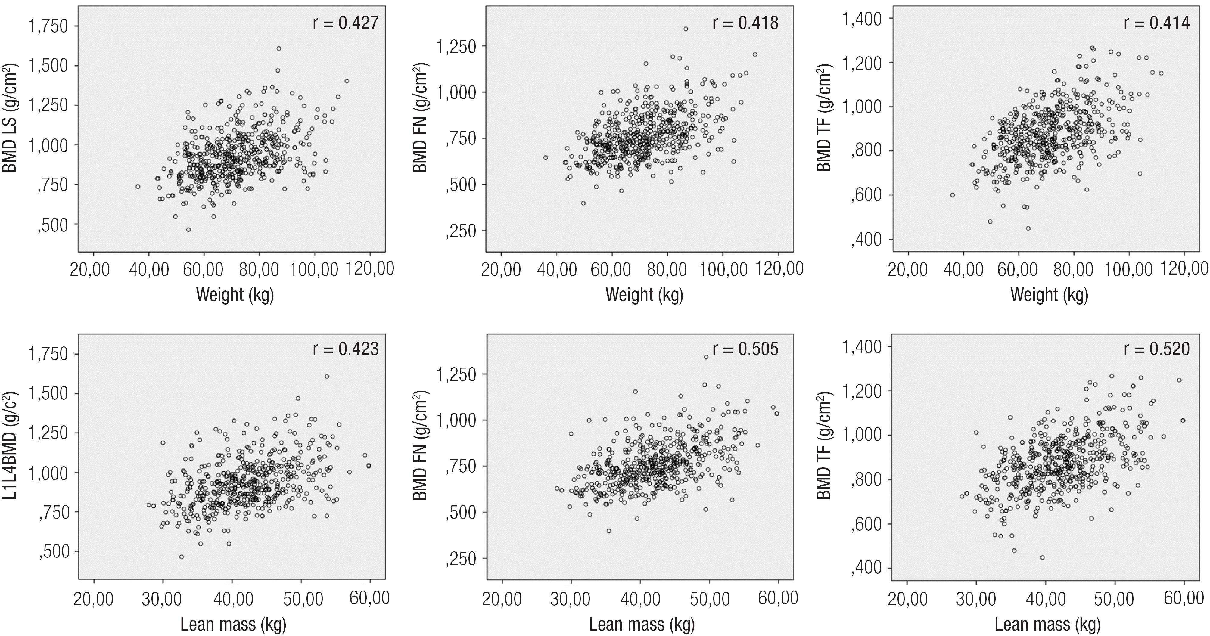
In women not treated for osteoporosis, among all the studied variables, the total body mass was the one that best correlated with all sites of bone mass: LS r = 0.427 (p = 0.000), FN r = 0.490 (p = 0.000) and TF r = 0.496 (p = 0.000). The correlation between the different sites of bone mass with height or BMI showed values ranging from r = 0.120 to r = 0.430 (p = 0.000). For the same 513 women, the lean mass was the variable that best correlated with all sites of bone mineral density, r = 0.423 (p = 0.000) at LS, r = 0.505 (p = 0.000) at FN and r = 0.520 (p = 0.000) at TF (Figure 1). The associations with fat mass were also significant but showed less expressive results in all sites [r = 0.361 (p = 0.000) for LS; r = 0.433 (p = 0.000) for FN and r = 0.430 (p = 0.000) for TF].
Correlation charts between the bone sites, total body mass and lean mass of untreated women for osteoporosis.
After performing the individual analyzes, we started to build statistical models to determine the influence of lean and fat mass on bone mass in the 513 untreated women and the 430 women treated for osteoporosis.
In untreated women, the coefficients of BMD determination found for lean mass models were better than the ones found for the fat mass: 17.9% in BMD of LS; 32.3% for FN and 30.2% of TF. On the other hand, models showed that fat mass could explain 13.2% of LS BMD; 27.7% of FN BMD and 23.4% of TF BMD (Table 2).
Results of the multiple linear regression analysis between the sites of BMD and lean body mass and fat mass variables of 513 untreated women for osteoporosis
In women treated with active drugs for osteoporosis, the correlation between BMD and body mass became weaker. In this group, the models for lean mass explained only 6.7% of LS BMD; 15.2% of FN BMD and 16% of TF BMD (Table 3), while the model for fat mass explained 8.1% of LS BMD, 17.9% of FN BMD and 17.6% of FT BMD. Therefore, treatment of osteoporosis appears to modify the relation between bone mass and the anthropometric variables, decreasing the great influence of these parameters on the BMD.
Results of the multiple linear regression analysis between the sites of BMD and lean body mass and fat mass variables of 430 treated women for osteoporosis
DISCUSSION
The total body mass in adults is one of the biological variables that most consistently correlate with bone mass and fracture risk (88. Camargo MB, Cendoroglo MS, Ramos LR, de Oliveira Latorre Mdo R, Saraiva GL, Lage A, et al. Bone mineral density and osteoporosis among a predominantly Caucasian elderly population in the city of São Paulo, Brazil. Osteoporos Int. 2005;16(11):1451-60.). In our study, this phenomenon was detected with greater relevance among women without treatment for osteoporosis. Treatment with specific drugs for osteoporosis made the relationship between BMD and body mass less relevant, probably because these drugs directly interfere on bone remodeling, mitigating the local influence of body mass (22. Radominski SC, Bernardo W, Paula AP, Albergaria B, Moreira C, Fernandes CE, et al. Diretrizes brasileiras para o diagnóstico e tratamento da osteoporose em mulheres na pós-menopausa. Rev Bras Reumatol. 2017;57(2):452-66.). In a similar study with Italian Caucasian postmenopausal women, authors (1313. Gnudi S, Sitta E, Fiumi N. Relationship between body composition and bone mineral density in women with and without osteoporosis: relative contribution of lean and fat mass. J Bone Miner Metab. 2007;25(5):326-32.) concluded that both fat and lean masses might affect bone mass but depending on the osteoporotic status. In non-osteoporotic women, only lean mass was associated with BMD. In osteoporotic women treated for osteoporosis, however, the lean and fat masses had the same importance.
Lewin and cols. (1414. Lewin S, Gouveia CH, Marone MMS, Wehba S, Malvestiti LF, Bianco AC. Densidade mineral óssea vertebral e femoral de 724 mulheres brancas brasileiras: influência da idade e do peso corporal. Rev Assoc Med Bras. 1997;43(2):127-36.) also studied a population of Brazilian Caucasian women and observed that heavier girls reached bone mass peak earlier, besides having higher BMD values. In addition, the bone loss caused by aging was reduced in those women with higher total body mass.
In our study, the correlation of total body mass was more relevant with the proximal femur than with the lumbar spine, what could translate influence of the mechanical load and physical activity on bone mass of lower limbs. To contribute to this theory, in our results the lean mass was the component that best correlated with bone density in different places, especially the proximal femur.
Reid (33. Reid IR. Fat and bone. Arch Biochem Biophys. 2010;503(1):20-7.), in a review study, emphasizes that total body mass is a main determinant of bone mineral density as well as fracture risk, and concludes that fat mass would be the main contributor to this relation. Controversely, other authors (66. Xiang J, Chen Y, Wang Y, Su S, Wang X, Xie B, et al. Lean Mass and Fat Mass as Mediators of the Relationship Between Physical Activity and Bone Mineral Density in Postmenopausal Women. J Womens Health (Larchmt). 2017;26(5):461-6.,1515. Ho-Pham LT, Nguyen ND, Lai TQ, Nguyen TV. Contributions of lean mass and fat mass to bone mineral density: a study in postmenopausal women. BMC Musculoskelet Disord. 2010;11:59.–1717. Li S, Wagner R, Holm K, Lehotsky J, Zinaman MJ. Relationship between soft tissue body composition and bone mass in perimenopausal women. Maturitas. 2004;47(2):99-105.) sustain that a higher amount of lean mass would be beneficial and have a stronger influence on bone mass. Both statements have physiological plausibility. The fat tissue would represent the influence of neuroendocrine factors, as well as the metabolism of sex steroids (1717. Li S, Wagner R, Holm K, Lehotsky J, Zinaman MJ. Relationship between soft tissue body composition and bone mass in perimenopausal women. Maturitas. 2004;47(2):99-105.) on bone density. The lean mass, on the other hand, would represent the mechanical and physical stimulation effect on bone tissue. Li and cols. (1717. Li S, Wagner R, Holm K, Lehotsky J, Zinaman MJ. Relationship between soft tissue body composition and bone mass in perimenopausal women. Maturitas. 2004;47(2):99-105.), analysing perimenopausal women (average of 49.6 years), revealed that both fat mass and lean mass had positive relations with BMD of lumbar spine and femur. However, using multiple regression analysis, authors observed that only lean mass and ethnicity remained significant predictors of BMD of femoral neck. The lean mass was the only predictor of BMD of the total femur, explaining in 38% the BMD at this site, while fat mass was not a significant predictor of BMD in any of the analyzed sites.
The influence of total body mass and its different components on bone mass apears to vary according to age, gender and skeletal site of the studied populations (Table 4). In most studies, the lean mass apears to have a greater influence on bone density than fat mass, specialy at proximal femoral sites. In younger men, however, fat mass can have negative effects on bone mass (44. Cui LH, Shin MH, Kweon SS, Park KS, Lee YH, Chung EK. Relative contribution of body composition to bone mineral density at different sites in men and women of South Korea. J Bone Miner Metab. 2007;25(3):165-71.). With aging, changes in body composition components induce an increment of fat mass followed by a decrement of lean mass. Considering postmenopausal American women, Chen and cols. (1818. Chen Z, Lohman TG, Stini WA, Ritenbaugh C, Aickin M. Fat or lean tissue mass: which one is the major determinant of bone mineral mass in healthy postmenopausal women? J Bone Miner Res. 1997;12(1):144-51.) also observed that lean mass exerts a stronger influence on the BMD of differente bone sites.
Description of published studies which investigated relationships between body compartments and bone density in different populations
In a Korean rural population (19 to 80 years old, both genders), lean mass was an important determinant of BMD either for young and elderly population, but fat mass showed a dual effect. High fat mass showed negative influence on bone mass in younger, however, with positive influence in postmenopausal women and older men (44. Cui LH, Shin MH, Kweon SS, Park KS, Lee YH, Chung EK. Relative contribution of body composition to bone mineral density at different sites in men and women of South Korea. J Bone Miner Metab. 2007;25(3):165-71.).
The physiological mechanisms that would explain this important correlation between bone mass and body mass are not completely defined yet. Some experimental studies suggest the existence of a bone remodeling central control that would work via neuropeptides and neurotransmitters, such as serotonin (2121. Borba VZC, Kulak CAM, Lazaretti-Castro M. Controle neuroendócrino da massa óssea: Mito ou verdade? Arq Bras Endocrinol Metab. 2003;47(4):453-7.,2222. Ducy P, Amling M,Takeda S, Priemel M, Schilling AF, Beil FT, et al. Leptin inhibits bone formation through a hypothalamic relay: a central control of bone mass. Cell. 2000;100(2):197-207) in addition to adipokines, as leptin, which would connect fat tissue to bone metabolism (2222. Ducy P, Amling M,Takeda S, Priemel M, Schilling AF, Beil FT, et al. Leptin inhibits bone formation through a hypothalamic relay: a central control of bone mass. Cell. 2000;100(2):197-207). Karsenty (2323. Karsenty G. Convergence between bone and energy homeostases: Leptin regulation of bone mass. Cell Metab. 2006;4(5):341-8.) postulate that bone tissue has an endocrine role in the regulation of energy metabolism, especially decarboxylated osteocalcin, acting in the regulation of glucose homeostasis and insulin sensitivity (2121. Borba VZC, Kulak CAM, Lazaretti-Castro M. Controle neuroendócrino da massa óssea: Mito ou verdade? Arq Bras Endocrinol Metab. 2003;47(4):453-7.–2828. Lee NK, Karsenty G. Reciprocal regulation of bone and energy metabolism. J Musculoskelet Neuronal Interact. 2008;8(4):351.).
It is well established that mechanical loading has a powerful anabolic effect on bone tissue, an effect coordinated by osteocytes (2929. Ocarino NM, Serakides R. Efeito da atividade física no osso normal e na prevenção e tratamento da osteoporose. Rev Bras Med Esporte. 2006;12(3):164-8.). This can be confirmed by increased bone mass observed in athletes, when compared with sedentary controls (3030. Lazzoli JK, Oliveira MAB, Leitão MB, Nóbrega ACL, Nahas RM, Rezende L, et al. Posicionamento Oficial da Sociedade Brasileira de Medicina do Esporte sobre: esporte competitivo em indivíduos acima de 35 anos. Rev Bras Med Esporte. 2001;7(3):83-92.). On the other hand, immobility and inactivity are considered great causes for low bone mass, risk of falls and fractures (3131. Zazula FC, Pereira MAS. Fisiopatologia da osteoporose e o exercício físico como medida preventiva. Arq Cienc Saúde Unipar. 2003;7(3):269-75.). Thus, lean mass measured by DXA can be interpreted as a marker of bone health, which means a greater mechanical load on the skeleton and would justify our findings. This gives better basis for the importance of encouraging physical activity to maintain muscle mass in preventing osteoporosis (3232. Klein-Nulend J, Bacabac RG, Bakker AD. Mechanical loading and how it affects bone cells: the role of the osteocyte cytoskeleton in maintaining our skeleton. Eur Cell Mater. 2012;24:278-91.).
In conclusion, revious studies, as well as the present one, confirm that several variables contribute to the association between lean mass, fat mass and bone mass. Among these variables we highlight the treatment for osteoporosis, different ages and stages of life, gender and ethnicity. Our data revealed an important relationship between total body mass and all bone mass sites in postmenopausal women without osteoporosis treatment. However, in women being treated for osteoporosis these correlations lose their relevance. Among the different BC components, we found that lean mass was the one that presented the best correlation with bone mineral density, mainly on the proximal femur. In multiple variables model, when lean mass was corrected by the age of women without treatment for osteoporosis it explained about 30% of proximal femur bone mass. These results suggest that maintaining a healthy muscle mass can contribute to decrese the risk for osteoporosis. Results like ours also stimulate the search for mechanisms that explain this phenomenon, as well as the relevance of BC parameters on bone mass during treatment of osteoporosis.
REFERENCES
-
1Marin RV, Pedrosa MA, Moreira-Pfrimer LD, Matsudo SM, Lazaretti-Castro M. Association between lean mass and handgrip strength with bone mineral density in physically active postmenopausal women. J Clin Densitom. 2010;13(1):96-101.
-
2Radominski SC, Bernardo W, Paula AP, Albergaria B, Moreira C, Fernandes CE, et al. Diretrizes brasileiras para o diagnóstico e tratamento da osteoporose em mulheres na pós-menopausa. Rev Bras Reumatol. 2017;57(2):452-66.
-
3Reid IR. Fat and bone. Arch Biochem Biophys. 2010;503(1):20-7.
-
4Cui LH, Shin MH, Kweon SS, Park KS, Lee YH, Chung EK. Relative contribution of body composition to bone mineral density at different sites in men and women of South Korea. J Bone Miner Metab. 2007;25(3):165-71.
-
5Gillette-Guyonnet S, Nourhashemi F, Lauque S, Grandjean H, Vellas B. Body composition and osteoporosis in elderly women. Gerontology. 2000;46(4):189-93.
-
6Xiang J, Chen Y, Wang Y, Su S, Wang X, Xie B, et al. Lean Mass and Fat Mass as Mediators of the Relationship Between Physical Activity and Bone Mineral Density in Postmenopausal Women. J Womens Health (Larchmt). 2017;26(5):461-6.
-
7Binder EF, Kohrt WM. Relationships between body composition and bone mineral content and density in older women and men. Clinical Exercise Physiology. 2000;2:84-91.
-
8Camargo MB, Cendoroglo MS, Ramos LR, de Oliveira Latorre Mdo R, Saraiva GL, Lage A, et al. Bone mineral density and osteoporosis among a predominantly Caucasian elderly population in the city of São Paulo, Brazil. Osteoporos Int. 2005;16(11):1451-60.
-
9Moreira LD, Fronza FC, Dos Santos RN, Zach PL, Kunii IS, Hayashi LF, et al. The benefits of a high-intensity aquatic exercise program (HydrOS) for bone metabolism and bone mass of postmenopausal women. J Bone Miner Metab. 2014;32(4):411-9.
-
10Camargo MB, Kunii LS, Hayashi LF, Muszkat P, Anelli CG, Marin-Mio RV, et al. Modifiable factors of vitamin D status among a Brazilian osteoporotic population attended a public outpatient clinic. Arq Bras Endocrinol Metabol. 2014;58(5):572-82.
-
11Nascimento NA, Moreira PF, Marin RV, Moreira LD, Castro ML, Santos CA, et al. Relation among 25(OH)D, aquatic exercises, and multifunctional fitness on functional performance of elderly women from the community. J Nutr Health Aging. 2016;20(4):376-82.
-
12Heyward V, Stolarczyk LM. Anthropometric method. Applied body composition assessment. Ed. Champaign: Human Kinetics; 1996. p. 76-85.
-
13Gnudi S, Sitta E, Fiumi N. Relationship between body composition and bone mineral density in women with and without osteoporosis: relative contribution of lean and fat mass. J Bone Miner Metab. 2007;25(5):326-32.
-
14Lewin S, Gouveia CH, Marone MMS, Wehba S, Malvestiti LF, Bianco AC. Densidade mineral óssea vertebral e femoral de 724 mulheres brancas brasileiras: influência da idade e do peso corporal. Rev Assoc Med Bras. 1997;43(2):127-36.
-
15Ho-Pham LT, Nguyen ND, Lai TQ, Nguyen TV. Contributions of lean mass and fat mass to bone mineral density: a study in postmenopausal women. BMC Musculoskelet Disord. 2010;11:59.
-
16Gjesdal CG, Halse JI, Eide GE, Brun JG, Tell GS. Impact of lean mass and fat mass on bone mineral density: The Hordaland Health Study. Maturitas 2008;59(2):191-200.
-
17Li S, Wagner R, Holm K, Lehotsky J, Zinaman MJ. Relationship between soft tissue body composition and bone mass in perimenopausal women. Maturitas. 2004;47(2):99-105.
-
18Chen Z, Lohman TG, Stini WA, Ritenbaugh C, Aickin M. Fat or lean tissue mass: which one is the major determinant of bone mineral mass in healthy postmenopausal women? J Bone Miner Res. 1997;12(1):144-51.
-
19Zhu K, Hunter M, James A, Lim EM, Walsh JP. Associations between body mass index, lean and fat body mass and bone mineral density in middle-aged Australians: The Busselton Healthy Ageing Study. Bone. 2015;74:146-52.
-
20Taaffe DR, Villa ML, Holloway L, Marcus R. Bone mineral density in older non-Hispanic Caucasian and Mexican-American women: relationship to lean and fat mass. Ann Hum Biol. 2000;27(4):331-44.
-
21Borba VZC, Kulak CAM, Lazaretti-Castro M. Controle neuroendócrino da massa óssea: Mito ou verdade? Arq Bras Endocrinol Metab. 2003;47(4):453-7.
-
22Ducy P, Amling M,Takeda S, Priemel M, Schilling AF, Beil FT, et al. Leptin inhibits bone formation through a hypothalamic relay: a central control of bone mass. Cell. 2000;100(2):197-207
-
23Karsenty G. Convergence between bone and energy homeostases: Leptin regulation of bone mass. Cell Metab. 2006;4(5):341-8.
-
24Sharma S, Tandon VR, Mahajan S, Mahajan V, Mahajan A. Obesity: Friend or foe for osteoporosis. J Midlife Health. 2014;5(1):6-9.
-
25Lee S, Rhee Y. Bone and energy metabolism. J Korean Diabetes. 2013;14(4):174-7
-
26Yadav VK, Karsenty G. Leptin-dependent co-regulation of bone and energy metabolism. Aging (Albany NY). 2009;1(11):954-6.
-
27Araújo TF, Guimarães DF, Ferreira F, Luz JCM, Spini VBMG. Leptina e o controle neuroendócrino do peso corporal. Rev Bras Med. 2009;66(10):325-30.
-
28Lee NK, Karsenty G. Reciprocal regulation of bone and energy metabolism. J Musculoskelet Neuronal Interact. 2008;8(4):351.
-
29Ocarino NM, Serakides R. Efeito da atividade física no osso normal e na prevenção e tratamento da osteoporose. Rev Bras Med Esporte. 2006;12(3):164-8.
-
30Lazzoli JK, Oliveira MAB, Leitão MB, Nóbrega ACL, Nahas RM, Rezende L, et al. Posicionamento Oficial da Sociedade Brasileira de Medicina do Esporte sobre: esporte competitivo em indivíduos acima de 35 anos. Rev Bras Med Esporte. 2001;7(3):83-92.
-
31Zazula FC, Pereira MAS. Fisiopatologia da osteoporose e o exercício físico como medida preventiva. Arq Cienc Saúde Unipar. 2003;7(3):269-75.
-
32Klein-Nulend J, Bacabac RG, Bakker AD. Mechanical loading and how it affects bone cells: the role of the osteocyte cytoskeleton in maintaining our skeleton. Eur Cell Mater. 2012;24:278-91.
Publication Dates
-
Publication in this collection
Aug 2018
History
-
Received
30 Sept 2017 -
Accepted
02 Apr 2018