 VTH BRAZILIAN CONSENSUS CONFERENCE ON HELICOBACTER PYLORI INFECTION
ABSTRACT
Background: Helicobacter pylori infection remains highly prevalent, affecting approximately 40% of the global population. Individuals with infection continue to be at risk for its various clinical consequences, including chronic gastritis, peptic ulcer disease, and gastric cancer.
Objective: The Brazilian Helicobacter pylori and Microbiota Study Group and the Brazilian Federation of Gastroenterology present the Fifth Brazilian Consensus on this topic, following the inaugural edition in 1995. Significant advances across various areas have emerged since the publication of the last consensus in 2018. These advances are particularly relevant to bacterial eradication, as treatment encounters mounting challenges due to rising antimicrobial resistance.
Methods: For this update, five working groups were established to address: 1) epidemiology; 2) associated diseases and treatment indications; 3) diagnosis; 4) gastric cancer; and 5) treatment and microbiota.
Conclusion: The groups developed 34 evidence-based recommendations to guide clinicians in managing this infection within the Brazilian population.
Keywords:
Helicobacter pylori; H. Pylori; Consensus
HIGHLIGHTS
• The consensus brought together 26 experts from all regions of Brazil, who developed 34 evidence-based recommendations to guide infection management in Brazil population.
• Removing the bacterium is recommended for all infected individuals and is crucial in cases of chronic gastritis, peptic ulcer disease, MALT lymphoma, and gastric precancerous conditions.
• The ¹³C-urea breath test is the non-invasive gold standard for diagnosis and treatment monitoring, while histology is the invasive gold standard for mucosal assessment.
• In response to increasing antimicrobial resistance, the consensus recommends 14-day treatments, emphasizing quadruple bismuth therapy and the use of P-CABs (such as vonoprazan) to maximize effectiveness.
• Eradication of H. pylori is crucial for the primary and secondary prevention of gastric cancer, particularly among first-degree relatives of patients with the disease and following resection of early tumors.
Resumo
Conexto: A infecção por Helicobacter pylori é ainda muito frequente e acomete aproximadamente 40% da população mundial, que segue exposta a suas diferentes consequências clínicas como gastrite crônica, úlcera péptica e câncer gástrico, entre outras.
Objetivo: O Núcleo Brasileiro para Estudo do Helicobacter pylori e Microbiota e a Federação Brasileira da Gastroenterologia promovem agora seu V Consenso Brasileiro sobre o tema, cuja primeira edição ocorreu em 1995. Importantes avanços, em diferentes tópicos, foram observados desde a publicação de seu último consenso em 2018, especialmente no tocante a erradicação da bactéria, cujo tratamento enfrenta dificuldades crescentes pelo aumento da resistência antimicrobiana.
Métodos: Para esta atualização foram criados cinco grupos de trabalhos abordando: 1) epidemiologia; 2) doenças associadas e indicações de tratamento; 3) diagnóstico; 4) câncer gástrico e, 5) tratamento e microbiota.
Conclusão: Foram elaboradas 34 recomendações fundamentadas nas melhores evidências para auxiliar os médicos no manuseio desta infecção na população brasileira.
Palavras-Chave:
Helicobacter pylori; H. pylori; Consenso
INTRODUCTION
Helicobacter pylori (H. pylori) infection affects the stomach of over 40% of the global population and can cause a wide spectrum of digestive diseases, such as chronic gastritis, peptic ulcer disease, gastric adenocarcinoma, and gastric MALT lymphoma1. To date, four consensus meetings on its management have been held in Brazil, namely in 19952, 20043, 20124, and 20185. The global rise in resistance to antibiotics commonly used in eradication therapy (e.g., clarithromycin, levofloxacin, and metronidazole) reduces expected treatment benefits. Coupled with the lack of new antimicrobials targeting this bacterium6, these factors raise growing concerns regarding infection management. However, significant developments have occurred in novel therapeutic regimens that use potent antisecretory agents combined with new dosing schedules, necessitating a new national consensus. Furthermore, despite the limited phenotypic and genotypic studies evaluating H. pylori characteristics in the Brazilian setting7,8, initial results are now available from well-structured, real-world studies encompassing all Brazilian regions. These data were obtained through the Brazilian Registry on H. pylori Management (Hp-BraReg), created in 2021 in partnership with the European Registry on H. pylori Management (Hp-EuReg), which together now constitute the World Registry on H. pylori Management (WorldHpReg)9,10. Although the Kyoto Consensus11 established that every individual with an infection should be treated, the primary treatment indications have been expanded and detailed with new recommendations. Various diagnostic advances are discussed, including non-invasive, endoscopic, and histopathological methods, alongside an arsenal of molecular tests specifically designed to detect antimicrobial resistance. These advances aim to introduce an as-yet elusive antimicrobial stewardship policy into routine clinical practice. The growing role of the gastric and intestinal microbiota and the potential use of probiotics during treatment are discussed, considering current knowledge, along with different treatment strategies for primary and secondary gastric cancer prevention. Accordingly, the Brazilian Helicobacter pylori and Microbiota Study Group (Núcleo Brasileiro para Estudo do Helicobacter pylori e Microbiota; NBEHPM, per its Portuguese acronym), supported by the Brazilian Federation of Gastroenterology (Federação Brasileira de Gastroenterologia; FBG), held its fifth meeting in Bento Gonçalves, Rio Grande do Sul, from October 9 to 11, 2025. A total of 26 panelists-including gastroenterologists, endoscopists, and pathologists from all five of the country’s regions-participated in the meeting. Participants were selected based on their expertise and contributions to the field.
Consensus Development Process
The Organizing Committee adopted the Delphi method12. A total of 34 clinical questions were selected and divided among five working groups focused on 1) epidemiology; 2) associated diseases and treatment indications; 3) diagnosis; 4) gastric cancer; and 5) treatment and microbiota. The 26 delegates were assigned to these groups based on their expertise, with one member from each group invited to serve as a moderator. Each delegate prepared one or two draft statements with references pertaining to up to two clinical questions, delivering oral presentations along with their recommendations during two separate virtual meetings restricted to their respective group. The draft statements and recommendations were discussed, modified when necessary, and edited electronically during the meetings by FBG staff, followed by anonymous voting. Subsequently, in a third meeting-now an in-person plenary session in Bento Gonçalves with all participants present-all recommendations were voted on again, re-discussed, and modified as needed. Voting allowed for five options: 1) strongly agree; 2) agree with reservations; 3) undecided; 4) disagree; and 5) strongly disagree. Decisions were based on the risk of bias in the studies, evidence of publication bias, heterogeneity among studies, indirectness of the evidence, and precision of the effect estimate.
The consensus threshold for the final approval of each recommendation was 80% of voters who responded with either “strongly agree” or “agree with reservations”. The level of evidence and the strength of the recommendations were classified using the GRADE system (Grading of Recommendations Assessment, Development and Evaluation13-15), summarized below:
Quality of Evidence
-
High quality Further research is very unlikely to change our confidence in the estimate of effect.
-
Moderate quality Further research is likely to have an important impact on our confidence in the estimate of effect and may change the estimate.
-
Low quality Further research is very likely to have an important impact on our confidence in the estimate of effect and is likely to change the estimate.
-
Very low quality.
Any estimate of effect is very uncertain.
The strength of the recommendation was classified as strong (recommendations) or weak (suggestions), depending on the quality of evidence, cost-benefit ratio, feasibility, and costs.
Strength of Recommendation
-
Strong recommendation Most patients should receive the recommended intervention.
-
Weak recommendation.
Clinicians should recognize that different choices are appropriate for different patients, and they must help each patient arrive at a management decision consistent with the patient’s values and preferences.
Working group 1: epidemiology
Statement 1
The prevalence of H. pylori is primarily related to socioeconomic and hygiene conditions, as well as cultural habits. Transmission within the family is possible.
Agreement: 100%
GRADE: 1A | Strength of recommendation: strong
Quality of evidence: high
Two large systematic reviews and meta-analyses have evaluated the global prevalence of H. pylori infection over the past decades1,16. Li Y et al. assessed infection prevalence between 1980 and 2022 across 224 studies from 71 countries involving nearly 3,000,000 individuals. They estimated a reduction in infection rates from 58.2% in the 1980s to 43.1% in the 2011-2022 period. Infection prevalence shows marked geographical heterogeneity, with a higher infection load observed in developing countries and rural areas compared to urban or developed regions1. Another large study assessing H. pylori prevalence and gastric cancer incidence across 1,748 studies confirmed this trend, showing a reduction in prevalence from 52.6% in adults before 1990 to 43.9% in the 2015-2022 period. The study estimated a 15.9% reduction over the past 3 decades in adults, though the decline was less significant in children, where prevalence rates remained as high as 35.1%16. As with most endemic infectious diseases, a decline in prevalence is more closely tied to improvements in population hygiene and sanitation than to individualized case-by-case treatment since, in most countries (except Japan, for example), only a minority of individuals with infection receive treatment17. Over 40% of the world’s population continues to carry the infection and remains at risk for potential clinical sequelae. The Brazilian studies included in these two systematic reviews, along with recent studies, confirm this downward trend in adult prevalence, even when accounting for regional socioeconomic and sanitary variations18,19. A cross-sectional study using the 13C-urea breath test as a diagnostic tool in 161 children (mean age 7.8 years) in a southeastern Brazilian city found an H. pylori prevalence of 20.5%20.
H. pylori infection is largely considered a familial disease. Person-to-person transmission within families, especially from mothers and siblings with the infection, is common in developing countries21. Genotyping studies have shown that strain concordance was detected in 10 of 18 (56%) mother-child pairs and in zero of 17 father-child pairs. Concordant strains were also observed among siblings in 29 of 36 (81%) families22. However, transmission between couples or spouses remains controversial23,24. Large-scale Chinese epidemiological studies based on family-level treatment indicate this strategy contains intrafamilial infection spread in highly endemic areas25,26.
Statement 2
Risk groups for H. pylori infection include populations with poor basic sanitation, rural communities, households with documented cases, healthcare professionals, and patients who are immunocompromised or those with comorbidities. H. pylori transmission occurs primarily in childhood, with the fecal-oral route being the most relevant.
Agreement: 100%
GRADE: 1A | Strength of recommendation: strong
Quality of evidence: high
Sanitary, socioeconomic, and environmental factors represent the primary determinants for acquiring H. pylori infection27. However, other population groups and clinical conditions also convey an elevated risk for acquiring and/or harboring the infection. Childhood is considered the critical period for acquisition, with most cases occurring before 10 years of age, likely due to greater exposure in environments with poor hygiene and closer interpersonal contact28. In older adults, the lifetime cumulative prevalence is associated with an increased risk of complications, such as atrophic gastritis, intestinal metaplasia, and gastric cancer29. Transmission patterns are predominantly person-to-person, although foodborne, waterborne, animal-to-human, and occupational pathways are well-documented30-35. Person-to-person transmission within households, particularly from mothers with the infection and siblings, is common in developing countries30. There are several transmission routes for H. pylori, with the fecal-oral and oral-oral routes being the most likely. Other described routes include gastro-oral, anal-oral, and genital-oral31,33,34. Nonetheless, further studies on transmission pathways and their relative importance are still needed30. Documented household infection, especially in the mother-child pattern, represents a significant transmission route, and family screening has proven beneficial in preventing associated complications25,26. Certain health conditions also confer increased vulnerability. Patients who are immunocompromised, such as those undergoing chemotherapy, transplant recipients, or people living with HIV, demonstrate heightened susceptibility and impaired infection control36. Furthermore, patients with chronic diseases, such as diabetes and cardiovascular conditions, appear to have a higher likelihood of acquiring the infection, though this remains controversial37. Racial and ethnic disparities have also been documented in the United States, where rates are higher among Black and Hispanic individuals than among White individuals. These differences may be attributed to both genetic factors and unfavorable socioeconomic and environmental conditions38. While the occupational risk remains controversial, a systematic review of 98 studies suggested that healthcare professionals, especially those in the gastrointestinal field, face a higher risk, indicating an iatrogenic transmission route39. The detection of H. pylori DNA in vomit, saliva, dental plaque, gastric juice, and stool supports its potential transmission via endoscopic procedures40-43. A recent Brazilian study found no differences in infection prevalence between clinical gastroenterologists and GI endoscopists, highlighting the effectiveness of current universal disinfection and protection protocols18.
Statement 3
Recurrence of H. pylori infection in adults is low in developed countries, with reinfection being more frequent in regions with higher prevalence.
Agreement: 88%
GRADE: 1A | Strength of recommendation: strong
Quality of evidence: high
Based on clinical and temporal criteria, two types of H. pylori infection recurrence after eradication therapy can be identified44,45:
Recrudescence - A relapse occurring within one year post-treatment. This process involves the resurgence of the original strain that was temporarily suppressed; thus, the bacterium responsible for the recurrence is genetically identical to the strain identified prior to eradication therapy.
Reinfection - Defined as an infection by a new H. pylori strain following successful eradication. Some authors posit a temporal criterion, suggesting reinfection is responsible when a diagnostic test yields a positive result at least 1 year after the initial eradication44,45.
Definitive confirmation, however, is achievable only through molecular analysis comparing strains isolated before and after relapse, a capability practically unavailable in routine clinical practice. Recrudescence is more common when low-efficacy therapies are utilized. Reinfection requires re-exposure and is therefore more likely in countries with high H. pylori prevalence and poor sanitation44. The annual rate of reinfection or recrudescence following successful eradication is low (<2%) among adults in developed countries, but higher (5-10%) among adults in developing countries and among children30. Several randomized clinical trials have demonstrated that family-based H. pylori screen-and-treat strategies reduce recurrence rates more effectively than treating individuals alone30. A systematic review and meta-analysis encompassing 31 studies (16,797 participants) revealed a pooled H. pylori recurrence rate of 9% using a random-effects model, showing an upward trend as time elapsed since eradication46. At 1 year of follow-up, the H. pylori recurrence rate was 4%; at 2 years, 6%; at 3 years, 8%; and at 4 years or beyond, it reached 12%, eventually exhibiting a stabilization trend. The H. pylori recurrence rate was inversely related to the Human Development Index.
Brazilian studies on this topic are scarce. A study conducted in the state of Minas Gerais analyzed 150 patients with duodenal ulcers who achieved successful H. pylori eradication and were followed for a median period of 6.4 years (range: 305 days to 8.9 years). Twenty of the 150 patients (13.3%) reverted to a positive result on the 14C-urea breath test, yielding an annual recurrence rate of 2.1%47. In the state of São Paulo, two studies were conducted: one identified a recurrence rate of 5.7% while following 194 patients over a 10-year period48. Another study involving 147 patients demonstrated an annual recurrence rate of 1.8% over a 5-year follow-up, with no recurrence at 1 year49.
Working group 2: associated diseases and treatment indications
Table 1 shows the main indications of H. pylori treatment.
Statement 4
Eradication therapy is recommended for all cases of H. pylori-associated chronic gastritis.
Agreement: 96%
GRADE: 1A | Strength of recommendation: strong
Quality of evidence: high
H. pylori infection is considered the leading cause of chronic gastritis, which drives persistent inflammation of the gastric mucosa, with potential progression to tissue damage and an elevated risk for severe complications such as peptic ulcer disease, gastric adenocarcinoma, and gastric MALT lymphoma11,50. For most patients, apparent clinical symptoms are absent despite structural and functional abnormalities resulting from mucosal inflammation50,51. The interaction among different H. pylori genotypes, the duration of infection, host genetics, and environmental factors leads to distinct outcomes and complications52.
H. pylori infection universally causes gastric mucosal inflammation, characterized by neutrophilic and lymphoplasmacytic infiltrates. The combination of neutrophil-mediated injury and a cytotoxic immune response-often induced by cagA+ strain polymorphisms-can lead to loss of tissue integrity (ulceration) and glandular depletion (atrophy). This atrophy, in turn, establishes the groundwork for gastric cancer development53.
While H. pylori infection is the leading cause of chronic gastritis, other causes encompass communicable (infectious) conditions, non-communicable host-related conditions such as autoimmune diseases (autoimmune gastritis), and immune-mediated conditions (e.g., Crohn’s disease), and drug-induced injuries (e.g., immunotherapy)11. Brazil presents a broad epidemiological spectrum of H. pylori gastritis, varying both regionally (and micro-regionally) and by age group, reflecting disparities in population access to basic sanitation and eradication therapy. Consequently, the etiological diagnosis of gastritis must be established to ensure an appropriate therapeutic approach.
Histology is a simple, cost-effective, and widely available test that facilitates H. pylori identification and structural assessment of the mucosa. The density of H. pylori distribution across the gastric mucosa is variable and influenced by anatomical site, atrophy/metaplasia, and medication use. Therefore, when an upper GI endoscopy is indicated, systematic tissue sampling following established protocols is recommended. The updated Sydney System recommends biopsies from the lesser and greater curvatures of the antrum and corpus (four samples) and a fifth from the incisura angularis54. More recently, the MAPS II guidelines recommend two biopsies from the antrum and two from the corpus55. Areas with an atrophic appearance and suspicious lesions warrant targeted biopsies. Samples must be placed in separate vials by site and properly identified. H. pylori is easily identified using hematoxylin and eosin (H&E) staining. Special staining techniques (such as Giemsa) and immunohistochemistry increase the probability of detection but entail higher costs and longer diagnostic turnaround times. They are preferentially indicated in special situations, such as MALT lymphoma, active chronic gastritis, and chronic atrophic gastritis, where infection is suspected, but the bacterium remains unidentified by H&E56. The histology report must include the gastritis etiology (probable or suspected), the intensity of inflammation, and the staging of atrophy and metaplasia.
H. pylori eradication eliminates inflammatory activity, halts the progression of tissue damage, and reduces the risk of future complications. Furthermore, it improves the control of dyspeptic symptoms and reduces the risk of bacterial transmission. Thus, eradication therapy is indicated for all patients with H. pylori-associated chronic gastritis51.
Statement 5
H. pylori eradication is recommended in patients with gastric or duodenal peptic ulcers, as long as therapeutic options remain available.
Agreement: 100%
GRADE: 1A | Strength of recommendation: strong
Quality of evidence: high
Multiple studies demonstrate a declining prevalence of H. pylori-associated peptic ulcer disease in recent decades57-59. This decline is aligned with the overall global reduction in H. pylori prevalence1, population-based treatment in some countries, and improved living conditions, despite the concurrent upward trend in ulcers associated with the use of aspirin and NSAIDs59. Despite the decreasing prevalence trend, H. pylori remains the leading cause of gastric or duodenal ulcers. A population-based prospective cohort study comprising 2416 adults from Denmark showed an odds ratio of 4.3 (95%CI 2.2-8.3) for the association between H. pylori infection and gastric or duodenal peptic ulcers60. In a prospective German study, the risk of developing a duodenal ulcer and a gastric ulcer was 18.4 and 2.9 times higher, respectively, among individuals with cagA+ H. pylori infections61. A recent Korean multicenter study involving 26,785 individuals (38.8% over age 65 years) described the main risk factors for peptic ulcer disease as H. pylori infection (41.8%), use of aspirin or NSAIDs (36.1%), and undetermined causes in 22.1% of cases62.
H. pylori infection promotes two distinct types of gastritis. The first involves the gastric antrum (antrum-predominant gastritis) and causes acid hypersecretion by interfering with acid regulatory pathways63. Increased acid secretion delivers a higher acid load to the duodenum, elevating duodenal susceptibility by inducing the development (or extension, if already present) of gastric metaplasia areas in the duodenum. This allows H. pylori colonization, rendering the duodenal epithelium more vulnerable and prone to ulceration. The second gastritis pattern associated with H. pylori involves the entire stomach (pangastritis) and is linked to gastric atrophy, reduced acid secretion, and gastric cancer. H. pylori is considered responsible for 70% to 80% of benign gastric ulcerations. The lower prevalence of the bacterium in gastric ulcers compared to duodenal ulcers is related to the higher frequency of NSAID-induced ulcers. Gastric ulcers tend to occur in non-acid-secreting mucosa or near the junction with non-secreting mucosa. Even when they occur high on the lesser curvature, they arise in non-secreting mucosa. Under these circumstances, H. pylori-induced pangastritis drives the metaplastic changes that convert secreting mucosa into non-secreting mucosa. Refluxed duodenal contents, especially bile and lecithin, damage the gastric mucosa, making it more sensitive to acid-induced injury, even in small amounts. Furthermore, H. pylori infection can alter the mucin layer coating the gastric epithelium and compromise mucosal defense mechanisms64,65.
H. pylori eradication promotes ulcer healing, reduces the risk of recurrence, and prevents severe complications such as bleeding and gastric cancer, rendering it a universal recommendation5,11,17,38,51. Among patients with successful eradication, the annual ulcer recurrence rate is approximately 0% to 2.3% for gastric ulcers and 0% to 1.6% for duodenal ulcers during a follow-up period of up to 9.8 years66,67. In contrast, patients with untreated H. pylori infections faced recurrence rates of 52% for gastric ulcers and 64% for duodenal ulcers68.
Statement 6
The “test-and-treat” strategy is recommended for patients aged 45 years or younger with uninvestigated dyspepsia and no alarm features.
Agreement: 95%
GRADE: 1A | Strength of recommendation: strong
Quality of evidence: high
Dyspepsia represents a common clinical condition with an estimated prevalence of approximately 20% in the general population69. It is characterized by pain or discomfort in the upper gastrointestinal tract (the gastroduodenal region) and classified as investigated or uninvestigated70. According to the Rome IV consensus, after investigation via upper GI endoscopy, dyspepsia can be classified as70:
-
Organic: when symptoms are explained by endoscopic findings, such as an ulcer or gastric cancer.
-
H. pylori-associated: when infection is present, and H. pylori eradication leads to sustained resolution of dyspeptic symptoms for over 6 months.
-
Functional: when relevant endoscopic abnormalities or H. pylori infection are absent, or, if infected, symptoms persist even after eradication therapy. This category comprises the vast majority of dyspepsia cases.
For individuals aged 45 years or younger presenting with dyspepsia without alarm features, management strategies have been evaluated in clinical trials and meta-analyses. Approaches include empirical treatment with proton pump inhibitors (PPIs), immediate endoscopy, the “test-and-treat” strategy for H. pylori, or testing followed by endoscopy if positive. The “test-and-treat” strategy has proved to be the most effective and cost-effective approach, particularly in regions with a high prevalence of infection, such as Brazil51,71-78.
A systematic review and network meta-analysis by Eusebi et al. evaluated 15 randomized controlled trials with 6,162 patients, comparing all strategies directly and indirectly. “Test-and-treat” ranked highest for symptom reduction, in both intention-to-treat and per-protocol analyses. Immediate endoscopy performed similarly, but the “test-and-treat” strategy led to a significantly lower number of endoscopies during the 12-month follow-up, offering substantial cost-saving benefits. The neoplasia detection rate was very low (0.4%) across all treatment arms78. Additional benefits of the “test-and-treat” strategy for uninvestigated dyspepsia include a reduction in the future incidence of peptic ulcer disease and gastric cancer, as well as reduced management costs for these conditions79.
Statement 7
H. pylori eradication therapy is recommended as the first-line therapeutic option for patients with investigated dyspepsia and provides a modest but significant benefit for symptom improvement. Symptom persistence or relapse despite eradication characterizes functional dyspepsia.
Agreement: 93%
GRADE: 1A | Strength of recommendation: strong
Quality of evidence: high
Functional dyspepsia, defined by the Rome IV consensus as a clinical syndrome characterized by recurrent and chronic dyspeptic symptoms without underlying structural or metabolic lesions to explain them, accounts for nearly 80% of dyspepsia cases70. While the condition does not impact life expectancy, it is a chronic disorder with a fluctuating course that profoundly impacts patients’ quality of life and carries a high financial burden70,80. Its pathophysiology is complex and multifactorial, with several proposed mechanisms, including alterations in the gastroduodenal microbiota70.
Among patients with dyspepsia, once H. pylori infection is confirmed, eradication treatment should be the first therapy instituted. Recent high-quality meta-analyses, systematic reviews, and randomized clinical trials have consistently shown that H. pylori eradication leads to a modest but statistically significant improvement in dyspepsia symptoms. The Number Needed to Treat (NNT) ranges from 9 to 15 for symptom improvement and from 11 to 21 for a cure81-83. The treatment benefit is most pronounced among patients with successful eradication81.
However, sustained symptom resolution following eradication characterizes the condition as H. pylori-associated dyspepsia, rather than functional dyspepsia, per Rome IV criteria70. Selecting initial H. pylori eradication in functional dyspepsia, particularly in highly endemic regions, is also advocated by major international consensus guidelines due to its cost-effectiveness11,51,84. One consideration is that eradication treatment carries a higher rate of side effects, though severe adverse events are rare82,83. Unanswered questions persist in this area, including whether symptomatic improvement results from gastroduodenal microbiota modulation by antibiotics, which patient subgroups benefit the most, what the long-term impact on quality of life entails, and what the true role of genetic and regional factors involves.
Statement 8
H. pylori eradication is recommended in all cases of precancerous conditions (atrophic gastritis and intestinal metaplasia) and precancerous lesions (dysplasia) of the gastric mucosa, given the likelihood of reducing gastric carcinoma risk.
Agreement: 100%
GRADE: 1A | Strength of recommendation: strong
Quality of evidence: high
The progression of normal mucosa toward non-hereditary gastric carcinoma (GC) involves multiple stages, starting with chronic gastritis. Prolonged, non-self-limiting inflammation in response to persistent H. pylori infection, as well as the direct action of the bacterium itself, facilitates adaptive responses such as atrophy and metaplasia. In a smaller subset of patients-especially individuals with a positive family history, smoking habits, and high dietary sodium-the mutation rate is higher in the metaplastic epithelium, from which dysplasia and GC, predominantly of the intestinal type, arise50. Gastric atrophy (GA) is characterized by glandular loss and may or may not be associated with gastric intestinal metaplasia (GIM), defined by the replacement of the foveolar epithelium with cells exhibiting an intestinal phenotype. GIM can be subdivided into complete and incomplete, reflecting small intestine and colonic phenotypes, respectively. Absorptive cells with a brush border and Paneth cells define the complete type, whereas non-absorptive columnar cells, alongside goblet cells, characterize the incomplete pattern, which conveys a higher risk of malignant transformation and is often seen in extensive GIM cases50,85. There is also a GIM classification based on mucin histochemical profiling, subtyping it into I, II, and III, with type III harboring the highest carcinogenic potential86. However, due to reagent toxicity, this classification is not recommended in routine practice87.
Dysplasia is phenotypically characterized by reduced mucosecretory differentiation and increased cell proliferation, displaying cytologic/architectural atypia without signs of stromal invasion. Based on its complexity, it is classified as low-grade and high-grade, conveying a progressive cancer risk. When the histopathological picture cannot be distinguished from regenerative changes, the case is deemed indefinite for dysplasia. A repeat biopsy is recommended, as only 9% of cases remain indefinite upon a second biopsy88,89.
GA and GIM progressively increase the risk for GC but possess the potential for regression following H. pylori treatment, defining these changes as precancerous conditions50,90. Notably, the likelihood of regression drops significantly when these conditions extensively involve the corpus and antrum91. In contrast, dysplasia is designated as a precancerous lesion, as it exhibits a higher risk of evolving into invasive GC and a lower chance of regression following antibiotic therapy50,87.
Long-term studies demonstrate that H. pylori eradication in patients with GA and GIM leads to significant regression of these conditions and reduces their progression to more advanced stages92-94. An enhanced benefit is anticipated when treatment occurs in the early stages. However, long-term improvement can occur even with extensive GA/GIM, when endoscopic surveillance is indicated90 (see recommendation 22). For precancerous lesions, the chance of regression is lower, even in low-grade dysplasia, but eradication still reduces the GC risk, even in high-grade dysplasia cases94,95. It is recommended to test for H. pylori using non-invasive methods in extensive GA/GIM, as the bacterial population is typically reduced in these settings, hindering its detection in histological sections.
Statement 9
Patients with gastric MALT lymphoma and H. pylori infection must receive eradication treatment. H. pylori-negative patients may benefit from empirical treatment.
Agreement: 100%
GRADE: 1A | Strength of recommendation: strong
Quality of evidence: high
MALT lymphoma is an extranodal low-grade B-cell lymphoma characterized by the clonal expansion of marginal zone cells of mucosa-associated lymphoid tissue (MALT). As a neoplasm that reflects the behavioral, cytoarchitectural, and immunophenotypic features of normal MALT96, it accounts for 30-60% of primary gastric lymphomas97. In addition, its development is strongly linked to H. pylori infection, which is present in up to 90% of cases98. Recent studies indicate a decreasing incidence of gastric MALT lymphoma and a lower proportion of cases associated with H. pylori99.
Testing for H. pylori infection must accompany the neoplasm diagnosis. Therefore, in addition to sampling the suspected neoplastic area, systematic gastric mucosa sampling following established protocols is recommended (see section on chronic gastritis). If H&E staining cannot identify the bacterium, infection must be ruled out by another method, including immunohistochemistry, urea breath test, or stool antigen testing.
Diagnosis of the disease must follow the current WHO classification96 and requires adequate tissue sampling for histological evaluation100,101. Histological diagnosis involves characterizing cellular composition, architecture, and identifying lymphoepithelial lesions96. While there is no single immunohistochemical marker, this evaluation allows for the immunophenotypic determination of cell composition, exclusion of other small cell lymphomas (mantle cell and follicular), better architectural characterization, and identification of lymphoepithelial lesions. The immunophenotype resembles that of marginal zone B-cells: CD20+, BCL2+, BCL6-, Cyclin D1-, CD5-, CD10-96.
H. pylori eradication therapy should be administered to all patients with H. pylori-positive MALT lymphoma, regardless of disease stage102,103. Treatment outcomes must be verified via breath testing or stool antigen testing at least 6 weeks after initiating therapy, at least 2 weeks after stopping PPIs, and 4 weeks after discontinuing antibiotics102. Histology during patient follow-up aims to assess the lymphoma’s histological response, graded by the GELA system, with the initial evaluation occurring 3 to 6 months post-eradication104. Complete Remission (CR) or Probable Minimal Residual Disease (pMRD) is considered a complete response, and regular endoscopic surveillance is recommended to ensure long-term lymphoma control. Responding Residual Disease (rRD) or No Change (NC) indicates persistent disease; when accompanied by symptomatic disease or signs of progression, specific treatment is required. For patients with clinical and endoscopic remission but microscopic lymphoma, it is reasonable to wait at least 12 months before initiating another treatment102.
Among H. pylori-negative cases, lymphoma regression following antibiotic treatment is less likely, and immediate initiation of specific anti-lymphoma treatments warrants consideration. Nevertheless, an empirical anti-H. pylori therapy trial can be justified, since a significant proportion of patients respond to treatment-possibly due to false-negative testing, infection by other Helicobacter species, or a direct antineoplastic effect105,106. Specific therapy should be considered if no endoscopic/histological response occurs 3 to 6 months post-eradication therapy102. For H. pylori-negative cases or those refractory to eradication, evaluation for chromosomal translocations (t11;18) via FISH is recommended107.
Statement 10
H. pylori eradication is recommended in all infected patients following endoscopic or surgical resection of gastric cancer.
Agreement: 100%
GRADE: 1A | Strength of recommendation: strong
Quality of evidence: high
For patients with a history of gastric cancer (GC), the residual stomach was potentially exposed to molecular driver events that promote carcinogenesis without necessarily developing all the alterations required for GC occurrence. However, this creates a high-risk environment for a new cancer. The field cancerization theory posits that carcinogenic injuries are not restricted to tumor cells but also affect areas adjacent to the cancer. Thus, even in the absence of identifiable histological lesions, molecular changes that increase cancer risk may be present in the residual stomach108-111. In this environment, hypothetically, eliminating the H. pylori infection reduces cancer risk by interrupting the cascade of events necessary for carcinogenesis. Particularly because of its interactions with other molecular driver mechanisms-such as DNA replication errors, microbiota shifts, and germline conditions that potentially affect the residual stomach-eradicating H. pylori lowers GC risk108. In cases of endoscopically resected early GC, the stomach is preserved and retains a microenvironment susceptible to persistent infection, and thus, to the risk of a new GC109.
Among post-gastrectomy patients, the microenvironment undergoes alterations and becomes less conducive to persistent infection; however, this increases susceptibility to other carcinogenic mechanisms, such as biliary reflux, elevated pH, and microbiota modifications. Additionally, diagnosing and treating H. pylori is more challenging in this new environment. Nonetheless, there is robust theoretical support for eradication to eliminate an additional risk factor108,112.
These molecular mechanisms are corroborated by clinical studies recommending H. pylori eradication in both settings. Following the endoscopic resection of early GC, randomized clinical trials and meta-analyses demonstrate a significant reduction in the incidence of metachronous neoplasms113,114. In 2025, Ford et al.115 evaluated three randomized clinical trials and two observational studies in patients undergoing endoscopic mucosal resection, showing that H. pylori eradication significantly lowers the risk of subsequent gastric cancer for these patients. These data bolster the strong recommendation, supported by high-quality evidence under GRADE methodology, for eradicating H. pylori after the endoscopic resection of early gastric cancer.
Conversely, patients undergoing surgical resection are evaluated primarily through observational studies and prospective cohorts. These studies indicate an association between H. pylori persistence and chronic inflammation of the residual mucosa, intestinal metaplasia, and the development of metachronous cancer112,116,117. Despite the lack of randomized trials in this scenario, the data align with established pathophysiological mechanisms, justifying a conditional recommendation with moderate-quality evidence.
Statement 11
First-degree relatives (parents, siblings, and children) of patients with gastric cancer should undergo testing and receive treatment if positive for H. pylori infection. Individuals aged 20 to 29 years should undergo bacterial testing using non-invasive methods, when available. Individuals aged 30 years and older should undergo an upper GI endoscopy with biopsies of the antrum and corpus to diagnose the infection and achieve adequate staging for follow-up.
Agreement: 92%
GRADE: 2C | Strength of recommendation: weak
Quality of evidence: low
Randomized studies and meta-analyses demonstrate that family members of patients with gastric cancer (GC) exhibit a 2 to 3 times higher risk of developing the disease compared to the general population118,119. Compared to the general population, they also show a higher incidence of H. pylori infection and more severe precancerous histological changes, likely due to two shared risk factors for GC: environmental exposure (H. pylori infection) and genetics120,121. A prospective, randomized, placebo-controlled study involving 1676 family members of GC patients, with a median follow-up of 9.2 years, demonstrated a 55% lower cancer risk in the group receiving antibiotic treatment versus placebo. In this same study, GC risk was 73% lower in the group with confirmed eradication compared to those with persistent H. pylori infection118. Other observational studies demonstrated that eradication substantially reduced cancer risk compared to persistent infection120,121. Thus, various international consensuses and guidelines recommend eradicating H. pylori in first-degree relatives of gastric cancer patients5,17,51,122. A Chinese study further demonstrated that eradication is cost-effective compared to non-eradication123. The recommended age to screen for H. pylori in family members of GC patients remains unstandardized in international guidelines, but there is a growing consensus that screening should ideally occur before precancerous changes develop124,125. The recommendation adopted here, with a slight adjustment regarding the minimum age (30 years) to begin endoscopic H. pylori screening in family members of GC patients, aligns with the recent guidelines from the European Societies of Gastrointestinal Endoscopy, Helicobacter pylori and Microbiota, and Pathology87.
Statement 12
Patients undergoing treatment with aspirin (ASA), nonsteroidal anti-inflammatory drugs (NSAIDs), and antiplatelet agents may encounter an elevated risk of developing gastric lesions and gastrointestinal bleeding. In these cases, if possible before initiating treatment, patients should be tested for H. pylori, and the infection eradicated.
Agreement: 100%
GRADE: 1A | Strength of recommendation: strong
Quality of evidence: high
In patients with peptic ulcers, the use of NSAIDs and low-dose ASA constitutes a risk factor for gastrointestinal hemorrhage126. Furthermore, H. pylori infection is also statistically associated with a significant increase in bleeding in these cases. H. pylori infection among patients receiving concomitant antithrombotic therapy increases the risk of gastrointestinal bleeding by approximately 1.7 times, and by roughly 8.4 times among patients utilizing antiplatelet agents51,127. Consequently, treating the H. pylori infection is a crucial step in reducing bleeding risk in these cases.
Major gastroenterology consensus guidelines, such as Maastricht VI/Florence, have emphasized that H. pylori testing should be prioritized, especially in long-term ASA users51. This phenomenon has been analyzed in studies such as the HEAT trial, which included 30,166 patients taking daily doses of ≤325 mg of aspirin and demonstrated a significant reduction in gastrointestinal bleeding with routine H. pylori eradication128. However, certain study limitations warrant consideration, such as the transient effect of this approach, as the benefit was lost after 2.5 years of follow-up; also, only a small fraction of the studied patients (0.7%) were prescribed dual therapy with antiplatelet and anticoagulant agents. Thus, some controversies persist regarding the ideal population (more or less severe cases) and the most appropriate timing to implement a routine test-and-treat strategy. Regarding patients treated with antiplatelet agents (other than ASA) in the presence of H. pylori infection, the increased risks of peptic ulcer bleeding are notable. However, these risks do not manifest when antiplatelet agents are administered in the absence of H. pylori129. Anticoagulants do not cause ulcers, but they increase bleeding risks, and the presence of H. pylori appears to exacerbate this vulnerability130.
Statement 13
Testing for and eradicating H. pylori are recommended for adults with unexplained iron deficiency anemia and/or immune thrombocytopenic purpura (ITP).
Agreement: 94%
GRADE: 1A | Strength of recommendation: strong
Quality of evidence: high
Iron deficiency is the most common micronutrient deficiency worldwide, affecting nearly 1 in 6 individuals, or approximately 16.7% of the population131. Several studies have demonstrated a correlation between chronic H. pylori infection and iron deficiency anemia. The mechanisms are diverse and include: a potential interference with iron absorption secondary to hypo- or achlorhydria or reduced gastric ascorbic acid levels; increased hepcidin via inflammation, which reduces iron absorption by enterocytes; bacterial competition for intraluminal iron; and the occurrence of micro-bleeding132.
Current clinical guidelines and studies support testing for H. pylori infection in patients with unexplained iron deficiency anemia (after investigation with upper GI endoscopy, colonoscopy, and the exclusion of other causes) and recommend treatment if the bacterium is detected38,133.
A meta-analysis including 15 observational studies134 demonstrated that H. pylori infection was a risk factor for diminished body iron stores, and infected individuals exhibited a higher risk of developing iron deficiency anemia (odds ratio of 2.8). Another meta-analysis showed that patients with H. pylori infections had a 1.72 times higher risk of developing iron deficiency anemia compared to uninfected individuals135. Regarding treatment, four interventional meta-analyses suggest that in patients with H. pylori infections and iron deficiency anemia, bacterial eradication combined with iron administration is more effective than iron therapy alone. This greater effectiveness translated into higher serum ferritin and hemoglobin levels among patients who received treatment for the infection136-139. Therefore, the convergence of biological plausibility and clinical evidence of hematological benefit post-eradication supports the treatment recommendation133-139.
In 1998, Gasbarrini reported a significant increase in platelet counts following H. pylori eradication in 8 out of 11 patients with ITP, proposing a pathophysiological link between the infection and ITP140. Since then, meta-analyses, prospective series, and observational studies have reported a sustained platelet increase in about 40% to 50% of H. pylori-positive patients with ITP undergoing eradication therapy141,142. A 2018 meta-analysis evaluating 6 clinical trials with 241 patients showed that those who had the infection treated had a higher overall platelet response rate compared to the control group (OR 1.93)142. The data show that H. pylori eradication facilitates an increase in platelet counts in patients with ITP. However, the benefit is not universal, with variable responses across different geographical regions, which may be tied to strain differences and host-related factors. Brazilian studies in adults and children show benefits in certain patient subgroups143,144.
International guidelines recommend investigating H. pylori in patients with ITP, especially in refractory or recent-onset forms145. When the test is positive, bacterial eradication is indicated, given its low cost, good safety profile, and potential hematological response, which may prevent or delay the need for immunosuppression.
Finally, studies investigating the link between vitamin B12 deficiency and H. pylori infection, first reported in 1984146, have yielded variable results regarding malabsorption147-150. High-quality studies linking eradication to the resolution of vitamin B12 deficiency anemia are scarce151. However, given the potential mechanism (infection-driven atrophy resulting in impaired absorption), its eradication is still recommended by some51,152, though not all, international consensuses17,38.
Statement 14
H. pylori testing and eradication are recommended among patients with GERD on chronic antisecretory therapy (PPIs and P-CABs).
Agreement: 100%
GRADE: 1A | Strength of recommendation: strong
Quality of evidence: high
H. pylori eradication does not alter the response to antisecretory drugs153, but whether H. pylori eradication triggers or worsens pre-existing GERD remains a controversial topic154,155. Notably, the chronic use of antisecretory agents, such as PPIs, for the maintenance treatment of patients with GERD infected with H. pylori facilitates bacterial migration from the antrum to the gastric corpus, leading to corpus-predominant gastritis. Gastric corpus atrophy scores can increase by approximately two to three times when comparing H. pylori (+) versus H. pylori (-) patients utilizing PPIs156. These are two high-risk conditions for gastric cancer development, which can be effectively prevented by early H. pylori eradication157-159.
Further complicating this clinical scenario, hypochlorhydria induced by antisecretory drugs, or parietal cell mass loss induced by gastric atrophy associated with chronic H. pylori infection, has also been linked to changes in the gastric microbiome unrelated to H. pylori. Among patients with gastric H. pylori infection, the interactions between changes in the gastric microbiome, altered gastritis topography with subsequent gastric mucosal atrophy, and precancerous development remain to be fully elucidated51,160. There appears to be no clinically significant difference in long-term safety between PPIs and P-CABs regarding gastric cancer risk post-eradication161.
From a clinical practice standpoint, H. pylori eradication improves gastritis among patients who require chronic antisecretory therapy156,162,163.
Statement 15
Upper GI endoscopy and an H. pylori diagnostic test should precede bariatric surgery, regardless of the type of surgery to be performed, and eradication of the infection is indicated, followed by confirmation of cure.
Agreement: 100%
GRADE: 2C | Strength of recommendation: weak
Quality of evidence: very low
A recent systematic review that included 208 studies totaling 472,511 individuals with obesity found a global estimated H. pylori prevalence of 32.3%164. Analyzing the 16 Brazilian studies included in this review, covering 2036 individuals, H. pylori prevalence was 48.9%, well above the global average. Major international societies for metabolic and bariatric surgery, such as the International Federation for the Surgery of Obesity and Metabolic Disorders (IFSO) and the American Society for Metabolic and Bariatric Surgery (ASMBS), recommend performing preoperative upper GI endoscopy, even among asymptomatic individuals. This approach aims to guide the treatment of modifiable conditions before surgery-such as H. pylori infection-alter surgical planning, or even contraindicate the procedure165,166. In a survey of 96 bariatric surgery experts from 46 countries, two-thirds of the participating clinicians performed endoscopy with biopsy and H. pylori eradication, when positive, prior to surgery, and verified eradication167. A study involving three bariatric centers in Italy, France, and Lebanon found abnormal preoperative endoscopy findings in 75.6% of patients168.
Two recent meta-analyses evaluating the impact of H. pylori infection on postoperative complications of sleeve gastrectomy reported conflicting results: one found no statistically significant difference between groups for overall complications (P=0.08)169, while the other concluded that H. pylori infection was correlated with higher overall postoperative complication hazard ratios (P=0.007), although noting that definitive causality remains unclear170. A study comparing patient-reported postoperative outcomes via questionnaires after sleeve gastrectomy revealed that H. pylori infection can negatively impact patient symptoms and experiences, such as more frequent bloating and regurgitation, and exacerbated reflux and postprandial heartburn symptoms, despite medical treatment171.
Marginal ulcers represent a persistent concern following gastric bypass. A meta-analysis evaluating predictive factors for this outcome demonstrated that H. pylori was a significant predictor for ulcer development (OR=4.97)172. In a recent systematic review and meta-analysis assessing the effect of H. pylori on bariatric and metabolic surgery complications, the complication prevalence between H. pylori-positive/negative patients was not significantly different (P>0.05)173. However, among patients with H. pylori lacking preoperative eradication, the odds ratio (OR) for bleeding was 1.48, for ulcers 6.88, for fistulas 1.73, for strictures 1.13, and for abscesses 3.01. The authors concluded that H. pylori infection is associated with potential postoperative complications and therefore requires adequate treatment prior to surgery.
Conversely, an ASMBS publication states that the role of H. pylori in marginal ulcer development is not clearly defined, with no strong specific recommendation for or against H. pylori testing before metabolic and bariatric surgery to prevent them174.
Statement 16:
H. pylori eradication is recommended if desired by the patient.
Agreement: 100%
GRADE: 2C Strength of recommendation: weak
Quality of evidence: very low
The vast majority of patients who seek medical care following a positive H. pylori diagnostic test prefer eradication therapy. The World Gastroenterology Organization Global Guideline on Helicobacter pylori states, as a good practice point, that the decision to test for H. pylori should only be made with therapeutic intent. The guideline incorporates patient preference, after full consultation with the clinician, as one of the treatment indications17. Acceptance necessitates weighing future risks (especially gastric cancer and peptic ulcer disease), acknowledging the possibility of antibiotic adverse events, and committing to performing a test to confirm bacterial eradication. Shared decision-making is recommended for older adults with frailty, when the anticipated clinical benefit is marginal, or when deciding whether to halt or pursue further therapeutic attempts after multiple previous failures, balancing incremental benefit against the inconvenience and re-exposure to antibiotics and acid suppression175.
Working Group 3: diagnosis
Statement 17:
The 13C-urea breath test (13C-UBT) is the gold standard for non-invasive diagnosis and for confirming the eradication of H. pylori infection. Monoclonal stool antigen testing is a valid alternative. Serological testing may be indicated in epidemiological studies and specific clinical conditions but has no role in confirming infection eradication.
Agreement: 100%
GRADE: 1A | Strength of recommendation: strong
Quality of evidence: high
The 13C-UBT is the gold standard among non-invasive methods for the diagnosis and confirmation of eradication of H. pylori infection, and it has been validated in Brazil for adults and pediatric patients older than 6 years176-178. It is easy to perform, features variable costs, and exhibits the highest accuracy among non-invasive methods179-181. The use of citric acid as the test meal is recommended because it delays gastric emptying and prolongs the substrate’s contact with the bacterial urease51,182. 13C-labeled urea developed in Brazil has shown good performance183. The 14C-urea breath test (14C-UBT) has slightly lower accuracy and should be avoided among pediatric and pregnant women179-181.
Stool antigen testing using ELISA with monoclonal antibodies is a valid alternative to the 13C-UBT, with good accuracy in both adults and pediatric patients, and can be used for both diagnosis and eradication confirmation179,180,184,185. This assay outperforms tests utilizing polyclonal antibodies and chromatography; the latter may be useful for rapid infection diagnosis-such as pre-recruitment screening followed by confirmatory testing in population-wide treatment programs-but it is not indicated for confirming eradication185,186. The stool antigen test requires local validation, and its performance can be compromised by inadequate storage or diarrhea184,185,187.
Serological testing is based on detecting anti-H. pylori antibodies, primarily IgG and IgA, in blood or urine188,189. It also requires local validation and can be performed using various techniques; however, the most accurate test is the measurement of IgG in the blood using the ELISA method188,190. Serology cannot detect active infection, and its main indication is for epidemiological studies. It should not be used to confirm the eradication of H. pylori infection51,180,188,190. IgG testing via ELISA may be useful as a complementary diagnostic tool in situations involving a reduced bacterial load, such as peptic ulcer bleeding, gastric MALT lymphoma, gastric cancer, gastric atrophy, or recent use of proton pump inhibitors, antibiotics, or bismuth190,191.
Statement 18
Upper GI endoscopy with gastric biopsies for the diagnosis of H. pylori infection and mucosal evaluation is indicated among individuals with alarm features; patients aged 40 years and older with dyspeptic symptoms; patients who have experienced failure with antisecretory therapy or the “test-and-treat” strategy; and family members aged 30 years and older of patients with gastric cancer.
Agreement: 100%
GRADE: 2B | Strength of recommendation: weak
Quality of evidence: moderate
The indications and techniques for performing endoscopy with biopsies and H. pylori testing are defined in global guidelines. They include patients with dyspepsia, with a minimum age of 45 or 55 years, individuals with alarm features such as weight loss, iron deficiency anemia, gastrointestinal bleeding, a palpable mass, and dysphagia, patients with a family history of gastric cancer in first-degree relatives, and patients who have experienced failure with antisecretory therapy or the “test-and-treat” strategy51,192. However, although the overall incidence of gastric cancer has been declining, recent studies have shown an upward trend in early-onset gastric cancer before age 40 years in some countries, especially in Latin America (e.g., Mexico, Colombia, Venezuela, and Ecuador)193. Considering Brazil’s epidemiological diversity regarding H. pylori prevalence and gastric cancer incidence, and recognizing that the timing of H. pylori eradication is essential to reduce gastric cancer risk, the indication for upper GI endoscopy in patients with dyspepsia has been suggested at a younger age in certain populations194,195.
Statement 19
Histology is the gold standard for diagnosing H. pylori and evaluating the structural integrity of the gastric mucosa. Immunohistochemistry is reserved for special cases. It is recommended to collect at least four biopsies: two from the antrum and two from the gastric corpus, placed in separate, properly labeled specimen containers. Biopsy of the incisura angularis is desirable. The rapid urease test alone can be an alternative diagnostic in resource-limited settings but is not recommended for confirming H. pylori eradication. Susceptibility testing (culture or PCR) is reserved for cases of therapeutic resistance.
Agreement: 100%
GRADE: 2B | Strength of recommendation: weak
Quality of evidence: moderate
Upper GI endoscopy combined with biopsy collection is the most reliable diagnostic procedure for evaluating patients with alarm symptoms51. Histopathological evaluation offers 95% sensitivity and 99% specificity for diagnosing H. pylori infection51,196. Additionally, it provides complementary information on the gastric mucosa, enabling targeted biopsies of suspicious areas. However, it has limitations, including high costs, inter-observer variability, accuracy affected by the use of proton pump inhibitors or antibiotics, and the need for qualified professionals197. Two samples from the antrum, obtained from the lesser and greater curvatures 2 to 3 cm from the pylorus, and two samples from the corpus, from the lesser curvature about 4 cm from the incisura and from the mid-portion of the greater curvature about 8 cm from the cardia, are recommended54.
Although the incisura angularis is recognized as the site with the highest incidence of intestinal metaplasia according to the modified Sydney System54, sampling this area remains optional. According to the European MAPS III guidelines and the REGAIN study, additional sampling at this anatomical site provides a low diagnostic yield in identifying patients at high risk (OLGA/OLGIM stage III/IV) and does not impact risk classification for intestinal metaplasia or gastric atrophy50,87. H. pylori tends to colonize areas of the gastric mucosa that are still relatively well-preserved. In regions with atrophy, intestinal metaplasia, ulcers, or erosions, bacterial density drops considerably. Consequently, a biopsy taken solely from these altered areas may yield false-negative results11,50. Samples must be placed in two separate vials, properly labeled with their topographical location. Additional biopsies from lesions or any other visible mucosal abnormalities should be placed in separate, properly labeled vials as well11,50,87,198.
The rapid urease test (RUT) is an alternative to histopathology, especially in resource-constrained environments199. Two samples (antrum and corpus) are collected, with sensitivity ranging from 80% to 100% and specificity from 97% to 99%200. However, sensitivity may decrease among patients undergoing gastrectomy or bariatric surgery, among individuals experiencing active bleeding, and when verifying H. pylori eradication therapy (sensitivity drops to around 60%). For this reason, the urease test is not recommended as a stand-alone method for diagnosis in these patient groups200,201.
Bacterial culture is considered the gold standard for assessing antibiotic susceptibility. To perform this assay, biopsies are collected from the antrum and corpus and must be placed in a sterile container with an appropriate transport medium201. The method’s main limitations include the extended duration required to obtain results, low sensitivity for H. pylori diagnosis, and limited availability to a few laboratories51.
Molecular methods based on nucleic acid amplification via PCR to detect H. pylori genetic material from gastric biopsies demonstrate 95% sensitivity and specificity, while also allowing the detection of mutations associated with antibiotic resistance. This type of analysis generally requires additional biopsies but can be performed using samples collected for the rapid urease test202. However, the application of these methods in Brazil is still restricted, demanding specialized laboratories, higher costs, and greater technical complexity5. It is important to highlight that all invasive methods can produce false-negative results in settings of low bacterial density, recent use of antibiotics, bismuth, or proton pump inhibitors, or in cases of active bleeding203,204.
Recent studies employing Endofaster-a device that provides real-time biochemical analysis of ammonia concentrations and pH in gastric juice aspirated during endoscopy-have shown good accuracy and, particularly, a high negative predictive value205.
Statement 20
It is recommended that H2-receptor antagonists be discontinued 2 h prior to diagnostic testing, PPIs and P-CABs 14 days prior, and antibiotics and bismuth compounds 4 weeks prior. Prior discontinuation of the use of sucralfate, alginate, and antacids is not required.
Agreement: 100%
GRADE: 1A | Strength of recommendation: strong
Quality of evidence: high
Various methods are used to detect H. pylori, but accuracy can be significantly compromised by medications that reduce bacterial load and/or interfere with urease activity. Antibiotics are the primary cause of false-negative results, as they diminish the active presence of the microorganism. For urea breath tests, recent antibiotic use can cause temporary bacterial suppression or eradication, reducing urease production and compromising test sensitivity. Similarly, in stool antigen testing, decreased excretion of bacterial antigens leads to false-negative results. In invasive methods such as histology, the rapid urease test, and culture, the reduced bacterial density hampers direct detection and can inhibit growth in culture206. For this reason, various guidelines and consensuses recommend discontinuing antibiotics for at least 4 weeks before performing these tests in both adults and children/adolescents5,11,17,38,51,122,207.
Regarding bismuth salts, their mechanism of action against H. pylori is not entirely clear, but they are believed to bind to essential metalloproteins and metalloenzymes, disrupting multiple bacterial pathways208. These compounds can exert a bactericidal or bacteriostatic effect, leading to temporary bacterial suppression and reduced urease activity, which facilitates false-negative results in diagnostic tests. A Colombian study estimated that colloidal bismuth subcitrate can cause between 45% to 55% of false-negative results in the 13C-UBT and 10% to 15% in stool antigen testing209. Thus, it is recommended to avoid bismuth use for at least 4 weeks before performing non-invasive and endoscopic tests, including histology and the rapid urease test38.
Proton pump inhibitors (PPIs) and potassium-competitive acid blockers (P-CABs) also exert a direct effect on H. pylori. While rarely achieving eradication on their own, they reduce bacterial load and interfere with diagnostic accuracy. In a prospective randomized study, after 14 days of omeprazole 20 mg/daily or esomeprazole 40 mg/day, the sensitivity of the 13C-UBT varied from 77.1% to 85.4%, while that of the stool antigen test dropped to 83%, remaining unchanged in the control group on antacids210. A Japanese study showed that both vonoprazan and lansoprazole significantly reduced urease activity detected in the 13C-UBT, with no changes in the control group211. Previous studies and current guidelines recommend stopping PPIs and, by extension, P-CABs for at least 2 weeks prior to breath tests, stool antigen tests, and endoscopic exams, including the urease test, histology, and culture38,212,213.
H2-receptor antagonists do not exhibit significant anti-H. pylori activity, and while their precautionary discontinuation a few days before non-invasive tests has been suggested, this practice is not uniform38,51,214-216.
Sucralfate can also generate false-negative results by reducing bacterial density and urease activity, as well as by forming a protective barrier over the mucosa that hinders substrate contact with the bacteria. The limited available studies offer conflicting views on its interference with H. pylori detection tests. While some studies show up to an 80% reduction in urease activity during its use-reversing upon discontinuation217-and potential inhibition of bacterial mucus-degrading enzymes that could compromise histological detection218, other studies found no significant difference in 13C-UBT results after 14 days of sucralfate alone219 or in the urease test following 1.0 g four times daily for 6 weeks in patients with ulcers220. Major guidelines make no mention of stopping it prior to H. pylori detection tests5,17,38,51,122. More studies are needed in this area. Alginate works by forming a physical barrier over the gastric contents without altering bacterial density or urease activity. Antacids, due to their low acid-neutralizing capacity and short duration of action, do not significantly affect the sensitivity of breath or stool antigen tests. Neither alginate nor antacids need to be discontinued prior to H. pylori diagnostic testing210.
Working Group 4: gastric cancer
Statement 21
Serological tests for gastrin-17 and pepsinogens (PGI, PGII, and the PGI/PGII ratio) are non-invasive tools useful for identifying atrophic gastritis and stratifying gastric cancer risk. When combined with anti-H. pylori antibodies in populations at high risk for neoplasia, they display good diagnostic accuracy and may be indicated for population screening and clinical monitoring, though they do not replace upper GI endoscopy with biopsy as the gold standard.
Agreement: 92%
GRADE: 2B | Strength of recommendation: weak
Quality of evidence: moderate
Serum levels of pepsinogens (PGI, PGII, and the PGI/PGII ratio) and gastrin play a relevant role in evaluating gastric atrophy, whether secondary to H. pylori infection or autoimmunity. They function as non-invasive methods for screening and stratifying populations at risk for gastric cancer. Pepsinogen I is primarily produced by the chief cells located in the mucosa of the gastric corpus and fundus, while pepsinogen II is secreted throughout the stomach and duodenum. In gastric corpus atrophy-a hallmark of autoimmune cases-there is a significant drop in PGI levels and the PGI/PGII ratio, reflecting the loss of oxyntic cells. Values of PGI <70 μg/L and a PGI/PGII ratio <3.0 are highly suggestive of extensive corpus atrophy, exhibiting high sensitivity and specificity, though this can vary depending on the population studied197,221-224.
Gastrin, especially gastrin-17, is primarily produced by antral G cells. In cases of gastric corpus atrophy, the resulting hypochlorhydria triggers a compensatory increase in serum gastrin levels through a feedback mechanism-the classic pattern for patients with autoimmune atrophic gastritis. On the other hand, antral atrophy may present with reduced gastrin-17 levels, which can limit accurate atrophy assessment in cases of atrophic pangastritis, typically seen with H. pylori infection. Thus, combining these markers (PGI, PGII, PGI/PGII ratio, and gastrin-17) facilitates the identification of atrophy, suggesting its location (antrum vs. corpus) and inferring its extent and severity197,221-223.
Diagnostic accuracy is highest for moderate to severe corpus atrophy and is limited for mild atrophy or atrophy restricted to the antrum-specifically the patients who exhibit the lowest risk of neoplasia223. The presence of H. pylori infection can influence the levels of these markers, which must be factored into result interpretation224.
By combining the evaluation of these three markers with anti-H. pylori antibodies (the so-called “serological panel”), sensitivity can reach up to 75% and specificity up to 95% for diagnosing gastric atrophy. This is useful for stratifying gastric cancer risk and identifying patients who should undergo endoscopy. Its accuracy appears limited in populations with a low risk of neoplasia, and ideally, these markers should be validated locally223,225-228.
Given this evidence, gastrin and pepsinogen tests represent valuable tools in clinical practice. Although they do not replace endoscopy with biopsies from the antrum, corpus, and incisura-the gold standard for diagnosis and staging-they offer a cost-effective alternative for population screening, post-treatment monitoring, and epidemiological studies in regions with high gastric cancer incidence. Furthermore, they are highly useful in settings where endoscopy is unavailable or as an adjunct to endoscopic evaluation197,225.
Statement 22
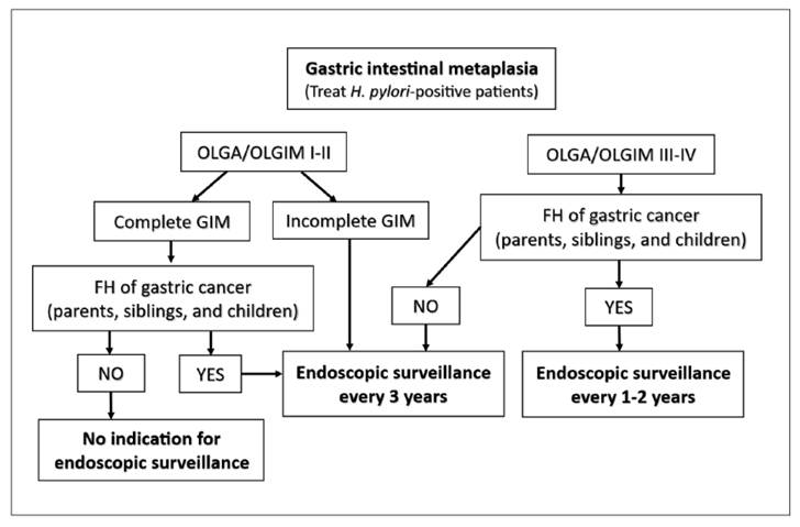
Patients with high-risk gastric atrophy and/or gastric intestinal metaplasia (GIM) (OLGA/OLGIM III or IV and/or incomplete GIM) are candidates for endoscopic surveillance every 3 years. This interval should be reduced to 1 to 2 years if they have a family history of gastric cancer.
Agreement: 96%
GRADE: 1B | Strength of recommendation: strong
Quality of evidence: moderate
The risk of malignant transformation, especially for intestinal-type gastric carcinoma (GC), progressively increases following the development of gastric atrophy (GA) and gastric intestinal metaplasia (GIM) in the context of chronic gastritis, regardless of GC incidence in the population studied. Meta-analyses and prospective studies with long follow-up periods have demonstrated that these precancerous conditions act as potential monitoring parameters for the secondary prevention of GC229,230, including the diffuse type231.
Chronic gastritis staging systems have been proposed based on the identification of GA and GIM (the OLGA system - Operative Link on Gastritis Assessment) or solely GIM (the OLGIM system - Operative Link on Gastric Intestinal Metaplasia). In both systems, it is mandatory to evaluate at least two biopsy specimens collected from each anatomical site (antrum and corpus), submitted in separate vials. The parameter initially observed is the extent of each process (GA and/or GIM) in each anatomical site separately. It is classified as mild when present in less than 1/3 of the sample, moderate between 1/3 and 2/3, and severe when over 2/3 of the mucosa is involved. For final staging (0 to IV), the extent across both anatomical sites is considered collectively, with more extensive processes classified into categories III and IV231,232.
Despite the good correlation between staging via both systems, GIM detection shows better intra- and inter-observer agreement compared to GA, favoring the use of OLGIM over OLGA233. Accurate histological investigation of GA requires examining the full thickness of the mucosa, from the superficial foveolae down to the muscularis mucosae, necessitating proper specimen orientation during paraffin embedding. GIM evaluation does not require such measures, facilitating verification under the microscope. Regardless, standardized endoscopic evaluation and biopsy collection protocols enhance the likelihood of detecting these conditions in the gastric mucosa234.
Long-term follow-up of patients with GA and GIM demonstrated an elevated probability of progression to dysplasia and malignant transformation in cases classified as OLGA/OLGIM III-IV, justifying endoscopic surveillance in these groups229,230. In the fourth edition of the Brazilian Consensus on H. pylori infection, endoscopies every 2 years were recommended for these patients 5. However, based on more recent findings87,90,235, a 3-year interval is now recommended for individuals with high-risk GIM, namely: a) OLGA/OLGIM III-IV; b) extensive GIM; c) incomplete-type GIM. Even if present in a single specimen, the incomplete pattern is considered an independent risk factor for GC in two recent meta-analyses86,236. If any of these patients has a family member with GC (parents, siblings, or children), it is suggested that the interval be reduced to 1 to 2 years, since family history also represents an independent risk factor. Additionally, if the patient has a family history of GC but their OLGA/OLGIM staging is I-II, endoscopy is suggested every 3 years237,238. A flowchart summarizing the indications and intervals for endoscopic surveillance in patients with GIM is depicted in Figure 1.

Flowchart for endoscopic surveillance in patients with gastric intestinal metaplasia. OLGA: operative Link on gastritis Assessment. OLGIM: operative link on gastric intestinal metaplasia. FH: family history. GIM: gastric intestinal metaplasia.
Statement 23
The use of an endoscopic classification of atrophy (Kimura-Takemoto) or gastric metaplasia (EGGIM - Endoscopic Grading of Gastric Intestinal Metaplasia) is recommended for the endoscopic staging of precancerous conditions and to stratify the risk of progression to gastric adenocarcinoma. Endoscopic classification must be integrated with histopathological staging.
Agreement: 94%
GRADE: 2B | Strength of recommendation: weak
Quality of evidence: moderate
Driven by technological advancements and the development of modern endoscopic equipment, upper GI endoscopy plays a fundamental role in visualizing, analyzing, and interpreting the findings that facilitate the visual staging and tissue sampling of the stages comprising the Correa cascade-from the initial infection to the development of intestinal-type gastric adenocarcinoma239. The staging of these phases, which include gastritis, atrophy, intestinal metaplasia, dysplasia, and cancer, is achievable through endoscopic examination, aided by increasingly precise visual documentation.
In the absence of H. pylori infection, the gastric mucosa presents typical features on white-light endoscopy, namely: a) antrum: flat, smooth mucosa with a light pink hue; b) corpus and fundus: more pronounced folding, especially along the greater curvature, more evident pink coloration, and the presence of a mucus pool; and c) regular arrangement of Collecting Venules (RAC): frequently observed in the gastric corpus. The finding of RAC exhibits a sensitivity of approximately 94% and specificity of 63% for excluding H. pylori infection240.
H. pylori infection can affect both the antrum and the corpus. When it involves both, the progressive risk of developing intestinal-type gastric adenocarcinoma is elevated239. When the infection involves the antrum, the mucosa displays erythema, a speckled pattern (spotty redness), and, among young patients, surface nodularity. With gastric corpus involvement, active inflammation can cause thickening and erythema of the folds, making them tortuous, with thick mucus accumulation. In the endoscopic staging of precancerous lesions, it is essential to consider all stages of the Correa cascade previously outlined241.
For histological evaluation, the Sydney System, published in 1991 and updated in 1996, standardizes biopsy collection (two from the antrum, one from the incisura angularis, and two from the gastric corpus), correlating etiology, topography, and morphology. This facilitates the assessment of chronic gastritis, location (antrum/corpus), and the presence of atrophy, metaplasia, and H. pylori54. To stratify the risk of progression to gastric cancer, the OLGA (Operative Link on Gastritis Assessment) system is used, quantifying the degree and extent of atrophy employing the Sydney System231.
The Kimura-Takemoto classification, introduced in 1969242, proposes a visual analysis of the gastric mucosa based initially on identifying the “endoscopic atrophic border”-normal mucosa has a pinkish color, whereas atrophic mucosa is pale, with a white/yellowish appearance. It proposes two main types: closed (C) Type - C1: atrophy restricted to the antrum; C2: atrophy extends along the lesser curvature up to the incisura angularis; and C3: extends along the lesser curvature of the corpus without reaching the cardia. Open (O) Type - O1: atrophy reaches the cardia; O2: extends to the anterior wall of the gastric corpus; and O3: reaches the greater curvature. Notably, the extension is parabolic, not linear. The higher the degree of open (O) atrophy, the greater the dysplasia risk. For validation, it is recommended to collect one biopsy from each area designated as C and O.
For the endoscopic evaluation of intestinal metaplasia: using white-light endoscopes with magnification and electronic chromoendoscopy, intestinal metaplasia can be identified as elevated areas (ridges), whitish or grayish in color, displaying a mosaic pattern and mild vascular irregularity. Another technique, in the absence of electronic chromoendoscopy, is the instillation of 1.5% acetic acid, producing an acetowhitening effect on metaplastic areas with high accuracy and specificity243.
The EGGIM (Endoscopic Grading of Gastric Intestinal Metaplasia) classification55,244: uses NBI (Narrow-Band Imaging) to map and score the stomach in three regions (antrum, incisura, and corpus), considering the extent of intestinal metaplasia (≥30% or <30% of the area)245.
The OLGIM system (Operative Link on Gastric Intestinal Metaplasia): inspired by OLGA, OLGIM evaluates the extent and distribution of intestinal metaplasia rather than atrophy232. Crucially, isolated atrophy or metaplasia in the antrum implies a low risk of neoplasia. However, if it involves the gastric corpus, the risk for intestinal adenocarcinoma progressively increases depending on the extent246,247.
Dysplasia can be detected visually within areas exhibiting altered coloration or focal irregularities, or randomly, through biopsies in areas of atrophy or metaplasia. When identified, the area should be demarcated using magnification techniques alongside electronic chromoendoscopy90,248. Chromoendoscopy with 1.5% acetic acid and 1.5% indigo carmine can also be utilized243,245,249.
The most recent guidelines from European Societies (MAPS III, 2025)87 recommend that every upper GI endoscopy with chromoendoscopy technology should include screening for early diagnosis of gastric cancer and risk stratification of precancerous conditions. To this end, the use of validated visual classifications such as Kimura-Takemoto (for atrophy) and EGGIM (for metaplasia) is recommended for assessing endoscopic gastric neoplastic risk.
Statement 24
In Brazil, population-based H. pylori screening for gastric cancer prevention is not recommended. In high-risk groups, screening and treatment of the infection are recommended.
Agreement: 100%
GRADE: 1A | Strength of recommendation: strong
Quality of evidence: high
H. pylori eradication is associated with a significant reduction in gastric cancer incidence and mortality115. Population-level testing and treatment of H. pylori (“screen-and-treat”) has been considered an important strategy for primary prevention. However, several questions remain regarding cost-effectiveness, bacterial resistance, and feasibility56,250. Over time, randomized clinical trials with long-term follow-up, combined with cost-effectiveness studies conducted in East Asia-an area with high H. pylori and gastric cancer prevalence-have strengthened the evidence base, prompting guidelines to become more assertive in recommending this strategy56.
Randomized clinical trials in China with up to 25 years of follow-up demonstrated that treating asymptomatic individuals reduced gastric cancer risk by 43% compared to placebo. The benefit was most pronounced among individuals without premalignant changes at the start of the study and with confirmed eradication251. In another randomized Chinese trial involving 180,284 individuals aged 25 to 54 years, with up to 11.8 years of follow-up, a significant reduction in gastric cancer incidence was observed in the group that eradicated The bacterium, with no serious adverse events occurring252. In the Matsu Islands population program in Taiwan, H. pylori prevalence fell from 64% to 15% over 14 years, accompanied by a 53% reduction in incidence and a 25% reduction in gastric cancer mortality compared to historical controls. No relevant changes in antimicrobial resistance rates or increased risks for other digestive tract neoplasms were identified253. Projections based on mathematical models show that H. pylori eradication therapy is cost-effective for reducing long-term gastric cancer incidence in regions where the neoplasm is highly prevalent254.
Population-based eradication is recommended in high-risk regions (annual gastric cancer incidence >20 cases per 100,000) with elevated H. pylori prevalence. In this context, a universal approach in adults is indicated, utilizing the urea breath test or stool antigen test, treatment tailored to local resistance profiles, universal confirmation of eradication, endoscopic surveillance for individuals with precancerous lesions, and continuous cost-effectiveness evaluation38,51,56,195.
In regions with an intermediate risk of gastric cancer (between 10 and 20 cases per 100,000), there is still no robust evidence to justify a population-level intervention strategy. It is recommended to target H. pylori screening and treatment to vulnerable groups, including first-degree relatives of patients with gastric cancer. Clinicians should consider screening and treating adult family members of patients positive for H. pylori and integrating national cancer screening programs-such as for colorectal cancer-with gastric cancer and H. pylori screening to optimize resources38,56,195,255.
In Brazil, the Brazilian National Cancer Institute (INCA) data256 for the 2023-2025 triennium show that gastric cancer remains among the cancers with the highest incidence among men, featuring a heterogeneous distribution. Regions such as the North (e.g., State of Pará) and Northeast (e.g., State of Ceará) present high H. pylori prevalence and intermediate cancer incidence. This requires individualized strategies, recently systematized for the American continent257.
Statement 25
Although no approved vaccines exist to prevent the acquisition of H. pylori infection or eliminate it in individuals with existing infections, the development of new technologies, such as multi-epitope vaccines, has shown promise in recent preclinical studies.
Agreement: 96%
GRADE: 2C | Strength of recommendation: weak
Quality of evidence: low
Human studies indicate that candidate vaccines against H. pylori can induce an immune response and are safe, but they have yet to demonstrate consistent efficacy in preventing or eradicating the infection258,259. The development of an effective and long-lasting H. pylori vaccine remains an unmet need due to the bacterium’s various immune evasion mechanisms and its profound genetic variability, which hinder the creation of a durable protective response in humans through any vaccine260-263.
The search for new adjuvants in H. pylori vaccines has expanded, yet many gaps remain in the successful development of an adjuvanted H. pylori vaccine260-261. Although research and development of H. pylori vaccines have achieved some progress over recent decades264,265, further research is necessary to overcome the immunological and technical challenges identified in clinical studies to date. The development of new vaccine technologies, such as multi-epitope-based vaccines against H. pylori, is currently under investigation in experimental trials, providing novel insights for advancing H. pylori vaccine development266-268.
Working Group 5: treatment And microbiota
Statement 26
During the initial patient evaluation, consider: (1) previously used antimicrobial regimens, avoiding the repeated use of clarithromycin and levofloxacin; (2) history of penicillin allergy to clarify questionable cases of true allergy; (3) caution regarding the use of clarithromycin and levofloxacin among patients with QT interval prolongation; (4) choice of antisecretory agent; (5) smoking cessation; and (6) encouragement of treatment adherence.
Agreement: 100%
GRADE: 1B | Strength of recommendation: strong
Quality of evidence: moderate
The success rate of conventional eradication therapies has declined over the past 2 decades due to rising bacterial resistance, insufficient acid suppression, and poor treatment adherence269,270. The therapeutic choice must consider allergies, medication availability and cost, and the assessment of local effectiveness data and bacterial resistance, adhering to the principle of avoiding unnecessary drug use. The targeted eradication benchmark is above 90%.
Firstly, a history of prior antimicrobial use, particularly macrolides and quinolones, increases the probability of bacterial resistance. A Chinese population-based study of patients treated with clarithromycin-based triple therapy found an increased risk of retreatment among individuals with a history of prior antibiotic use, especially macrolides, with the risk rising alongside longer antibiotic exposure time271. A Korean population study confirmed these findings, revealing significant prior macrolide use over the past decade among patients with clarithromycin-resistant H. pylori272. Carothers et al. found that levofloxacin resistance was associated with any prior quinolone use within the past 10 years and increased with a higher number of uses273.
Among the antibiotics used in eradication treatment, amoxicillin is one of the most effective, exhibiting low resistance rates274. However, approximately 10% of the general population report having a penicillin allergy, yet fewer than 5% of these possess a true allergy274,275. Patients at moderate and high risk encompass individuals with a history of urticaria, anaphylactic reactions, positive skin tests, recurrent reactions upon use, or hypersensitivity to multiple beta-lactam antibiotics275. Patients at low risk include individuals experiencing gastrointestinal symptoms, headaches, or itching without a rash, or those with a history of unknown or doubtful reactions that occurred more than 10 years ago. Skin sensitivity testing and/or referral to an allergist should be indicated for patients considered to be at moderate and high risk275.
The use of macrolides or quinolones can be associated with significant QT interval prolongation, a condition linked to potentially fatal ventricular arrhythmias. Consider obtaining an ECG for patients at high risk for QT interval prolongation (e.g., patients with cardiac conditions, individuals with a family history, older adults, patients with electrolyte imbalances, or individuals using antiarrhythmics, antipsychotics, or antidepressants)274.
The choice of an antisecretory agent and its proper dosing regimen are essential to optimize treatment efficacy. Effective acid suppression triggers bacterial proliferation, increases antibiotic concentration in the stomach, and prolongs its action on H. pylori269,276. A systematic review and meta-analysis showed that individuals with a rapid metabolizer phenotype for CYP2C19, the enzyme responsible for 80% to 90% of first-generation PPI metabolism, were associated with an 82% higher probability of therapeutic failure270. Jiang et al. suggest that eradication regimens based on P-CABs yielded the optimal eradication rates276.
Consistent data show that smoking reduces eradication rates. In a meta-analysis, patients who smoked exhibited an almost twofold higher probability of therapeutic failure than non-smokers277. Another meta-analysis confirmed these findings and showed an elevated risk of failure associated with a greater smoking intensity and current use during treatment278. Smoking alters CYP2C19 metabolism, increases acidity (which raises the proportion of bacteria in a non-replicative state), and reduces mucosal blood flow, ultimately lowering the efficacy and local availability of antibiotics278. In this meta-analysis, no higher risk of failure was observed in studies utilizing vonoprazan. The effect of alcohol consumption remains controversial: a meta-analysis involving 40 studies found no association between alcohol consumption and eradication, except among Asian individuals-a population known to feature altered alcohol metabolism due to aldehyde dehydrogenase deficiency279. Alcohol activates gastric acid secretion, reduces amoxicillin absorption, and strongly induces the CYP2C19 enzyme279.
Adherence is also a fundamental factor for treatment success. Common barriers include the complexity of eradication regimens, medication intolerance, and poor communication between clinicians and patients175. A meta-analysis showed increased eradication rates and treatment adherence when patient education programs were implemented280. Two Chinese studies showed significant improvements in adherence and eradication rates utilizing social media and text messages sent to cell phones281,282.
Statement 27
Proton pump inhibitors (PPIs) remain recommended, and the type and dose must be individualized to maximize acid suppression and the consequent therapeutic efficacy. When available, potassium-competitive acid blockers (P-CABs), such as vonoprazan at a dose of 20 mg twice daily, constitute a preferred alternative, especially in treatment regimens containing clarithromycin, amoxicillin, and levofloxacin.
Agreement: 100%
GRADE: 2B | Strength of recommendation: weak
Quality of evidence: moderate
The use of antisecretory agents, such as proton pump inhibitors (PPIs) and potassium-competitive acid blockers (P-CABs), is fundamental in the treatment of H. pylori infection, underpinned by pathophysiological and pharmacokinetic mechanisms. Elevating the intragastric pH (above 4-6) reduces the degradation of acid-sensitive antibiotics such as clarithromycin and amoxicillin, increasing their stability and concentration in the stomach, which enhances their action against H. pylori. This bacterium survives at pH 4-8 and multiplies at pH 6-8, a range where it becomes more vulnerable to antibiotics283.
PPIs, both first-generation (omeprazole, lansoprazole, dexlansoprazole, and pantoprazole) and second-generation (rabeprazole and esomeprazole), are essential adjuvants in H. pylori infection treatment. PPI potency is linked to eradication efficacy, treatment duration, and the choice of antibiotic regimen. It is universally agreed that high doses increase eradication rates, particularly in dual or triple regimens lasting 14 days, especially in populations with a high prevalence of rapid CYP2C19 metabolizers284,285. The equivalence among different PPIs has been evaluated to define dose potency (Table 2).
P-CABs, such as vonoprazan and tegoprazan, among others, have demonstrated faster, more potent, and more prolonged acid suppression compared to PPIs. They are stable in an acidic environment, can be used independently of meals, and do not require conversion to their active form, providing a more immediate effect. With a half-life of 6 to 9 h, in contrast to the 1 to 2 h of PPIs, P-CABs remain available longer, contributing to a more prolonged and effective control of gastric acidity. Furthermore, P-CABs are unaffected by genetic polymorphisms of CYP2C19, being metabolized primarily by CYP3A4/5161,286.
Direct head-to-head studies have analyzed the efficacy of vonoprazan compared to PPIs in clinical trials. A large trial conducted in the US and Europe compared vonoprazan with lansoprazole in triple therapy (vonoprazan or lansoprazole + amoxicillin + clarithromycin) and dual therapy (vonoprazan + amoxicillin) regimens for 14 days. The results demonstrated that both triple and dual therapies containing vonoprazan were superior to PPI-based triple therapy among patients with clarithromycin-resistant strains (eradication rates of 66% to 70% vs 32%) and among the general population (80.8% and 77.2% vs 68.5%)287. Additional randomized studies and meta-analyses, especially from Asia, reinforced the advantage of vonoprazan and tegoprazan in the context of clarithromycin resistance, although they also showed benefits for the general population288-290.
Currently, no head-to-head studies compare PPIs and P-CABs in combination with levofloxacin and rifabutin for second-line treatments. In bismuth-based quadruple therapy regimens, vonoprazan has demonstrated efficacy similar to PPIs, or with slightly higher eradication rates, and a comparable safety profile291-294. Vonoprazan has been commercially available since 2020, although it has not been officially approved specifically for anti-H. pylori therapy everywhere. In Brazil, its off-label use has been documented in a real-world study developed by the Brazilian Registry on H. pylori Management, utilized in 15% of first-line treatments and 26% of retreatments among the 2132 patients analyzed, with promising results9,295. The recommended doses for vonoprazan and tegoprazan are 20 mg and 50 mg, respectively, administered twice daily286,288,293,295.
Statement 28
For first-line treatment, the following are indicated: (1) Bismuth quadruple therapy (BQT) with bismuth, metronidazole and tetracycline combined with a PPI or P-CAB (when available) for 10 to 14 days; (2) Triple therapy with clarithromycin and amoxicillin for 14 days, optimized by adding bismuth and/or by substituting the PPI with a P-CAB (when available), twice daily; (3) If bismuth is unavailable, dual therapy with amoxicillin 3 to 4 g/days, divided into three or four doses, combined with a PPI (at a high dose, three or four times daily) or, preferably, a P-CAB (vonoprazan 20 mg, twice daily) when available, for 14 days; (4) Concomitant therapy with a PPI or P-CAB (when available) for 14 days may be an option.
Agreement: 100%
GRADE: 2C | Strength of recommendation: weak
Quality of evidence: low
Empirical triple therapy (TT) with a PPI has yielded unsatisfactory results292,297 due to growing bacterial resistance298. In the “Brazilian Registry on H. pylori Management”, it was used in 95% of the 1557 first-line treatments, with an eradication rate of 78%10. PPI-based TT has been discouraged, except where clarithromycin resistance is <15% or susceptibility is confirmed via prior testing299,300. Vonoprazan-based TT had higher eradication rates (85% to 89%) than PPI-based TT (71% to 79%) in an Asian meta-analysis and a US/European RCT (66% vs 32%) when treating resistant strains287,292.
A meta-analysis and two real-world studies demonstrated that the addition of bismuth to TT (PPI, bismuth-clarithromycin-amoxicillin) optimized first-line outcomes, increasing eradication rates to >90%, comparable to classic bismuth quadruple therapy, even for resistant strains297,299,301.
Two meta-analyses and one real-world study concluded that classic bismuth quadruple therapy (PPI, bismuth, tetracycline, and metronidazole)-administered separately for 14 days or in a single capsule for 10 days consistently yields eradication rates >90% as a first-line treatment, overcoming bacterial resistance297,301,302. Another meta-analysis found no differences in effectiveness when comparing 10 or 14 days of treatment for bismuth quadruple therapies, using either a PPI or a P-CAB as the antisecretory agent303. Both bismuth quadruple therapies (classic and amoxicillin-clarithromycin-bismuth) are used in Europe, Asia, and Latin America as preferred first-line options300,301,304, bypassing the need for susceptibility testing even in settings with high bacterial resistance302. Aside from frequently reported adverse effects301, bismuth salts are unavailable in many countries298 and, in Brazil, can still only be obtained exclusively through compounding pharmacies.
Dual therapy (DT) pairs high-dose amoxicillin with a PPI (high dose, three or four times daily)-known as high-dose dual therapy (HDDT)305-or vonoprazan (20 mg twice daily)306. The amoxicillin dosing is 750 to 1000 mg three or four times daily for 10 to 14 days306,307. Particularly for patients with obesity, 1 g of amoxicillin four times daily should be used308.
HDDT has been employed with favorable results across various regions worldwide, being studied most intensively in Asia. Two recent meta-analyses-one evaluating 14 randomized controlled studies involving 5121 patients-both showed intention-to-treat (ITT) eradication rates of 86% to 87%305,307. In Latin America, a Colombian randomized study309 achieved an 89% eradication rate by ITT, and a Chilean observational study involving 126 patients achieved a 92% eradication rate by ITT310. In Europe, a Portuguese observational study involving 100 patients treated with HDDT as a first-line or retreatment regimen observed a 96% eradication rate by ITT analysis311.
Vonoprazan-based DT has also shown highly favorable performance as a first-line option in Asia. Two recent meta-analyses, mostly involving randomized controlled studies, found ITT eradication rates between 86% and 89%306,312. The efficacy of HDDT305 and vonoprazan-based DT306,312 was comparable to that of bismuth quadruple therapies. Both HDDT313 and vonoprazan-based DT287,293 are largely unaffected by bacterial resistance. Advantages of DT include the use of only two drugs, a low rate of adverse events, and a lower cost compared to the other regimens considered305-307,312. Studies remain scarce on vonoprazan-based DT in the West. A US/European randomized controlled study showed 77.2% eradication with vonoprazan-based DT for 14 days287 and, interestingly, two large recent Taiwanese randomized studies showed eradication rates between 83% and 84% with this regimen288,314. A potential limitation regarding the widespread recommendation for DT is the report of emerging amoxicillin resistance >70% in some African countries and >15% in some Asian countries (Vietnam and Iran)298.
Concomitant therapy with clarithromycin, amoxicillin, and metronidazole for 14 days is an option where bismuth and susceptibility testing are unavailable, and where clarithromycin resistance is high300. This approach demonstrated eradication rates >90% in two large real-world registries315,316, with good performance against resistant strains299. However, dual resistance to clarithromycin and metronidazole can compromise effectiveness299. Concomitant therapy utilizes one or two potentially ineffective drugs, which may contribute to increased local resistance300.
Sequential and hybrid/reverse therapies also warrant consideration297, alongside a new kit containing omeprazole, rifabutin, and amoxicillin, approved for first-line treatment in the US317 (Table 3).
Statement 29
For second-line treatment, it is recommended not to repeat the same regimen used in the first line. The following are indicated: (1) bismuth quadruple therapy with metronidazole and tetracycline combined with a PPI or P-CAB (when available) for 10 to 14 days; (2) amoxicillin-levofloxacin-bismuth regimen combined with a PPI or P-CAB (when available) for 14 days; (3) if bismuth is unavailable, dual therapy with amoxicillin 3 to 4 g/day divided into three or four doses combined with a PPI (at a high dose, three or four times daily) or, preferably, a P-CAB (vonoprazan 20 mg, twice daily) when available, for 14 days.
Agreement: 100%
GRADE: 2C | Strength of recommendation: weak
Quality of evidence: low
Following the failure of clarithromycin-based triple therapy, the most recommended therapeutic option is levofloxacin-amoxicillin-PPI (LA-PPI) therapy for 10 to 14 days51. A European systematic review on retreatment found the 14 days therapy superior to the 10 days therapy (87% vs 72%)318. To overcome rising levofloxacin resistance, adding bismuth to the regimen has been proposed319. A systematic review with network meta-analysis evaluating different second-line therapies showed that levofloxacin-bismuth quadruple therapy for at least 10 days yielded optimal results, backed by moderate-level evidence, when compared to various 7 days triple therapies, with or without quinolones320. Another systematic review and meta-analysis included 16 second-line studies post-failure of non-bismuth quadruple regimens (sequential or concomitant) and evaluated the outcomes of different quinolone therapies321. In this review, only two studies evaluated the levofloxacin-bismuth quadruple regimen: one for 14 days using LA-PPI-bismuth (93% efficacy) and another using levofloxacin-tetracycline-PPI-bismuth for 10 days, achieving a 96% cure rate, though both featured very small sample sizes (69 and 24 patients, respectively)321. Data from the European Registry-though not direct comparisons-involving over 5000 patients on second-line therapies showed that the LA-PPI-bismuth regimen reached 88% effectiveness, whereas LA-PPI therapy reached 81%322. In Brazil, results from the Hp-BraReg analyzing 386 second-line retreatments showed that the LA-PPI regimen for 10 or 14 days had suboptimal results (57% and 73%, respectively)9. Conversely, adding bismuth to the 14 days LA-PPI regimen optimized results, achieving a 100% cure rate (95%CI 88-100%)323. High doses (>500 mg) and low doses (≤500 mg) of levofloxacin per day tend to show similar results323.
For retreatment, empirical BQT is recommended, particularly in areas with high quinolone resistance38,51. The growing rate of levofloxacin resistance and warnings from regulatory agencies led the North American consensus to recommend BQT as the preferred second-line therapy, reserving levofloxacin for susceptibility-guided therapy38,324. BQT can be prescribed with the drugs individually or in a single capsule (“three-in-one”) containing bismuth, metronidazole, and tetracycline325. An Asian meta-analysis of retreatment studies showed that BQT for 10 to 14 days had an eradication rate of 82% vs 76% for 7 days326. The ideal duration of this regimen has yet to be fully defined. Few studies have compared BQT regimens of 14 and 10 days. A recent meta-analysis demonstrated that the eradication rate of the 10 days BQT regimen was statistically similar to the 14 days one (RR 0.98; 95%CI 0.95-1.00)303. A single-center, non-randomized US-based first-line study found 14 days BQT eradication rates of 87%, whereas 10 days BQT reached 77%327.
BQT regimens can cause adverse effects in up to 40% of cases, generally mild and short-lived38. Adverse effects and treatment adherence are comparable between 10 and 14 days therapies303.
A European systematic review found a 75.6% success rate for BQT in the second line318. In that study, the single-capsule BQT regimen for 10 days, currently unavailable in Brazil, achieved higher rates (89%, 95%CI 87.4-91.0% by intention-to-treat) than those obtained by traditional BQT (76%, 95%CI 66.1-85.1%)318.
Studies on BQT indicate a trend towards better efficacy with tetracycline compared to doxycycline. In the first line, eradication was 67% to 70% with doxycycline and 85% with tetracycline328. In the Hp-EuReg, third-line rates were 65% versus 76%, trending statistically in favor of tetracycline328.
When the regimens suggested above are unavailable, dual therapy with high-dose amoxicillin can be considered an option, although few studies provide confirmatory results in the Western world. In a Portuguese study involving patients undergoing retreatment, dual therapy with high-dose amoxicillin (3 g/ day divided into four doses) and a PPI (esomeprazole 40 mg twice daily) for 14 days outperformed the 10 days BQT regimen (100% and 62.5%, respectively; P=.028 per protocol and by intention-to-treat)311. In the Brazilian Registry, out of 386 patients undergoing retreatment evaluated, 24 received dual therapy with amoxicillin (≥3 g/day in three or more doses) and vonoprazan 20 mg twice daily, with a 75% cure rate9. These results must be further evaluated in Western studies with larger sample sizes (Table 4).
Statement 30
For rescue regimens, it is recommended not to repeat previously used regimens containing clarithromycin or levofloxacin. The following are indicated: (1) bismuth quadruple therapy composed of bismuth, tetracycline, and metronidazole combined with a PPI or P-CAB (when available) for 10 to 14 days; (2) triple therapy with rifabutin and amoxicillin combined with a PPI or P-CAB (when available) for 14 days, especially when bismuth quadruple therapy is unavailable or has already been used; (3) in the absence of bismuth and rifabutin, dual therapy with amoxicillin 3 to 4 g/day divided into three or four doses combined with a PPI (at a high dose, three or four times daily) or, preferably, a P-CAB (vonoprazan 20 mg, twice daily) when available, for 14 days, though Western confirmatory studies are still lacking.
Agreement: 100%
GRADE: 2C | Strength of recommendation: weak
Quality of evidence: low
Following the failure of first- and second-line treatments to eradicate H. pylori, the choice of rescue regimen must account for previously used antibiotic regimens, allergies, local drug availability, costs, and, if possible, local and individual resistance profiles. The main options include:
1 - Bismuth quadruple therapy (see recommendations 28 and 29): this is the preferred option for patients with a history of prior treatment, particularly when the resistance profile is unknown or multiple past failures have occurred. Due to the low rates of bacterial resistance to these antibiotics in the regimen, it can even be repeated as a rescue treatment by utilizing a high dose of metronidazole. The lack of commercially available bismuth in standard retail pharmacies (though available via compounding pharmacies) remains a problem affecting regions where approximately one billion people live298.
2 - Rifabutin triple therapy: comprising a PPI, amoxicillin, and rifabutin for 10 to 14 days, this is an alternative when bismuth quadruple therapy is unavailable, was previously used, or in cases of tetracycline allergy. In a recent randomized study of patients who underwent two prior therapeutic regimens, rifabutin triple therapy demonstrated eradication rates of 89.0% by intention-to-treat (ITT) and 94.0% per protocol (PP)-results non-inferior to BQT, even in populations with high resistance to clarithromycin, levofloxacin, and metronidazole. Adherence was superior, and total and moderate/severe adverse events were significantly lower in the rifabutin group (26.4% and 14.3%, respectively) compared to BQT (54.4% and 28.6%)329. Three systematic reviews reinforce the efficacy of the rifabutin, amoxicillin, and PPI combination in rescue therapies, demonstrating average ITT eradication rates of 71% to 80%330-332. Initial studies with small sample sizes suggest that combining vonoprazan with rifabutin and amoxicillin is effective and safe, despite methodological limitations9,333,334. A pilot study showed that adding bismuth subcitrate to the rifabutin triple regimen significantly increased the eradication rate without relevant adverse effects335. The most heavily studied regimens consist of rifabutin 150 mg twice daily, amoxicillin 1 g twice daily, and a PPI, for 10 to 14 days, with 14 days being the recommended duration329-331. Rifabutin resistance remains rare, typically under 1%335-337. The regimen is well-tolerated, with fever, nausea, vomiting, and reversible bone marrow suppression being the most relevant side effects. A complete blood count is recommended during treatment331,337. Within the Brazilian context, rifabutin is not available on the Brazilian market, necessitating importation. However, it is listed on the Brazilian Unified Health System (SUS) National List of Essential Medicines (RENAME), though its use is strategically restricted to treating resistant mycobacterial infections338.
3 - Dual therapy with a PPI or vonoprazan and amoxicillin: recent Asian studies have shown variable results utilizing the combination of high-dose amoxicillin and a PPI as a rescue therapy. Randomized studies and Asian systematic reviews show ITT eradication rates between 73% and 94%307,339,340, with a low rate of adverse effects, even among patients who underwent multiple previous treatment failures. However, Western studies on this rescue regimen remain limited. In a German randomized non-inferiority trial involving patients with prior treatment failure and persistent infection by clarithromycin-resistant and metronidazole-susceptible strains, dual therapy using omeprazole 40 mg and amoxicillin 750 mg, both four times daily, showed eradication rates of 75.6%, non-inferior to bismuth quadruple therapy341. A real-world study coordinated by the European Registry on H. pylori Management analyzed 41 patients undergoing rescue treatment (2nd to 6th line) with amoxicillin 3.0 g/day and PPIs in varying formulations and doses for 14 days, achieving an ITT effectiveness of only 51%342.
While dual therapy with a PPI and amoxicillin can be considered a simplified alternative for treating H. pylori in rescue contexts, the most robust available evidence refers to the use of vonoprazan alongside high-dose amoxicillin. In a Chinese real-world retrospective study involving 186 patients with up to seven prior failures, the eradication rate using vonoprazan (20 to 40 mg/day) combined with amoxicillin (3.0 g/day) for 14 days was 92.5% (95%CI: 87.4-95.7%) with mild, self-limiting adverse effects (7.5%), regardless of the number of previous treatments or the regimens previously utilized343. A randomized multicenter study with patients who underwent multiple eradication attempts showed that dual therapy with high-dose vonoprazan and amoxicillin for 14 days was non-inferior to BQT, achieving an ITT eradication rate of 73.8% alongside good tolerability344. A recent meta-analysis confirms these findings in Asian populations, showing eradication rates above 90% even in rescue scenarios, paired with a favorable safety profile306. To date, robust Western studies specifically evaluating dual therapy with vonoprazan and amoxicillin in rescue treatments are absent. In Brazil, interim real-world data from the Hp-BraReg showed that 36 out of 186 patients who underwent three or more H. pylori eradication attempts were treated with vonoprazan and high-dose amoxicillin, yielding a modified ITT of 100%, alongside good tolerability and mild adverse effects9,295 (Table 5).
Statement 31
Triple therapy with PPI-amoxicillin-clarithromycin and PPI-amoxicillin-levofloxacin should ideally be employed following susceptibility testing, where available. After three therapeutic failures, susceptibility testing should be used when available.
Agreement: 100%
GRADE: 2B | Strength of recommendation: weak
Quality of evidence: moderate
The increase in H. pylori resistance rates demands new treatment strategies, including methods to evaluate antibiotic susceptibility298. However, controversies remain regarding the superiority of susceptibility-guided therapy over empirical therapy and whether it is cost-effective302,345.
Among phenotypic techniques, the agar dilution method is considered the gold standard. However, it is labor-intensive, time-consuming, and ill-suited for infections caused by a small number of strains, complicating routine use346. Alternative methods include disk diffusion and the E-test, the latter of which is increasingly recommended347,348.
Yet, phenotypic tests still face controversies. The European Committee on Antimicrobial Susceptibility Testing validated the E-test and standardized the minimum inhibitory concentration values for six antimicrobials (amoxicillin, clarithromycin, levofloxacin, metronidazole, rifampicin, and tetracycline)349. In the United States, the Clinical and Laboratory Standards Institute (CLSI) solely recommends the agar dilution test and has only established resistance criteria for clarithromycin350. In Brazil, no governmental body has established resistance criteria for phenotypic tests. Beyond the methodological hurdles, phenotypic tests suffer from limited availability, even in developed countries298.
As alternatives to culture, genotypic tests employ molecular methods to identify point mutations in H. pylori associated with resistance (polymerase chain reaction-PCR or next-generation sequencing)351. These methods are faster and can utilize gastric biopsies, gastric aspirates, or stool samples, although stool sampling remains unvalidated298. Fecal genotypic testing is under development and could be employed as a non-invasive molecular method. A meta-analysis of 26 studies found that despite a specificity of 96%, sensitivity was only 71%352. The primary limitation of the stool method appears to be the presence of PCR inhibitors, leading to an increase in false-negative results. Genotypic methods are not yet commercially available in many countries, including Brazil298.
Phenotypic and genotypic methods have been compared and show good correlation, particularly regarding clarithromycin and levofloxacin resistance-the drugs that most impact therapeutic failure352,353.
Clarithromycin resistance is typically driven by point mutations in the 23S rRNA gene, the most important being A2143G, A2142G, and A2142C354. In a meta-analysis comparing guided versus empirical therapy, results for clarithromycin triple therapy favored guided therapy (87% vs 78%), especially in areas with clarithromycin resistance >20%302.
Mutations in the gyrA and gyrB genes confer levofloxacin resistance, detectable via genotypic methods355. In a meta-analysis, levofloxacin-based triple regimens showed 81% efficacy against susceptible strains and 36% against mutants323. Rifabutin resistance involves the rpoB gene; amoxicillin resistance stems from mutations in PBP1A; and tetracycline resistance involves 16S rRNA. Resistance levels for rifabutin, amoxicillin, and tetracycline remain low and stable355. Metronidazole resistance usually stems from rdxA mutations, but doses >1.5 g/day within a 14 days bismuth quadruple therapy are capable of overcoming it356. In a meta-analysis, bismuth quadruple therapy showed similar efficacy whether empirical or guided, and it was minimally affected by prior antibiotic use, largely due to the rarity of tetracycline and amoxicillin resistance and the limited impact of metronidazole resistance357.
Among patients with a history of prior exposure to various antibiotics, particularly macrolides and quinolones for other infections, susceptibility testing may be considered, given the well-established risk of higher resistance358.
Beyond bacterial resistance, other factors implicated in therapeutic failure include treatment adherence, inadequate acid suppression, smoking, the expression of efflux pumps, and biofilm formation175. Strategies to overcome biofilm formation have been attempted, such as using N-acetylcysteine (NAC)359, though a meta-analysis showed no superiority when NAC was added to the standard therapeutic regimen360. In practice, overcoming the biofilm is optimally achieved by employing bismuth, maximizing optimized acid suppression, ensuring treatment adherence, and avoiding the repetition of antimicrobial regimens prone to higher resistance, such as clarithromycin and quinolones361.
Statement 32
For patients with a penicillin allergy, the recommended first-line treatments are: a) Bismuth quadruple therapy with metronidazole and tetracycline combined with a PPI or P-CAB (when available) for 10 to 14 days; b) Dual therapy with tetracycline and a P-CAB (when available) for 14 days. For second-line treatment, one of the previously unused options can be employed, or alternatively, a 14 days triple therapy using a PPI, quinolone, and clarithromycin.
Agreement: 100%
GRADE: 2B | Strength of recommendation: weak
Quality of evidence: moderate
Amoxicillin is a component of the most effective antimicrobial regimens for H. pylori eradication. As such, penicillin allergy is a major clinical challenge.
The global prevalence of penicillin allergy is 9.4% (95% CI 8.4% to 10.4%), with Latin American countries underrepresented in the research362. Confirmatory tests for true immune-mediated hypersensitivity-such as skin tests and specific IgE testing-are rarely performed and offer limited diagnostic accuracy363,364. For patients with a history of penicillin allergy, treatment options must exclude amoxicillin and other penicillins.
Another limitation is the rapid global rise in resistance to clarithromycin, fluoroquinolones, and nitroimidazoles298, already demonstrated across the five geographic regions of Brazil7.
For first-line treatment among patients with a penicillin allergy, classic 14 days bismuth quadruple therapy is the most consistent recommendation, demonstrating eradication rates around 89% to 90%51,365-367 and is considered adequate in regions with high clarithromycin and metronidazole resistance51. Another novel option investigated the comparison of the rapid and potent acid inhibition of vonoprazan versus rabeprazole within a modified quadruple therapy containing bismuth, metronidazole, and doxycycline. The outcome was non-inferiority, with ITT eradication rates of 90.4% versus 71.1%, respectively293. Just as dual therapy with amoxicillin and a P-CAB has been used for patients without an amoxicillin allergy history, the first randomized clinical trial in 300 patients with a penicillin allergy utilized vonoprazan and tetracycline (VT) for 14 days. The ITT eradication rate for VT versus classic BQT was 92% vs 89.3%, respectively, demonstrating non-inferiority alongside fewer adverse events368.
For second-line treatment among patients with a penicillin allergy, after reviewing the initial antibiotic regimen used, the options are classic bismuth quadruple therapy and triple therapy containing a PPI, clarithromycin, and a quinolone (e.g., levofloxacin), demonstrating eradication rates ranging from 64% in a multicenter prospective study369 to 80% to 100% in reviews of retrospective studies365,366.
Following a second failure, in rescue treatments, triple therapy with clarithromycin and metronidazole is indicated, as is the possibility of repeating classic triple therapy using high-dose metronidazole (1.5 to 1.6 g/day) for 14 days370.
Amoxicillin-free empirical treatments have used various antibiotic regimens, especially in regions with high antibiotic resistance and limited medication availability, relying on agents such as cefuroxime, rifabutin, and furazolidone365,366,371,372.
Among cases of refractoriness, whenever possible, the penicillin allergy should be confirmed through testing (skin tests and specific IgE)362,363. Clinicians must ascertain which antibiotics were previously used and, whenever possible, perform antimicrobial susceptibility testing or cultures to guide antibiotic selection366 (Table 6).
Statement 33
Specific probiotic strains may exert a beneficial effect during anti-H. pylori treatment by increasing eradication rates and reducing side effects caused by antibiotics. However, further studies are needed to better define the specific probiotic strains, required dosage, ideal timing, and duration of supplementation.
Agreement: 91%
GRADE: 2B | Strength of recommendation: weak
Quality of evidence: moderate
Various therapeutic regimens, relying on the combination of antibiotics and antisecretory drugs, are conventionally recommended for H. pylori treatment; however, in recent years, a significant reduction in bacterial eradication rates has been observed5,51. Consequently, recent research has explored alternative options, and several options have shown significant promise, such as adding probiotics to traditional anti-H. pylori regimens38,373.
Experimental studies have demonstrated that certain probiotics can modulate the gastric microbiota, secrete antibacterial substances, compete with H. pylori for adhesion to the gastric mucosa, and stimulate immune responses and mucin production374,375. Despite these findings, studies demonstrated that probiotics alone offer minimal eradication efficacy (averaging 14%) and are not indicated as monotherapy; their primary role is as an adjunct to classic antibiotic treatment376.
Several randomized clinical trials, meta-analyses, and systematic reviews have consistently demonstrated that adding probiotics to different anti-H. pylori regimens improves eradication rates (by 8% to 16%) and significantly reduces the incidence of side effects such as diarrhea, nausea, and abdominal pain377-381. Across multiple clinical studies, the reduction in adverse effects ranged from 30% to 59%378. These results are consistent across different populations and geographical regions378,379.
Favorable results were described when using specific strains (either single- or multi-strain formulations)377-381. Some authors noted more robust effects, both in therapeutic efficacy and side-effect reduction, with multi-strain probiotic regimens, especially those containing Lactobacillus, Bifidobacterium, and Saccharomyces377-381. Some evidence suggests that higher doses and a longer duration of probiotic supplementation (>2 weeks) yield better outcomes375,379.
More recent studies have found even more favorable results, concluding that the adjunctive use of certain probiotic strains can enhance efficacy and improve tolerability across different H. pylori eradication regimens379-381. Notable among these is the study by Casas Deza et al., which presented data from the European Registry on H. pylori Management and provided the largest cohort analyzed to date (2013 to 2021) regarding the addition of probiotics to eradication regimens in clinical practice379. The authors highlighted the genera and strains linked to the highest therapeutic efficacy and identified which eradication regimens benefit most from the supplementation.
Despite these recent, highly favorable data, the topic remains controversial due to the vast heterogeneity in the strains and doses utilized, the timing (before and/or during and/or after antibiotics), and the duration of supplementation5,38,51. We conclude that probiotic supplementation is a plausible and highly promising adjunctive strategy. Specific probiotic strains can confer a beneficial effect during anti-H. pylori treatment, driving higher eradication rates and lowering antibiotic-induced side effects. However, it remains necessary to identify the ideal genera/strains and combinations to justify a strong recommendation for probiotic supplementation in H. pylori treatment51,38,375.
Statement 34
H. pylori eradication partially restores gastric microbiota diversity. However, the antibiotic therapy used for this purpose can cause transient or, rarely, persistent gut dysbiosis.
Agreement: 91%
GRADE: 2B | Strength of recommendation: weak
Quality of evidence: moderate
H. pylori infection undeniably modifies the gastric microbiota. This modification, however, is directly related to the specific bacterial strain, its aggressiveness, the characteristics of the host’s stomach, and the inflammatory response.
Classically, initial H. pylori infection drives a gastrin increase. This can induce hydrochloric acid hypersecretion among patients with a high parietal cell mass and low production of interleukin-1 beta and TNF-alpha (these cytokines are potent antisecretory agents). This patient group will experience a reduction in gastric microbiota diversity, driven by the strong bactericidal effect of HCl. Conversely, among patients with a reduced number of oxyntic cells and elevated production of the aforementioned cytokines, acid secretion undergoes little change and may even decrease over time due to eventual antral and fundic atrophy. In this scenario, the elevated pH promotes an increase in the number and diversity of the gastric microbiota. Gastric bacterial overgrowth is a known risk factor for neoplasm development382,383.
The presence of hypochlorhydria also ultimately impacts the intestinal microbiota, potentially fostering small intestinal bacterial overgrowth and its associated complications384.
H. pylori eradication can alter gastric secretory capacity due to the reasons outlined above, exacerbated by the antimicrobial effect of the medications used in eradication therapy. This acutely reduces bacterial diversity and richness, leading to changes in the composition of microbial communities, including a decrease in Enterococcus and an increase in Lactobacillus, Bifidobacterium, and Bacteroides385. These alterations tend to be most pronounced in the first few weeks post-treatment, followed by a gradual recovery of microbial diversity and composition over months, generally returning to baseline patterns within 1 year386,387. Reports indicate minor changes in some genera that may persist in isolated cases388. The concomitant use of specific probiotic strains or combinations can attenuate antibiotic-induced dysbiosis, promoting a faster recovery of the intestinal microbiota385,389.
Beyond bacterial alterations, evidence suggests that H. pylori eradication also affects the intestinal virome, reducing viral diversity and altering phage-bacterium interactions-effects that may persist for up to 6 months, especially among patients who underwent multiple courses of antibiotics390,391.
ACKNOWLEDGEMENTS
The authors would like to thank the employees of the Brazilian Federation of Gastroenterology Martha Sylvio, Caio Cesar C. Maria, Adriana Januário and Karine Boaventura for their invaluable technical and digital support during all stages of the Consensus. The authors also thank Dr. José Vitor Zir for logistical support in Bento Gonçalves, RS.
REFERENCES
- 1 Li Y, Choi H, Leung K, et al. Global prevalence of Helicobacter pylori infection between 1980 and 2022: a systematic review and meta-analysis. Lancet Gastroenterol Hepatol. 2023;8:553-64.
- 2 Coelho LGV, Barros CAS, Lima DCA, et al. Consenso Nacional sobre H. pylori e afecções associadas. GED Gastroenterol Endosc Dig. 1996;15:53-8.
- 3 Coelho LG, Zaterka S e Representantes indicados pela Federação Brasileira de Gastroenterologia e Núcleo Brasileiro para o Estudo do Helicobacter II Consenso Brasileiro sobre H. pylori. Arq Gastroenterol. 2005;42:128-32.
- 4 Coelho LG, Maguilnik I, Zaterka S, et al. 3rd Brazilian Consensus on H. pylori. Arq Gastroenterol. 2013;50:81-96.
- 5 Coelho LGV, Marinho JR, Genta R, et al. IVth Brazilian Consensus Conference on Helicobacter Pylori Infection. Arq Gastroenterol. 2018;55:97-121.
- 6 Vita NA, Anderson SM, LaFleur MD, et al. Targeting Helicobacter pylori for antibacterial drug discovery with novel therapeutics. Curr Opin Microbiol. 2022;70:102203.
- 7 Sanches BS, Martins GM, Lima K, et al. Detection of Helicobacter pylori resistance to clarithromycin and fluoroquinolones in Brazil: A national survey. World J Gastroenterol. 2016;22:7587-94.
- 8 Benigno TGDS, Ribeiro Junior HL, Azevedo OGR, et al. Clarithromycin-resistant H. pylori primary strains and virulence genotypes in the Northeastern region of Brazil. Rev Inst Med Trop Sao Paulo. 2022;64:e47.
- 9 Sanches BSF, Nyssen OP, Chaves SR, et al. Empirical Rescue Eradication Therapy for Helicobacter pylori Infection in Second and Subsequent Treatment Lines: Experience From 500 Cases of the Brazilian Registry on H. pylori Management (Hp-BrazilReg). Helicobacter. 2025;30:e70077.
- 10 Chaves SR, Nyssen OP, Veloso JCS, et al. First-line empirical eradication therapy for Helicobacter pylori infection: experience from 1,500 cases of the Brazilian Registry on H. pylori Management (Hp-BrazilReg/WorldHpReg). Gastroenterology. 2026;170:S-2664.
- 11 Sugano K, Tack J, Kuipers EJ, et al. Kyoto global consensus report on Helicobacter pylori gastritis. Gut. 2015;64:1353-67.
- 12 Barrett D, Heale R. What are Delphi studies? Evid Based Nurs. 2020;23:68-9.
- 13 Guyatt GH, Oxman AD, Vist GE, et al. GRADE: an emerging consensus on rating quality of evidence and strength of recommendations. BMJ. 2008;336:924-6.
- 14 Guyatt GH, Oxman AD, Kunz R, et al. Going from evidence to recommendations. BMJ. 2008;336:1049-51.
- 15 Sultan S, Falck-Ytter Y, Inadomi JM. The AGA Institute process for developing clinical practice guidelines part one: grading the evidence. Clin Gastroenterol Hepatol. 2013;11:329-32.
- 16 Chen YC, Malfertheiner P, Yu HT, et al. Global Prevalence of Helicobacter pylori Infection and Incidence of Gastric Cancer Between 1980 and 2022. Gastroenterology. 2024;166:605-19.
- 17 Katelaris P, Hunt R, Bazzoli F, et al. Helicobacter pylori World Gastroenterology Organization Global Guideline. J Clin Gastroenterol. 2023;57:111-26.
- 18 Coelho LGV, Chinzon D, Ribeiro LT, et al. Prevalence of Helicobacter pylori infection among gastroenterologists and gastroendoscopists in Brazil. Arq Gastroenterol. 2025;62:e25019.
- 19 Bezerra TMO, Xavier KVM, Luz ACO, et al. Prevalence of cagA, cagM, vacA and oipA genes in isolates of Helicobacter pylori obtained from hospital patients in Northeast Brazil. Braz J Microbiol. 2024;55:2631-41.
- 20 Carlos ABM, Costa VE, Kobayasi R, et al. Prevalence of Helicobacter pylori infection among asymptomatic children in southeastern Brazil: a cross-sectional study. Sao Paulo Med J. 2022;140:719-22.
- 21 Weyermann M, Rothenbacher D, Brenner H. Acquisition of Helicobacter pylori infection in early childhood: independent contributions of infected mothers, fathers, and siblings. Am J Gastroenterol. 2009;104:182-9.
- 22 Kivi M, Tindberg Y, Sörberg M, et al. Concordance of Helicobacter pylori strains within families. J Clin Microbiol. 2003;41:5604-08.
- 23 Georgopoulos SD, Mentis AF, Spiliadis CA, et al. Helicobacter pylori infection in spouses of patients with duodenal ulcers and comparison of ribosomal RNA gene patterns. Gut. 1996;39:634-8.
- 24 Luman W, Zhao Y, Ng HS, et al. Helicobacter pylori infection is unlikely to be transmitted between partners: evidence from genotypic study in partners of infected patients. Eur J Gastroenterol Hepatol. 2002;14:521-8.
- 25 Ding SZ, Du YQ, Lu H, et al. Chinese Consensus Report on Family-Based Helicobacter pylori Infection Control and Management (2021 Edition). Gut. 2022;71:238-53.
- 26 Zhou XZ, Lyu NH, Zhu HY, et al. Large-scale, national, family-based epidemiological study on Helicobacter pylori infection in China: the time to change practice for related disease prevention. Gut. 2023;72:855-69.
- 27 Kayali S, Manfredi M, Gaiani F, et al. Helicobacter pylori, transmission routes and recurrence of infection: state of the art. Acta Biomed. 2018;89:72-6.
- 28 Huang AF, He C, Sheng JW, et al. The epidemiological study of family-based Helicobacter pylori screening and its benefits: a cross-sectional study. Sci Rep. 2025;15:5553.
- 29 Alfaray RI, Saruuljavkhlan B, Ansari S, et al. Review: Epidemiology of Helicobacter pylori infection. Microb Health Dis. 2022;4:e733.
- 30 Malfertheiner P, Camargo MC, El-Omar E, et al. Helicobacter pylori infection. Nat Rev Dis Primers. 2023;9:19.
- 31 Duan M, Li Y, Liu J, et al. Transmission routes and patterns of Helicobacter pylori. Helicobacter. 2023;28:e12945.
- 32 Liu M, Gao H, Miao J, et al. Helicobacter pylori infection in humans and phytotherapy, probiotics, and emerging therapeutic interventions: a review. Front Microbiol. 2024;14:1330029.
- 33 Elbehiry A, Abalkhail A, Anajirih N, et al. Helicobacter pylori: Routes of Infection, Antimicrobial Resistance, and Alternative Therapies as a Means to Develop Infection Control. Diseases. 2024;12:311.
- 34 Almashhadany DA, Zainel MA, AbdulRahman TT. Review of foodborne helicobacteriosis. Ital J Food Saf. 2024;13:12176.
- 35 Wang X, Xu K, Zhang T, et al. Is Helicobacter pylori infection associated with animal contact? A systematic review and meta-analysis. J Public Health (Oxf). 2025;47:e298-e308.
- 36 Nkuize M, De Wit S, Demetter P, et al. Helicobacter pylori and Human Immunodeficiency Virus co-infection: potential implications for future gastric cancer risk. Microorganisms. 2023;11:887.
- 37 Maleki N, Mohammadzadeh A, Mardaneh J, et al. Helicobacter pylori infection and association with chronic diseases: A focus on cardiovascular disease, MASLD, and type 2 diabetes. Metabol Open. 2025;27:100385.
- 38 Chey WD, Howden CW, Moss SF, et al. ACG Clinical Guideline: Treatment of Helicobacter pylori infection. Am J Gastroenterol. 2024;119:1730-53.
- 39 Kheyre H, Morais S, Ferro A, et al. The occupational risk of Helicobacter pylori infection: a systematic review. Int Arch Occup Environ Health. 2018;91:657-4.
- 40 Makristathis A, Pasching E, Schütze K, et al. Detection of Helicobacter pylori in stool specimens by PCR and antigen enzyme immunoassay. J Clin Microbiol. 1998;36:2772-4.
- 41 Banatvala N, Lopez CR, Owen R, et al. Helicobacter pylori in dental plaque. Lancet. 1993;341:380.
- 42 Dowsett SA, Kowolik MJ. Oral Helicobacter pylori: can we stomach it? Crit Rev Oral Biol Med. 2003;14:226-33.
- 43 Vaira D, Vakil N. Blood, urine, stool, breath, money, and Helicobacter pylori. Gut. 2001;48:287-9.
- 44 Xu L, Li XT, Ur-Rahman I, et al. Global H. pylori recurrence, recrudescence, and re-infection status after successful eradication in pediatric patients: a systematic review and meta-analysis. J Gastroenterol. 2024;59:668-81.
- 45 Ierardi E, Losurdo G, Mileti A, et al. The Puzzle of Coccoid Forms of Helicobacter pylori: Beyond Basic Science. Antibiotics (Basel). 2020;9:293.
- 46 Zhao H, Yan P, Zhang N, et al. The recurrence rate of Helicobacter pylori in recent 10 years: A systematic review and meta-analysis. Helicobacter. 2021;26:e12852.
- 47 Coelho LG, Passos MCF, Cunha FAF, et al. H. pylori reinfection rates and serological evaluation in a 6.4 year follow-up study from a developing country. Gut. 1999;45:110.
- 48 Eisig JN, Zaterka S, Silva FM, et al. Helicobacter pylori recurrence in patients with duodenal ulcer: Clinical, endoscopic, histologic, and genotypic aspects. A 10-year Brazilian series. Helicobacter. 2006;11:431-5.
- 49 Silva FM, Navarro-Rodriguez T, Barbuti RC, et al. Helicobacter pylori reinfection in Brazilian patients with peptic ulcer disease: a 5-year follow-up. Helicobacter. 2010;15:46-52.
- 50 Rugge M, Genta RM, Malfertheiner P, et al. RE.GA.IN.: the Real-world Gastritis Initiative-updating the updates. Gut. 2024;73:407-41.
- 51 Malfertheiner P, Megraud F, Rokkas T, et al. Management of Helicobacter pylori infection: the Maastricht VI/Florence consensus report. Gut. 2022;327745.
- 52 Conteduca V, Sansonno D, Lauletta G, et al. H. pylori infection and gastric cancer: state of the art (review). Int J Oncol. 2013;42:5-18.
- 53 Maruta LM, Furukawa A, Seidler HBK, et al. Validation of the ABC Method for Gastric Cancer Risk Stratification Across Helicobacter pylori Infections With Diverse CagA Status and Subtypes in Brazil. Cancer Med. 2025;14:e71016.
- 54 Dixon MF, Genta RM, Yardley JH, et al. Classification and grading of gastritis. The updated Sydney System. International Workshop on the Histopathology of Gastritis, Houston 1994. Am J Surg Pathol. 1996;20:1161-81.
- 55 Pimentel-Nunes P, Libânio D, Marcos-Pinto R, et al. Management of epithelial precancerous conditions and lesions in the stomach (MAPS II): European Society of Gastrointestinal Endoscopy (ESGE), European Helicobacter and Microbiota Study Group (EHMSG), European Society of Pathology (ESP), and Sociedade Portuguesa de EndoscopiaDigestiva (SPED) guideline update 2019. Endoscopy. 2019;51:365-88.
- 56 Park JY, editor. Population-based Helicobacter pylori screen-and-treat strategies for gastric cancer prevention: guidance on implementation. Lyon, France: International Agency for Research on Cancer;2025.
- 57 Xia B, Xia HH, Ma CW, et al. Trends in the prevalence of peptic ulcer disease and Helicobacter pylori infection in family physician-referred uninvestigated dyspeptic patients in Hong Kong. Aliment Pharmacol Ther. 2005;22:243-9.
- 58 Azhari H, King JA, Coward S, et al. The Global Incidence of Peptic Ulcer Disease Is Decreasing Since the Turn of the 21st Century: A Study of the Organisation for Economic Co-Operation and Development (OECD). Am J Gastroenterol. 2022;117:1419-27.
- 59 Xie X, Ren K, Zhou Z, et al. The global, regional and national burden of peptic ulcer disease from 1990 to 2019: a population-based study. BMC Gastroenterol. 2022;22:58.
- 60 Rosenstock S, Jørgensen T, Bonnevie O, et al. Risk factors for peptic ulcer disease: a population based prospective cohort study comprising 2416 Danish adults. Gut. 2003;52:186-93.
- 61 Schöttker B, Adamu MA, Weck MN, et al. Helicobacter pylori infection is strongly associated with gastric and duodenal ulcers in a large prospective study. Clin Gastroenterol Hepatol . 2012;10:487-93.
- 62 Choi YJ, Kim TJ, Bang CS, et al. Changing trends and characteristics of peptic ulcer disease: A multicenter study from 2010 to 2019 in Korea. World J Gastroenterol . 2023;29:5882-93.
- 63 El-Omar EM, Penman ID, Ardill JE, et al. Helicobacter pylori infection and abnormalities of acid secretion in patients with duodenal ulcer disease. Gastroenterology. 1995;109:681-91.
- 64 Coelho LGV, Arantes VN, Guedes LR. Úlcera péptica. In: Averbach M, et al, eds. Tratado Ilustrado de Endoscopia Digestiva. Rio de Janeiro: Thieme Revinter; 2018:295-310.
- 65 Vakil N. Peptic Ulcer Disease: A Review. JAMA. 2024;332:1832-42.
- 66 Miwa H, Sakaki N, Sugano K, et al. Recurrent peptic ulcers in patients following successful Helicobacter pylori eradication: a multicenter study of 4940 patients. Helicobacter. 2004;9:9-16.
- 67 Van der Hulst RW, Rau’s EA, Köycü B, et al. Prevention of ulcer recurrence after eradication of Helicobacter pylori: a prospective long-term follow-up study. Gastroenterology. 1997;113:1082-86.
- 68 Ford AC, Gurusamy KS, Delaney B, et al. Eradication therapy for peptic ulcer disease in Helicobacter pylori-positive people. Cochrane Database Syst Rev. 2016;4:CD003840.
- 69 Ford AC, Marwaha A, Sood R, et al. Global prevalence of, and risk factors for uninvestigated dyspepsia: a meta-analysis. Gut. 2015;64:1049-57.
- 70 Stanghellini V, Chan FKL, Hasler WL, et al. Gastroduodenal Disorders. Gastroenterology. 2016;150:1380-92.
- 71 Gisbert JP, Calvet X. Helicobacter Pylori “Test-and-Treat” Strategy for Management of Dyspepsia: A Comprehensive Review. Clin Transl Gastroenterol. 2013;4:e32.
- 72 Delaney BC, Qume M, Moayyedi P, et al. Helicobacter pylori test and treat versus proton pump inhibitor in initial management of dyspepsia in primary care: multicentre randomised controlled trial (MRC-CUBE trial). BMJ. 2008;336:651-4.
- 73 Koletzko L, Macke L, Schulz C, et al. Helicobacter pylori eradication in dyspepsia: New evidence for symptomatic benefit. Best Pract Res Clin Gastroenterol. 2019;40-41:101637.
- 74 Delaney B, Ford AC, Forman D, et al. WITHDRAWN: Initial management strategies for dyspepsia. Cochrane Database Syst Rev . 2009:CD001961.
- 75 Chey WD, Leontiadis GI, Howden CW, et al. ACG clinical guideline: treatment of Helicobacter pylori Infection. Am J Gastroenterol. 2017;112:212-38.
- 76 Beresniak A, Malfertheiner P, Franceschi F, et al. Helicobacter pylori “Test-and-Treat” strategy with urea breath test: a cost-effective strategy for the management of dyspepsia and the prevention of ulcer and gastric cancer in Spain-Results of the Hp-Breath initiative. Helicobacter. 2020;25:e12693.
- 77 Eusebi LH, Black CJ, Howden CW, et al. Effectiveness of management strategies for uninvestigated dyspepsia: systematic review and network meta-analysis. BMJ. 2019;367:l6483.
- 78 Ford AC, Marwaha A, Sood R, et al. Global prevalence of, and risk factors for, uninvestigated dyspepsia: a meta-analysis. Gut. 2015;64:1049-57.
- 79 Ford A, Tsipotis E, Yuan Y, et al. Efficacy of Helicobacter pylori eradication therapy for functional dyspepsia: updated systematic review and meta-analysis. Gut. 2022;71:1967-75.
- 80 Du L, Chen B, Kim J, et al. Helicobacter pylori eradication therapy for functional dyspepsia: Systematic review and meta-analysis. World J Gastroenterol . 2016;22:3486-95.
- 81 Kang S, Park B, Shin C. Helicobacter pylori Eradication Therapy for Functional Dyspepsia: A Meta-Analysis by Region and H. pylori Prevalence. J Clin Med. 2019;8:504.
- 82 Zhao B, Zhao J, Cheng W, et al. Efficacy of Helicobacter pylori Eradication Therapy on Functional Dyspepsia: A Meta-Analysis of Randomized Controlled Studies With 12-Month Follow-up. J Clin Gastroenterol. 2014;48:241-7.
- 83 Mazzoleni L, Sander G, Francesconi C, et al. Helicobacter pylori eradication in functional dyspepsia: HEROES trial. Arch Intern Med. 2011;171:1929-36.
- 84 Moayyedi P, Lacy BE, Andrews CN, et al. ACG and CAG clinical guideline: management of dyspepsia. Am J Gastroenterol. 2017;112:988-1013.
- 85 Shao L, Li P, Ye J, et al. Risk of gastric cancer among patients with gastric intestinal metaplasia. Int J Cancer. 2018;143:1671-7.
- 86 Du S, Yang Y, Fang S, et al. Gastric Cancer Risk of Intestinal Metaplasia Subtypes: A Systematic Review and Meta-Analysis of Cohort Studies. Clin Transl Gastroenterol. 2021;12:e00402.
- 87 Dinis-Ribeiro M, Libânio D, Uchima H, et al. Management of epithelial precancerous conditions and early neoplasia of the stomach (MAPS III): European Society of Gastrointestinal Endoscopy (ESGE), European Helicobacter and Microbiota Study Group (EHMSG) and European Society of Pathology (ESP) Guideline update 2025. Endoscopy. 2025;57:504-54.
- 88 Cho YS, Chung IK, Jung Y, et al. Risk stratification of patients with gastric lesions indefinite for dysplasia. Korean J Intern Med. 2021;36:1074-82.
- 89 Yim K, Shin JH, Yoo J. Novel pathologic factors for risk stratification of gastric “indefinite for dysplasia” lesions. Gastroenterol Res Pract. 2020;2020:9460681.
- 90 Dinis-Ribeiro M, Shah S, El-Serag H, et al. The road to a world-unified approach to the management of patients with gastric intestinal metaplasia: a review of current guidelines. Gut. 2024;73:1607-17.
- 91 Rugge M, Genta RM, Di Mario F, et al. Gastric Cancer as Preventable Disease. Clin Gastroenterol Hepatol . 2017;15:1833-43.
- 92 Yan L, Chen Y, Chen F, et al. Effect of Helicobacter pylori Eradication on Gastric Cancer Prevention: Updated Report From a Randomized Controlled Trial With 26.5 Years of Follow-up. Gastroenterology. 2022;163:154-62.
- 93 Ford AC, Yuan Y, Moayyedi P. Helicobacter pylori eradication therapy to prevent gastric cancer: systematic review and meta-analysis. Gut. 2020;69:2113-21.
- 94 Fu Q, Yu H, Liu M, et al. Effect of Helicobacter pylori eradication on gastric cancer risk in patients with intestinal metaplasia or dysplasia: a meta-analysis of randomized controlled trials. Front Microbiol. 2025;16:1530549.
- 95 Piazuelo MB, Bravo LE, Mera RM, et al. The Colombian Chemoprevention Trial: 20-Year Follow-Up of a Cohort of Patients With Gastric Precancerous Lesions. Gastroenterology. 2021;160:1106-17.
- 96 WHO Classification of Tumours Editorial Board. Digestive System Tumours. 5th ed. Lyon, France: IARC; 2019.
- 97 Schreuder MI, van den Brand M, Hebeda KM, et al. Novel developments in the pathogenesis and diagnosis of extranodal marginal zone lymphoma. J Hematop. 2017;10:91-107.
- 98 Wotherspoon AC, Ortiz-Hidalgo C, Falzon MR, et al. Helicobacter pylori-associated gastritis and primary B-cell gastric lymphoma. Lancet. 1991;338:1175-6.
- 99 Luminari S, Cesaretti M, Marcheselli L, et al. Decreasing incidence of gastric MALT lymphomas in the era of anti-Helicobacter pylori interventions. Ann Oncol. 2010;21:855-9.
- 100 Takeshita K, Ashikawa T, Watanuki S, et al. Endoscopic and clinicopathological features of primary gastric lymphoma. Hepatogastroenterology. 1993;40:485-90.
- 101 Ono S, Kato M, Ono Y, et al. Target biopsy using magnifying endoscopy in clinical management of gastric mucosa-associated lymphoid tissue lymphoma. J Gastroenterol Hepatol. 2011;26:1133-38.
- 102 Zucca E, Arcaini L, Buske C, et al. Marginal zone lymphomas: ESMO Clinical Practice Guidelines for diagnosis, treatment and follow-up. Ann Oncol. 2020;31:17-29.
- 103 Wotherspoon AC, Doglioni C, Diss TC, et al. Regression of primary low-grade B-cell gastric lymphoma of mucosa-associated lymphoid tissue type after eradication of Helicobacter pylori. Lancet. 1993;342:575-7.
- 104 Copie-Bergman C, Gaulard P, Lavergne-Slove A, et al. Proposal for a new histological grading system for post-treatment evaluation of gastric MALT lymphoma. Gut. 2003;52:1656.
- 105 Zullo A, Hassan C, Ridola L, et al. Eradication therapy in Helicobacter pylori-negative, gastric low-grade mucosa-associated lymphoid tissue lymphoma patients: a systematic review. J Clin Gastroenterol. 2013;47:824-7.
- 106 Xie YL, He CY, Wei SQ, et al. Clinical efficacy of the modified Helicobacter pylori eradication therapy for Helicobacter pylori-negative gastric mucosa-associated lymphoid tissue lymphoma: a meta analysis. Chin Med J (Engl). 2020;133:1337-46.
- 107 Lima KS, Albuquerque W, Arantes VN, et al. Helicobacter pylori and t(11;18)(q21;q21) translocation in gastric MALT lymphoma. Arq Gastroenterol. 2014;51:84-9.
- 108 Assumpção PP, Genta RM, Camargo MC, et al. The Landscape of Helicobacter pylori-related Gastric Carcinogenesis. J Gastrointestin Liver Dis. 2024;33:524-34.
- 109 Salvatori S, Marafini I, Laudisi F, et al. Helicobacter pylori and Gastric Cancer: Pathogenetic Mechanisms. Cell Oncol (Dordr). 2024;47:1523-36.
- 110 Tomasetti C, Li L, Vogelstein B. Stem cell divisions, somatic mutations, cancer etiology, and cancer prevention. Science. 2017;355:1330-4.
- 111 Slaughter DP, Southwick HW, Smejkal W. Field cancerization in oral stratified squamous epithelium; clinical implications of multicentric origin. Cancer. 1953;6:963-8.
- 112 Park S, Chun HJ. Helicobacter pylori infection following partial gastrectomy for gastric cancer. World J Gastroenterol . 2014;20:2765-70.
- 113 Choi J, Kook M, Kim Y, et al. Helicobacter pylori Therapy for the Prevention of Metachronous Gastric Cancer. N Engl J Med. 2018;378:1085-95.
- 114 Fukase K, Kato M, Kikuchi S, et al. Effect of eradication of Helicobacter pylori on incidence of metachronous gastric carcinoma after endoscopic resection. Lancet. 2008;372:392-7.
- 115 Ford AC, Yuan Y, Park JY, et al. Eradication Therapy to Prevent Gastric Cancer in Helicobacter pylori-Positive Individuals: Systematic Review and Meta-Analysis of Randomized Controlled Trials and Observational Studies. Gastroenterology. 2025;169:261-76.
- 116 Zhao Z, Zhang R, Chen G, et al. Anti-Helicobacter pylori Treatment in Patients With Gastric Cancer After Radical Gastrectomy. JAMA Netw Open. 2024;7:e243812.
- 117 Zhou L, Sung JJ, Lin SR, et al. A five-year follow-up study on the eradication of Helicobacter pylori in patients after gastric cancer surgery. Chin Med J (Engl). 2019;132:2531-7.
- 118 Choi IJ, Kim CG, Lee JY, et al. Family History of Gastric Cancer and Helicobacter pylori Treatment. N Engl J Med. 2020;382:427-36.
- 119 Ligato I, Dottori L, Sbarigia C, et al. Systematic review and meta-analysis: Risk of gastric cancer in patients with first-degree relatives with gastric cancer. Aliment Pharmacol Ther. 2024;59:606-15.
- 120 Marcos-Pinto R, Dinis-Ribeiro M, Carneiro F, et al. First-degree relatives of early-onset gastric cancer patients show a high risk for gastric cancer: phenotype and genotype profile. Virchows Arch. 2013;463:391-9.
- 121 Marcos-Pinto R, Carneiro F, Dinis-Ribeiro M, et al. First-degree relatives of patients with early-onset gastric carcinoma show even at young ages a high prevalence of advanced OLGA/OLGIM stages and dysplasia. Aliment Pharmacol Ther. 2012;35:1451-9.
- 122 Leung WK, Cheung KS, Sham PCO, et al. Consensus recommendations for the screening, diagnosis, and management of Helicobacter pylori infection in Hong Kong. Hong Kong Med J. 2023;29:532-41.
- 123 Zheng H, Xie Q, Zhan M, et al. Cost-effectiveness Analysis of Helicobacter pylori Eradication Therapy in First-Degree Relatives of Patients with Gastric Cancer. Patient Prefer Adherence. 2021;15:77-85.
- 124 Quach DT, Luu MN, Nguyen DTN, et al. Interaction Effect of Helicobacter pylori Infection and Family History of Gastric Cancer on the Detection Age of Significant Gastric Precancerous Conditions. Dig Dis. 2025;43:400-10.
- 125 Graham DY, Lee YC. Commentary on: the management of patients with gastric intestinal metaplasia. Gut. 2025;74:699-700.
- 126 Huang JQ, Sridhar S, Hunt RH. Role of Helicobacter pylori infection and non-steroidal anti-inflammatory drugs in peptic-ulcer disease: a meta-analysis. Lancet. 2002;359:14-22.
- 127 Wärme J, Sundqvist MO, James S, et al. Screening for Helicobacter pylori infection in patients with cardiovascular and gastrointestinal disease. Nat Rev Cardiol. 2024;21:593.
- 128 Hawkey C, Avery A, Coupland CAC, et al. Helicobacter pylori eradication for primary prevention of peptic ulcer bleeding in older patients prescribed aspirin in primary care (HEAT): a randomised, double-blind, placebo-controlled trial. Lancet. 2022;400:1597-606.
- 129 Venerito M, Schneider C, Costanzo R, et al. Contribution of Helicobacter pylori infection to the risk of peptic ulcer bleeding in patients on nonsteroidal anti-inflammatory drugs, antiplatelet agents, anticoagulants, corticosteroids and selective serotonin reuptake inhibitors. Aliment Pharmacol Ther. 2018;47:1464-71.
- 130 Suceveanu AI, Suceveanu AP, Parepa I, et al. Reducing upper digestive bleeding risk in patients treated with direct oral anticoagulants and concomitant infection with Helicobacter pylori. Exp Ther Med. 2020;20:205.
- 131 Lee S, Son Y, Hwang J, et al. Global, regional and national burden of dietary iron deficiency from 1990 to 2021: a Global Burden of Disease study. Nat Med. 2025;31:1809-29.
- 132 Pu S, Zhuang Z, Liu N, et al. Research progress on the relationship between Helicobacter pylori infection and iron deficiency anemia. Front Microbiol. 2025;16:1552630.
- 133 Ko CW, Siddique SM, Patel A, et al. AGA Clinical Practice Guidelines on the Gastrointestinal Evaluation of Iron Deficiency Anemia. Gastroenterology. 2020;159:1085-94.
- 134 Muhsen K, Cohen D. Helicobacter pylori infection and iron stores: a systematic review and meta-analysis. Helicobacter. 2008;13:323-40.
- 135 Hudak L, Jaraisy A, Haj S, et al. An updated systematic review and meta-analysis on the association between Helicobacter pylori infection and iron deficiency anemia. Helicobacter. 2017;22:e12330.
- 136 Qu XH, Huang XL, Xiong P, et al. Does Helicobacter pylori infection play a role in iron deficiency anemia? A meta-analysis. World J Gastroenterol . 2010;16:886-96.
- 137 Huang X, Qu X, Yan W, et al. Iron deficiency anaemia can be improved after eradication of Helicobacter pylori. Postgrad Med J. 2010;86:272-78.
- 138 Zhang ZF, Yang N, Zhao G, et al. Effect of Helicobacter pylori eradication on iron deficiency. Chin Med J (Engl). 2010;123:1924-30.
- 139 Arnold DM, Bernotas A, Nazi I, et al. Platelet count response to H. pylori treatment in patients with immune thrombocytopenic purpura: a systematic review. Haematologica. 2009;94:850-6.
- 140 Gasbarrini A, Franceschi F, Tartaglione R, et al. Regression of autoimmune thrombocytopenia after eradication of Helicobacter pylori. Lancet. 1998;352:878.
- 141 Lee A, Hong J, Chung H, et al. Helicobacter pylori eradication affects platelet count recovery in immune thrombocytopenia. Sci Rep. 2020;10:9370.
- 142 Kim BJ, Kim HS, Jang HJ, et al. Helicobacter pylori Eradication in Idiopathic Thrombocytopenic Purpura: A Meta-Analysis of Randomized Trials. Gastroenterol Res Pract . 2018;2018:6090878.
- 143 Barbosa AMC, Ribeiro RA, Silva CÍSM, et al. Platelet count response to Helicobacter pylori eradication for idiopathic thrombocytopenic purpura in northeastern Brazil. Hematol Transfus Cell Ther. 2018;40:12-17.
- 144 Brito HS, Braga JA, Loggetto SR, et al. Helicobacter pylori infection & immune thrombocytopenic purpura in children and adolescents: A randomized controlled trial. Platelets. 2015;26:336-41.
- 145 Neunert C, Terrell DR, Arnold DM, et al. American Society of Hematology 2019 guidelines for immune thrombocytopenia. Blood Adv. 2019;3:3829-66.
- 146 O’Connor HJ, Axon AT, Dixon MF. Campylobacter-like organisms unusual in type a (pernicious anaemia) gastritis. Lancet. 1984;2:1091.
- 147 Stabler SP. Vitamin B12 deficiency. N Engl J Med. 2013;368:2041-2.
- 148 Sarari AS, Farraj MA, Hamoudi W, et al. Helicobacter pylori, a causative agent of vitamin B12 deficiency. J Infect Dev Ctries. 2008;2:346-9.
- 149 Shuval-Sudai O, Granot E. An association between Helicobacter pylori infection and serum vitamin B12 levels in healthy adults. J Clin Gastroenterol. 2003;36:130-3.
- 150 Osmola M, Chapelle N, Vibet MA, et al. Iron and Vitamin B12 Deficiency in Patients with Autoimmune Gastritis and Helicobacter pylori Gastritis. Dig Dis. 2024;42:145-53.
- 151 Mwafy SN, Afana WM. Hematological parameters, serum iron and vitamin B12 levels in hospitalized Palestinian adult patients infected with Helicobacter pylori: a case-control study. Hematol Transfus Cell Ther. 2018;40:160-5.
- 152 Jaroń K, Pietrzak A, Daniluk J, et al. Diagnostic and therapeutic recommendations on Helicobacter pylori infection. Prz Gastroenterol. 2023;18:225-48.
- 153 Moayyedi P, Bardhan C, Young L, et al. Helicobacter pylori eradication does not exacerbate reflux symptoms in gastroesophageal reflux disease. Gastroenterology. 2001;121:1120-26.
- 154 Yılmaz S, Gisi K, Cetinkaya A, et al. Relationship between Helicobacter pylori infection and gastroesophageal reflux disease. Prz Gastroenterol. 2024;16:423-7.
- 155 Wang H, Qu Y, Lin Y, et al. Helicobacter pylori Infection and Eradication in Relation to Gastroesophageal Reflux Disease. J Gastroenterol Hepatol. 2025;40:2391-401.
- 156 Lundell L, Vieth M, Gibson F, et al. Systematic review: the effects of long-term proton pump inhibitor use on serum gastrin levels and gastric histology. Aliment Pharmacol Ther. 2015;42:649-63.
- 157 Mukaisho K, Hagiwara T, Nakayama T, et al. Potential mechanism of corpus-predominant gastritis after PPI therapy in Helicobacter pylori-positive patients with GERD. World J Gastroenterol . 2014;20:11962-5.
- 158 Romano M, Gravina AG, Eusebi LH, et al. Management of Helicobacter pylori infection: Guidelines of the Italian Society of Gastroenterology (SIGE) and the Italian Society of Digestive Endoscopy (SIED). Dig Liver Dis. 2022;54:1153-61.
- 159 Moayyedi P, Wason C, Peacock R, et al. Changing patterns of Helicobacter pylori gastritis in long-standing acid suppression. Helicobacter. 2008;5:206-14.
- 160 Minalyan A, Gabrielyan L, Scott D, et al. The gastric and intestinal microbiome: role of proton pump inhibitors. Curr Gastroenterol Rep. 2017;19:42.
- 161 Patel A, Laine L, Moayyedi P, et al. AGA Clinical Practice Update on Integrating Potassium-Competitive Acid Blockers Into Clinical Practice: Expert Review. Gastroenterology. 2024;167:1228-38.
- 162 Malfertheiner P, Kandulski A, Venerito M. Proton-Pump inhibitors: understanding the complications and risks. Nat Rev Gastroenterol Hepatol. 2017;14:697-710.
- 163 Li Z, Wu C, Li L, et al. Effect of long-term proton pump inhibitor administration on gastric mucosal atrophy: A meta-analysis. Saudi J Gastroenterol. 2017;23:222-28.
- 164 Sadeghi A, Nouri F, Taherifard E, et al. Estimates of global and regional prevalence of Helicobacter pylori infection among individuals with obesity: a systematic review and meta-analysis. Infection. 2024;52:1223-34.
- 165 Brown WA, Johari Halim Shah Y, Balalis G, et al. IFSO Position Statement on the Role of Esophago-Gastro-Duodenal Endoscopy Prior to and after Bariatric and Metabolic Surgery Procedures. Obes Surg. 2020;30:3135-53.
- 166 Campos GM, Mazzini GS, Altieri MS, et al. ASMBS position Statement on the rationale for performance of upper gastrointestinal endoscopy before and after metabolic and bariatric surgery. Surg Obes Relat Dis. 2021;17:837-47.
- 167 Chiappetta S, Stier C, Ghanem OM, et al. Perioperative Interventions to Prevent Gastroesophageal Reflux Disease and Marginal Ulcers After Bariatric Surgery - an International Experts’ Survey. Obes Surg. 2023;33:1449-62.
- 168 Abdallah H, El Skalli M, Mcheimeche H, et al. Indications for upper gastrointestinal endoscopy before bariatric surgery: a multicenter study. Surg Endosc. 2023;37:1342-8.
- 169 Dantas ACB, Jayme VR, Filardi KFXC, et al. Impact of Helicobacter pylori on early postoperative complications after sleeve gastrectomy: a systematic review and meta-analysis. Arq Bras Cir Dig. 2024;36:e1788.
- 170 Marcolin P, Machado Berleze M, Polettini J, et al. The Impact of Helicobacter pylori on Laparoscopic Sleeve Gastrectomy Postoperative Complications: a Systematic Review and Meta-analysis. Obes Surg. 2023;33:3649-57.
- 171 Amaniera I, Petcka NL, Robinson V, et al. Does Helicobacter pylori infection impact reflux symptoms after sleeve gastrectomy? A pilot study using patient reported outcome measures (PROMs). Surg Endosc. 2025;39:5976-84.
- 172 Beran A, Shaear M, Al-Mudares S, et al. Predictors of marginal ulcer after gastric bypass: a systematic review and meta-analysis. J Gastrointest Surg. 2023;27:1066-77.
- 173 Kermansaravi M, Valizadeh R, ShahabiShahmiri S, et al. Effect of Helicobacter pylori infection on metabolic and bariatric surgical complications: a systematic review and meta-analysis. Updates Surg. 2025;77:2121-34.
- 174 Vosburg RW, Nimeri A, Azagury D, et al. American Society for Metabolic and Bariatric Surgery literature review on risk factors, screening recommendations, and prophylaxis for marginal ulcers after metabolic and bariatric surgery. Surg Obes Relat Dis. 2025;21:101-8.
- 175 Shah SC, Iyer PG, Moss SF. AGA Clinical Practice Update on the Management of Refractory Helicobacter pylori Infection: Expert Review. Gastroenterology. 2021;160:1831-41.
- 176 Coelho LG, Reber M, Passos MC, et al. Application of isotope-selective non-dispersive infrared spectrometry for the evaluation of the 13C-urea breath test: comparison with three concordant methods. Braz J Med Biol Res. 1999;32:1493-97.
- 177 Kawakami E, Machado RS, Reber M, et al. 13C-urea breath test with infrared spectroscopy for diagnosing H. pylori infection in children and adolescents. J Pediatr Gastroenterol Nutr. 2002;35:39-43.
- 178 Leal YA, Flores LL, Fuentes-Pananá EM, et al. 13C-urea breath test for the diagnosis of H. pylori infection in children: a systematic review and meta-analysis. Helicobacter. 2011;16:327-37.
- 179 Omar M, Abu-Salah R, Agbareia R, et al. A comparative systematic review and meta-analysis on the diagnostic accuracy of non-invasive tests for Helicobacter pylori detection in elderly patients. Front Med (Lausanne). 2023;10:1-15.
- 180 Best L, Takwoingi Y, Siddique S, et al. Non-invasive diagnostic tests for Helicobacter pylori infection. Cochrane Database Syst Rev . 2016:CD012080.
- 181 Lemos F, de Castro C, Luz M, et al. Urea breath test for Helicobacter pylori infection in adult dyspeptic patients: a meta-analysis of diagnostic test accuracy. World J Gastroenterol . 2024;30:579-98.
- 182 Graham DY, Runke D, Anderson SY, et al. Citric acid as the test meal for the 13C-urea breath test. Am J Gastroenterol. 1999;94:1214-17.
- 183 Coelho LG, Sant’Ana CR, Oliveira RB, et al. Performance of the 13C-urea breath test for the diagnosis of H. pylori infection using a substrate synthesized in Brazil: a preliminary study. Clinics (Sao Paulo). 2018;73:e16553.
- 184 Zhou X, Su J, Xu G, et al. Accuracy of stool antigen test for the diagnosis of H. pylori infection in children: a meta-analysis. Clin Res Hepatol Gastroenterol. 2014;38:629-38.
- 185 Gisbert JP, de la Morena F, Abraira V. Accuracy of monoclonal stool antigen test for the diagnosis of H. pylori infection: a systematic review and meta-analysis. Am J Gastroenterol. 2006;101:1921-30.
- 186 Sanches BSF, Martins GM, Pinto HAF, et al. Evaluation of stool antigen test on initial diagnosis and control test of Helicobacter pylori eradication. Helicobacter. 2013;18:P10.14.
- 187 Raguza D, Machado RS, Ogata SK, et al. Validation of a monoclonal stool antigen test for diagnosing Helicobacter pylori infection in young children. J Pediatr Gastroenterol Nutr. 2010;50:400-3.
- 188 Ho B, Marshall B. Accurate diagnosis of Helicobacter pylori: serologic testing. Gastroenterol Clin North Am. 2000;29:853-62.
- 189 Gong Y, Li Q, Yuan Y. Accuracy of testing for anti-Helicobacter pylori IgG in urine for H. pylori infection diagnosis: a systematic review and meta-analysis. BMJ Open. 2017;7:e013248.
- 190 Imperial M, Tan K, Fjell C, et al. Diagnosis of Helicobacter pylori infection: serology vs. urea breath test. Microbiol Spectr. 2024;12:e01084-24.
- 191 Kokkola A, Rautelin H, Puolakkainen P, et al. Diagnosis of Helicobacter pylori infection in patients with atrophic gastritis: comparison of histology, 13C-urea breath test, and serology. Scand J Gastroenterol. 2000;35:138-41.
- 192 Beg S, Ragunath K, Wyman A, et al. Quality standards in upper gastrointestinal endoscopy: a position Statement of the British Society of Gastroenterology (BSG) and Association of Upper Gastrointestinal Surgeons of Great Britain and Ireland (AUGIS). Gut. 2017;66:1886-99.
- 193 Li Y, Hahn A, Laszkowska M, et al. Global burden of young-onset gastric cancer: a systematic trend analysis of the global burden of disease study 2019. Gastric Cancer. 2024;27:684-700.
- 194 Curado MP, Mendonça e Silva DR, Oliveira MM, et al. Disparities in Epidemiological Profile of Adenocarcinoma in Selected Cities of Brazil. Asian Pac J Cancer Prev. 2019;20:2253-8.
- 195 Liou JM, Malfertheiner P, Lee YC, et al. Screening and eradication of Helicobacter pylori for gastric cancer prevention: the Taipei Global consensus. Gut. 2020;69:2093-2112.
- 196 Lee JY, Kim N. Diagnosis of Helicobacter pylori by invasive test: histology. Ann Transl Med. 2015;3:10.
- 197 Shah SC, Piazuelo MB, Kuipers EJ, et al. AGA Clinical Practice Update on the Diagnosis and Management of Atrophic Gastritis: Expert Review. Gastroenterology. 2021;161:1325-32.
- 198 Urgessa NA, Geethakumari P, Kampa P, et al. A Comparison Between Histology and Rapid Urease Test in the Diagnosis of Helicobacter Pylori in Gastric Biopsies: A Systematic Review. Cureus. 2023;15:e39360.
- 199 Uotani T, Graham DY. Diagnosis of Helicobacter pylori using the rapid urease test. Ann Transl Med. 2015;3:9.
- 200 Nishikawa K, Sugiyama T, Kato M, et al. A prospective evaluation of new rapid urease tests before and after eradication treatment of Helicobacter pylori, in comparison with histology, culture and 13C-urea breath test. Gastrointest Endosc. 2000;51:164-8.
- 201 Miller JM, Binnicker MJ, Campbell S, et al. A Guide to Utilization of the Microbiology Laboratory for Diagnosis of Infectious Diseases: 2018 Update by the Infectious Diseases Society of America and the American Society for Microbiology. Clin Infect Dis. 2018;67:e1-e94.
- 202 Jeon JS, Kim JK, Kim GY. Detection of clarithromycin-resistant Helicobacter pylori by polymerase chain reaction using residual samples from rapid urease test. Indian J Med Microbiol. 2017;35:406-9.
- 203 Bordin DS, Voynovan IN, Andreev DN, et al. Current Helicobacter pylori diagnostics. Diagnostics. 2021;11:1458-69.
- 204 Dore MP, Pes GM. What is new in Helicobacter pylori diagnosis: An Overview. J Clin Med. 2021;10:2091-2105.
- 205 Zullo A, Annibale B, Dinis-Ribeiro M, et al. Gastric juice analysis in clinical practice: why, how, and when. The experience with EndoFaster. Eur J Gastroenterol Hepatol. 2024;36:264-70.
- 206 McConaghy JR, Decker A, Nair S. Peptic Ulcer Disease and H. pylori Infection: Common Questions and Answers. Am Fam Physician. 2023;107:165-72.
- 207 Homan M, Jones NL, Bontems P, et al. Updated joint ESPGHAN/NASPGHAN guidelines for management of Helicobacter pylori infection in children and adolescents (2023). J Pediatr Gastroenterol Nutr . 2024;79:758-85.
- 208 Rosário JDS, Moreira FH, Rosa LHF, et al. Biological Activities of Bismuth Compounds: An Overview. Molecules. 2023;28:5921.
- 209 Bravo LE, Realpe JL, Campo C, et al. Effects of acid suppression and bismuth medications on the performance of diagnostic tests for Helicobacter pylori infection. Am J Gastroenterol. 1999;94:2380-3.
- 210 Gatta L, Vakil N, Ricci C, et al. Effect of proton pump inhibitors and antacid therapy on 13C urea breath tests and stool test for Helicobacter pylori infection. Am J Gastroenterol. 2004;99:823-9.
- 211 Takimoto M, Tomita T, Yamasaki T, et al. Effect of Vonoprazan, a Potassium-Competitive Acid Blocker, on the 13C-Urea Breath Test in Helicobacter pylori-Positive Patients. Dig Dis Sci. 2017;62:739-45.
- 212 Graham DY, Opekun AR, Hammoud F, et al. Studies regarding the mechanism of false negative urea breath tests with proton pump inhibitors. Am J Gastroenterol. 2003;98:1005-9.
- 213 Keller J, Hammer HF, Afolabi PR, et al. European guideline on indications, performance and clinical impact of 13 C-breath tests in adult and pediatric patients: An EAGEN, ESNM, and ESPGHAN consensus, supported by EPC. United European Gastroenterol J. 2021;9:598-25.
- 214 Medhioub M, Chtourou L, Kchaou M, et al. Tunisian consensus on the management of Helicobacter Pylori infection. Tunis Med. 2023;101:657-69.
- 215 Savarino V, Tracci D, Dulbecco P, et al. Negative effect of ranitidine on the results of urea breath test for the diagnosis of Helicobacter pylori. Am J Gastroenterol. 2001;96:348-52.
- 216 Dulbecco P, Gambaro C, Bilardi C, et al. Impact of long-term ranitidine and pantoprazole on accuracy of [13C]urea breath test. Dig Dis Sci. 2003;48:315-21.
- 217 Banerjee S, El-Omar E, Mowat A, et al. Sucralfate suppresses Helicobacter pylori infection and reduces gastric acid secretion by 50% in patients with duodenal ulcer. Gastroenterology. 1996;110:717-24.
- 218 Slomiany BL, Piotrowski J, Slomiany A. Effect of sucralfate on the degradation of human gastric mucus by Helicobacter pylori protease and lipases. Am J Gastroenterol. 1992;87:595-9.
- 219 Murakami K, Sato R, Okimoto T, et al. Influence of anti-ulcer drugs used in Japan on the result of (13)C-urea breath test for the diagnosis of Helicobacter pylori infection. J Gastroenterol. 2003;38:937-41.
- 220 Winter TA, Louw JA, Marks IN, et al. Effect of sucralfate on Helicobacter pylori and severity of gastritis in patients with duodenal ulcer. Scand J Gastroenterol Suppl. 1995;210:85-8.
- 221 Kishikawa H, Nakamura K, Ojiro K, et al. Relevance of pepsinogen, gastrin, and endoscopic atrophy in the diagnosis of autoimmune gastritis. Sci Rep. 2022;12:4202.
- 222 Zhang T, Zhou X, Meng X, et al. The potential value of serum pepsinogen and gastrin-17 for the diagnosis of chronic atrophic gastritis at different stages of severity: a clinical diagnostic study. BMC Gastroenterol. 2025;25:428.
- 223 Nasrollahzadeh D, Aghcheli K, Sotoudeh M, et al. Accuracy and cut-off values of pepsinogens I, II and gastrin 17 for diagnosis of gastric fundic atrophy: influence of gastritis. PLoS One. 2011;6:e26957.
- 224 Miftahussurur M, Waskito LA, Aftab H, et al. Serum pepsinogens as a gastric cancer and gastritis biomarker in South and Southeast Asian populations. PLoS One. 2020;15:e0230064.
- 225 Zagari RM, Rabitti S, Greenwood DC, et al. Systematic review with meta-analysis: diagnostic performance of the combination of pepsinogen, gastrin-17 and anti-Helicobacter pylori antibodies serum assays for the diagnosis of atrophic gastritis. Aliment Pharmacol Ther. 2017;46:657-67.
- 226 Huang RJ, Park S, Shen J, et al. Pepsinogens and Gastrin Demonstrate Low Discrimination for Gastric Precancerous Lesions in a Multi-Ethnic United States Cohort. Clin Gastroenterol Hepatol . 2022;20:950-2.
- 227 Coelho MCF, Ribeiro HG, Gomes CGO, et al. Helicobacter pylori chronic gastritis on patients with premalignant conditions: OLGA and OLGIM evaluation and serum biomarkers performance. Arq Gastroenterol. 2021;58:39-47.
- 228 Mattar R, Marques SB, Ribeiro IB, et al. Diagnostic accuracy of Gastropanel® for atrophic gastritis in Brazilian subjects and the effect of proton pump inhibitors. Arq Gastroenterol. 2020;57:154-60.
- 229 Rugge M, Genta RM, Fassan M, et al. OLGA gastritis staging for the prediction of gastric cancer risk: a long-term follow-up study of 7436 patients. Am J Gastroenterol. 2018;113:1621-8.
- 230 Lee JWJ, Zhu F, Srivastava S, et al. Severity of gastric intestinal metaplasia predicts the risk of gastric cancer: a prospective multicentre cohort study (GCEP). Gut. 2022;71:854-63.
- 231 Wang JE, Kim SE, Lee BE, et al. The risk of diffuse-type gastric cancer following diagnosis with gastric precancerous lesions: a systematic review and meta-analysis. Cancer Causes Control. 2022;33:183-91.
- 232 Rugge M, Meggio A, Pennelli G, et al. Gastritis staging in clinical practice: the OLGA staging system. Gut. 2007;56:631-6.
- 233 Capelle LG, de Vries AC, Haringsma J, et al. The staging of gastritis with the OLGA system by using intestinal metaplasia as an accurate alternative for atrophic gastritis. Gastrointest Endosc. 2010;71:1150-8.
- 234 Isajevs S, Liepniece-Karele I, Janciauskas D, et al. Gastritis staging: interobserver agreement by applying OLGA and OLGIM systems. Virchows Arch. 2014;464:403-7.
- 235 Marcos P, Gonçalves N, Gama J, et al. Standardizing endoscopic protocols increases detection of gastric cancer during surveillance of patients with precancerous conditions: a systematic review and meta-analysis. Dig Liver Dis. 2025;57:1588-1602.
- 236 Dhingra R, Natov NS, Daaboul Y, et al. Increased risk of progression to gastric adenocarcinoma in patients with non-dysplastic gastric intestinal metaplasia versus a control population. Dig Dis Sci. 2020;65:3316-23.
- 237 Wei N, Zhou M, Lei S, et al. A meta-analysis and systematic review on subtypes of gastric intestinal metaplasia and neoplasia risk. Cancer Cell Int. 2021;21:173.
- 238 Huang Y, Chen J, Guo Y, et al. Staging of operative link on gastritis assessment and operative link on gastric intestinal metaplasia systems for risk assessment of early gastric cancer: a case-control study. J Clin Pathol. 2025;78:117-22.
- 239 Chen M, Liu XL, Zhu XJ, et al. Endoscopic grading of gastric atrophy and histological gastritis staging on risk assessment for early gastric cancer: A case-control study. J Dig Dis. 2023;24:262-70.
- 240 Correa P, Piazuelo MB. The gastric precancerous cascade. J Dig Dis. 2012;13:2-9.
- 241 Kato T, Yagi N, Kamada T, et al. Diagnosis of Helicobacter pylori infection in gastric mucosa by endoscopic features: a multicenter prospective study. Dig Endosc. 2013;25:508-18.
- 242 Quach DT, Hiyama T. Assessment of endoscopic gastric atrophy according to the Kimura-Takemoto classification and its potential application in daily practice. Clin Endosc. 2019;52:321-7.
- 243 Kimura K, Takemoto T. An endoscopic recognition of the atrophic border and its significance in chronic gastritis. Endoscopy. 1969;1:87-7.
- 244 Song KH, Kim GH, Park DY, et al. Acetic acid chromoendoscopy for determining the extent of gastric intestinal metaplasia. Gastrointest Endosc. 2017;85:349-56.
- 245 Esposito G, Pimentel-Nunes P, Angeletti S, et al. Endoscopic grading of gastric intestinal metaplasia (EGGIM): a multicenter validation study. Endoscopy. 2019;51:515-21.
- 246 Rugge M, Genta RM, Graham DY, et al. Chronicles of a cancer foretold: 35 years of gastric cancer risk assessment. Gut. 2016;65:721-5.
- 247 Kawamura M, Uedo N, Yao K, et al. Endoscopic and histological risk stratification for gastric cancer using gastric intestinal metaplasia. J Gastroenterol Hepatol. 2024;39:1910-6.
- 248 Lyu D, Zhao J, Jin HF, et al. The Role of Endoscopic Grading of Gastric Intestinal Metaplasia (EGGIM) in Assessing the Extent and Degree of Gastric Intestinal Metaplasia. J Dig Dis. 2025;26:129-34.
- 249 Morgan DR, Corral JE, Li D, et al. ACG Clinical Guideline: Diagnosis and Management of Gastric Premalignant Conditions. Am J Gastroenterol. 2025;120:709-37.
- 250 Cassaro M, Rugge M, Gutierrez O, et al. Topographic patterns of intestinal metaplasia and gastric cancer. Am J Gastroenterol. 2000;95:1431-8.
- 251 Liu Z, Xu H, You W, et al. Helicobacter pylori eradication for primary prevention of gastric cancer: progresses and challenges. J Natl Cancer Cent. 2024;4:299-310.
- 252 Yan L, Chen Y, Chen F, et al. Effect of Helicobacter pylori Eradication on Gastric Cancer Prevention: Updated Report From a Randomized Controlled Trial With 26.5 Years of Follow-up. Gastroenterology. 2022;163:154-62.
- 253 Pan KF, Li WQ, Zhang L, et al. Gastric cancer prevention by community eradication of Helicobacter pylori: a cluster-randomized controlled trial. Nat Med. 2024;30:3250-60.
- 254 Chiang TH, Chang WJ, Chen SL, et al. Mass eradication of Helicobacter pylori to reduce gastric cancer incidence and mortality: a long-term cohort study on Matsu Islands. Gut. 2021;70:243-50.
- 255 Wang Z, Han W, Xue F, et al. Nationwide gastric cancer prevention in China, 2021-2035: a decision analysis on effect, affordability and cost-effectiveness optimisation. Gut. 2022;71:2391-400.
- 256 Shah SC, Wang AY, Wallace MB, et al. AGA Clinical Practice Update on Screening and Surveillance in Individuals at Increased Risk for Gastric Cancer in the United States: Expert Review. Gastroenterology. 2025;168:405-16.
-
257 INSTITUTO NACIONAL DE CÂNCER (INCA). Estimativa 2023-2025: Incidência de Câncer no Brasil. Rio de Janeiro: INCA; 2023. Available from: https://www.inca.gov.br/estimativa
» https://www.inca.gov.br/estimativa - 258 Riquelme A, Abnet CC, Goodman KJ, et al. Recommendations for gastric cancer prevention and control in the Americas. Lancet Reg Health Am. 2023;27:100608.
- 259 Hasanzadeh Haghighi F, Menbari S, Mohammadzadeh R, et al. Developing a potent vaccine against Helicobacter pylori: critical considerations and challenges. Expert Rev Mol Med. 2024;27:e12.
- 260 Sutton P, Boag JM. Status of vaccine research and development for Helicobacter pylori. Vaccine. 2019;37:729-39.
- 261 Gong J, Wang Q, Chen X, et al. Helicobacter pylori Vaccine: Mechanism of Pathogenesis, Immune Evasion and Analysis of Vaccine Types. Vaccines (Basel). 2025;13:526.
- 262 Li S, Zhao W, Xia L, et al. How Long Will It Take to Launch an Effective Helicobacter pylori Vaccine for Humans? Infect Drug Resist. 2023;16:3787-05.
- 263 Malfertheiner P, Selgrad M, Wex T, et al. Efficacy, immunogenicity, and safety of a parenteral vaccine against Helicobacter pylori in healthy volunteers challenged with a Cag-positive strain: a randomised, placebo-controlled phase 1/2 study. Lancet Gastroenterol Hepatol. 2018;3:698-707.
- 264 Sirit Is, Peek Jr RM. Decoding the ability of Helicobacter pylori to evade immune recognition and cause disease. Cell Mol Gastroenterol Hepatol. 2025;19:101470.
- 265 Zeng M, Mao XH, Li JX, et al. Efficacy, safety, and immunogenicity of an oral recombinant Helicobacter pylori vaccine in children in China: a randomised, double-blind, placebo-controlled, phase 3 trial. Lancet. 2015;386:1457-64.
- 266 Yunle K, Tong W, Jiyang L, et al. Advances in Helicobacter pylori vaccine research: From candidate antigens to adjuvants - A review. Helicobacter. 2023;28:e13034.
- 267 Cui M, Ji X, Guan F, et al. Design of a Helicobacter pylori multi-epitope vaccine based on immunoinformatics. Front Immunol. 2024;15:1432968.
- 268 Bakiera A, Solarz A, Kowalczyk M, et al. Challenges and Prospects for Eradication of Helicobacter pylori: Targeting Virulence Factors, Metabolism, and Vaccine Innovation. Pathogens. 2025;14:619.
- 269 Souza Guimarães AV, Santos AM, Araújo JO, et al. Unveiling potential Helicobacter pylori vaccine candidates: A comprehensive multi-epitope approach. Comput Biol Med. 2026;202:111441.
- 270 Cheng J, Fan C, Li Z, et al. Real-World Situation of Eradication Regimens and Risk Factors for Helicobacter pylori Treatment in China: A Retrospective Single-Center Study. Clin Exp Gastroenterol. 2024;17:191-200.
- 271 Shah SC, Tepler A, Chung CP, et al. Host genetic determinants associated with Helicobacter pylori eradication treatment failure: A systematic review and meta-analysis. Gastroenterology. 2021;165(5):1443-59.
- 272 Guo CG, Jiang F, Cheung KS, et al. Timing of prior exposure to antibiotics and failure of Helicobacter pylori eradication: a population-based study. J Antimicrob Chemother. 2022;77:517-23.
- 273 Kwon YM, Kim SJ, Lee JG, et al. Effects of prior antibiotic use on clarithromycin resistance in Helicobacter pylori. Helicobacter. 2023;28:e12974.
- 274 Carothers JJ, Bruce MG, Hennessy TW, et al. The relationship between previous fluoroquinolone use and levofloxacin resistance in Helicobacter pylori infection. Clin Infect Dis. 2007;44:e5-8.
- 275 Nguyen CT, Davis KA, Nisly SA, et al. Treatment of Helicobacter pylori in Special Patient Populations. Pharmacotherapy. 2019;39:1012-22.
- 276 Shenoy ES, Macy E, Rowe T, et al. Evaluation and Management of Penicillin Allergy: A Review. JAMA. 2019;321:188-99.
- 277 Jiang Y, Zhang R, Fang Y, et al. P-CAB versus PPI in the eradication of Helicobacter pylori: a systematic review and network meta-analysis. Ther Adv Gastroenterol. 2024;17:1-20.
- 278 Suzuki T, Matsuo K, Ito H, et al. Smoking increases the treatment failure for Helicobacter pylori eradication. Am J Med. 2006;119:217-24.
- 279 Yu J, Yang P, Qin X, et al. Impact of smoking on the eradication of Helicobacter pylori. Helicobacter. 2022;27:e12860.
- 280 Yu J, Lv Y, Yang P, et al. Alcohol increases treatment failure for Helicobacter pylori eradication in Asian populations. BMC Gastroenterol. 2023;23:365.
- 281 Zha J, Li Y, Qu J, et al. Effects of enhanced education for patients with the Helicobacter pylori infection: A Systematic Review and Meta-Analysis. Helicobacter. 2022;27:e12880.
- 282 Luo M, Hao Y, Tang M, et al. Application of a social media platform as a patient reminder in the treatment of Helicobacter pylori. Helicobacter. 2020;25:e12682.
- 283 Wang T, Yang X, Li Y, et al. Twice daily short-message-based re-education could improve Helicobacter pylori eradication rate in young population: A prospective randomized controlled study. Helicobacter. 2019;24:e12569.
- 284 Scott DR, Sachs G, Marcus EA. The role of acid inhibition in Helicobacter pylori eradication. F1000Res. 2016;5:F1000.
- 285 Pabón-Carrasco M, Keco-Huerga A, Castro-Fernández M, et al. Role of proton pump inhibitors dosage and duration in Helicobacter pylori eradication treatment: Results from the European Registry on H. pylori management. United European Gastroenterol J. 2024;12:122-38.
- 286 Graham DY, Lu H, Dore MP. Relative potency of proton-pump inhibitors, Helicobacter pylori therapy cure rates, and meaning of double-dose PPI. Helicobacter. 2019;24:e12554.
- 287 Kagami T, Sahara S, Ichikawa H, et al. Potent acid inhibition by vonoprazan in comparison with esomeprazole, with reference to CYP2C19 genotype. Aliment Pharmacol Ther. 2016;43:1048-59.
- 288 Chey WD, Mégraud F, Laine L, et al. Vonoprazan Triple and Dual Therapy for Helicobacter pylori Infection in the United States and Europe: Randomized Clinical Trial. Gastroenterology. 2022;163:608-19.
- 289 Hsu PI, Chen CL, Shih CA, et al. Vonoprazan High-Dose Dual, Vonoprazan Triple, and Rabeprazole Reverse Hybrid Therapies for First-Line Treatment of Helicobacter pylori Infection: A Multicenter Randomized Trial. Am J Gastroenterol. 2025.
- 290 Kanu JE, Soldera .. Treatment of Helicobacter Pylori With Potassium Competitive Acid Blockers: A Systematic Review and Meta-Analysis. World J Gastroenterol . 2024;30:1213-23.
- 291 Jin T, Wu W, Zhang L, et al. The efficacy and safety of Vonoprazan and Tegoprazan in Helicobacter pylori eradication: a comprehensive systematic review and meta-analysis of randomized controlled trials. Therap. Ther Adv Gastroenterol. 2025;18:17562848251314801.
- 292 Cheng Y, Yang L, Xu S, et al. Vonoprazan Is Not Inferior to Proton Pump Inhibitors in Bismuth-Containing Quadruple Therapy for Helicobacter Pylori Eradication: A Meta-Analysis of 10 Studies From East Asia. J Clin Gastroenterol. 2024;58:950-6.
- 293 Benito BM, Nyssen OP, Gisbert JP. Efficacy and Safety of Vonoprazan in Dual/Triple/Quadruple Regimens Both in First-Line and Rescue Therapy for Helicobacter pylori Eradication: A Systematic Review With Meta-Analysis. Helicobacter. 2024;29:e13148.
- 294 Yan T, Wang J, Zhu R, et al. Vonoprazan improves the efficacy of bismuth quadruple therapy containing doxycycline and metronidazole as first-line Helicobacter pylori treatment in penicillin-allergic patients: a randomized controlled trial. J Antimicrob Chemother. 2025;80:927-34.
- 295 Yan TL, Wang JH, Zhu RK, et al. Vonoprazan Improves Efficacy of Bismuth Quadruple Therapy for Helicobacter pylori Rescue Treatment: A Multicenter Randomized Controlled Trial. Helicobacter. 2025;30:e70056.
- 296 Chaves SR, Nyssen OP, Sanches BS, et al. Vonoprazan is highly effective and safe as an adjuvant in different regimes in first- and rescue-line therapies for H. pylori infection in Brazil. Gastroenterology. 2025;169:S773-4.
- 297 Kirchheiner J, Glatt S, Fuhr U, et al. Relative potency of proton-pump inhibitors-comparison of effects on intragastric pH. Eur J Clin Pharmacol. 2009;65:19-31.
- 298 Yeo YH, Shiu SI, Ho HJ, et al. First-line Helicobacter pylori eradication therapies in countries with high and low clarithromycin resistance: a systematic review and network meta-analysis. Gut. 2018;67:20-27.
- 299 Schulz C, Liou JM, Alboraie M, et al. Helicobacter pylori antibiotic resistance: a global challenge in search of solutions. Gut. 2025;74:1561-70.
- 300 Bujanda L, Nyssen OP, Ramos J, et al. Effectiveness of Helicobacter pylori Treatments According to Antibiotic Resistance. Am J Gastroenterol . 2024;119:646-54.
- 301 Gisbert JP, Malfertheiner P. Considerations for choice of Helicobacter pylori treatment regimens. In: Park JY, editor. Population-based Helicobacter pylori screen-and-treat strategies. Lyon: IARC; 2025:338-57.
- 302 Olmedo L, Calvet X, Gené E, et al. Evolution of the use, effectiveness and safety of bismuth-containing quadruple therapy for Helicobacter pylori infection between 2013 and 2021: results from the European registry on H. pylori management (Hp-EuReg). Gut. 2024;74:15-25.
- 303 Nyssen OP, Espada M, Gisbert JP. Empirical vs. Susceptibility-Guided Treatment of Helicobacter pylori Infection: A Systematic Review and Meta-Analysis. Front Microbiol. 2022;13:913436.
- 304 Duan M, Kong Q, Wang H, et al. Optimal Duration of Bismuth-Containing Quadruple Therapy for Helicobacter pylori Eradication: A Systematic Review and Meta-Analysis. Helicobacter. 2024;29:e13144.
- 305 Otero RW, Riquelme A, Remes-Troche JM, et al. Actualización en el tratamiento de Helicobacter pylori: revisión del LATAMGCHMSG. Rev Gastroenterol Peru. 2024;44:359-73.
- 306 Zhou BG, Mei YZ, Zhang M, et al. High-dose dual therapy versus bismuth-containing quadruple therapy for Helicobacter pylori eradication: a systematic review and meta-analysis with trial sequential analysis. Ther Adv Gastroenterol. 2023;16:17562848221147756.
- 307 Zhang J, Zhang H, Zhu XJ, et al. Efficacy and safety of vonoprazan and high-dose amoxicillin dual therapy in eradicating Helicobacter pylori: A systematic review and meta-analysis. Int J Antimicrob Agents. 2024;64:107331.
- 308 Shih CA, Wu DC, Shie CB, et al. Dual Therapies Containing an Antibiotic Plus a Proton Pump Inhibitor or Vonoprazan for Helicobacter pylori Infection: A Systematic Review. Microorganisms. 2025;13:715.
- 309 Laudanno O, Ahumarán G, Gollo P, et al. Tailored Helicobacter pylori eradication therapy in obese patients undergoing bariatric surgery. Rev Esp Enferm Dig. 2021;113:345-7.
- 310 Castaño Llano R, Piñeres A, Toro Calle J, et al. First-Line Eradication of Helicobacter pylori with High-Dose Dual Therapy Versus Quadruple Therapy with Bismuth for 14 Days: A Multicenter, Prospective and Randomized Study]. Acta Gastroenterol Latinoam. 2022;52:241-50.
- 311 von Muhlenbrock C, Cordova A, Nuñez P, et al. Eradication rate and adherence with high-dose amoxicillin and proton pump inhibitor as first-line treatment for Helicobacter pylori infection: Experience from University Hospital in Chile. Helicobacter. 2024;29:e13052.
- 312 Macedo Silva V, Lima Capela T, Freitas M, et al. A “new” option in Helicobacter pylori eradication: High-dose amoxicillin dual therapy outperforms bismuth quadruple therapy in a high dual resistance setting. Helicobacter. 2023;28:e12962.
- 313 Zhang MM, Wang MD, Yang SY, et al. The efficacy and safety of vonoprazan-based high-dose dual therapy for eradication of Helicobacter pylori: A systematic review and meta-analysis. J Infect Public Health. 2025;18:102768.
- 314 Yang J, Zhang Y, Fan L, et al. Eradication Efficacy of Modified Dual Therapy Compared with Bismuth-Containing Quadruple Therapy as a First-Line Treatment of Helicobacter pylori. Am J Gastroenterol . 2019;114:437-45.
- 315 Hsu PI, Chen KY, Tai WC, et al. Hybrid, High-Dose Dual and Bismuth Quadruple Therapies for First-Line Treatment of Helicobacter pylori Infection in Taiwan: A Multicenter, Open-Label, Randomized Trial. Am J Gastroenterol . 2023;118:1184-95.
- 316 Nyssen OP, Bordin D, Tepes B, et al. European Registry on Helicobacter pylori management (Hp-EuReg): patterns and trends in first-line empirical eradication prescription and outcomes of 5 years and 21 533 patients. Gut. 2021;70:40-54.
- 317 Merchant SA, Badalov JM, Shah SC, et al. Comparative Effectiveness of Empiric Treatment for Helicobacter pylori in a Large, Diverse Population in the United States. Clin Gastroenterol Hepatol . 2026;24:92-102.
- 318 Graham DY, Canaan Y, Maher J, et al. Rifabutin-Based Triple Therapy (RHB-105) for Helicobacter pylori Eradication: A Double-Blind, Randomized, Controlled Trial. Ann Intern Med. 2020;172:795-802.
- 319 De Francesco V, Zullo A, Manta R, et al. Helicobacter pylori eradication following first-line treatment failure in Europe: What, how and when chose among different standard regimens? A systematic review. Eur J Gastroenterol Hepatol. 2021;33:e66-e70.
- 320 Gisbert JP, Romano M, Gravina AG, et al. Helicobacter pylori second-line rescue therapy with levofloxacin- and bismuth-containing quadruple therapy, after failure of standard triple or non-bismuth quadruple treatments. Aliment Pharmacol Ther. 2015;41:768-75.
- 321 Yeo YH, Hsu CC, Lee CC, et al. Systematic review and network meta-analysis: Comparative effectiveness of therapies for second-line Helicobacter pylori eradication. J Gastroenterol Hepatol. 2019;34:59-67.
- 322 Marin AC, Nyssen OP, McNicholl AG, et al. Efficacy and Safety of Quinolone-Containing Rescue Therapies After the Failure of Non-Bismuth Quadruple Treatments for Helicobacter pylori Eradication: Systematic Review and Meta-Analysis. Drugs. 2017;77:765-76.
- 323 Nyssen OP, Vaira D, Pérez Aísa Á, et al. Empirical Second-Line Therapy in 5000 Patients of the European Registry on Helicobacter pylori Management (Hp-EuReg). Clin Gastroenterol Hepatol . 2022;20:2243-57.
- 324 Chen PY, Wu MS, Chen CY, et al. Systematic review with meta-analysis: the efficacy of levofloxacin triple therapy as the first- or second-line treatments of Helicobacter pylori infection. Aliment Pharmacol Ther. 2016;44:427-37.
- 325 FDA Drug Safety Communication. FDA updates warnings for oral and injectable fluoroquinolone antibiotics due to disabling side effects. FDA; 2016.
- 326 Gisbert JP. Helicobacter pylori eradication: A new, single-capsule bismuth-containing quadruple therapy. Nat Rev Gastroenterol Hepatol. 2011;8:307-9.
- 327 Yang HJ, Jung HK, Kang SJ, et al. Salvage Regimens after Failure of Previous Helicobacter pylori Eradication Therapy: A Systematic Review and meta-analysis. Korean J Helicobacter Upper Gastrointest Res. 2021;21:59-71.
- 328 Alsamman MA, Vecchio EC, Shawwa K, et al. Retrospective Analysis Confirms Tetracycline Quadruple as Best Helicobacter pylori Regimen in the USA. Dig Dis Sci. 2019;64:2893-8.
- 329 Nyssen OP, Perez-Aisa A, Rodrigo L, et al. Bismuth quadruple regimen with tetracycline or doxycycline versus three-in-one single capsule as third-line rescue therapy for Helicobacter pylori infection: Spanish data of the European Helicobacter pylori Registry (Hp-EuReg). Helicobacter. 2020;25:e12722.
- 330 Chen J, Guo Y, Huang Y, et al. Rifabutin-Containing Triple Therapy Versus Bismuth Quadruple Therapy for Helicobacter pylori Rescue Treatment: A Multicenter, Randomized Controlled Trial. J Infect Dis. 2023;228:511-8.
- 331 Xirouchakis E, Georgopoulos SD. Evaluating treatments with rifabutin and amoxicillin for eradication of Helicobacter pylori infection in adults: a systematic review. Expert Opin Pharmacother. 2022;23:201-10.
- 332 Gingold-Belfer R, Niv Y, Levi Z, et al. Rifabutin triple therapy for first-line and rescue treatment of Helicobacter pylori infection: a systematic review and meta-analysis. J Gastroenterol Hepatol. 2021;36:1392-1402.
- 333 De Moraes Andrade PV, Monteiro YM, Chehter EZ. Third-line and rescue therapy for refractory Helicobacter pylori infection: A systematic review. World J Gastroenterol . 2023;29:390-409.
- 334 Hirata Y, Yamada A, Niikura R, et al. Efficacy and safety of a new rifabutin-based triple therapy with vonoprazan for refractory Helicobacter pylori infection: A prospective single-arm study. Helicobacter. 2020;25:e12719.
- 335 Gunaratne AW, Hamblin H, Clancy A, et al. Combinations of antibiotics and vonoprazan for the treatment of Helicobacter pylori infections-Exploratory study. Helicobacter. 2021;26:e12830.
- 336 Ciccaglione AF, Tavani R, Grossi L, et al. Rifabutin Containing Triple Therapy and Rifabutin with Bismuth Containing Quadruple Therapy for Third-Line Treatment of Helicobacter pylori Infection: Two Pilot Studies. Helicobacter. 2016;21:375-81.
- 337 Kim Y, Lee JH, Na HK, et al. Efficacy of Rifabutin-Based Helicobacter pylori Eradication Regimen in Korea With High Prevalence of Tuberculosis Infection. Helicobacter. 2025;30:e70011.
- 338 Gisbert JP. Rifabutin for the Treatment of Helicobacter Pylori Infection: A Review. Pathogens. 2020;10:15.
-
339 Brasil. Recomendações para o diagnóstico e tratamento de micobactérias tuberculosas. [Internet]. 2021. Available from: https://bvsms.saude.gov.br/bvs/publicacoes/recomendacoes_diagnostico_micobacterias_tuberculosas_Brasil.pdf
» https://bvsms.saude.gov.br/bvs/publicacoes/recomendacoes_diagnostico_micobacterias_tuberculosas_Brasil.pdf - 340 Wang C, Wang HJ, Li K, et al. Clinical Impact of High-dose Esomeprazole-amoxicillin Dual Therapy as Rescue Treatment for Helicobacter pylori Infection: A Prospective, Multicenter, Randomized Trial. J Clin Gastroenterol. 2025;59:833-41.
- 341 Ding ZH, Huang Y, Chen JN, et al. A randomized superiority clinical trial: metronidazole improved the efficacy of high-dose dual therapy in Helicobacter pylori rescue treatment. J Antimicrob Chemother. 2023;78:828-31.
- 342 Miehlke S, Kirsch C, Schneider-Brachert W, et al. A prospective, randomized study of quadruple therapy and high-dose dual therapy for treatment of Helicobacter pylori resistant to both metronidazole and clarithromycin. Helicobacter. 2003;8:310-19.
- 343 Fernández-Salazar L, Campillo A, Rodrigo L, et al. Effectiveness and Safety of High-Dose Dual Therapy: Results of the European Registry on the Management of Helicobacter pylori Infection (Hp-EuReg). J Clin Med. 2022;11:3544.
- 344 Gao W, Xu Y, Liu J, et al. A real-world exploratory study on the feasibility of vonoprazan and tetracycline dual therapy for the treatment of Helicobacter pylori infection in special populations with penicillin allergy or failed in previous amoxicillin-containing therapies. Helicobacter. 2023;28:e12947.
- 345 Zhang J, Wang X, Song S, et al. Efficacy and Safety of Vonoprazan and High-Dose Amoxicillin Dual Therapy for Rescue Treatment of Helicobacter pylori Infection: A Multicenter Randomized Controlled Trial. United European Gastroenterol J. 2025;13:1541-9.
- 346 Furuta T, Shirai N, Kodaira M, et al. Pharmacogenomics-based tailored versus standard therapeutic regimen for eradication of H. pylori. Clin Pharmacol Ther. 2007;81:521-8.
- 347 Mégraud F, Lehours P. Helicobacter pylori detection and antimicrobial susceptibility testing. Clin Microbiol Rev. 2007;20:280-322.
- 348 Mishra KK, Srivastava S, Garg A, et al. Antibiotic susceptibility of Helicobacter pylori clinical isolates: comparative evaluation of disk-diffusion and E-test methods. Curr Microbiol. 2006;53:329-34.
- 349 Xiong M, Mohammed Aljaberi HS, Khalid Ansari N, et al. Phenotype and genotype analysis for Helicobacter pylori antibiotic resistance in outpatients: a retrospective study. Microbiol Spectr. 2023;11:e0055023.
- 350 Alarcón T, Urruzuno P, Martínez MJ, et al. Antimicrobial susceptibility of 6 antimicrobial agents in Helicobacter pylori clinical isolates by using EUCAST breakpoints compared with previously used breakpoints. Enferm Infecc Microbiol Clin. 2017;35:278-82.
- 351 Clinical and Laboratory Standards Institute. Methods for antimicrobial dilution and disk susceptibility testing of infrequently isolated or fastidious bacteria. 3rd ed. CLSI Guideline M45. Wayne, PA; Clinical and Laboratory Standards Institute. 2016.
- 352 Graham DY, Moss SF. Antimicrobial Susceptibility Testing for Helicobacter pylori Is Now Widely Available: When, How, Why. Am J Gastroenterol . 2022;117:524-8.
- 353 Khadangi F, Yassi M, Kerachian MA. Review: Diagnostic accuracy of PCR-based detection tests for Helicobacter Pylori in stool samples. Helicobacter. 2017;22.
- 354 Binyamin D, Pastukh N, On A, et al. Phenotypic and genotypic correlation as expressed in Helicobacter pylori resistance to clarithromycin and fluoroquinolones. Gut Pathog. 2017;9:48.
- 355 Egli K, Wagner K, Keller PM, et al. Comparison of the Diagnostic Performance of qPCR, Sanger Sequencing, and Whole-Genome Sequencing in Determining Clarithromycin and Levofloxacin Resistance in Helicobacter pylori. Front Cell Infect Microbiol. 2020;10:596371.
- 356 Hasanuzzaman M, Bang CS, Gong EJ. Antibiotic Resistance of Helicobacter pylori: Mechanisms and Clinical Implications. J Korean Med Sci. 2024;39:e44.
- 357 Graham DY, Lee SY. How to Effectively Use Bismuth Quadruple Therapy: The Good, the Bad, and the Ugly. Gastroenterol Clin North Am . 2015;44:537-63.
- 358 Gisbert JP. Empirical or susceptibility-guided treatment for Helicobacter pylori infection? A comprehensive review. Ther Adv Gastroenterol. 2020;13:1756284820968736.
- 359 McNulty CA, Lasseter G, Shaw I, et al. Is Helicobacter pylori antibiotic resistance surveillance needed and how can it be delivered? Aliment Pharmacol Ther. 2012;35:1221-30.
- 360 Cammarota G, Branca G, Ardito F, et al. Biofilm demolition and antibiotic treatment to eradicate resistant Helicobacter pylori: a clinical trial. Clin Gastroenterol Hepatol . 2010;8:817-20.
- 361 Biswas DP, Tk DS. The efficacy of adjuvant N acetyl cysteine for the eradication of H pylori infections: A systematic review and meta-analysis of randomized clinical trials. Clin Res Hepatol Gastroenterol . 2022;46:101832.
- 362 Dore MP, Lu H, Graham DY. Role of bismuth in improving Helicobacter pylori eradication with triple therapy. Gut. 2016;65:870-8.
- 363 Luintel A, Healy J, Blank M, et al. The global prevalence of reported penicillin allergy: A systematic review and meta-analysis. J Infect. 2025;90:106429.
- 364 Sousa-Pinto B, Tarrio I, Blumenthal KG, et al. Accuracy of penicillin allergy diagnostic tests: A systematic review and meta-analysis. J Allergy Clin Immunol. 2021;147:296-308.
- 365 AAAAI. Penicillin Allergy Evaluation Should Be Performed Proactively in Patients With a Penicillin Allergy Label. J Allergy Clin Immunol Pract. 2023;11:3626-8.
- 366 El Boury K, Boudarf H, Adoud I, et al. Clinical Effectiveness of Penicillin-Free Therapies in First-Line and Rescue Treatments for Helicobacter pylori: A Systematic Review. Antibiotics. 2025;14:476.
- 367 Dutta AK, Phull PS. Treatment of Helicobacter pylori infection in the presence of penicillin allergy. World J Gastroenterol . 2021;27:7661-8.
- 368 Liu L, Nahata MC. Treatment of Helicobacter pylori infection in patients with Penicillin Allergy. Antibiotics. 2023;12:737.
- 369 Gao W, Liu J, Wang X, et al. Simplified Helicobacter pylori therapy for patients with penicillin allergy: a randomised controlled trial of vonoprazan-tetracycline dual therapy. Gut. 2024;73:1414-20.
- 370 Gisbert JP, Barrio J, Molina-Infante J, et al. Helicobacter Pylori First-Line and Rescue Treatments in the Presence of Penicillin Allergy. Dig Dis Sci. 2015;60:458-64.
- 371 Nyssen OP, Pérez-Aisa Á, Tepes B, et al. Helicobacter pylori first-line and rescue treatments in patients allergic to penicillin: Experience from the European Registry on H pylori management (Hp-EuReg). Helicobacter. 2020;25:e12686.
- 372 Wang H, Kong Q, Zhang Q, et al. Efficacy and Safety of Cefuroxime-Tetracycline-Containing Bismuth Quadruple Therapy for Helicobacter pylori Eradication in Penicillin-Allergic Patients: A Multicenter Randomized Controlled Trial. Helicobacter. 2025;30:e70033.
- 373 Nyssen OP, Vaira D, Saracino IM, et al. Experience with Rifabutin-Containing Therapy in 500 Patients from the European Registry on Helicobacter pylori Management (Hp-EuReg). J Clin Med. 2022;11:1658.
- 374 Savitri CMA, Fauzia KA, Alfaray RI, et al. Opportunities for Helicobacter pylori Eradication beyond Conventional Antibiotics. Microorganisms. 2024;12:1986.
- 375 Homan M, Orel R. Are probiotics useful in Helicobacter pylori eradication? World J Gastroenterol . 2015;21:10644-53.
- 376 Yuan L, Yang C, Han Y, et al. Beyond antibiotics: probiotics as a promising ally against Helicobacter pylori. Front Pharmacol. 2025;16:1620870.
- 377 Losurdo G, Cubisino R, Barone M, et al. Probiotic monotherapy and Helicobacter pylori eradication: A systematic review with pooled-data analysis. World J Gastroenterol . 2018;24:139-49.
- 378 Yang Z, Zhou Y, Han Z, et al. The effects of probiotics supplementation on Helicobacter pylori standard treatment: an umbrella review of systematic reviews with meta-analyses. Sci Rep. 2024;14:10069.
- 379 Tanashat M, Abuelazm M, Abouzid M, et al. Efficacy of probiotics regimens for Helicobacter pylori eradication: A systematic review, pairwise, and network meta-analysis of randomized controlled trials. Clin Nutr ESPEN. 2025;65:424-44.
- 380 Casas Deza D, Alcedo J, Lafuente M, et al. Probiotics Prescribed With Helicobacter pylori Eradication Therapy in Europe: Usage Pattern, Effectiveness, and Safety. Results From the European Registry on Helicobacter pylori Management (Hp-EuReg). Am J Gastroenterol . 2025;120:2644-59.
- 381 Mishra V, Dash D, Panda AK, et al. Efficacy of Lactobacillus spp. Supplementation in Helicobacter pylori Eradication: A Systematic Meta-Analysis of Randomized Controlled Trials With Trial Sequential Analysis. Helicobacter. 2024;29:e70006.
- 382 Li M, Xie Y. Efficacy and safety of Saccharomyces boulardii as an adjuvant therapy for the eradication of Helicobacter pylori: a meta-analysis. Front Cell Infect Microbiol . 2025;15:1441185.
- 383 Troost E, Hold GL, Smith MG, et al. The role of interleukin-1beta and other potential genetic markers as indicators of gastric cancer risk. Can J Gastroenterol. 2003;17:8B-12B.
- 384 Zeng R, Gou H, Lau HCH, et al. Stomach microbiota in gastric cancer development and clinical implications. Gut. 2024;73:2062-73.
- 385 Zhu D, Wang XL, Dai Y, et al. Influence of Helicobacter pylori infection and its eradication treatment on small intestinal bacterial overgrowth. Zhonghua Yi Xue Za Zhi. 2022;102:3382-7.
- 386 Wu L, Wang Z, Sun G, et al. Effects of anti-H. pylori triple therapy and a probiotic complex on intestinal microbiota in duodenal ulcer. Sci Rep. 2019;9:12874.
- 387 Liou JM, Chen CC, Chang CM, et al. Long-term changes of gut microbiota, antibiotic resistance, and metabolic parameters after Helicobacter pylori eradication: a multicentre, open-label, randomised trial. Lancet Infect Dis. 2019;19:1109-20.
- 388 Du L, Chen B, Cheng F, et al. Effects of Helicobacter pylori Therapy on Gut Microbiota: A Systematic Review and Meta-Analysis. Dig Dis. 2024;42:102-12.
- 389 Jakobsson HE, Jernberg C, Andersson AF, et al. Short-term antibiotic treatment has differing long-term impacts on the human throat and gut microbiome. PLoS One. 2010;5:e9836.
- 390 Albush A, Yassine F, Abbas H, et al. The impact of Helicobacter infection and eradication therapies on gut microbiota: a systematic review of microbial dysbiosis and its implications on gastric carcinogenisis. Front Cell Infect Microbiol . 2025;15:1-13.
- 391 Wang L, Yao H, Morgan DC, et al. Altered human gut virome in patients undergoing antibiotics therapy for Helicobacter pylori. Nat Commun. 2023;14:2196.