Abstracts
CONTEXT: Peripheral neuropathy is one of the chronic complications of diabetes mellitus and is directly related to gastrointestinal consequences of the disease. Myenteric neurons are affected in some pathological conditions such as diabetic neuropathy. The imbalance between cellular antioxidants and free radicals, leading to an increase in oxidative stress, is considered one of the main factors responsible for neuronal damages in diabetes. Drugs that reduce the oxidative stress may play a significant role in the treatment of neurological complications of diabetes mellitus. OBJECTIVE: To evaluate the effect of L-glutamine supplementation on the myenteric neurons from the cecum and duodenum of Wistar rats with streptozotocin-induced diabetes mellitus. METHODS: The animals were divided in four groups (n = 5): non-treated normoglycemics, normoglycemics treated with L-glutamine, non-treated diabetics and diabetics treated with L-glutamine from the 4th day of diabetes induction on. The amino acid L-glutamine was added to their diet at 1%. Giemsa's technique was employed to stain the myenteric neurons. We determined the cell body area of 500 neurons in each group studied. The quantitative analysis was performed by sampling in an area of 16.6 mm² in the cecum and 3.6 mm² in the duodenum of each animal. RESULTS: After the supplementation with L-glutamine in the duodenum, we observed a preservation of neuronal density in groups normoglycemic and diabetic (P<0.05). We also observed a preservation of the cell bodies area in diabetic animals (group treated with L-glutamine) (P<0.05). In the cecum, that preservation was not evident. CONCLUSION: Supplementation with L-glutamine (1%) promoted a neuroprotective effect on the myenteric neurons from the duodenum of rats, both in terms of natural aging and of diabetes mellitus.
Glutamine; Myenteric plexus; Diabetes mellitus, experimental; Diabetic neuropathies; Oxidative stress; Cecum; Duodenum; Rats
CONTEXTO: Os neurônios entéricos são afetados em condições patológicas, como a neuropatia diabética. A neuropatia periférica é uma das complicações crônicas do diabetes mellitus e está diretamente relacionada com as manifestações gastrointestinais da doença. O desequilíbrio entre antioxidantes celulares e radicais livres, com o consequente aumento do estresse oxidativo, é considerado um dos principais responsáveis pelas alterações neuronais provocadas pelo diabetes. Drogas que reduzem o estresse oxidativo podem ter papel relevante no tratamento das complicações neurológicas do diabetes mellitus. OBJETIVO: Avaliar os efeitos da suplementação com L-glutamina sobre os neurônios mioentéricos do ceco e duodeno de ratos Wistar com diabetes mellitus induzido pela estreptozootocina. MÉTODOS: Os animais foram divididos em quatro grupos (n = 5): normoglicêmicos, normoglicêmicos suplementados com L-glutamina, diabéticos, diabéticos suplementados com L-glutamina a partir do 4º dia da indução do diabetes. O aminoácido L-glutamina foi adicionado à ração na quantidade de 1%. A técnica de Giemsa foi utilizada para evidenciar os neurônios mioentéricos. Foram avaliadas as áreas de corpos celulares de 500 neurônios em cada grupo estudado. A análise quantitativa foi realizada em uma área de 16,6 mm² no ceco e 3,6 mm² no duodeno de cada animal. RESULTADOS: Após suplementação com L-glutamina verificou-se no duodeno a preservação da densidade neuronal tanto nos animais normoglicêmicos quanto nos animais diabéticos (P<0,05), e também o restabelecimento da área do corpo celular nos animais diabéticos (P<0,05). No ceco esta preservação e restabelecimento não foram evidenciados. CONCLUSÃO: A suplementação com L-glutamina (1%) teve efeito neuroprotetor sobre os neurônios mioentéricos do duodeno tanto em condições de envelhecimento natural como no diabetes mellitus.
Glutamina; Plexo mientérico; Diabetes mellitus, experimental; Neuropatias diabéticas; Estresse oxidativo; Ceco; Duodeno; Ratos
EXPERIMENTAL GASTROENTEROLOGY GASTROENTEROLOGIA EXPERIMENTAL
Effects of L-glutamine supplementation on the myenteric neurons from the duodenum and cecum of diabetic rats
Efeitos da suplementação com L-glutamina sobre os neurônios mioentéricos do duodeno e jejuno de ratos diabéticos
Jacqueline Nelisis Zanoni; Eleandro Aparecido Tronchini; Sheila Alves Moure; Ivan Domicio da Silva Souza
Department of Morphological Sciences, Universidade Estadual de Maringá, Maringá, PR, Brazil
Correspondence Correspondence: Dr. Jacqueline Nelisis Zanoni Av. Colombo, 5790 87020-900 Maringá, PR, Brazil E-mail: jnzanoni@uem.br
ABSTRACT
CONTEXT: Peripheral neuropathy is one of the chronic complications of diabetes mellitus and is directly related to gastrointestinal consequences of the disease. Myenteric neurons are affected in some pathological conditions such as diabetic neuropathy. The imbalance between cellular antioxidants and free radicals, leading to an increase in oxidative stress, is considered one of the main factors responsible for neuronal damages in diabetes. Drugs that reduce the oxidative stress may play a significant role in the treatment of neurological complications of diabetes mellitus.
OBJECTIVE: To evaluate the effect of L-glutamine supplementation on the myenteric neurons from the cecum and duodenum of Wistar rats with streptozotocin-induced diabetes mellitus.
METHODS: The animals were divided in four groups (n = 5): non-treated normoglycemics, normoglycemics treated with L-glutamine, non-treated diabetics and diabetics treated with L-glutamine from the 4th day of diabetes induction on. The amino acid L-glutamine was added to their diet at 1%. Giemsa's technique was employed to stain the myenteric neurons. We determined the cell body area of 500 neurons in each group studied. The quantitative analysis was performed by sampling in an area of 16.6 mm2 in the cecum and 3.6 mm2 in the duodenum of each animal.
RESULTS: After the supplementation with L-glutamine in the duodenum, we observed a preservation of neuronal density in groups normoglycemic and diabetic (P<0.05). We also observed a preservation of the cell bodies area in diabetic animals (group treated with L-glutamine) (P<0.05). In the cecum, that preservation was not evident.
CONCLUSION: Supplementation with L-glutamine (1%) promoted a neuroprotective effect on the myenteric neurons from the duodenum of rats, both in terms of natural aging and of diabetes mellitus.
Headings: Glutamine. Myenteric plexus. Diabetes mellitus, experimental. Diabetic neuropathies. Oxidative stress. Cecum. Duodenum. Rats.
RESUMO
CONTEXTO: Os neurônios entéricos são afetados em condições patológicas, como a neuropatia diabética. A neuropatia periférica é uma das complicações crônicas do diabetes mellitus e está diretamente relacionada com as manifestações gastrointestinais da doença. O desequilíbrio entre antioxidantes celulares e radicais livres, com o consequente aumento do estresse oxidativo, é considerado um dos principais responsáveis pelas alterações neuronais provocadas pelo diabetes. Drogas que reduzem o estresse oxidativo podem ter papel relevante no tratamento das complicações neurológicas do diabetes mellitus.
OBJETIVO: Avaliar os efeitos da suplementação com L-glutamina sobre os neurônios mioentéricos do ceco e duodeno de ratos Wistar com diabetes mellitus induzido pela estreptozootocina.
MÉTODOS: Os animais foram divididos em quatro grupos (n = 5): normoglicêmicos, normoglicêmicos suplementados com L-glutamina, diabéticos, diabéticos suplementados com L-glutamina a partir do 4º dia da indução do diabetes. O aminoácido L-glutamina foi adicionado à ração na quantidade de 1%. A técnica de Giemsa foi utilizada para evidenciar os neurônios mioentéricos. Foram avaliadas as áreas de corpos celulares de 500 neurônios em cada grupo estudado. A análise quantitativa foi realizada em uma área de 16,6 mm2 no ceco e 3,6 mm2 no duodeno de cada animal.
RESULTADOS: Após suplementação com L-glutamina verificou-se no duodeno a preservação da densidade neuronal tanto nos animais normoglicêmicos quanto nos animais diabéticos (P<0,05), e também o restabelecimento da área do corpo celular nos animais diabéticos (P<0,05). No ceco esta preservação e restabelecimento não foram evidenciados.
CONCLUSÃO: A suplementação com L-glutamina (1%) teve efeito neuroprotetor sobre os neurônios mioentéricos do duodeno tanto em condições de envelhecimento natural como no diabetes mellitus.
Descritores: Glutamina. Plexo mientérico. Diabetes mellitus, experimental. Neuropatias diabéticas. Estresse oxidativo. Ceco. Duodeno. Ratos.
INTRODUCTION
The enteric nervous system (ENS), a division of the autonomic nervous system, represents a neural network distributed throughout the alimentary tract(18). Cells in ENS are organized as two major plexuses, the myenteric and the submucous plexus, and their main function is to control most of the gastrointestinal functions(33). Among these functions, we can mention the regulation of enteroendocrine and paracrine secretion, absorption of nutrients and fluids, vascular tone, sensorial information and motility(6, 11, 14).
Several studies have reported degenerative characteristics in the nervous tissue of the gastrointestinal tract caused by diseases that affect the digestive canal or the ENS itself. For example, Monckton and Pehowich(29), described morphometric changes in the myenteric plexus of diabetic rats. According to Lin et al.(26), there was loss of myenteric neurons in rats with induced colitis. Buttow et al.(8) and Zanoni et al.(38) found, respectively, a reduction in density of myenteric neurons in the duodenum and in the cecum of diabetic rats.
Diabetes mellitus is defined as a chronic disease characterized by metabolic disorders in which hyperglycemia and glucose intolerance are the main characteristics(22, 43). Hyperglycemia disrupts the normal metabolism of cells, inducing the formation of reactive oxygen species, and also decreases the levels of cellular glutathione, an endogenous antioxidant(34).
The metabolic abnormalities involve important disorders of the metabolism of carbohydrates, lipids and proteins. The main metabolic complications are retinopathy, nephropathy, peripheral vascular disease and neuropathy of peripheral and autonomic nervous system(28).
Diabetic neuropathy is a heterogeneous group of disorders that causes a variety of abnormalities(37), which may affect the autonomic and peripheral nervous system harming quality of life(1, 20, 36). The most common complications include dysphagia, reflux, constipation, abdominal pain, nausea, vomiting, and diarrhea(16, 43). The development and severity of diabetic neuropathy is related to the duration of diabetes and reduction of metabolic control(21).
Another factor that leads to neuronal degeneration is the accumulation of sorbitol and its metabolites, due to an increase in the activity of aldose reductase enzyme on polyol pathway as a result of hyperglycemia(31, 35, 40). The excessive activation of the polyol pathway decreases the nicotinamide adenine dinucleotide phosphate (NADPH) in the cytosol, once it is consumed in the conversion of glucose into sorbitol(35). Because of the elevated consumption of NADPH, there is a reduction in the ratio NADPH:NADP+(19). The conversion of oxidized glutathione into reduced glutathione, by glutathione reductase, is compromised with this NADPH reduction. For this reason, during DM there are low levels of glutathione(35), which increases the susceptibility of endothelial cells to oxidative stress(19). Since glutamine is a precursor of glutathione, its supplementation may possibly maintain the levels of glutathione in order to prevent oxidative stress damages(2).
L-glutamine is the most abundant free amino acids in the body being mainly stored in skeletal muscle, promoting and maintaining the function of various organs and cells, such as: kidneys, intestines, heart, neurons, lymphocytes, macrophages(13). L-glutamine is metabolized by glutaminase resulting in L-glutamate and ammonia(30). Glutamate is transported to the cytosol and can be used in the synthesis of glutathione(2) thus, keeping glutathione concentration in the cells.
The objective of this work was to evaluate the effect of L-glutamine supplementation on the quantitative and morphometric aspects of the population of myenteric neurons in the duodenum and cecum of rats with streptozotocin-induced DM.
METHODS
All experiments described in this study were reviewed and approved by the Committee of Ethics in Animal Experimentation of Universidade Estadual de Maringá, PR, Brazil. Twenty-five 90-day-old male Wistar rats (Rattus norvegicus) were used. At 88 days-old the animals were transferred to the sectorial vivarium, kept under controlled temperature (22 ± 2ºC) and light (12 h light/dark). At 90 days-old, the animals of diabetic group (D) and diabetic group treated with L-glutamine (DG) received streptozotocin (35 mg/kg of body weight, IV) for induction of diabetes, starting the experimental period. Glycemia(7) of animals in groups D and DG was evaluated on the 4th day after diabetes onset, resulting in a mean value of 486.9 ± 14.25 mg/dL.
The animals were then distributed in four groups, each one containing five animals: normoglycemic group (C), normoglycemic group treated with L-glutamine from the 4th day of the trial period on (CG); diabetic group (D); diabetic group treated with L-glutamine from the 4th day of the trial period on (DG). L-glutamine was added (1%, w/w) to the diet of groups CG and DG.
It was measured the intake of water and food, as well as the volume of eliminated urine, for comparison among the groups. These data were collected in 5 consecutive days every month, considering an experimental period of 120 days (equivalent to 4 months).
At 210 days-old, the animals were weighed, anesthetized (with sodium thiopental, 40 mg/kg of body weight) and killed. Duodenums and cecums of these animals were collected, washed out and fixed with Giemsa's fixative. It was performed the outlining of the cecums on a piece of paper and the drawing obtained (corresponding to the area of the median longitudinal section of the cecum) was measured using the image analysis software Image-Pro Plus 4.5.0.29 (Media Cybernetics, Silver Spring, MD, USA). The region of the body of the cecum was separated and cut in the mesocolic fold and in the medial line opposite to it (anti-mesocolic), resulting in two equal membrane samples per animal. Duodenal-jejunal flexure was considered as a final delineator to duodenum.
Whole-mount preparations were made by microdissection under stereomicroscope, removing mucous and the submucous layers. In order to mark the general population of myenteric neurons in tunic muscle, the staining technique described by Barbosa(4) which is based on Giemsa's stain, was employed. Slides for microscopy were mounted with synthetic resin (Permount®).
Quantitative and morphometrical analysis
Cecum whole-mount slides were divided in three regions, with the aid of a ballpoint pen, and neurons were quantified in the two regions proximal to the anti-mesocolic line. In duodenum, the quantification was performed in the intermediate region (60º-120º and 240º-300º) of the duodenal circumference of each animal, considering 0º as the mesenteric insertion(41). The quantitative analysis was performed by sampling as much microscopic fields as necessary to obtain a coefficient of variation below 30%. Quantification was performed in 16.6 mm2 and 3.6 mm2 of tissue for the cecum and duodenum, respectively. Results were presented as mean number of neurons per 1 cm2.
To perform morphometrical analysis, images of the two segments analyzed were captured by a high-resolution camera (Olympus QColor 3, Melville, NY, USA) coupled to a light microscope (Olympus BX 41, Tokyo, Japan) at 400X magnification, digitalized in a microcomputer using the software QCapture Pro 5.1.1.14 (Media Cybernetics) and recorded in flash drive. The image analysis software Image-Pro Plus was used to perform morphometrical analysis. It was measured 500 myenteric neurons cell bodies in each group of each segment.
Statistical analysis
Data were statistically analyzed using Statistica 7.1 (StatSoft, Tulsa, OK, USA) and GraphPad Prism 5.01 (GraphPad Software, San Diego, CA, USA) and were expressed as mean ± standard error. Morphometric data were set in delineation blocks followed by Tukey's test. For all the other data, we applied one-way analysis of variance (ANOVA) followed by Tukey's test. Values of P<0.05 were considered statistically significant.
RESULTS
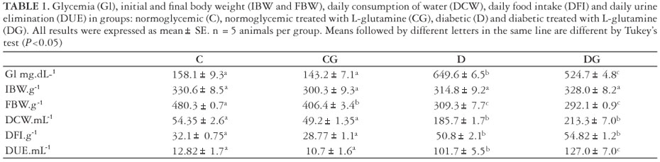
Hyperglycemia was confirmed in groups D and DG (P<0.05) when compared to normoglycemics (C and CG). At the end of the experiment, the animals in diabetic groups (D and DG) had lower body weight (P<0.05) than normoglycemic animals (C and CG). The diabetic syndrome could also be confirmed by their metabolic consequences such as: polydipsia, polyphagia and polyuria (P<0.05). L-glutamine supplementation did not restore the physiological normality of these symptoms (Table 1).
By measuring the area of median longitudinal sections of cecums, we observed that animals in groups D and DG presented a higher area than that of animals in groups C and CG (P<0.05). This result suggests an expansion of the cecum due to diabetes (Table 2).
In the cecum, we found a reduction of 46.0% and 36.9% in the neuronal density of diabetic animals (group D) when compared to animals in groups C and CG, respectively (P<0.05). In this segment, L-glutamine treatment was unable to significantly prevent the neuronal loss in group DG (P>0.05). In the duodenum, we observed a higher neuronal density in treated normoglycemic animals (group CG) when compared to animals of group C (P<0.05). In animals of group DG we observed a preservation of 29% in their neuronal density (P<0.05) when compared to group D (Figure 1).
We found an increase in neuronal cell body area (P<0.05) of diabetic animals (group D) when compared to normoglycemic animals (group C), both in the cecum and duodenum (Table 3). L-glutamine treatment prevented the cell body area increase in the duodenum of animals in group DG (P<0.05) when compared to group D (Table 3).
Most neurons in the duodenum showed an area ranging from 100 to 200 µm2 whereas in the cecum this range was between 100 and 300 µm2. We observed that the neuronal cell body area in duodenum ranged from 44 to 525 µm2 and in the cecum from 9 to 982 µm2 (Figure 2).
DISCUSSION
The metabolic abnormalities found (polyuria, polydipsia, polyphagia and weight loss) are consistent with the literature, in the case of diabetic animals(17, 39). When the potential for glycogen storage of the kidneys is exceeded, the high glucose level in blood promotes glucosuria. This glucosuria, on the other hand, leads to intense osmotic diuresis (polyuria) with great loss of water and electrolytes. The excessive elimination of water, associated with high plasma osmolarity due to hyperglycemia, stimulates the thirsty center in order to restore the osmotic balance (polydipsia). Insulin deficiency, on its turn, causes changes in the catabolic metabolism of lipids and proteins, resulting in an energetic imbalance, which enhances appetite (polyphagia). However, weight loss is eminent, since the proteolysis and lipolysis are very wide(24). The experimental model employed was efficient, since we observed hyperglycemia and the characteristic symptoms of the syndrome in the diabetic groups (P<0.05).
Macroscopic analysis of the cecum of the animals in groups D and DG allowed us to infer that the diabetic condition promotes the expansion of this segment (P<0.05), which may contribute to the deleterious effects of DM on the homeostasis of the gastrointestinal tract. Zanoni et al.(38) also found a similar expansion in the cecum of rats after 2 and 8 months from the administration of streptozotocin.
The Giemsa's staining method, adapted by Barbosa(4), was used in this experiment to stain the myenteric neurons. This method reveals the general population of myenteric neurons and consists in the reaction of methylene blue with the polyribosome complex (Nissl bodies) in the perikaryon of myenteric neurons.
In this study, the cecum (2131.25 ± 433.68 neurons/cm2) and duodenum (7944.44 ± 164.8 neurons/cm2) presented a neuronal density reduction of 46% and 15% in D, respectively, when compared to control group, confirming diabetic neuropathy condition (P<0.05). Diabetic neuropathy has been much studied in recent years, once, due to its onset, diabetic patients have a range of symptoms that cause great morbidity and mortality. Our research group has been studying different segments of the gastrointestinal tract for enteric neuropathies due to DM: Buttow et al.(8) found a neuronal loss of 29.9% in the duodenum after 60 days of diabetes; in the ileum, Zanoni et al.(40) observed a neuronal reduction of approximately 25% after 4 months of diabetes; finally, Zanoni et al.(38) found a neuronal loss of 34.6% in the cecum of rats submitted to 8 months of DM. There are several mechanisms that causes myenteric density reduction in function of diabetes and hyperglycemia, we can list: 1) the elevated production of sorbitol, since it causes cell death by increasing the osmolarity in the neuron leading to edema and to a reduction in the transmission of nerve impulses(9, 10); 2) the auto-oxidative glycosylation(25) and non-enzymatic glycation(23), that increase oxidative stress; 3) lipid peroxidation, which destroys the cell membranes(32), and finally, 4) the metabolic process described above, when coupled with frequent inflammatory responses, which help to generate reactive oxygen species(3, 12). The neuropathy, in diabetic condition, develops when the natural antioxidant systems cannot overcome the oxidative stress.
Treatment with L-glutamine promoted neuroprotection in duodenum by avoiding the loss of myenteric neurons as a consequence of natural aging (group CG: 14638.8 ± 1779.72 neurons/cm2). Group CG presented a neuronal density 35.9% higher (P<0.05) than group C (9383.33 ± 4525.22 neurons/cm2). We also observed this neuroprotection effect in group DG in the duodenum. In this case, neuronal preservation was positive once the neuronal density was higher than that observed in groups D (P<0.05) and C (P>0.05). These effects were not observed in the cecum (P>0.05). Glutathione is related to an important endogenous mechanism of antioxidant defense. During diabetic condition, this antioxidant system is impaired because the availability of glutathione is reduced. L-glutamine has proved to be very efficient in neuronal preservation since it is used by cells as a source for glutathione biogenesis, restoring its deficiency(2, 27). Fläring et al.(15) reported that supplementation with glutamine attenuated glutathione depletion in skeletal muscle of humans. Our group has observed that the loss of myenteric neurons as a consequence of diabetic neuropathy is not similar throughout the entire digestive tube, for instance, the colon is one of the most affected segments(5, 42). It is possible that some segments have a better antioxidant protection than others. In this study, we observed that the neuroprotection afforded by elevated levels of glutathione, due to L-glutamine supplementation, was more effective in the duodenum than in the cecum.
Our research group previously verified that probably due to the diabetic condition there is an increase in the cell body area of some specific neuronal subpopulations, like the submucous VIP-ergic neurons and the myenteric nitrergic neurons(40). This increase is probably related to an excessive production of neurotransmitters. However, in this study we cannot affirm that there was an increase in the synthesis of any type of neurotransmitter due to the nature of the technique employed.
The results observed allow us to conclude that the experimental model was effective, considering that hyperglycemia and metabolic abnormalities, such as: polyuria, polydipsia and polyphagia, were observed. The technique used allowed us to stain all the neuronal populations, consequently, we could observe the diabetic neuropathy through the loss of neuronal density of the diabetic groups in relation to the control groups, both in the cecum and in the duodenum. At the same time, the L-glutamine supplementation promoted the neuronal protection in CG and DG treated groups, as well as the preservation of the cell body area of DG group in the duodenum. This protection and preservation were not observed in the cecum of the CG or DG animals.
We conclude that L-glutamine supplementation presented better effects in the duodenum than in the cecum, because it preserved the myenteric neurons both in number and size.
Received 9/10/2009.
Accepted 19/8/2010.
- 1. Afzaal S, Singh M, Saleem I. Aetipathogenesis and management of diabetic neuropathy. J Assoc Physicians India. 2002;50:707711.
- 2. Amores-Sánchez MI, Medina MA. Glutamine, as a precursor of glutathione, and oxidative stress. Mol Genet and Metab. 1999;67:1005.
- 3. Babior BM. Phagocytes and oxidative stress. Am J Med. 2000;109:33-44.
- 4. Barbosa AJA. Técnica histológica para gânglios nervosos intramurais em preparados espessos. Rev Bras Pesq Méd Biol. 1978;11:95-97.
- 5. Belai A, Lincoln J, Milner P, Burnstock G. Differential effect of streptozotocin-induced diabetes on the innervation of the ileum and distal colon. Gastroenterology. 1991;100:102432.
- 6. Belkind-Gerson J, Graeme-Cook F, Winter H. Enteric nervous system disease and recovery, plasticity, and regeneration. J Pediatr Gastroenterol Nutr. 2006;42:34350.
- 7. Bergmeyer HU, Bernet E. In determination with glucose oxidase and peroxidase. Bergmeyer HU, editor. Methods of enzymatic analysis. 2nd ed. New York: Verlag Chemie Academic Press; 1974. p.1205-12.
- 8. Büttow NC, Miranda-Neto MH, Bazotte RB. Morphological and quantitative study of the myenteric plexus of the duodenum of streptozotocin-induced diabetic rats. Arq Gastroenterol. 1997;34:34-42.
- 9. Chung SS, Ho EC, Lam KS, Chung S. Contribution of polyol pathway to diabetes-induced oxidative stress. J Am Soc Nephrol. 2003;14:s2336.
- 10. Clements RS Jr, Bell DS. Diabetic neuropathy: peripheral and autonomic syndromes. Postgrad Med. 1982; 71:50-67.
- 11. Costa M, Brookes SJ, Hennig GW. Anatomy and physiology of the enteric nervous system. Gut. 2000;47:159.
- 12. Cui K, Luo X, Xu K, Ven Murthy MR. Role of oxidative stress in neurodegeneration: recent developments in assay methods for oxidative stress and nutraceutical antioxidants. Progr Neuro-Psychopharmacol Biol Psychiatry. 2004;28:771-99.
- 13. Curi R, Lagranha CJ, Doi SQ, Sellitti DF, Procopio J, Pithon-Curi TC, Corless M, Newsholme P. Molecular mechanisms of glutamine action. J Cell Physiol. 2005;204:392401.
- 14. De Giorgio R, Camilleri M. Human enteric neuropathies: morphology and molecular pathology. Neurogastroenterol Motil. 2004;16:51531.
- 15. Fläring UB, Rooyackers OE, Wernerman J, Hammarqvist F. Glutamine attenuates post-traumatic glutathione depletion in human muscle. Clin Sci. 2003;104:27582.
- 16. Freeman R. Autonomic peripheral neuropathy. Neurol Clin. 2007;25:277301.
- 17. Furlan MM, Molinari SL, Miranda-Neto MH. Morphoquantitative effects of acute diabetes on the myenteric neurons of the proximal colon of adults rats. Arq Neuropsiquiatr. 2002;60:576-81.
- 18. Gabella G. On the plasticity of form and structure of enteric ganglia. J Auton Nerv Syst. 1990;30:55966.
- 19. Giugliano D, Ceriello A, Paolisso G. Oxidative stress and diabetic vascular complication. Diabetes Care. 1996;19:25767.
- 20. Guo C, Quobatari A, Shangguan Y, Hong S, Wiley JW, Quobatari A. Diabetic autonomic neuropathy: evidence for apoptosis in situ in the rat. Neurogastroenterol Motil. 2004;16:33545.
- 21. Harati Y. Diabetic neuropathies: unanswered questions. Neurol Clin. 2007;25:30317.
- 22. Harris MI. Definition and classification of diabetes mellitus and the criteria for diagnosis. In: Leroith D, Olesky JM, Taylor SI. Diabetes mellitus: a fundamental and clinical text. 3rd ed. Philadelphia: Lippincott Williams & Wilkins; 2004. p.45761.
- 23. Kikuchi S, Shinpo K, Takeuchi M, Yamagishi S, Makita Z, Sasaki N, Tashiro K. Glycation a sweet tempter for neuronal death. Brain Res Brain Res Rev. 2003;41:306-23.
- 24. Kumar V, Abbas A, Fausto N, editores. Robbins & Cotran. Patologia. Bases patológicas da doença. 7Ş ed. Rio de Janeiro: Elsevier; 2005.
- 25. Kuyvenhoven JP, Meinders AE. Oxidative stress and diabetes mellitus pathogenesis of long-term complications. Eur J Int Med. 1999;10:9-19.
- 26. Lin Y, Berg AH, Iyengar P, Lam TKT, Giacca A, Combs, TP, Rajala MW, Du X, Rollman B, Li W, Hawkins M, Barzilai N, Rhodes CJ, Fantus IG, Brownlee M, Scherer PE. The hyperflycemia-induced inflammatory response in adipocytes: the role of reactive oxygen species. J. Biol. Chem. 2005;280:4617- 26.
- 27. Martín-Gállan P, Carrascosa A, Gussinyé M, Domínguez C. Biomarkers of diabetes-associated oxidative stress and antioxidant status in young diabetic patients with or without subclinical complications. Free Radic Biol Med. 2003;34:1563-74.
- 28. Mclaren EH. Diabetic neuropathy. Compr Ther. 1976;4:54-8.
- 29. Monckton G, Pehowich E. Autonomic neuropathy in the streptozotocin diabetic rat. Can J Neurol Sci. 1980;7:135-42.
- 30. Newsholme P, Lima MM, Procopio J, Pithon-Curi TC, Doi SQ, Bazotte RB, Curi R. Glutamine and glutamate as vital metabolites. Braz J Med Biol Res. 2003;36:15363.
- 31. Oates PJ. Aldose reductase, still a compelling target for diabetic neuropathy. Curr Drug Targets. 2008;9:1436.
- 32. Parthiban A, Vijayalingam S, Shanmugasundaram KR, Mohan R. Oxidative stress and the development of diabetic complications-antioxidants and lipid peroxidation en erythrocytes and cell membrane. Cell Biol Int. 1995;19:987-93.
- 33. Schemann M, Neunlist M. The human enteric nervous system. Neurogastroenterol Motil. 2004;16:559.
- 34. Sullivan KA, Feldman EL. New developments in diabetic neuropathy. Curr Opin Neurol. 2005;18:58690.
- 35. Vincent AM, Russel JW, Low P, Feldman EL. Oxidative stress in the pathogenesis of diabetic neuropathy. Endocr Rev. 2004;25:61228.
- 36. Vinik AI. Diabetic neuropathy: pathogenesis and therapy. Am J Med. 1999;107:1726.
- 37. Vinik AI, Mehrabyan A. Diabetic neuropathies. Med Clin North Am. 2004;88:94799.
- 38. Zanoni JN, de Miranda-Neto MH, Bazotte RB, de Souza RR. Morphological and quantitative analysis of the neurons of the myenteric plexus of the cecum of streptozotocin diabetic rats. Arq Neuropsiquiatr. 1997;55:696-702.
- 39. Zanoni JN, Hernandes L, Bazotte RB, Miranda-Neto MH. Terminal ileum submucous plexus: study of the VIP-ergic neurons of diabetic rats treated with ascorbic acid. Arq Neuropsiquiatr. 2002;60:32-7.
- 40. Zanoni JN, Buttow NC, Bazotte RB, Miranda Neto MH. Evaluation of the population of NADPH-diaphorase-stained and myosin-V myenteric neurons in the ileum of chronically streptozotocin-diabetic rats treated with ascorbic acid. Auton Neurosci. 2003;104:328.
- 41. Zanoni JN, De Freitas P, Pereira RV, Dos Santos Pereira MA, De Miranda-Neto MH. Effects of supplementation with ascorbic acid for a period of 120 days on the myosin-V and NADPHd positive myenteric neurons of the ileum of rats. Anat Histol Embryol. 2005;34:149-53.
- 42. Zanoni JN, Pereira RVF, Freitas P de. Effect of the ascorbic acid treatment on the NADHd-positive myenteric neurons of diabetic rats proximal colon. Braz Arch Biol Technol. 2007;50:31-8.
- 43. Zhao J, Yang J, Gregrersen H. Biomechanical and morphometric intestinal remodeling during experimental diabetics in rats. Diabetologia. 2003;46: 168897.
Publication Dates
-
Publication in this collection
25 Apr 2011 -
Date of issue
Mar 2011
History
-
Received
09 Oct 2009 -
Accepted
19 Aug 2010