Abstracts
OBJECTIVE: To functionally and oncologically assess the treatment of GCT on radius distal end (stage B3) following wide resection and reconstruction with avascular autologous graft from proximal fibular end. METHODS: The residual function was assessed using ISOLS score, measurement of the global residual arch of the operated wrist, residual percentage of the hand apprehension strength between thumb and index fingers. The oncologic monitoring was assessed by clinical examination of the operated limb and by imaging tests of the wrist and thorax. RESULTS: 17 patients were assessed: 10 females (58.8%) and seven males (41.2%), with aged ranging from 16 to 61 years (mean: 32.3 years), all of them right-handed. On the functional assessment (ISOLS), the results were as follows: 11 excellent, two good, and one poor. The three cases requiring arthrodesis evolved with excellent scores. The global residual arch was 196.2 ± 116.6º. The residual arch of the operated wrist corresponded to 58.9% of the control. The "pinch" percentage was 80.6 ± 14.8 % of control. We did not find recurrences or metastasis in this case series. CONCLUSION: This technique provided encouraging functional results, assuring patients' return to their usual activities. The absence of local recurrence and/or metastasis found even in the patients with longer follow-up times allow us to suggest this technique, which seems to be safe for oncologic control of the tumor.
Giant cells tumors; Radius; Surgical procedures
OBJETIVO: Avaliação funcional e oncológica do tratamento do TCG do segmento distal do rádio (estadio B3) mediante ressecção ampla e reconstrução com enxerto autólogo avascular da extremidade proximal da fíbula. MÉTODOS: A função residual foi avaliada mediante escore ISOLS, medida do arco residual global do punho operado, do percentual residual da força de preensão da mão e da preensão entre o polegar e o indicador. O controle oncológico foi avaliado mediante exame clínico do membro operado e avaliação por imagens do punho e do tórax. RESULTADOS: 17 pacientes avaliados, dez do sexo feminino (58,8%) e sete do sexo masculino (41,2%) com idades entre 16 e 61 anos (média de 32,3 anos), todos destros. Na avaliação funcional (ISOLS) observamos 11 resultados excelentes, dois bons e um ruim; os três casos que demandaram artrodese evoluíram com escore excelente. O arco residual global foi de 196,2 ± 116,6º. O arco residual do punho operada correspondeu a 58,9% do controle. A força de preensão da mão correspondeu a 55,4 ± 17,4% do controle. O percentual de "pinça" foi de 80,6 ± 14,8% do controle. Não constatamos recidiva ou metástases nesta casuística. CONCLUSÃO: A técnica propiciou resultados funcionais alentadores, assegurando o retorno dos pacientes às suas atividades. A ausência de recidiva local e/ou metástases, observada inclusive nos pacientes com seguimento mais longo, permite sugerir que a técnica parece ser segura no controle oncológico do tumor.
Tumores de celulas gigantes; Rádio; Procedimentos cirurgicos operatórios
ORIGINAL ARTICLE
Wide resection and fibular transposition in the treatment of GCT on radius distal end
Alex GuedesI; Pedro Péricles Ribeiro BaptistaII; Cláudio SantiliII; Eduardo Sadao YonamineII; Hélio Rubens Polido GarciaII; Emília Cardoso MartinezII
IOrthoapedics and Traumatology Service, Hospital Santa Izabel Santa Casa da Misericórdia da Bahia, Salvador, BA, Brazil
IIDepartment of Orthopaedics and Traumatology, Santa Casa da Misericórdia de São Paulo Pavilhão Fernandinho Simonsen, SP, Brazil
Correspondences to
SUMMARY
OBJECTIVE: To functionally and oncologically assess the treatment of GCT on radius distal end (stage B3) following wide resection and reconstruction with avascular autologous graft from proximal fibular end.
METHODS: The residual function was assessed using ISOLS score, measurement of the global residual arch of the operated wrist, residual percentage of the hand apprehension strength between thumb and index fingers. The oncologic monitoring was assessed by clinical examination of the operated limb and by imaging tests of the wrist and thorax.
RESULTS: 17 patients were assessed: 10 females (58.8%) and seven males (41.2%), with aged ranging from 16 to 61 years (mean: 32.3 years), all of them right-handed. On the functional assessment (ISOLS), the results were as follows: 11 excellent, two good, and one poor. The three cases requiring arthrodesis evolved with excellent scores. The global residual arch was 196.2 ± 116.6º. The residual arch of the operated wrist corresponded to 58.9% of the control. The "pinch" percentage was 80.6 ± 14.8 % of control. We did not find recurrences or metastasis in this case series.
CONCLUSION: This technique provided encouraging functional results, assuring patients' return to their usual activities. The absence of local recurrence and/or metastasis found even in the patients with longer follow-up times allow us to suggest this technique, which seems to be safe for oncologic control of the tumor.
Keywords: Giant cells tumors; Radius/ anatomy; Surgical procedures.
INTRODUCTION
Giant cell tumor (GCT) constitutes an aggressive benign bone neoplasia, first described by Cooper (1818), who named it as "fungus medullary exostosis".1,2
Jaffe et al., in 1940, described its appearance on the X-ray, anatomical distribution, histology and variable and sometimes locally aggressive clinical behavior.1,3
It more commonly affects individuals in the age group of 18 to 40 years, with peak incidence in the third decade of life, being more usually found in women (1,5 - 2 : 1), being uncommon in children.
It is more frequently found in Southeastern Asia populations, accounting for 21% of the benign bone tumors, while among the Western population, its incidence ranges from 4 to 9.5% 2.
It affects mostly the epiphyseal segments of long bones.(3,4) In children (2-6% of the cases), its location can be metaphyseal or diaphyseal.2,3
The most frequently affected segments are, sequentially: distal femoral, tibial proximal and radius distal epiphyses.2,3,5-11 The radius distal epiphysis is affected in 10% of the cases.2,10
Although classified as a benign neoplasia, multicentre involvement (1% of the cases) or metastasis to lungs and mediastinum (1-9% of the cases) may occur, usually associated to primary injuries located at the radius distal segment.2,3,9
Its signs and symptoms will depend on the size, site and how fast the lesion progresses. Pain, either associated to an increased volume or not, is frequent. Pathologic fracture can be the first sign of the process, in up to 15% of the cases. Effusion and limited local mobility denote a lesion near a joint surface.12
X-ray findings will depend on the evolution time until diagnosis. It starts as an eccentric, lytic, insufflating and bone-restricted epiphysiometaphyseal lesion and, in advanced stages, is more aggressive, presenting cortical and subchondral rupture, becoming extra-compartmental.2
Radius GCT at Enneking's B1 stage is classically treated with curettage and filling with methylmetacrylate4,12,13, allowing early mobilization and facilitating the visualization of recurrences on the bone-cement interface, if occurring. However, these lesions, of small size and potentially treatable by this method, are usually asymptomatic and, thus, undiagnosed at this initial phase.
In more advanced stages, we notice a thinner bone cortical of the radius distal epiphysis (Enneking's B2 stage). With the cortical being ruptured, tumor will be found on soft parts and subchondral bone destruction (Enneking's B3 stage).
Most of the patients with GCT are composed by young adults, in their most productive age. The purpose of treatment includes the full tumor removal and function preservation.8 When treating B3-stage tumors, curettage, with oncologic criteria, is technically complex. A wide resection and joint reconstruction are necessary in order to maintain function and minimize the potential for tumor recurrence.3,4
Wrist reconstruction, due to the topographic proximity from neurovascular and tendinous structures and to the limited coverage of soft parts, constitutes a challenge for surgeons.3,4,5,14 The techniques employed for that end should provide stability and joint mobility enough to meet the functional demand of the distal segment of the upper limb.11,15,16
The key solutions for joint reconstruction after oncologic resection of the radius distal end are: arthrodesis, in its several modalities (arthrodesis with autologous bone graft; ulnar centralization; distal translocation of the ulna; arthrodesis with vascularized fibular graft, and partial fibulo-scapholunate arthrodesis) and arthroplastic reconstructions (avascular or vascularized fíbula pro radio, prosthetic radius replacement, microvascular flap of iliac crest, and homologous radius graft).
The first arthroplastic reconstruction of the wrist using avascular fibular bone-joint graft was described by Walther in 1911, who employed it in the treatment of osteosarcoma of the radius distal third.16 Since then, the anatomical similarity between proximal fibular segment and the distal portion of the radius has been recognized by other authors, who started using this procedure to treat GCT at that site.
The results of this reconstruction technique are strong, especially from a functional perspective, encouraging professionals to use it for treating lesions compromising radius distal segment.
Our study targets the evaluation of the functional and oncologic result when treating patients with giant cells tumor on the distal radius segment (Enneking's B3 stage), submitted to wide resection and reconstruction with avascular autologous graft collected from proximal fibular segment.
CASE SERIES AND METHODS
Our study was evaluated and approved by the Committee of Ethics in Research of Santa Casa de São Paulo.
Between July 1985 and May 2006, 25 patients with GCT on the distal third of the radius were operated using the wide resection and reconstruction technique with avascular autologous graft from proximal fibula at the Department of Orthopaedics and Traumatology, Santa Casa de São Paulo - Pavilhão Fernandinho Simonsen, and at the Orthopaedics and Traumatology Service, Santa Casa de Salvador - Hospital Santa Izabel.
Of that total, we could contact and assess 17 patients (Chart 1), 10 females (58.8%) and seven males (41.2%) whose ages at baseline ranged from 16 and 61 years (mean: 32.2 years).
The right side was affected in eight patients (47.1%) while the left side in nine patients (52.9%), all of them being right-handed.
All patients presented with clinical and X-ray aspects consistent with Enneking's B3 stage and none had metastasis at baseline screening. All patients were submitted to biopsy prior to surgery, confirming GCT diagnosis.
All patients were treated with a modified surgical technique for tumor resection and radius reconstruction with fibular avascular autologous graft as therapy for GCT of the distal segment of the radius. Modifications on the technique lie on the employed access, details on the fibular graft uptake, and on the kind of capsuloplasty.
Description of the surgical technique
Following diagnosis, and by establishing the accurate staging, we proceeded with the surgical planning.
X-ray images were taken of the forearm and of the graft donator leg (ipsilateral), aiming to assess the integrity of the donator bone and of the joint surface of the proximal fibular epiphysis. X-ray images must be taken with rule, enabling to measure the radius segment to be resected and of the corresponding fibula required for reconstruction.
Under general anesthesia, the affected upper limb and the donating lower limb are disinquined with degerming solution. The patient is positioned in horizontal supination with a cushion beneath the gluteus region at the side of the donor end, allowing for a subtle internal rotation of the lower limb, facilitating graft removal.
The ends are prepared and isolated with sterilized surgical drapes. The upper limb must be exposed from the axil, and the lower limb from the inguinal fold.
Two tables are prepared; one to attend the radius segment resection and the other to uptake the fibula and reconstruct the wrist. Surgical instruments exchange between the tables must not occur.
Venous depletion of the forearm is conducted by lifting the limb or with mechanical procedures with the use of Esmarch's band. Should the latter is selected, care must be taken to not to compress the lesion area. After depletion, a pneumatic garrote or the Esmarch's band is applied.
The surgery starts with the "in block" resection of the injured radial segment. An arch-like dorsal incision is made starting at 1.5 cm distal to the ulnar styloid apophysis towards the radial styloid apophysis. At the level of the skin protuberance of the radial styloid, the incision is curved following the longitudinal axis of the radius, at a proportional length to the segment to be resected just sufficient for adapting the dynamic compression plate that will fixate the fibular graft on the proximal radius segment; Special attention must be paid to the exeresis of the biopsy scar, if present. (Figure 1)
Then, the antebrachial fascia is opened, preceded by skin and subcutaneous tissue hemostasis. Following, the retinacula of the extensors and tendon sheaths of the long abductor and short extensor muscles of the thumb (first tunnel); short and long radial extensors of the carpus (second tunnel); long extensor of the thumb (third tunnel) and long extensors of the fingers and the index (fourth tunnel) are opened.
Brachiostyloradial muscle tendon is identified and sectioned near its insertion, respecting the oncologic resection margins. The extensor tendons are medially displaced, thus exposing the affected radial segment. The dorsal surface of the radius distal third is dissected at a sufficient length for resection, respecting the oncologic margins. (Figure 2)
The osteotomy site is determined according to the proximal limit gross and radiographic of the affected bone and the required margin for each individual case. The delimitation is performed with electric knife, perpendicular to the longitudinal axis of the radius. At this moment, with the forearm at neutral position, another mark is made perpendicular to the latter and located on the lateral radius surface. That mark will serve as a control for forearm rotation when fixating the graft. (figure 2)
Osteotomy is performed and the radius distal segment is grasped with bone tweezers and carefully lifted in order to facilitate its release. The square pronator muscle should be resected in conjunction with the bone, as an oncologic margin. (Figure 2)
Arthrotomy of the distal radius-ulna joint is then provided with or without a small ulnar fragment, as well as the arthrotomy of the radiocarpal joint. Joint capsules are preserved as much as possible (meeting the oncologic criteria), facilitating capsuloplasty between fibular graft and the carpus. (Figure 2)
The resected piece's length is measured. This measurement will serve as a control for the appropriate measurement of the fibular graft to be used for reconstruction. The garrote is then removed and hemostasis is reviewed.
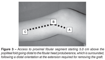
After venous depletion and garroting of the donor lower limb, a cushion is placed beneath the knee, keeping it at 45° of flexion. We initiate the posterolateral access at five centimeters above the joint interline, posterior to the crural biceps tendon, continuing at distal direction over the skin protuberance of the proximal fibular segment. (Figure 3)
The fibular nerve is identified, isolated, rebated and protected. Fibula is dissected, releasing soleus, long fibular, long fingers extensor muscles and the interbone membrane long enough to reconstruct the resected radius. The femoral biceps tendon and the lateral collateral ligament of the knee are sectioned close to their insertions on the fibula, preserving about half centimeter of the length of these structures' stumps inserted into the resected segment, which will serve as anchors to the remaining ligaments of the radiocarpal joint. It is important to protect posterior popliteal vessels and the anterior tibial vessels penetrating the distal interbone membrane to the fibular neck.
By means of blunt dissection, the posterolateral capsule of the knee is exposed between the lateral head of the gastrocnemius and the soleus muscle. Tibiofibular arthrotomy is performed by sectioning the origin of anterior, posterior and a portion of the arched ligaments. (Figure 4)
The length required for radius reconstruction is measured, adding 1.5 2 cm as adjustment margin for reconstruction. (Figure 4) The fibula is osteotomized, protecting soft parts with two Bennett retractors. After the graft is removed, fibular joint is prepared, adjusting it to capsulorrhaphy. A spongy graft is removed from the tibia through a port made on the proximal tibiofibular joint.
The lateral collateral ligament is reinserted, suturing it to tibial periosteum.
The garrote is now removed and hemostasis is reviewed. The wound is closed by planes, and an aspirating drain and a sterile bandage is placed.
Radius reconstruction starts with capsulorrhaphy, suturing the remnants of the radiocarpal capsule to the stumps of the femoral biceps tendon and of the lateral collateral ligament, preserved at the end of the fibular graft with simple stitches using non-absorbable monofilament nylon 2-0. With the wrist positioned at forced volar flexion, the capsule is sutured by the volar surface, continuing in a circumferential fashion, reaching the lateral, dorsal and medial surfaces. The joint cartilage of fibular head is positioned so as to form a hinge with the scaphoid; this adaptation is made in order to avoid dorsal or volar carpal dislocation. (Figure 5)
After capsuloplasty, the hand is slightly pulled, forcing the radial displacement of the wrist and adjusting the graft proportionally to the length of the bone failure on the radius. The length of the fibular segment must provide adequate support to carpus, without excessive tension to the level of its attachment to the fibula.
The graft is placed with the forearm at neutral position, using the longitudinal mark made on the lateral surface of the radial stump as a parameter; once adjusted, a mark is made on the graft, continuous to that of the radial segment. This mark will make the checking of graft position easier at the moment of fixation. The graft is fixated on the radial segment with a dynamic compression plate of small AO fragments with six screws. (Figure 6)
Fixation can be supplemented with 2.0-mm Kirschner wire, transfixing the new fibuloulnar joint, when instability persists after capsuloplasty.
At this moment, the spongy graft obtained from the tibia is placed on the attachment between the fibular segment and the proximal radius fragment. (Figure 6)
The wound is then closed by planes, paying special attention to the coverage of the plate with the soft parts. Whenever possible, extensor tunnels reconstruction is attempted, especially that of the tendon of the long extensor of the thumb, which loses the support of the Lister tubercle due to radius resection. The aspirating drain is inserted and, following the application of bandages, the limb is immobilized with axilopalmar splint, with the forearm at neutral position.
Bandages are refreshed on a daily basis, and the stitches are removed on the 15th postoperative day.
The axilopalmar splint is kept for three weeks, and replaced by an antebrachiopalmar plastered device (plastered glove), being kept for three additional weeks, releasing the elbow and enabling exercises for prono-supination gain.
In cases where an additional fixation of the new fibulo-ulnar joint with Kirschner wire is necessary, we keep the axilopalmar splint for six weeks, when the wire is removed and the elbow mobilization is initiated.
After the external immobilization is removed, motor rehabilitation is initiated with no load, in order to gain range of joint motion.
After six weeks, exercises for strength gain are allowed, releasing full load between six months and one year, depending on each patient's evolution.
The X-ray monitoring is performed on a monthly basis until the joint site between the fibular graft and the proximal radius segment is united.
Functional and Oncologic Assessment
For the functional assessment, we used the wrist functional score standardized by the International Society on Limb Salvage (ISOLS).17 (Chart 2)
The flexion-extension range and the radial and ulnar displacements of the wrists, as well as the prono-supination arch of the forearms were measured with a goniometer, including the operated and the intact limb.
Hand apprehension strength has been measured with a JAMAR dynamometer (Sammons Preston, Bolingbook, IL) as kgf., always comparing it to the contralateral side, using a maximum effort measure as a parameter.
The "grasp" strength between the thumb and the index finger was measured with a "grasp" dynamometer (B & L Engineering, Santa Fe, CA), as kgf., always comparing it to the contralateral side, using a maximum effort measure as a parameter.
The oncologic evaluation was made upon detailed physical examination of the operated limb, pursuing to detect any residual tumor or palpable masses and upon imaging tests of the operated wrist and of the thorax, pursuing to identify suggestive findings of local recurrence.
The classificatory variables were presented as tables containing absolute frequencies (n) and relative frequencies (%). Quantitative variables were descriptively presented as tables containing mean, standard deviation, median, minimum and maximum values.
The operated side and the control were compared with the Wilcoxon's non-parametric test of signs. P values <0.05 were regarded as statistically significant.
RESULTS
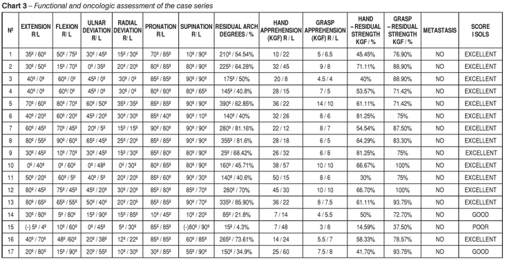
In the functional assessment, using the wrist functional score standardized by ISOLS, the patients in which arthroplastic reconstruction was preserved by follow-up (three were submitted to arthrodesis during postoperative follow-up period), we found 11 excellent results (Figure 7) and two good results. The patient whose functional outcome was regarded as poor has been scheduled for fixation review because of an aseptic loosening of the synthesis material.
The patients submitted to arthrodesis, after complications of the primary procedure, had excellent functional outcomes. Despite being submitted to joint fusion, they presented signs of apprehension strength that were similar to the rest of the individuals of the group, with global range of motion above 120° in the last evaluation visit. (Chart 3)
The comparative measures of the range of motion and apprehension strength of the hand and the grasping between index and thumb between the operated and the intact limbs of the assessed patients are presented on Table 1 and Graphs 1 to 4.


The analysis of the residual arch measure shows mean values of 196.2 + 116.6 degrees, with median of 175 degrees. The assessment of the residual percentage shows that, in average, the operated hand kept 58.9% of the range of joint motion on the control limb.
The residual apprehension strength percentage of the hand was, in average, 55.4 + 17.4%, and the grasp between the thumb and the index fingers of the operated hand was 80.6 + 14.8%.
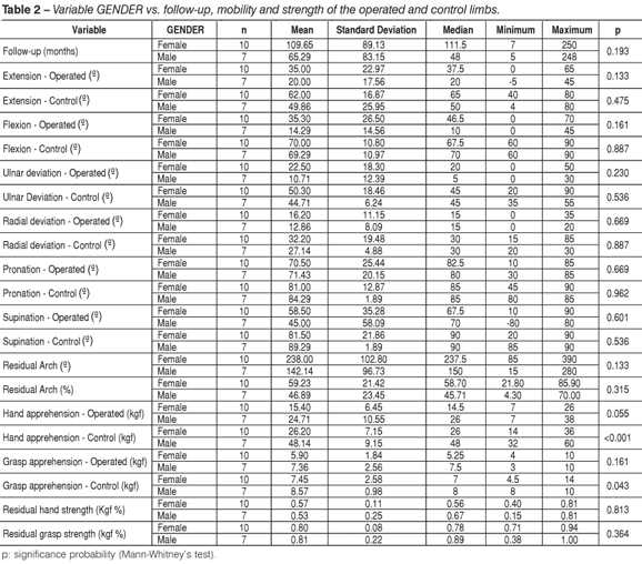
The comparative analysis between genders showed that, despite the apprehension and grasp strength of the hand are stronger in males, as shown by Table 2, the residual strength percentage is similar for both genders.
When we compare the operated side, we can see by Table 3 that the apprehension and grasp strength of the hand, as well as the residual strength percentage are shown to be identical, regardless of dominance.
In all cases but one, there was fibular graft integration and consolidation of the attachment between the proximal radius segment and fibula.
No tumor recurrence or signs of lung metastasis were documented in routine clinical and X-ray evaluation or in the last assessment.
Among the complications associated to reconstruction, we saw three cases of late infection. In the first case, surgical cleaning was performed five months after the procedure, with improvement of the infection; 11 months later, the patient was submitted to wrist arthrodesis, combined with Darrach procedure. In the second case, nine months after the primary procedure, surgical cleaning was provided, s well as the removal of the synthesis material, evolving to clinical improvement. In the third case, aseptic loosening of the synthesis material was seen three months after the primary procedure, in which case we decided to provide a graft review using the contralateral fibula; the patient evolved with infection, being submitted to surgical cleaning, removal of the infected graft and wrist arthrodesis with tricortical iliac graft combined with Darrach surgery. All patients evolved with clinical improvement, with no signs of infection in the last assessment, and returning to their previous activities with no limitations.
One patient evolved with simple fall and fracture of the graft on the third postoperative year. She was treated with plastered cast for three months, unsuccessfully. She evolved with union delay and pain, with the wrist arthrodesis being performed combined with the Darrach procedure, with improvement of the clinical picture and return to her daily life activities.
Pseudoarthrosis on the attachment between the proximal radius segment and the fibular graft was seen in one case. Despite of the indication of review with grafting, the patient is now satisfied with the result, which does not imply on occupational restraints, and motivating him to not to undergo surgery.
There was aseptic loosening of the synthesis material in one patient at seven months postoperatively, which evolved with significant functional deficit. He is now scheduled for osteosynthesis review.
Morbidity at the donor site was identified in two cases. In the first case, common fibular paresis occurred, with spontaneous recovery; in the second one, the patient evolved with restraint to hallux extension.
Three sub-dislocation cases were documented: two volar and one dorsal, evolving with subtle restraint to wrist dorsiflexion and mild pain at strong efforts.
Degenerative changes on the fibulocarpal "joint" occurred in most of the patients with longer follow-up periods, but this did not cause bias to residual function or was related to complaints from the patient.
DISCUSSION
The preferential treatment for Enneking's B3 stage GCT located on the distal radius third, consists of wide resection associated to forearm reconstruction, targeting to preserve wrist and hand function.18
Several techniques have been developed to reconstruct the forearm after oncologic resection of the distal radius third. We will classify these procedures concerning postoperative mobility of the wrist as stiff and arthroplastic.
Among the stiff procedures, the following are included: arthrodesis with avascular autologous bone graft; centralization of the ulna; distal ulnar translocation; arthrodesis with vascularized graft from fibula, rib or iliac, and fibuloscapholunar arthrodesis of the carpus.
When the whole joint capsule, a portion of the carpus or the ulna needs to be included as surgical margin, we should not indicate functional arthroplasty. In this situation, arthrodesis is functionally superior to arthroplastic reconstruction7,11,16,19 constituting an alternative of choice for treatment. Some authors7,11,19 indicate arthrodesis in elderly patients or performing stronger activities, not requiring modulation of fine movements in their activities.
However, fusion and integration of a graft must occur at the arthrodesis site and on the transition with the bone, increasing the risk of developing paseudoarthrosis.12 Ben Amor et al.6, in their series of five resection/arthrodesis cases for treating radius GCT, found two pseudoarthrosis cases requiring review with grafting. The stiff fixation of grafts with plate and screws, especially on the attachment with the proximal radius segment, seems to reduce such possibility, but bias on extensor tendons function may occur, resulting in rupture by rubbing.9,13
Graft fracture is frequent when resection/ arthrodesis are selected. Ben Amor et al.6 found fractures in three of their five cases; two patients were submitted to a new procedure with fixation review, while the remaining one decided to not to undergo surgery again.
"In block" resection brings the advantage of oncologic control when associated to wrist arthrodesis, but its disadvantage is the loss of wrist movements.12,18
In an attempt to facilitate reconstruction, some authors decide for centralization or distal translocation of the ulna. These procedures are successful in this objective, but the inconvenience of limited wrist mobility remains. In ulnar centralization, flexion-extension and prono-supination movements of the forearm are totally lost. The distal translocation of the ulna preserves prono-supination, but flexion-extension of the wrist is lost.16
Arthrodesis with vascularized fibular graft provides the theoretical advantage of reducing the possibility of union and integration delay, which are common complications when using avascular bone grafts. However, it still has the disadvantage of wrist movements' loss, worsened by the need of a microsurgical technique and the challenges inherent to it. There are reports of pseudoarthrosis and local recurrence associated to the use of this technique.
A certain joint range may be preserved after arthrodesis, since the midcarpal joint is saved by the exclusive fusion of the proximal row of the carpus to the graft.7 With this in mind, some authors most recently have selected partial arthrodesis of the carpus with the fixation of a fibular diaphyseal vascularized segment to the scaphoid and semilunar. After the union of the arthrodesis core, an increment of the midcarpal mobility is seen, which allows for functional flexion-extension range and radio-ulnar displacement of the wrist. However, residual range of joint motion is unpredictable, additionally to the fact that the fusion between graft and the proximal carpal row is more difficult, for contact limitations between both.7
Among the arthroplastic procedures, the following are included: prosthetic replacement of radius distal third, homologous radius graft, vascularized iliac graft and bone-joint autologous graft, or avascular graft of the proximal fibula.
The use of endoprosthesis in the reconstruction of radius distal third presents as advantages a shorter surgical time, no postoperative immobilization required, and a functional outcome that is theoretically comparable to that of the arthroplastic reconstruction.19 However, its costs are high and, although effective regarding insertion time19, it usually fails in young individuals, leading to discouraging results. Painful stiffness and loosening become frequent, demanding early review of the implant material.12,19
The advanced orthopaedic oncology has prioritized the search for long-lasting solutions for bone-joint reconstruction after oncologic resection, which, at the same time, could provide the best outcomes under a functional perspective. Thus, the investigation of "biological solutions" has taken the lead in scientific research. With this in mind, the reconstruction of the affected radial segment may be provided with homologous radius graft, vascularized fibular or iliac graft, or vascular fibular graft.
The wrist has been regarded as one of the most appropriate sites for reconstruction with homologous graft, because it does not constitute a load joint7. The use of these grafts became more consistent as the methods employed for its preparation and preservation evolved.
This method has a number of theoretical advantages, including: function preservation; restoration of the anatomy, rich graft supply (tissue libraries), and the fact of avoiding morbidity associated to autologous graft uptake. There are, though, many potential problems associated to its use, including donor selection, method for acquiring and preserving grafts, and graft coaptation. Despite of the increment on midcarpal joint mobility after surgery, rehabilitation process is lengthy, with a progressive apprehension strength reduction when compared to the intact side.18
Homologous grafts have antigenic properties and are composed by totally dead bones, changed by radiation, freezing or both. After surgery, risks of graft infection, reabsorption and fracture must be considered, as well as joint collapse, longer time required for graft integration, and the compromising of a definitive result regarding wrist stability and mobility.5
The most frequent complication is fracture. The thin cortical of the distal radius epiphysis facilitates a strong remodeling, increasing the incidence of this complication. Kocher et al..5, in their series of 20 radius GCT cases submitted to reconstruction with homologous graft, reviewed one third of the grafts, most of them secondarily to fracture.
Harness and Mankin3 reported the evolution of 15 patients treated with wide resection and reconstruction with radius allograft (mean follow-up of 19 + 4 years). Review due to local recurrence was reported for two cases, 16 procedures for treating different complications and progressive functional deterioration in six patients submitted to this modality of reconstruction.
Reconstruction using vascularized fibula presents as advantages: union and integration of the graft regardless of the local perfusion status13; enables early rehabilitation19, maintain mechanical properties and bone resistance. It is regarded by some authors as the technique allowing the best functional outcome.12,15
In practice, however, final joint mobility is similar to that obtained when using avascular autologous fibular graft.19
When treating neoplasias located at the distal segment of the radius, the optimal resection margins of which exceeding eight centimeters from the length of that bone, there is advantage in using the vascularized fibula. This resource reduces the incidence of pseudoarthrosis associated to the use of longer avascular grafts, usually secondary to fracture or union delay of the transposed segment.19
In the treatment of radius GCT, resecting more than eight centimeters of its length is seldom necessary to provide appropriate oncologic treatment. The resection limit is situated immediately proximally to the margin outlined by preoperative examinations and gross aspect. Thus, it is required to resect an additional length of the intact radius in order to adjust the vascularized fibular segment, since the fibular epiphysis is nourished by the shaft, being required at least 10 centimeters of the fibular proximal segment in order to provide a proper vascularization of the graft.15
Another disadvantage inherent to the method is the need of performing a microsurgical technique and, as a result, a longer surgical time, thus increasing the risks of postoperative infection.19
The collection of a vascularized fibular graft may lead to morbidity to the donor site16; discomfort may be experienced on the ankle or leg, as well as reduced muscular strength, injury of the common fibular nerve, contraction of the long flexor muscles of the hallux and toes with "claw" deformities, fibular osteoporosis, and changes on gait analysis.
Vascularized fibula assures a feasible joint cartilage, but does not avoid subdislocation and degenerative arthritis associated to the incongruence of the "joint" between the fibula and the carpus.14
Complications such as union delay, fractures (due to a longer transplanted bone segment), infection, local recurrence, graft reabsorption, early degeneration of the fibulocarpal joint, and carpal subdislocation have been reported and are common to this procedure and other arthroplastic reconstruction methods for the radius distal third.
Radius reconstruction with vascularized iliac graft15 attenuated the disadvantages of vascularized fibular graft: there is no need of additional resection of the radius to be reconstructed the pedicled iliac graft measures between three and five centimeters; integration is comparably faster; shorter surgical time and lower morbidity associated to graft removal. However, it still requires a microsurgical technique, with the use of a graft presenting the typical curvature of the iliac and where joint cartilage is not available4,8, being associated to a high incidence of dorsal subdislocation.15
We routinely used the wide resection technique associated to reconstruction with avascular proximal fibular graft through dorsal arch-like approach for the treatment of Enneking's B3 stage GCT located at the distal third of the radius.
Arthroplasty with bone-joint avascular autologous graft removed from proximal fibula constitutes an excellent reconstruction method after wide resection of the GCT on the distal third of the radius. In series with follow-up periods longer than five years, whenever employed14, constituted a long-lasting and effective solution for restoring mobility and strength on the wrist and hand.
In our series, we found degeneration of the fibulocarpal "joint" in patients with longer follow-up periods, with no functional or symptomatic correlation. This change is frequent8, with no corresponding damages to wrist function.5,12,15 They are present in reconstructions with homologous and autologous grafts (either vascularized or not). Mattar Júnior et al.12 attribute this fact to three factors: 1) joint incongruence between fibula and the proximal carpal row; 2) avascular necrosis of the fibular epiphysis due to vascular failure; 3) changes caused by the absence of joint innervation (Charcot joint).
Literature describes problems associated to the fixation of a fibular graft on the radius18, usually related to insufficient fixation. Pseudoarthrosis seems to be associated to the kind of fixation employed, and cannot be avoided when using a vascularized graft. Although the union of a vascularized graft occurs rapidly, issues with the avascular graft may be minimized with the use of stiff internal fixation.14
Aithal and Bhaskanarand14, when reviewing 30 cases of GCT on distal radius treated with four different fixation methods, finding better results with the use of stiff fixation with dynamic compression plate (DCP); only one case (6%) evolved with pseudoarthrosis in that group, being successfully treated with bone grafting. The incorporation and union of avascular grafts were found to be dependent on the method of fixation employed, being lower when stiff osteosynthesis was used.
Maruthainar et al.20, in their series of 13 cases, used avascular fibular graft with stiff fixation, obtaining union in all cases within a period ranging from 9 to 12 weeks. Aithal and Bhaskanarand14, using avascular fibular graft with stiff fixation reported a mean incorporation time of 5.2 months.
It is reasonable to assume that our understanding about the need of stiff fixation is not only globally recognized as mandatory. All patients in our study but one showed union at the attachment between the proximal radius segment and the fibular graft. All cases experienced incorporation of the graft.
Graft fractures caused by stress or trauma are described in literature.12,15 A patient of ours experienced a simple fall, evolving with graft fracture after three years; she was unsuccessfully treated with the conservative approach, being therefore submitted to arthrodesis.
We found carpal subdislocation in three of our patients, who evolved with limitation of the wrist flexion-extension and mild pain at stronger efforts. This event constitutes an inherent complication to the kind of reconstruction employed, once fibulocarpal congruence varies according to the anatomical characteristics of each individual patient, not always associated to corresponding function changes. Aiming to minimize the potential for subdislocation, some authors recommend a deeper fibular joint concavity, making it more congruent to the convexity of the proximal row of the carpus. Aithal and Bhaskanarand14 decided to pass a Kirschner wire through the third metacarpal, transfixing the fibulocarpal joint.
We disagree with adding further trauma to the articular cartilage of the graft. We decided to use the ipsilateral fibula, because it adjusts better than the contralateral one because of its format and curvature, consistently to the convexity of the proximal row of the carpus, as well as mimicking the styloid process of the radius and the Lister tubercle.
We disagree with the principle employed on "step" and oblique osteotomies, of which objective is to enlarge the contact area and/or enhance stability between the fibular graft and the proximal radius segment. We believe that a successful surgery lies on a careful capsular suture, which promotes the required stability to a good functional outcome. Only after suture we adjust the rotation and length of the graft until totally adapted, with no tension applied to the carpus and radius. Performing a non-transverse osteotomy makes the adjustment between the graft and the proximal radius segment after capsuloplasty difficult or even impossible.
The reconstruction of soft parts is critical for a successful procedure, since joint instability leads to deterioration of the results in the long term. Although most of the authors do not mention reconstruction of soft parts, some report capsule or radial collateral ligaments repair, associating extrinsic tendons on capsuloplasty. Radial collateral ligament repair is the minimum requirement for joint stability.
GCT on the distal segment of the radius presents the strongest potential for local recurrence, with a risk rate of approximately 25%. Recurrence occurs early, usually within the first two years in 95% of the cases2, which increases the risk of metastasis by about six fold. Such risk may be associated to repeated attempts of local control after primary treatment failure.9 It occurs previously in 54-83% of the patients presenting lung metastasis, and the distal segment of the radius is the most common primary site of a metastatic tumor.2
We didn't find local recurrences or metastasis in our series, which can be attributable to the modality of resection employed on the affected segment. Radius excision with large margins, including the squared pronator in its volar surface11 and the extraperitoneal incision of the extensor tendons' sheaths, keeping peritentinous structures close to its dorsal surface avoids recurrences.8
The arched access port allows for an excellent exposure of the wrist. The distal curvature of the incision enables an easier access to the distal segment of the radius, especially in larger tumors, thank to the distribution of repairs for skin incision (ulnar and radial styloid apophyses), which facilitates the reproduction of this technique, even when local anatomy is deformed.
According to Ihara et al..7, preserving more than 50º of range of motion without instability imply in superior results compared to arthrodesis. There is a consensus regarding excellent mobility, when this exceeds 120º of range of joint motion.
We had encouraging results concerning our patients' function. In cases where arthroplastic reconstruction was preserved throughout the follow-up, 11 functional results were regarded as excellent, two as good, and only one poor result. Even in patients submitted to arthrodesis, functional results were regarded as excellent, with global range of motion above 120º in the last assessment. The median for residual range of motion was 175º, with 55.4 + 17.4 % of the hand apprehension strength and 80.6 + 14.8 % of the "grasp" apprehension being preserved.
Finally, we suggest the technique described here for the treatment of B3-stage GCT of the distal radius epiphysis for being reproducible, providing an excellent exposure of the segment, even when a larger local compromise is seen, and for providing a favorable and long-lasting functional outcome, not compromising the prognosis.
CONCLUSIONS
The functional evaluation of patients submitted to reconstruction of the distal segment of the radius shows encouraging results, assuring the return of patients to their previous activities and daily activities, as evidenced by our findings:
a) In the group where arthroplastic reconstruction was preserved, we had, according to the functional criteria by ISOLS, 11 excellent results, 2 good results and one poor result.
b) The analysis of global residual arch angles shows mean data of 196.2 + 116.6º, with 175º as median, and a mean value of 58.9% for joint range of motion on the control limb.
c) The residual hand apprehension strength percentage was, in average, 55.4 + 17.4 %, while the residual "grasp" percentage between the index and the thumb was 80.6 + 14.8 %.
We achieved oncologic control in all patients, which was enabled by the arch-like access, providing an extensive exposure of the lesion, allowing for a better control of surgical margins and reproducibility even in the presence of large anatomical distortions.
Although the follow-up of five of our patients is below 2 years, the oncologic outcome achieved in the remaining 12 cases the follow-up of which ranged from 46 and 250 months allow us to suggest that the technique seems to be safe for local control of the tumor.
ACKNOWLEDGEMENTS
We acknowledge the Publication Support Nucleus of the Medical Sciences School, Santa Casa de São Paulo (NAP-SC) for the technical-scientific support to the publication of this manuscript.
REFERENCES
- 1. McCarthy EF. Giant-cell tumor of bone: An historical perspective. Clin Orthop. 1980;(153):14-25.
- 2. James SL, Davies AM. Giant-cell tumours of bone of the hand and wrist: a review of imaging findings and differential diagnoses. Eur Radiol. 2005;15:1855-66.
- 3. Harness NG, Mankin HJ. Giant-cell tumor of the distal forearm. J Hand Surg Am. 2004;29:188-93.
- 4. Bajec J, Gang RK. Bone reconstruction with a free vascularized fibular graft after giant cell tumour resection. J Hand Surg Br. 1993;18:565-7.
- 5. Kocher MS, Gebhardt MC, Mankin HJ. Reconstruction of the distal aspect of the radius with use of an osteoarticular allograft after excision of a skeletal tumor. J Bone Joint Surg Am. 1998;80:407-19.
- 6. Ben Amor H, Zouari M, Karray S, Zehi K, Litaien T, Douik M. Tumeurs à cellules gèants de l'extrémité inférieure du radius traittés par résection-arthrodèse. Acta Orthop Belgica. 1988;64:41-6.
- 7. Ihara K, Doi K, Sakai K, Yamamoto M, Kanchiku T, Kawai S. Vascularized fibular graft after excision of giant cell tumor of the distal radius. A case report. Clin Orthop Relat Res. 1999;(359):189-96.
- 8. Cheng CY, Shih HN, Hsu KY, Hsu RW. Treatment of giant cell tumor of distal radius. Clin Orthop Relat Res. 2001;(383):221-8.
- 9. Athanasian EA. Aneurysmal bone cyst and giant cell tumor of bone of the hand and distal radius. Hand Clin. 2004;20:269-81.
- 10. Khan MT, Gray JM, Carter SR, Grimer RJ, Tillman RM. Management of giant-cell tumours of the distal radius. Ann R Coll Surg Engl. 2004;86:18-24.
- 11. Bianchi G, Donati D, Staals EL, Mercuri M. Osteoarticular allograft reconstruction of the distal radius after bone tumour resection. J Hand Surg Br. 2005;30:369-73.
- 12. Mattar Júnior RM, Azze RJ, de Camargo OP, Oliveira NR, Croci AT, Okane SY, Campos Filho R. Abordagem cirúrgica do tumor de células gigantes da extremidade distal do rádio. Rev Hosp Clín Fac Med São Paulo. 1994;49:95-9.
- 13. Vander Griend RA, Funderburk CH. The treatment of giant-cell tumors of the distal part of the radius. J Bone Joint Surg Am. 1993;75:899-908.
- 14. Aithal VK, Bhaskaranand K. Reconstruction of the distal radius by fíbula following excision of giant cell tumor. Int Orthop. 2003;27:110-3.
- 15. Leung PC, Chan KT. Giant cell tumor of the distal end of the radius treated by the resection and free vascularized iliac crest graft. Clin Orthop Relat Res. 1986;(202):232-6.
- 16. Hackbarth DA Jr. Resections and reconstructions for tumors of the distal radius. Orthop Clin North Am. 1991;22:49-64.
-
17International Society of Limb Salvage. Protocols and Guidelines. [online]. Wrist. 2004. [citado 01 jun 2007]. Disponível em: http://www.isols.org/files/protocols.cfm
- 18. Murray JA, Schlafly B. Giant-cell tumors in the distal end of the radius. Treatment by resection and fibular autograft interpositional arthrodesis. J Bone Joint Surg Am. 1986;68:687-94.
- 19. Mercuri M, Biagini R, Ferruzzi A, Calderoni P, Gamberini G, Campanacci M. Perone pro radio. Chir Organi Mov. 1987;72:63-8.
- 20. Maruthainar N, Zambakidis C, Harper G, Calder D, Cannon SR, Briggs TW. Functional outcome following excision of tumours of the distal radius and reconstruction by autologous non-vascularized osteoarticular fibula grafting. J Hand Surg Br. 2002;27:171-4.
Endereço de Correspondência:
Publication Dates
-
Publication in this collection
17 July 2009 -
Date of issue
2009
History
-
Accepted
08 Aug 2008 -
Received
08 Nov 2007