 Phenotypic variability in the M1 population of Citrullus lanatus (Thunb.) Matsum. & Nakai ‘Sugar Baby’ irradiated with Cobalt-60 gamma rays
ABSTRACT.
Citrullus lanatus (mini-watermelon type) ‘Sugar Baby’ seeds were irradiated with Cobalt-60 gamma rays at doses of 0 (control), 100, 200, 300, and 400 Gy to enhance genetic variability. Germination, anomalies, development, flowering, and fruiting were assessed through 29 quantitative and qualitative traits and 79 molecular markers (random amplified polymorphic DNA (RAPD), intersimple sequence repeat (ISSR), and simple sequence repeat (SSR)). Gamma radiation significantly influenced germination (p < 0.001), with low doses showing neutral or positive effects and higher doses reducing germination. The 50% lethal dose (LD50) was determined as 325.81 Gy. Of 25 evaluated traits, most showed highly significant differences (p < 0.001), with developmental improvements at 100 and 200 Gy and deleterious effects at 300 and 400 Gy. Phenotypic diversity increased with the dose, forming distinct clusters: three at 400 Gy, five at 300 Gy, and fewer at lower doses. Molecular analyses revealed changes induced by two RAPD markers, five ISSR markers, and one SSR marker. Seventeen morphotypes that differed from the control were identified. These results confirm the efficiency of gamma radiation in inducing significant phenotypic and genetic variability, enabling LD50 determination and the detection of previously unreported molecular alterations in mini watermelon.
Keywords:
watermelon; Co-60 ray irradiation; mutations; morphological variability; diversity
Introduction
Mutations and genetic recombination are the foundations of cultivated plant evolution and contribute to empirical selection and domestication, serving as a source of agricultural phenotypic diversity and playing an important role in the evolutionary diversification of crops (Datta et al., 2018; Gurushidze et al., 2017; Food and Agriculture Organization of the United Nations [FAO/IAEA], 2023). Mutations can be induced, representing one way to generate variability beyond what is naturally available, and they can be a source of new genetic diversity (Lundqvist et al., 2012; Maluszynski et al., 2009). This approach can be used in mutation breeding, a form of crop improvement through induced genetic alterations (Spencer-Lopes et al., 2018).
Researchers aim to unlock the vast genetic diversity present in agricultural species through the deliberate induction of mutations, such as through radiation and chemical mutagenic agents (Ahloowalia et al., 2004). Radiation-induced mutagenesis has been widely used to develop new cultivars (Ahloowalia et al., 2004). Mutagenesis in crop breeding enables the identification of improved phenotypes that can be used in breeding programs (Parry et al., 2009). Additionally, it is a leading technology for providing new genes in improved cultivars (Coretchi et al., 2018). This diversity is key to developing new traits, increasing yield, and enhancing the tolerance of crops to environmental stress (Jankowicz-Cieslak et al., 2016).
Mutation breeding takes advantage of the natural genetic variability inherent in crops, offering a non-transgenic approach (Shu, 2009) and provides an alternative method for developing new cultivars (Gurushidze et al., 2017). This is a random process, making it difficult to predict the outcome (Holme et al., 2019). Thus, prior knowledge of genes or their functions is not required to generate plants with different and improved traits (Jankowicz-Cieslak et al., 2016). Some studies in cucurbits highlight the potential of using mutagens to understand mutagenesis processes and develop new plant variants.
Mutagenesis studies in cucurbits have been conducted with positive results, contributing to our understanding of spontaneous mutations (Zhang et al., 1996). Research on the effects of Cobalt-60 (Co-60) radiation in post-harvest processes has shown that irradiated cells exhibit greater DNA extension (Marín-Huachaca et al., 2004). Studies have also determined the radiosensitivity of watermelon varieties, establishing appropriate dosage levels (Ernest et al., 2020). Mutagenesis effect libraries have been developed to identify heritable traits, such as leaf color, fruit shape, and pericarp width, induced by Co-60 gamma rays and EMS (Ethyl Methanesulfonate) treatments (Yin et al., 2023). Other studies have explored traits, such as pulp color, carotenoid content (Galpaz et al., 2013; Zhang et al., 2022), and dwarf plant architecture, which are agronomically valuable traits in watermelon breeding. This characteristic has the potential to increase fruit yield and reduce labor costs in watermelon cultivation (Wei et al., 2019).
The effects of Co-60 gamma rays on cucurbits are visually observable, especially in the leaves. For example, phenotypic variations in leaf morphology have been identified in melon (Sousa et al., 2025). Treatment effects can be detected at both phenotypic and genetic levels, making it an important tool for enhancing breeding efficiency as part of rapid improvement strategies (Ahmar et al., 2020). Therefore, to expand the diversity of commercial mini-watermelon and explore alternatives for pre-breeding, this study determined the effects irradiation with Co-60 gamma rays on the phenotypic variability and genetic changes in the M1 population of mini-watermelon.
Material and methods
Plant material
Watermelon seeds of the ‘Sugar Baby’ variety (open-pollinated) were obtained from Top Seed and had an 85% germination rate and 99% purity. The moisture content of the batch sent for irradiation was 9%. The seeds were separately packed in polyethylene bags and sent to the Center for Nuclear Energy in Agriculture (CENA) at the University of São Paulo (USP) (Piracicaba, São Paulo State, Brazil).
Seed treatment
‘Sugar Baby’ watermelon seeds were exposed to gamma radiation from a Cobalt-60 (⁶⁰Co) source, specifically a Gammacell-220 type, at four levels (Y1 = 100, Y2 = 200, Y3 = 300, and Y4 = 400 Gy), with an additional control group at dose 0.
Germination tests
Each treatment consisted of 4 replicates of 25 seeds sown in 200-cell trays filled with sterilized sand moistened with sterile distilled water (at a ratio of 600 mL 4 kg-1 of sand) (Brasil, 2009). The trays were kept in a greenhouse at 30°C and watered daily with sterile distilled water. Seed germination observations were recorded every 24h for 14 days, following seed analysis standards (Brasil, 2009). Seeds were considered germinated when their cotyledons were fully expanded. After 14 days, the following parameters were measured: hypocotyl length (HL), number of cotyledons (NCo), and number of true leaves (NL). Photographs of each treatment were taken to determine the cotyledon length (CoL), cotyledon width (CoW), and leaf length (LL).
The following germination-related parameters, as proposed by Ling et al. (2014) and adapted for watermelon germination characteristics, were also considered:
Germination Potential (GP) (%) = (Number of seeds germinated within 7 days / Total number of seeds) × 100%.
Germination Rate (GR) (%) = (Number of seeds germinated within 14 days / Total number of seeds) × 100%.
Germination Speed Index (GSI) = ∑ (Gt / Dt), where Gt represents the number of seeds germinated on day t and Dt represents the number of days to germination (Li et al., 2014).
Letal dose (LD50): Determined based on the shoot length, considering the dose that led to a 50% reduction in shoot length. It was estimated using a linear regression model fitted to a straight-line equation.
Mutation detection using molecular markers
DNA extraction
Young plants that showed significant changes were selected from each treatment. Fresh and healthy leaves were collected, and a bulk sample was formed with leaf segments from each plant per treatment. DNA was extracted following the protocol developed by Doyle and Doyle (1990). DNA quantification was performed on 1% (w/v) agarose gel to visually compare the intensity of DNA bands with known molecular weight bands of 1 kb (Cellco, São Paulo, Brasil). The DNA concentration was adjusted to 10 ng μL-1 for PCR reactions.
Analysis using RAPD, ISSR, and SSR markers
Amplification reactions with RAPD, ISSR, and SSR markers were carried out in a final volume of 12 µL, with 10 ng of DNA, 0.2 µM of ISSR primers, 5 µM of RAPD primers, each of the SSR markers (forward and reverse), 2 mM of each dNTP, 1X buffer solution (50 mM KCl; 200 mM Tris-HCl, pH 8.4), 1.5 mM MgCl₂, 5 µg of bovine serum albumin (BSA), and 1 U of Taq DNA polymerase. The sequences of the RAPD, ISSR, and SSR primers used are listed in Table 1 of the Annex (Available in the link https://www.mediafire.com/file/nnq2j28s83ndw78/APPENDIX_watermelon_mutants.docx/file).
The amplification protocol for the RAPD marker consisted of initial denaturation at 96°C for 1 min., followed by 40 cycles of denaturation at 94°C for 1 min., annealing at 40°C for 1 min., extension at 72°C for 2 min., and final extension at 72°C for 5 min. For ISSR markers, it consisted of initial denaturation at 94°C for 4 min., followed by 35 cycles of denaturation at 94°C for 40 s, annealing at 45-55°C for 40 s, extension at 72°C for 1 min., and final extension at 72°C for 5 min. For SSR markers, it consisted of initial denaturation at 96°C for 1 min., 40 cycles of denaturation at 94°C for 1 min., annealing at a specific temperature for each marker (depending on primer specificity) for 1 min., extension at 72°C for 2 min., and final extension at 72°C for 5 min.
PCR products were separated by agarose gel electrophoresis: 1.2% for RAPD, 2% for ISSR, and 3% for SSR. Gels were stained with ethidium bromide (0.5 μg mL-1) in 1× TBE buffer and run at 120 V for 2-3h. The bands were visualized under UV light using a gel photodocumentation system (Proteinsimple). The amplified fragment sizes were compared with 100-bp DNA Ladder Plus molecular weight marker (Cellco).
The results from RAPD and ISSR markers were analyzed using a binary system, where the presence of a band was scored as “1” and absence as “0”, considering only clear and reproducible bands. For SSR markers, polymorphisms were evaluated for each specific marker.
Evaluation of plant development
At 14 days after sowing, the plants were transplanted into 8-L buckets containing a mixture of soil and Topstrato substrate in a 3:1 ratio. The plants were maintained in a greenhouse at 37°C, with daily irrigation until field capacity was reached. At 38 days after sowing, the survival rate (SR) (%) was evaluated using the following formula: SR = (Number of surviving plants at 38 days × 100 / Total number of germinated plants). Additionally, plants were assessed at different developmental stages. The number of stems (NS) was recorded before pruning, and the plants were pruned twice to reduce the effects of chimeras. The first cut was made 1 cm above the first node of the main vine, and a second pruning was performed 8 days later on the secondary vine, which was maintained until fruiting (Spencer-Lopes et al., 2018).
The following quantitative variables were measured: average leaf width (LW), average leaf length (LL), days to flowering (DF), total number of anomalous flowers (TAF), number of petals with anomalies (NPA), number of anthers per flower (NA), frequency of anomaly presence (FAP), number of hermaphroditic flowers (NHF), days to flowering (DF), fruit weight (FW), pericarp width (PW), and number of seeds per fruit (NSF). The following qualitative variables were assessed: growth habit (GH), leaf lobulation degree (LLD), adult leaf color (ALC), presence of hermaphroditic flowers (HF), fruit shape (FS), fruit color distribution standard (FCDS), primary rind color (PRC), secondary rind color (SRC), and flesh color (FC). These variables were analyzed based on the minimum descriptors for watermelon (European Cooperative Programme for Plant Genetic Resources, 2008).
Statistical analysis
Quantitative data were subjected to analysis of variance (ANOVA) using F-tests, followed by Tukey’s multiple comparison test at a 95% probability level. For qualitative variables, frequency analysis was performed, followed by a Chi-square test (X²) with a 95% confidence level to evaluate the significance of the observed association between variables and the assessed levels within the contingency table. The Gower dissimilarity coefficient was calculated using all variables to construct a phenogram (Y= treatments (Y4 = 400 Gy, Y3 = 300 Gy, and Y2 = 200Gy, Y1 = 100Gy), R = repetitions (R1, R2, R3, and R4), P = plant (P1, P2, P3, etc…) in R software (version 3.0.0) by level.
Results and discussion
Effects of gamma rays on germination and seedling development
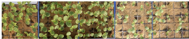
Gamma rays induce changes in variables associated with germination. At lower levels, the changes were positive or had averages similar to the control; at higher doses, a drastic reduction in germination parameters was observed (Figure 1). Some evaluated variables, such as Germination Rate (GR) and Germination Speed Index (GSI)were significantly affected (p < 0.001). The GP of the 300 and 400 Gy doses differed from the 100 and 200 Gy doses, remaining similar to the control (Figure 1), and the 100 and 200 Gy doses increased GP, with averages between 40 and 45%, compared to the control (21%). Lower values of GP than the control were obtained for the 300 and 400 Gy doses (Table 1). This result contrasts with previous studies on other watermelon varieties, such as ‘Arka Manik’ and ‘Arka Muthu’, whose GPs remained stable up to 300 Gy, with changes only occurring at 400 and 500 Gy doses (Gowda et al., 2022). However, TG in watermelon was also reduced by 5.88% at a 350 Gy dose (Yin et al., 2023).
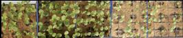
Effects of gamma ray doses on the germination of mini watermelon (‘Sugar Baby’ variety): (a) control, (b) 100 Gy, (c) 200 Gy, (d) 300 Gy, and (e) 400 Gy
The development of watermelon seedlings was affected by gamma ray doses. At dose of 200 Gy and higher, growth was reduced, with SL decreasing from 4.49 cm in the control to 1.57 cm at 400 Gy. A similar trend was observed for CoW, CoL, and NL, with reductions occurring only at 300 and 400 Gy. All doses resulted in a decrease in LL. In contrast to previous findings, NCo was not affected by gamma ray dosages. The SR of the plants at 15 days after transplant was also impacted by the applied doses, with a decline starting at 200 Gy (96.7%). At 300 Gy, survival dropped to 21.5% compared to 200 Gy, and at 400 Gy, it was reduced by 42.7% compared to 300 Gy (Table 1). Similar morphological effects have been observed in Phaseolus at 150 Gy (Ulukapi & Ozmen, 2018).
For the mini watermelon ‘Sugar Baby’, doses of 100 and 200 Gy did not negatively affect germination, maintaining a high GR of 97 and 90%, respectively. In contrast, at higher doses (300 and 400 Gy), GR dropped to 63 and 59%, respectively. The behavior at 100 and 200 Gy was similar to that observed in melon (Cucumis melo) (Sousa et al., 2025). The performance of ‘Sugar Baby’ was superior to reports on other watermelon varieties. Studies on ‘Kaolack’ and ‘Crimson Sweet’ varieties showed radiosensitivity at 100 and 200 Gy, with a TG of 83 and 60%, respectively. At 300 and 400 Gy, the results are similar to those for ‘Sugar Baby’, with GR values of 70 and 40% for Kaolack and Crimson Sweet (Ernest et al., 2020). Changes were only observed at 300 Gy in Arka Manik and Arka Muthu cultivars (Gowda et al., 2022).
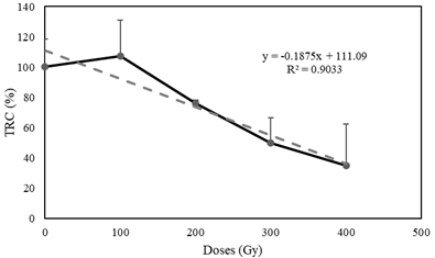
Regarding the SR, ‘Sugar Baby’ showed a high rate of 100% at 100 Gy and in the control group. However, SR decreased to 96.7% at 200 Gy. The response of ‘Sugar Baby’ at 200 Gy was similar to the control group of ‘Kaolack’ (96.66%) (Ernest et al. 2020), indicating the low radiosensitivity of ‘Sugar Baby’ to gamma rays. The LD50 of ‘Sugar Baby’ was 325.81 Gy, whereas Kaolack and Crimson Sweet had LD50 values of 225.40 and 221.56 Gy, respectively (Ernest et al., 2020). For Arka Manik and Arka Muthu varieties, the LD50 was 400 Gy (Gowda et al., 2022).
The growth curve showed a positive trend up to the 100 Gy dosage, with a slight increase. After this dosage, growth was severely affected, exhibiting a notable reduction (Figure 2). ANOVA revealed a statistically significant relationship between GRR and dosage (p < 0.05) with 95.0% confidence and a correlation coefficient of -0.98855, indicating a strong negative relationship between the variables. Thus, GRR followed a negative linear trend, demonstrating that for each dosage increase, growth tended to decrease significantly. According to the linear equation, the LD50 for ‘Sugar Baby’ watermelon was 325.81 Gy (Table 2).
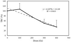
Correlation between the relative growth rate and different doses of gamma rays (Co-60) tested for the ‘Sugar Baby’ watermelon variety.
Phenotypic variations induced by Cobalt-60 gamma rays in watermelon
Gamma irradiation induces alterations in the morphological, developmental, and phenological traits of mini watermelons. Among the evaluated variables, only PW remained unchanged, whereas the remaining traits exhibited statistically significant differences (p < 0.001). Gamma ray application was correlated with modifications in plant architecture. All irradiation doses influenced NuS, leading to an increase beyond a single stem, with some plants developing up to four stems. The highest values were recorded at 200 and 400 Gy (Table 3). Although observed at a low frequency, alterations in GH were detected, with 0.58% of the total evaluated population exhibiting a determinate, shrub-like GH under the 100 and 300 Gy treatments (Table 2 of the Annex). This modification in GH has been previously documented in cucurbit mutants and is associated with a truncated protein lacking the conserved domain necessary for 2-oxoglutarate binding (Wei et al., 2019).
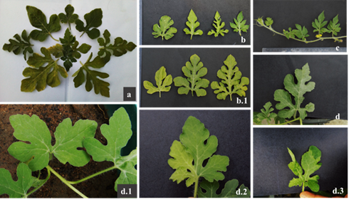
The leaf morphology variables exhibited variations in size; LW was smaller (8.1 cm) at the 300 Gy dose and larger (9.53 cm) at the 200 Gy dose. Similar variations in leaf size due to Co-60 gamma ray exposure have been observed in melon (Sousa et al., 2025). A comparable trend was observed for LW (8.11 cm), with the lowest values at the 300 Gy dose (Table 3). Morphological characteristics, such as LLD, demonstrated morphological diversity induced by gamma radiation (Figure 3b and b.1), including variations in leaf margins, changes in leaf type (Figure 3a and c), and leaves with deformities or chimeric color variations (Figure 3d.2). The latter was classified into two new categories, 2 and 3 (light green and dark green), with frequencies of 4.62% for light green across all doses and 0.87% for dark green at the 100 and 300 Gy doses (Table 2 of the Annex). In some mutants, plants that remain green throughout the growth period are classified as Chlorina (Raina et al., 2022).
The low chlorophyll levels observed within the evaluated population resulted in individuals with lighter green leaves, with frequencies of 0.58 and 0.29% at the 100 and 300 Gy doses, respectively. These changes may be associated with genes involved in chlorophyll biosynthesis (HEMA, HEMD, CHL1, CHLM, and CAO), potentially explaining the reduced chlorophyll pigment content. Similarly, variations in chlorophyll metabolism-related genes, such as PDS, ZDS, and VDE (Xu et al., 2023), may be linked to individuals with more intense green pigmentation, as observed in 4.62% of individuals in this study. This suggests possible differences in light utilization for chlorophyll synthesis, as observed in other cucurbit mutants (Zhang et al., 1996). Leaf variations, including changes in the color and margin structure, have been reported in EMS- and gamma ray-induced mutations, leading to yellowish, gray, chimeric, large, or small leaves at frequencies of 4.75% (Yin et al., 2023).
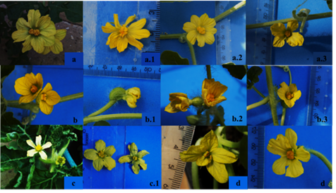
DF was affected, exhibiting an average increase of 5 days. Under normal conditions, DF was 60 days; however, at the 300 Gy dose, it fluctuated around 65 days (Table 3). In some cases, individuals at the 400 Gy dose had a DF of 80 days. Regarding morphology, certain anomalies in floral structure were observed (Figure 4). TAF was detected at all doses, with a higher incidence at 400 and 100 Gy. These included Siamese flowers, which were either both male, male and female, or both female (Figure 4b, b.1, and b.2). Additionally, NPA was recorded, with a significant increase at higher doses (300 and 400 Gy). Flowers with more than five petals were observed (Table 3), reaching up to nine petals, along with variations in petal color and morphology (Figure 5a and a.1). Some flowers exhibited a reduced petal count, ranging from three to four (Figure 4a.3). Similar morphological alterations have been previously reported in watermelon (Yin et al., 2023).
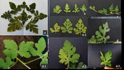
Influence of gamma rays on the leaf morphology of watermelon variety ‘Sugar Baby’. a) Diversity of leaf shapes; b) variations in the margins; b.1) degrees of lobing; c) changes in leaf type; d) leaf with lateral deformation; d.1-d.3) leaf with deformation without an apex segment; d.2-d.3) color variations.

Anomalies in flowers induced by gamma ray doses in watermelon variety ‘Sugar Baby’. a) Variations in the number of petals and anthers; a.1) more than five petals with an elongated shape; a.2) more than five petals with a rounded shape; a.3) fewer than five petals; b) formation of Siamese flowers; b.1) fusion of male and female flowers; b.2) fusion of female flowers; b.3) hermaphrodite flower; c) variations in petal shape and color; c.1) asymmetrical flowers; d) asymmetry or fusion of petals.
NA exhibited variations across all evaluated doses, with a reduction in number to two anthers at the 100 Gy dose and an increase to up to four anthers at the 300 and 400 Gy doses. For NHF, the quantity increased under 100 and 200 Gy doses (Table 3) (Figure 4b.3), while during flowering, HF was higher at 100, 200, and 300 Gy doses, totaling 5.22% (Table 2 of the Annex). Other anomalies associated with petal morphology were observed, with changes affecting all petals and resulting in thin petals with irregular edges (Figure. 4c and 4c.1) or irregular petal sizes, as observed in Figure 4d and d.1 (NPA). The 400 Gy dose accounted for up to 46.79% of the total anomalies induced by gamma rays (Table 2 of the Annex). The findings regarding floral structure, such as NA, provide new insights into the effects of gamma rays on watermelon. Variations related to flowering were also observed, including Siamese or clustered flowers and flowers with 2-4 anthers at the 300 and 400 Gy doses. These individuals could be useful for understanding the floral biology of watermelon. Lower radiation doses have previously been tested for haploid embryo induction, starting at 275 Gy (Taşkin et al., 2013).
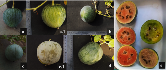
The applied doses also affected FW, with a lower average recorded at the 300 Gy dose, whereas the 200 Gy dose produced the highest FW (207 g) (Table 3). FS also varied, with the emergence of two new categories-flattened and elliptical-in addition to the typical round shape of the ‘Sugar Baby’ variety (Figure 5a, b, and d). Variations in fruit morphology are illustrated in Figure 6. Color distribution included an additional category (3) for treated individuals, corresponding to fruit with wide stripes at a frequency of 1.76% for the 200, 300, and 400 Gy doses (Table 2 of the Annex). Variations in fruit characteristics have been previously reported in watermelon, although with differences in frequency and category distribution (Yin et al., 2023).
The rind color was affected by gamma rays, resulting in two new PRC categories, 3 and 4 (light green and yellowish green). In the case of SRC, category 3 had a frequency of 0.87% (Figure 5c). The pulp color also exhibited changes, with the emergence of three new categories in treated plants, showing frequencies of 5.81, 3.78, and 5.52% (Table 2 of the Annex) corresponding to deep red, orange, and green, respectively (Figure 5e). PW showed no significant variations across doses, while SN decreased at the 300 Gy dose, corresponding to a lower FW (Table 3). Studies on other cucurbit species highlight the effects of mutagens on carotenoid pathway alterations, leading to pro-lycopene accumulation instead of β-carotene accumulation, which causes pulp color changes in mutants (Galpaz et al., 2013), similar to findings observed with EMS in watermelon (Zhang et al., 2022). The variation in green pulp color is novel for watermelon, as previous studies have only reported changes in the reddish spectrum (Yin et al., 2023).
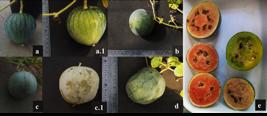
Influence of gamma rays on fruit morphological variation. a) Variations in fruit shape a-a1; b) reduction in fruit size; c) variations in fruit epidermis; d) increase in size and changes in shape; e) variations in pulp color.
In addition to responding well to the tested doses, the ‘Sugar Baby’ variety exhibited interesting phenotypic characteristics for genetic improvement. One of these was the change in growth habit. Considering that dwarf architecture is an important and valuable agronomic trait in watermelon breeding (Wei et al., 2019), gamma rays proved effective in inducing individuals with this characteristic. Phenotypically, the change in growth habits was more evident in seedlings and after the first month. When changes in leaf chlorophyll were associated with bushy growth habits, which negatively affected plant survival, lower radiation levels only resulted in a light green leaf color and bushy growth habits, with plants surviving for only a few days.
Phenotypic diversity induced by gamma rays
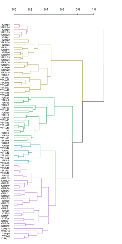
The phenotypic diversity resulting from gamma ray effects was grouped by treatment. The highest dose in this study, 400 Gy (Y4), generated four groups. Group I consisted of two individuals who were markedly distant from the remaining groups, with a dissimilarity of 0.8. These individuals (Y4R4P7 and Y4R4P8) exhibited various physiological disorders, such as yellowed leaves, reduced growth in both stems and leaves, and seedless fruit. Group II, composed of nine individuals, showed the fewest variations and maintained characteristics similar to the control, with only some floral anomalies. Groups III and IV exhibited developmental changes, with some individuals producing small fruit or short stems (Figure 6).

Phenogram based on Gower’s dissimilarity index grouping the phenotypic diversity related to morphological changes and physiological disorders in the 400 Gy (Y4) treatment, which was categorized into four phenotypic groups.
The 300 Gy dose, induced the most phenotypic diversity. Group I, consisting of five individuals, failed to produce seed-bearing fruit, and the flowers were generally anomalous, with a dissimilarity > 1. Groups II (5) and III (12), located at a distance of 0.8 from the other groups, were formed by individuals with variations in leaf morphology, color, and fruit with differentiated skin and reduced size. Groups IV (11), V (4), and VI (12) exhibited slight morphological changes, with a reduction in leaf size for groups IV and V, showing a distance of 0.6. Group VI showed some minor changes in flowers but retained many characteristics similar to the control (Figure 7).
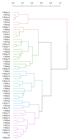
Phenogram based on Gower’s dissimilarity index grouping the phenotypic diversity related to morphological variations induced by the 300 Gy (Y3) treatment, which was categorized into six phenotypic groups.
For the 200 Gy dose, five groups were generated, with four of them being > 1 distant from Group I, corresponding to individuals with difficulties in flower and fruit production (6 individuals). Group II was at a distance of 0.8 from the other groups and consisted of 22 individuals exhibiting vigorous plants with large leaves, more than one stem, large fruit with higher FW values, and variation in FC. The remaining groups retained characteristics similar to the control, with some changes in leaf characteristics, lobulation, leaf color, and NHF (Figure 8).

Phenogram based on Gower’s dissimilarity index showing the phenotypic diversity of the 200 Gy (Y2) treatment in watermelon, which was grouped into five phenotypic groups.
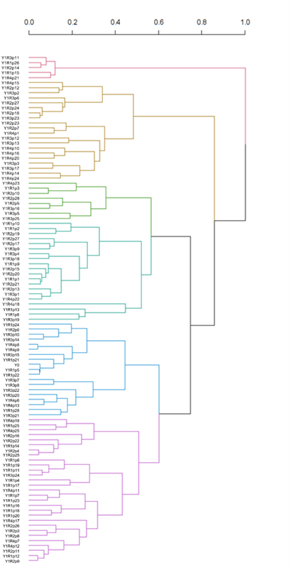
Similar to the 200 Gy dose, the 100 Gy dose produced six groups. Group I (5 individuals) exhibited variations in the number of flowers, especially hermaphroditic flowers, and was the most distant group (> 1). Group II was > 0.8 distance from the other groups and consisted of 20 individuals presenting variations in LLD and pulp color. Groups III and IV formed a distinctive group, showing differences from the control, such as secondary distribution of rind and pulp color. Individuals in Groups V and IV exhibited a phenotype similar to the control, with slight changes in flowering and development (Figure 9).
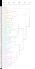
Phenogram based on Gower's dissimilarity index showing the phenotypic diversity of the 100 Gy (Y1) treatment in watermelon, which was grouped into six phenotypic groups.
Mutation detection using molecular markers
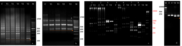
Of the 74 molecular markers evaluated, the RAPD markers from the OPA series did not provide relevant information nor did they show polymorphism. In the OPM series, only primer 02 showed a polymorphic band larger than 100 bp for the 100 and 200 Gy treatments compared to the control. In the OPD series, primer 08 exhibited polymorphic bands for the 100 and 200 Gy treatments, with fragments of 400 bp but with unreliable bands. Of the ISSR primers, only primers 1, 4, 6, 7, and 8 (DiCA3´YG, DiGA3'RC, DiGA5'CR, DiCA5'CY, and TriGTG) displayed polymorphic bands. For the evaluated SSR primers, only primer BVWS00106 showed polymorphism for the 400 Gy treatment compared to the control (C) (Figure 10). Primer BVWS00106 has been previously studied in watermelon genetic diversity and is related to growth habit and pulp color traits (Pal et al., 2020).
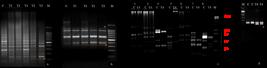
Agarose gel for amplification of RAPD, ISSR, and SSR markers aimed at detecting the effects of gamma rays at the molecular level comparing the gamma ray treatments (Y) and the control (C). a) Primer OPM 02 for 100 (Y1) and 200 Gy (Y2) with two repetitions; b) primer OPD08; c) primer ISSR 01-08 for the 400 Gy treatment (Y4) versus C; d) primer SSR BVWS00106 for the Y4 treatment versus C. The letter M represents the weight marker.
In the molecular marker analysis, only two RAPD primers displayed polymorphism and identified mutant individuals. In their genetic characterization of fig plants irradiated with gamma rays for Polianthes tuberosa L., the DNA polymorphism was dose-dependent (Hajizadeh et al., 2023), they also identified the limited efficiency of RAPD markers to detect mutated regions. Although few RAPD and SSR markers were polymorphic, a higher rate of polymorphism was observed for ISSR markers. This polymorphism was derived from ISSR amplification of variations in DNA sequences at the primer binding site. According to Pradeep Reddy et al. (2002), the origin of this polymorphism can be attributed to mutations at sites and/or insertion/deletion events in the SSR region. Therefore, ISSR markers demonstrate considerable discriminative power regarding mutants or mutant loci (and their genetic diversity), which is in agreement with similar studies (Amorim et al., 2012; Taheri et al., 2014). This method offers an alternative to other systems, enabling highly reproducible markers to be obtained without prior sequence information for various genetic analyses. Due to the abundance and rapid evolution of SSR regions, ISSR amplification has the potential to reveal a much larger number of polymorphic fragments per primer than any other marker system, such as restricted fragment length polymorphisms (RFLPs) and microsatellites. In the future, for individuals with promising traits, evaluations could be carried out to identify mutation sites and single nucleotide polymorphisms (SNPs) and insertion-deletion polymorphisms (InDels), as has been done for other members of the Cucurbitaceae (Zhu et al., 2022).
The use of Co-60 gamma rays was efficient, and the calculation of LD50 indicates that the ideal dose to generate variability in mini-watermelon variety ‘Sugar Baby’ is 325.81 Gy. This study generated 17 different morphotypes, which were easily detectable through variations in morphological characteristics in flowers, fruit, and leaves and molecular-level changes detectable with non-specific markers and microsatellites. Moreover, the potential use of ionizing radiation in Cucurbitaceae, specifically in melon and watermelon, is generally recommended to induce genetic variability, improve drought tolerance characteristics, and as a sustainable production method (Food and Agriculture Organization of the United Nations, 2023).
Conclusion
Ionizing radiation proved effective in generating morphological variability in mini-watermelon, resulting in morphotypes distinct from the control with potential for watermelon production. Additionally, variations, particularly in the green pulp color, were reported herein for the first time. Phenotypic variability was observed and detected using molecular markers. For the ‘Sugar Baby’ variety, doses below 325 Gy may be useful in generating phenotypic variability. Among the evaluated doses, 100 Gy promoted development, while 200 and 300 Gy doses generated individuals with morphological changes. Generational advancements and further studies will be conducted on individuals of interest.
Data availability
The data supporting the conclusions of this study are available in the article and also via the link https://www.mediafire.com/file/nnq2j28s83ndw78/APPENDIX_watermelon_mutants.docx/file.
References
-
Ahloowalia, B. S., Maluszynski, M. & Nichterlein, K.. (2004). Global impact of mutation-derived varieties. Euphytica, 135(2), 187-204. https://doi.org/10.1023/B:EUPH.0000014914.85465.4f
» https://doi.org/https://doi.org/10.1023/B:EUPH.0000014914.85465.4f -
Ahmar, S., Gill, R. A., Jung, K., Faheem, A. Q, Mubeen, M. & Zhou, W.. (2020). Conventional and molecular techniques from simple breeding to speed breeding in crop plants: recent advances and future outlook. International Journal of Molecular Sciences, 21(7), 1-24. https://doi.org/10.3390/ijms21072590
» https://doi.org/https://doi.org/10.3390/ijms21072590 -
Amorim, E. P., Pestana, R. K. N. & Oliveira e Silva, S.. (2012). Caracterização agronômica de mutantes de bananeira obtidos por meio da radiação gama. Bragantia, 71(1), 8-14. https://doi.org/10.1590/S0006-87052012005000002
» https://doi.org/https://doi.org/10.1590/S0006-87052012005000002 - Brasil (2009). Regras para análise de sementes. MAPA/ACS.
- Coretchi, L., Cliciuc, D. & Bondarenco, E. (2018). Molecular and genetic aspects of the resistance of leguminous plants to biotic stress factors. In International Symposium on Plant Mutation Breeding and Biotechnology. FAO/IAEA.
-
Datta, S., Jankowicz-Cieslak, J., Nielen, S., Ingelbrecht, I. & Till, B. J.. (2018). Induction and recovery of copy number variation in banana through gamma irradiation and low-coverage whole-genome Sequencing. Plant Biotechnology Journal, 16(9), 1644-1653. https://doi.org/10.1111/pbi.12901.
» https://doi.org/https://doi.org/10.1111/pbi.12901. - Doyle, J. J. & Doyle, J. L.. (1990). Isolation of plant DNA from fresh tissue. Focus, 12(1), 13-15.
-
Ernest, F. P., Noëlle, M. A. H., Godswill, N., Thiruvengadam, M., Simon, O. A., Bille, N. H., Rebezov, M. & Shariati, M. A.. (2020). Radiosensitivity of two varieties of watermelon (Citrullus lanatus) to different doses of gamma irradiation. Brazilian Journal of Botany, 43(4), 897-905. https://doi.org/10.1007/s40415-020-00659-8
» https://doi.org/https://doi.org/10.1007/s40415-020-00659-8 - European Cooperative Programme for Plant Genetic Resources (2008). Minimum descriptors for Cucurbita spp., cucumber, melon and watermelon. ECPGR.
- Food and Agriculture Organization of the United Nations (2023). Plant breeding & genetic newsletter. FAO/IAEA.
-
Galpaz, N., Burger, Y., Lavee, T., Tzuri, G., Sherman, A., Melamed, T., Eshed, R., Meir, A., Portnoy, V., Bar, E., Shimoni-Shor, E., Feder, A., Saar, Y., Saar, U., Baumkoler, F., Lewinsohn, E., Schaffer, A. A., Katzir, N. & Tadmor, Y.. (2013). Genetic and chemical characterization of an ems-induced mutation in Cucumis melo CRTISO gene. Archives of Biochemistry and Biophysics, 539(2), 117-25. https://doi.org/10.1016/j.abb.2013.08.006
» https://doi.org/https://doi.org/10.1016/j.abb.2013.08.006 -
Gowda, A. S., Prakash, B. G., Halesh, G. K., Prashant, M., Sood, M. & Krishna, H. C.. (2022). Estimation of lethal (LD50) doses of gamma ray irradiation in watermelon (Citrullus lanatus Thunb). International Journal of Plant & Soil Science, 34(20), 215-19. https://doi.org/10.9734/IJPSS/2022/V34I20365370
» https://doi.org/https://doi.org/10.9734/IJPSS/2022/V34I20365370 -
Gurushidze, M., Hiekel, S., Otto, I., Hensel, G. & Kumlehn, J. (2017). Site-directed mutagenesis in barley by expression of tale nuclease in embryogenic pollen. In J. Jankowicz-Cieslak, T. Tai, J. Kumlehn & B. Till (Eds.), Biotechnologies for plant mutation breeding. (pp. 113-128). Springer. https://doi.org/10.1007/978-3-319-45021-6_7
» https://doi.org/https://doi.org/10.1007/978-3-319-45021-6_7 -
Hajizadeh, H. S., Mortazavi, S. N., Ganjinajad, M. & Okatan, V.. (2023). Evaluation of the optimum threshold of gamma-ray for inducing mutation on Polianthes tuberosa cv. double and analysis of genetic variation with RAPD marker. International Journal of Radiation Biology, 99(8), 1204-1216. https://doi.org/10.1080/09553002.2023.2159566
» https://doi.org/https://doi.org/10.1080/09553002.2023.2159566 -
Holme, I. B., Gregersen, P. L. & Brinch-Pedersen, H.. (2019). Induced genetic variation in crop plants by random or targeted mutagenesis: convergence and differences. Frontiers in Plant Science, 10, 1-9. https://doi.org/10.3389/fpls.2019.01468
» https://doi.org/https://doi.org/10.3389/fpls.2019.01468 -
Jankowicz-Cieslak, J., Tai, T. H. & Kumlehn, J. (2016). Biotechnologies for plant mutation breeding: protocols. Springer Nature. https://doi.org/10.1007/978-3-319-45021-6
» https://doi.org/https://doi.org/10.1007/978-3-319-45021-6 -
Ling, L., Jiafeng, J., Jiangang, L., Minchong, S., Xin, H., Hanliang, S. & Yuanhua, D. (2014). Effects of cold plasma treatment on seed germination and seedling growth of soybean. Scientific Reports, 4(5859), 1-10. https://doi.org/10.1038/srep05859
» https://doi.org/https://doi.org/10.1038/srep05859 -
Lundqvist, U., Franckowiak, J. D. & Forster, B. P. (2012). Mutation categories. In Q. Y. Shu, B. P. Forster & H. Nakagawa (Eds.), Plant mutation breeding and biotechnology. (pp. 47-55). CABI Publishing. https://doi.org/10.1079/9781780640853.0047
» https://doi.org/https://doi.org/10.1079/9781780640853.0047 - Maluszynski, M., Szarejko, I., Bhatia, C. R., Nichterlein, K. & Lagoda, P. J. L. (2009). Methodologies for generating variability part 4: mutation techniques. In S. Ceccarelli, E. P. Guimarães & E. Weltzien (Eds.), Plant breeding and farmer participation. (pp. 159-194). FAO.
-
Marín-Huachaca, N. S., Mancini-Filho, J., Delincée, H. & Villavicencio, A. L. C. H.. (2004). Identification of gamma-irradiated papaya, melon and watermelon. Radiation Physics and Chemistry, 71(1-2), 193-196. https://doi.org/10.1016/j.radphyschem.2004.04.026
» https://doi.org/https://doi.org/10.1016/j.radphyschem.2004.04.026 -
Pal, S., Revadi, M., Thontadarya, R. N., Reddy, D. C. L., Pandey, C. D. & Rao, E. S.. (2020). Understanding genetic diversity, population structure and development of a core collection of indian accessions of watermelon (Citrullus lanatus (Thunb.) Matsum. and Nakai). Plant Genetic Resources: Characterisation and Utilisation, 18(5), 359-368. https://doi.org/10.1017/S1479262120000386
» https://doi.org/https://doi.org/10.1017/S1479262120000386 -
Parry, M. A. J., Madgwick, P. J., Bayon, C., Tearall, K., Hernandez-Lopez, A., Baudo, M., Rakszegi, M., Hamada, W., Al-Yassin, A., Ouabbou, H., Labhilili, M. & Phillips, A. L.. (2009). Mutation discovery for crop improvement. Journal of Experimental Botany, 60(10), 2817-2825. https://doi.org/10.1093/jxb/erp189
» https://doi.org/https://doi.org/10.1093/jxb/erp189 -
Pradeep Reddy, M., Sarla, N. & Siddiq, E. A.. (2002). Inter simple sequence repeat (ISSR) polymorphism and its application in plant breeding. Euphytica, 128(1), 9-17. https://doi.org/10.1023/A:1020691618797
» https://doi.org/https://doi.org/10.1023/A:1020691618797 -
Raina, A., Laskar, R. A., Wani, M. R., Jan, B. L., Ali, S. & Khan, S.. (2022). Comparative mutagenic effectiveness and efficiency of gamma rays and sodium azide in inducing chlorophyll and morphological mutants of cowpea. Plants, 11(10), 1-26. https://doi.org/10.3390/plants11101322
» https://doi.org/https://doi.org/10.3390/plants11101322 - Shu, Q. Y. (2009). Induced plant mutations in the genomics era. FAO/IAEA.
-
Sousa, W. K., Alpala, D. A. R., Cunha, E. S. P., Nunes, G. H. S., Tulmann Neto, A. & Holanda, I. S. A.. (2025). Response of melon accessions to doses of co60 gamma rays and their effects on the morphology of the M1 generation. Revista Ciência Agronômica, 56, 1-9. https://doi.org/10.5935/1806-6690.20250024
» https://doi.org/https://doi.org/10.5935/1806-6690.20250024 - Spencer-Lopes, M. M., Forster, B. P. & Jankuloski, L. (2018). Manual on mutation breeding (3rd ed.). FAO/IAEA.
-
Taheri, S., Abdullah, T. L., Ahmad, Z. & Abdullah, N. A. P.. (2014). Effect of acute gamma irradiation on curcuma alismatifolia varieties and detection of dna polymorphism through ssr marker. BioMed Research International, 2014, 1-18. https://doi.org/10.1155/2014/631813
» https://doi.org/https://doi.org/10.1155/2014/631813 -
Taşkin, H., Yücel, N. K. & Baktemur, G.. (2013). Effects of different genotypes and gamma ray doses on haploidization with irradiated pollen technique in watermelon (Citrullus lanatus L.). Canadian Journal of Plant Science, 93(6), 1165-1168. https://doi.org/10.4141/CJPS2013-059
» https://doi.org/https://doi.org/10.4141/CJPS2013-059 -
Ulukapi, K. & Ozmen, S. F.. (2018). Study of the effect of irradiation (60Co) on M1 plants of common bean (Phaseolus vulgaris l.) cultivars and determined of proper doses for mutation breeding. Journal of Radiation Research and Applied Sciences, 11(1-2), 157-161. https://doi.org/10.1016/j.jrras.2017.12.004
» https://doi.org/https://doi.org/10.1016/j.jrras.2017.12.004 -
Wei, C., Zhu, C., Yang, L., Zhao, W., Ma, R., Li, H., Zhang, Y., Ma, J., Yang, J. & Zhang, X.. (2019). A point mutation resulting in a 13 bp deletion in the coding sequence of cldf leads to a ga-deficient dwarf phenotype in watermelon. Horticulture Research, 6(132), 1-12. https://doi.org/10.1038/s41438-019-0213-8
» https://doi.org/https://doi.org/10.1038/s41438-019-0213-8 -
Xu, B., Zhang, C., Gu, Y., Cheng, R., Huang, D., Liu, X. & Sun, Y.. (2023). Physiological and transcriptomic analysis of a yellow leaf mutant in watermelon. Scientific Reports, 13(9647), 1-11. https://doi.org/10.1038/s41598-023-36656-6
» https://doi.org/https://doi.org/10.1038/s41598-023-36656-6 -
Yin, L., Hou, Y., Chen, X., Huang, X., Feng, M., Wang, C., Wang, Z., Yue, Z., Zhang, Y., Ma, J., Li, H., Zhang, X., Yu, R. & Wei, C.. (2023). Construction of watermelon mutant library based on 60Co γ-ray irradiation and EMS treatment for germplasm innovation. Horticulturae, 9(10), 1-13. https://doi.org/10.3390/horticulturae9101133
» https://doi.org/https://doi.org/10.3390/horticulturae9101133 -
Zhang, X. P., Rhodes, B. B., Baird, W. V., Skorupska, H. T. & Bridges, W. C.. (1996). Phenotype, inheritance, and regulation of expression of a new virescent mutant in watermelon: juvenile albino. Journal of the American Society for Horticultural Science, 121(4), 609-615. https://doi.org/10.21273/jashs.121.4.609
» https://doi.org/https://doi.org/10.21273/jashs.121.4.609 -
Zhang, J., Sun, H., Guo, S., Ren, Y., Li, M., Wang, J., Yu, Y., Zhang, H., Gong, G., He, H. & Zhang, C.. (2022). ClZISO mutation leads to photosensitive flesh in watermelon. Theoretical and Applied Genetics, 135(5), 1565-1578. https://doi.org/10.1007/s00122-022-04054-7
» https://doi.org/https://doi.org/10.1007/s00122-022-04054-7 -
Zhu, Y., Yuan, G., Wang, Y., An, G., Li, W. & Liu, J.. (2022). Mapping and functional verification of leaf yellowing genes in watermelon during whole growth period. Frontiers in Plant Science, 13, 1-17. https://doi.org/10.3389/fpls.2022.1049114
» https://doi.org/https://doi.org/10.3389/fpls.2022.1049114
Edited by
-
Associate Editor in charge:
Alessandro Lucca BracciniCarlos Alberto Scapim