VLP (Virus-Like Particles) Vaccines - Current Knowledge and Future Directions
Abstract
Viruses are acellular, potential pathogenic agents causing infections, ranging from acute to chronic. A wide range of conventional immunization methodologies have been employed to control viral diseases. But, their ability to mutate or remain inactive in environment empowers their survival. Hence, novel particles capable of mimicking such viruses to immunize susceptible organisms and not cause harm is the need-of-the-hour. Virus-like particles (VLPs) are virus-derived entities containing only antigenic determinant portion of target virus with the ability to self-assemble, mimic form and size of target virus but lack genetic material. The lack of genetic material renders them incapable of infecting hosts thereby serving as safe and reliable way to combat infectious diseases. However, expression of viral proteins can occur in various expression systems. VLPs mount immune responses similar to original viruses by inducing either humoral or cell-mediated immune responses or both. Recent advancements in biomedical engineering technologies have led to approved commercial use of VLP-based vaccines against three infectious agents namely, Hepatitis B virus, Human Papillomavirus and Hepatitis E virus due to production of efficacious and enduring immune response. However, enhancement of immunogenicity of antigenic determinants displayed on VLP surface is the main objective behind use of VLP-based vaccines. An increasing attention towards VLP-based vaccines has led to development of mono-, diand multi-valent VLP particles capable of combating many viral infectious agents simultaneously. This review provides an insight on the importance, characteristics, development, and application of VLP-based vaccines to combat emerging diseases, and as nanoparticles to deliver drugs against cancer.
Keywords:
Cancer vaccine; Infectious disease vaccine; Immune response; Nano-drug delivery system; Virus-like particles (VLPs).
HIGHLIGHTS
VLPs are antigenic determinant bearers lacking any viral genetic material.
They are highly efficient in immuno-stimulation compared to conventional viral vaccines.
The construction, expression, production and formulation of VLP-based vaccines is tedious and expensive.
VLP-based vaccines against various infectious diseases and cancers are in use for treatment purposes.
INTRODUCTION
Viruses are obligate intracellular pathogens infecting a wide range of organisms including both eukaryotes and prokaryotes. These pathogenic agents have a unique feature where their envelope, capsid and other structural proteins can independently or spontaneously self-assemble encompassing genetic material, either DNA or RNA, forming mature virions [1]. Although, vaccinations available against many viral infections are mostly based on attenuated or inactivated forms of live viruses, they may increase the risk of these entities reverting to their virulent forms leading to development of various diseases and their outbreaks. The property of self-assembly in viruses serve as one the principles in construction of Virus-like particles (VLP), that are avirulent subunit vaccines completely lacking genetic material of target viruses thereby reducing the risk of reversion and re-assortment into virulent forms [2]. VLPs are biocompatible molecules exhibiting structural flexibility during synthesis allowing for modification by chemical or molecular methods. They exhibit high uniformity, stability and functionality relative to other forms of viral vaccines [3], [4]. VLPs can be developed from many viruses by cloning their structural genes coding for desired viral proteins into an expression vector whose choice depends on expression system in which viral proteins will be expressed. Hence, when the expression system differs from the usual host of the target virus, the protein gets codon-optimized. Resultant chimeric DNA is either transformed/ transfected into appropriate expression system in which the DNA is transcribed and translated. The translated viral proteins are folded and assembled to form VLPs [5].
VLPs have wide variety of applications in the field of biomedical science, including: (i) Vaccine development against emerging diseases, (ii) As nanomaterials in site-specific drug delivery, (iii) In vivo imaging on tagging with fluorophores, (iv) In diagnostic tests as positive controls for infectious diseases, and (v)Therapies like, gene therapy and cancer treatment [1], [6]. Despite technological advancement and promising results in control of many diseases, a variety of challenges remain for the development of vaccines such as, effective defence against pathogenic agents for which vaccines have not yet been developed, as VLP-based vaccines cannot be produced for all virus types. reduction in cost of vaccine production, establish manufacturing platforms for worldwide supply and, increasing vaccine safety in order to comply with regulatory guidelines [2], [7]. As a result, the demand for improved vaccine safety at the level of manufacture has been increased by regulatory bodies such as, FDA or EMA and by antivaccination groups. Thus, new molecular techniques can help improve the safety of vaccines in its design and manufacturing process [7].
The application of molecular techniques has enabled in-vivo and in-vitro production of VLPs. VLPs produced in vivo are used as vaccines, drug delivery system and nanomaterials but, assembly of expressed capsid proteins into VLPs are accompanied by entrapment of host related contaminations hampering their use as these can mount unwanted immune response [8]. On the other hand, in in-vitro assembly, capsid proteins are expressed and purified from the expression systems through disassembly procedure that avoids entrapment of contaminants [9], [10]. The expressed proteins are incubated under defined chemical conditions promoting their assembly into VLPs. In-vitro VLPs are preferred than in-vivo as they provided possibilities to mix different antigenic determinants within the particle enabling their application against multiple viruses [11], and in-vitro VLP production technology also allows the possibility to control the number of different payloads during assembly of capsid proteins [8]. Therefore, this article highlights the various aspects including the classification, development, formulation, immunogenicity, application and future prospects of VLP-based vaccines.
Structural classification of VLPS
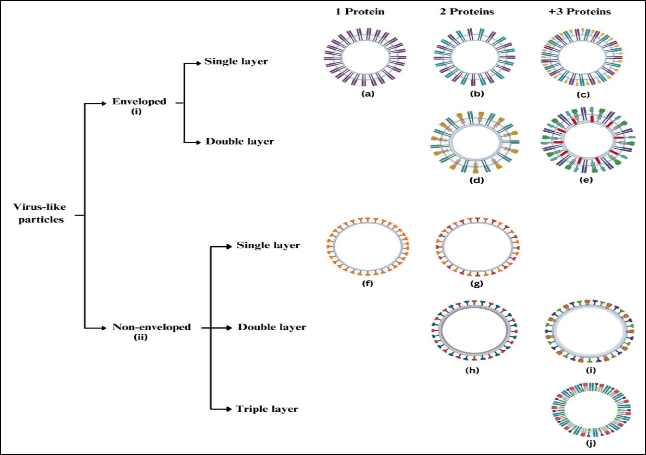
Spontaneous interaction and polymerization of one or more viral structural capsid proteins result in formation of VLPs that are structurally and visually similar to live viruses but lack viral genome. They can assume geometrical symmetries such as, icosahedral, spherical, or rod-like structures based on the source of viral protein. VLPs are largely classified into two groups based on their structural complexity including, occurrence of viral envelope and type of capsid protein (as in Figure 1). VLPs mainly constitutes of viral capsid protein which can be arranged in one, two or three layers of same (homogenous) or different (heterogenous) proteins. Single-protein VLPs have a relatively simple structure than multi-protein ones [12].
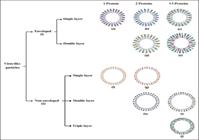
Structural classification of VLPs. (i) Enveloped VLPs (explained in section 2.2) comprising of a single layer made of (a) 1 protein, (b) 2 proteins, (c) greater than 3 proteins; and, double layer made of (d) 2 proteins, (e) greater than 3 proteins. (ii) Non-enveloped VLPs (explained in 2.1) comprising of single layer made of (f) 1 protein, (g) 2 proteins; double layer made of (h) 2 proteins, (i) greater than 3 proteins; and, triple layer made of (j) greater than 3 proteins.
Non-enveloped VLPs
This category of VLPs comprise of one, two or more components of a target virus having the ability to self-assemble into a complete particle that can elicit an immune response [13], [14]. They are often made of one or many self-assembled components of viral protein and lack lipid envelope usually derived from host cell membrane. Non-enveloped VLPs are smaller in size allowing them to cross body barriers with ease [15]. The non-enveloped VLPs are further classified into single-capsid or multi-capsid protein and, into single-layered, double-layered, and triple-layered VLPs [12].
The simplest VLPs of this kind are composed of a single capsid protein like Human Papilloma Virus (HPV) VLP vaccines, that can be expressed in different expression platforms, both eukaryotes and prokaryotes. Soluble forms of single capsid VLPs are first expressed in cell-based systems followed by assembly in cell-free environment to facilitate proper folding [16], [17], [18]. However, multiple capsid proteins expressed by various viral mRNAs like, Poliovirus and Rotavirus, form structurally complex VLPs consisting of concentric layers of different capsid proteins, commonly expressed in eukaryotic systems such as yeast, insect cells and plants. Otherwise, different proteins can be expressed and assembled in heterogenous hosts [19], [20], [21], [22], [23].
Enveloped VLPs
These nanostructures are composed of an envelope, cell membrane derived from host cell, containing one or many glycoprotein spikes embedded on the surface that acts as target for host immune system [2]. Often, enveloped VLPs contain a matrix protein situated inside the envelope where the antigenic glycoproteins are embedded [12]. VLP vaccines against influenza virus and retrovirus are examples of commonly available enveloped VLP vaccines [2]. They display high degree of flexibility due to their property to target antigenic epitopes from same or different viruses. For example, enveloped VLP vaccines derived from simian immunodeficiency virus (SIV) and human immunodeficiency virus (HIV) contained Gag protein and Env protein, respectively [24]. Their large size of greater than 100 nm make administration challenging as they might aggregate at site of injection and refrain from reaching lymph nodes thereby not serving the purpose of immunization [15].
Enveloped VLPs are subdivided into single-, doubleand multi-layer internal structures which lie beneath the envelope. These particles obtain their lipid membrane from host cell in which they are expressed, assembled and mature, fully formed particles are released by a process named budding. Thus, the glycoproteins acting as antigenic determinants get inserted into the lipid envelope during this process so that, the VLPs can be detected by the immune system to produce neutralizing immunoglobulins (or antibodies). Therefore, the nature, origin and composition of viral envelope differs amongst various viruses and depends on the source from which VLPS have been derived from. The source virus involved will also determine the assembly and budding of mature VLPs from host cell involved [12].
PRODUCTION, PURIFICATION AND CHARACTERIZATION OF VLPS
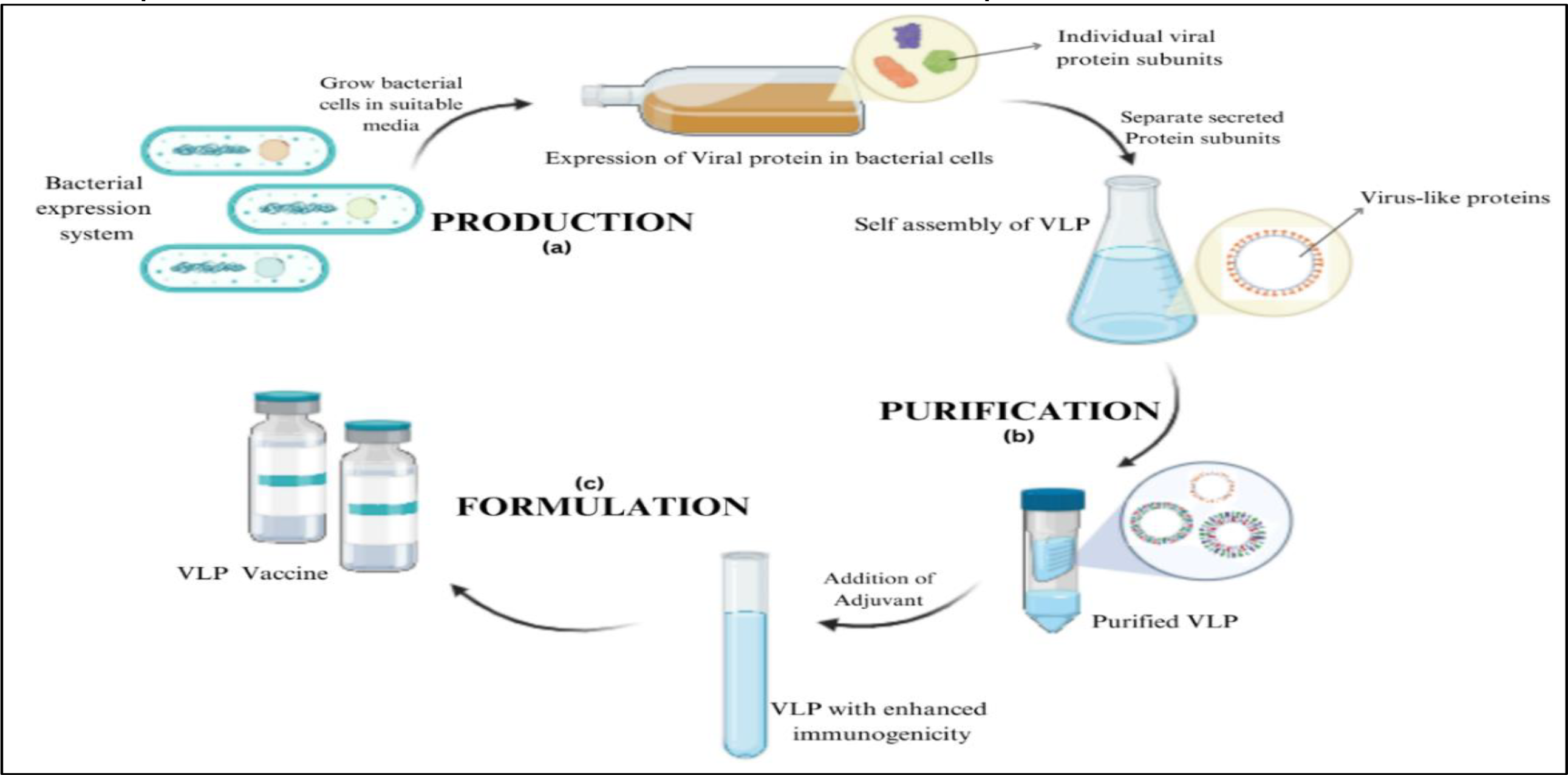
The manufacturing process consists of four sections namely, up-stream processing or production, down-stream processing or purification, formulation and characterization (as in Figure 2). Initially, viral gene of interest will be cloned, followed by its expression as self-assembling protein units in either prokaryotic or eukaryotic expression platforms occurs. The host cells are harvested, lysed and clarified in order to remove aggregates and contaminants [10], [25]. Techniques such as ultracentrifugation, ion-exchange chromatography can be employed to obtain purified VLPs [25]. Polishing is a step used to eliminate residues of host cell nucleic acids and proteins. Finally, sterile filtration and formulation, followed by characterization of the produce is performed to obtain safe, effective and efficient product.
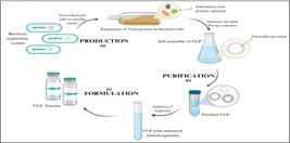
Schematic representation of an overview of VLP-based vaccines. (a) Production of VLP particles via bacterial expression systems, (b) Purification step involving various methodologies, and, (c) Formulation of produced VLPs with adjuvants and authorized excipients.
Cloning and expression of viral gene of interest
There are several fundamental roles to be followed for construction of recombinant VLPs: (a) the construction process involves several years of research on the target virus, (b) the coding nucleotide sequence for viral carrier coat protein and foreign functional peptides can be synthetically obtained from aligned oligonucleotides, (c) foreign peptides can be inserted or incorporated into the carrier without affecting the self-assembly property, (d) can be obtained from different hosts, (e) advantageous over native viruses due to easy accessibility, availability and, better functional properties [26].
The viral structural genes required for cloning can be obtained from sources like clinical specimens, environmental samples, etc., and their corresponding sequences can be deduced by comparing obtained sequence with existing ones available on National Centre for Biotechnology Information. This sequence, thus obtained, can be sufficiently used for gene synthesis in absence on infected source material. Often, such synthesis allows inclusion of a typical AA codons for heterologous host in target gene thereby enhancing the expression of desired target protein. All known and available VLPs are well characterized at levels of nucleotide sequence and genome organization. However, large genome size of each VLP makes analysis of cloning details of individual VLPs nearly impossible [26]. Moreover, depending on the target virus of VLP vaccine, different expression systems, such as bacteria, yeast, insect cells, plant cells, and mammalian cells must be employed for the expression of different viral protein subunits [27].
Upstream Processing
Batch and fed-batch bioreactors, that are directly related to cell densities, are robust processes when the culture conditions are monitored and controlled carefully. These bioreactors are best suitable for insect cells that have high growth rates in serum and/or protein-free medium cultures due to their ability to adapt to culture conditions enabling large-scale productions [28]. Features like convenience and rapid expression of desired proteins, allow control and monitor of culture condition thereby making Baculovirus Expression Vector System/ Insect Cellsbased (BEVS/ IC-based) bioreactors advantageous and attractive compared to mammalian platforms, as the latter require tedious culture adaptation procedures. In addition, it allows growth of insect cells in the absence of serum that would otherwise require intense purification in order to remove host cell impurities and serum proteins. BEVS/ IC system consists of a two-phase process where, insect cells are grown to desired viable cell density and then, infected by recombinant baculoviruses for expression of protein during log growth phase. The infection step requires determination of, multiplicity of infection (MOI), time of infection (TOI) and cell concentration at infection (CCI) [10]. The scalability of yield obtained is crucial to choose an expression platform providing higher yields such as bacteria or yeast, but these platforms don’t support the degree of complexity of VLPs as designed using BEVS/IC system. However, higher yields obtained as a result of increased expression levels can be attributed to the viruses’ ability to silence host gene transcription and facilitate overexpression of heterologous genes, that have been delivered to host cells by infection, by orchestrating cellular machinery required for the process [29].
The product yield if relatively higher than in eggor mammaliancell-based VLP production and production period can be reduced to about 12 weeks in contrast to the 20-24 weeks required for the latter [30]. The upstream processing technology employed offers numerous benefits including: (i) use single-use vessels to avoid contamination, (ii) cleaning and sterilization cost eliminated, (iii) residual validation cost, (iv) improved flexibility for process scale adjustment, (v) lower upfront investment, (vi) short turn-around time, (vii) ease of installation and little space utilization [31]. However, it is accompanied by the following drawbacks as part of using single-use or disposable vessels: (i) quality control (QC) data of limited extractable and /or leachable substance from disposable materials and manufacturers; (ii) handling difficulty at scales over 1000L; (iii) pressure limitation; (iv) high temperature limitation [31].
Though in fed-batch bioreactors the productivity can be improved across a wide range of MOIs, these are neither economical nor practical. The metabolome/proteome or fluxome tools can be employed to optimize upstream processing by integrated analysis of the metabolic fluxes that can drive online process control strategies so as to improve the productivity [32]. It can be coupled with mathematical models to analyse complex network of fluxes and identify limiting factors [33], and, thermodynamic analysis can be used to identify the most suitable environmental parameters for production and stability maintenance during purification and storage [31], [34].
In contrast to the former methods, continuous production system involving either chemostat, stressostat, turbidostat or morbidostat, could be employed to achieve highest productivity as it allows control over the specific growth rates of expression systems like bacteria, and product formation by monitoring change in either concentration of limiting substrate for specific strain, turbidity, stress levels or morbidity in the culture vessel. Moreover, this system offers minimal capital cost per unit product yield and is thus, often used for the production rabies virus VLPs in HEK293 (mammalian) cell system [35]. Despite such advantages, there can be high risk of contamination, high volumes of diluted culture medium and low concentration of product formed [36]. Moreover, the production of Measles virus VLPs is a challenging task as it causes host cell lysis leading to rapid accumulation of host cell protein and debris in the medium. To overcome this, continuous filtration during the production process could be performed but the clogging of filter pores by debris and proteins of host cell makes it difficult. Hence, either discontinuous filtration can be opted or repeated batch process can be considered most appropriate for measles virus and other large-sized viruses [37].
Downstream Processing
The purification process of virus derived VLPs involve the following steps: (a) release synthesized VLPs into solution by lysis of host cell, (b) clarification of solution to remove cell debris and large aggregates, (c) concentration step, (d) polishing to remove residual hostderived impurities [31]. Since mammalian and insect cell systems secrete VLPs into culture supernatants, cell lysis isn’t necessary but, those produced in non-secreting systems require use of treatment methods to prepare cell-free extract [31]. Treatment with detergent containing solutions can be used for eukaryotic cells while, strong mechanical treatment like ultrasonication, grinding with abrasives, French presses, enzymatic treatments, etc., can be applied to bacteria, yeast and plant cells [38]. During purification, VLPs can are often protected from oxidation and host protease activity using extraction buffers supplemented with reducing and chelating agents, and protease inhibitors, respectively. Host nucleic acids released into the extracts can be degraded by adding nucleases [31]. Following clarification using ultracentrifugation or low-speed centrifugation, the VLP solution is concentrated and purified. Precipitation with ammonium sulphate and/or polyethylene glycol is another approach used to a number of VLPs stable to withstand the process. However, this method remarkably decreases both the volume of and quantity of impurities in the extract thereby reducing the number of centrifugation and chromatography steps used for purification [26], [31]. VLP purification involving ultracentrifugation in cesium chloride or sucrose gradient is followed at laboratory-scale with successive rounds of lowand high-speed centrifugation processes. For industrial-scale processing, special chromatographic processes such as size-exclusion, ion-exchange and affinity chromatography are used depending on VLP properties as use of ultracentrifugation causes VLP aggregation, lack of scalability and also, enhances labor intensity [31]. For example, HIV-1 gag VLPs produced using mammalian expression platform was purified by clarifying and purifying the cell culture supernatant in anion-exchange monolith. This method, thus, showed an improvement of 220-fold in productivity in comparison to density gradient centrifugation process [39]. Similarly, plants platforms like lettuce-based production systems were used to generate Plant Made Pharmaceutical Proteins (PMPs) due to their low secondary metabolites level [40]. Purification procedures not only focus on removal of host derived impurities from VLPs but also, improves the functional properties of VLPs [31].
Characterization
Various properties of VLPs are used to detect, screen and characterize desired VLP particles synthesized using different expression systems. Precipitation of VLPs with high molecular weight leaving low molecular impurities in the solution can be achieved by applying polyethylene glycol or ammonium sulphate [41]. The precipitated VLP structural proteins can then be identified by SDS/PAGE, agarose gel retardation test of protein-nucleic acid complexes, which are applicable for VLPs of all size [42], [43] and, sucrose gradientand size-exclusionchromatography for high-molecular-weight VLPs [44]. In cases where precipitation could dissemble the particles, VLPs can be concentrated in the solution using ultracentrifugation methods. Serological methods such as ELISA, Western blot and immunodiffusion tests like Ouchterlony’s Double Diffusion or Radial Immune Diffusion in agarose could be used to identify VLPs formed on availability of monoor poly-clonal antibodies against the target viruses’ VLPs [45]. Since, these methods may incorrectly identify large, unstructured protein aggregates as Virus-Like Particles, electron microscopic analysis serves as a confirmatory test for the synthesized VLPs [26].
The composition of VLPs can be analysed with the aid of mass spectrometry that allows measurement of molecular mass of proteolytically degraded or post-translationally modified proteins in VLP [46], [47]. Lectin based glycan differentiation assays can be used for identification of structural modifications in proteins like glycosylation patterns of mammalian VLP and, their structural integrity can be demonstrated upon treatment with trypsin, a protease, in the presence or absence of detergents that could dissociate VLP forming subunits [48]. Likewise, Capillary isoelectric focusing can be used to measure the isoelectric points of proteins as used a study to help differentiate VLPs from different norovirus genogroups [49]. A technique using colloidal gold conjugated with antibodies in immunoelectron microscopy or, using electron cryomicroscopy offering high resolution can be employed to visualize and locate antigens on the surface of VLPs. Hence, high resolution images of foreign peptides displayed on VLP surface were observed using electron cryomicroscopy in Bluetongue, HIV-1, HCV, CHIKV [50], [51], [52], [53] and others. Then, resolution characterization of VLP properties can be conducted using crystallization and X-ray structure analysis [54], [55]. Stability tests of VLPs produced by different downstream processes are determined to identify the conditions required to support disassembly of particles for successive assembly and packaging. In order to obtain this data, the purified VLPs are incubated in various buffers and/or elevated temperatures. Data obtained can be validated using native agarose gel analysis for wide range of virus derived VLPs like plant virus Cowpea Chlorotic Mottle Virus (CCMV) and TNA bacteriophages Cb5, MS2, and PRR1, but, spectroscopic methods are mainly used for filamentous or empty, low molecular VLPs that can be separated and concentrated by ultracentrifugation [26], [43], [46], [56], [57]. For high-throughput analysis, structural changes in VLPs at elevated temperatures can be monitored with the aid of sypro-orange dye using qPCR system and DNA melting point determination program [46].
FORMULATION
Improved stability, efficacy and safety of VLP vaccines during storage and shipping until administration is the objective of vaccine formulation. Adjuvants and authorized excipients, otherwise called bulking agents, are added into the formulations to optimize the efficacy of VLP vaccines [23], [58]. Addition of bulking agents such as surfactant stabilizer, preservatives, buffers and other stabilizing chemical compounds such as 1-histidine, polysorbate 80, 2-phenoxyethanol and sodium borate/phosphate protects VLP vaccines from chemical and physical instability, and enzymatic degradation [23]. The distinct molecular and structural properties of most VLPs can stimulate the host immune system, however the use of adjuvants in the formulation tends to increase immunogenicity of the VLP vaccine and stimulate specific type of immune responses [23]. Various classes of adjuvants have been tested including; insoluble aluminium salts such as aluminum phosphate, aluminium hydroxide and aluminium hydroxyl-phosphate in commercialized VLP-based vaccines like Cervarix (HPV vaccine), Engerix-B (HBV vaccine) [59]; virosomal based adjuvants as in two commercialized vaccines, Inflexal (Influenza vaccine) and Epaxal (HEV vaccine) [60]; Pattern Recognition Receptors (PRRs) agonist adjuvants, like AS01/AS02, AS04, flagellin, TLR7, TLR7/8, that are compounds derived from Pathogen Associated Molecular Patterns (PAMPs) [23], [61]. The Cervarix VLP-based HPV vaccine contains AS04, a licensed TLR4 agonist adjuvant which is a combination of MPLA (3-O-desacyl-4’-monophosphoryl lipid A) with aluminum phosphate or hydroxide salts to enhance the immune response to the vaccine [62]. However, TLR7 and TLR7/8 agonist adjuvants have presented their ability to directly actuate Antigen Presenting Cells (APCs) and stimulate both type of cellular immune responses [61]. These characteristic properties of TLR agonist adjuvants have enabled their use in vaccines to prevent infectious diseases and cancers as well. Clinical trials conducted in various metastatic malignancies showed that, TLR3 stimulates immune responses to a level that offers clinical benefits and helps prolong the survival in patients. Hence, utilizing a TLR7 agonist adjuvant, Imiquimod, has been approved to be formulated along with a non-invasive bladder cancer VLP vaccine and a melanoma VLP vaccine [63]. Chitosan, a mucosal adjuvant, is capable of effectively delivering the administered VLPs to local phagocytic cells and thereby strongly induce both systemic and mucosal immune responses [61]. For example, norovirus VLP-based vaccine in conjunction with chitosan can be administered intranasally to offer immunization against Norwalk viral gastroenteritis and further infections [64]. A study conducted in mice revealed that, the application of cytokines, like IL-12 produced by APCs involved in immunoregulation, as adjuvants to Influenza virus (H3N2) VLP vaccine, not only enhanced antibody response but also, offered about 90% protection against the viral infection [65]. Moreover, the use of bacterial toxins like cholera toxin and heat-labile toxin as an adjuvant was studied in mice. Reduction in viral load in the lungs of infected mice was observed on administering Influenza VLP-based vaccine formulated with cholera toxin, while in another study, the use of B subunits of cholera toxin or heat-labile toxin as adjuvant to Rotavirus VLP helped to enhance specific immune responses [61], [66], [67].
CONSTRUCTION
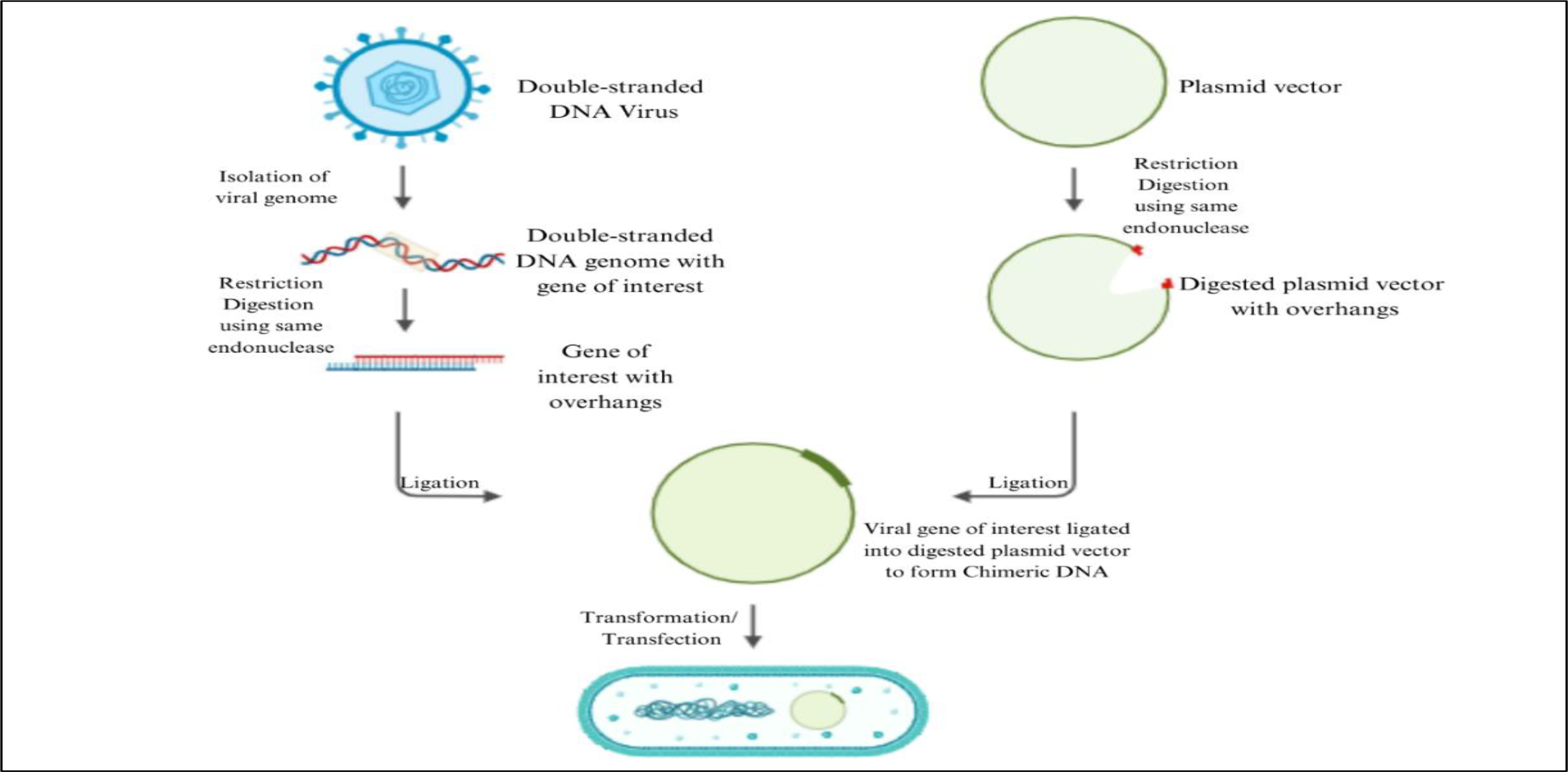
The construction of Chimeric Virus-like Particles serves as one of the prerequisites for the synthesis of Virus-like Particles (as in Figure 3). The Chimeric VLPs (cVLPs) is a complex multiprotein macrostructure containing antigenic determinants or epitopes of different viruses [68]. VLPs of this kind can be produced by designing a recombinant DNA molecule capable of encoding both relevant and foreign viral peptide or protein [69]. However, the synthesis and production of these viral peptides or proteins must be carried out in a suitable and stable expression platform such as bacteria, yeast, and so on. The genetically engineered Chimeric VLPs displaying numerous repetitive sequences on their surface can be added with exogenous antigens of other viruses by genetic fusion or chemical conjugation [69]. The resulting moieties have the ability to increase the antibody titer and corresponding immune response against foreign antigens by inducing a strong cytolytic T-lymphocyte immune responses [8]. Thus, these vaccines are also targeted against non-infectious diseases including diabetes, hypertension, allergies, nicotine addiction and Alzheimer’s [70].
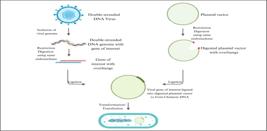
Construction of Chimeric DNA for expression in bacterial expression platform. The desired viral genes restriction digested and integrated into the plasmid vectors with the aid of recombinant DNA technology to yield chimeric DNA. The resulting chimeric DNA is inserted into bacterial expression systems for synthesis of desired VLP particles.
EXPRESSION PLATFORMS
Various platforms, including both prokaryotic and eukaryotic systems, can be used for the production of VLP vaccines [71], [72]. The choice of an appropriate expression system is crucial as it dictates proper protein folding and the quaternary structure of proteins due to difference in post translational modifications (PTMs) such as glycosylation and phosphorylation occurring in different systems. Therefore, the choice of expression system is also responsible for the degree of immunogenicity attributed by the administered VLP vaccine [73], [74]. Moreover, various advantages and disadvantages of the following expression platforms have been listed in Table 1.
Advantages and drawbacks of various expression systems used in the development of VLPs. (a) Bacteria; (b) Yeast; (c) Insect; (d) Plant; and, (e) Mammalian cell lines.
Bacteria
Bacterial platforms have been widely used as expression platforms for production of recombinant proteins including VLPs. Features such as easy manipulation, fast growth rate, high-level expression, genetic stability, and simplicity of expression makes it a safe and cost-effective platform for production [70], [75]. However, their inability to produce recombinant proteins with PTMs, inability to create proper disulfide bonds, issues with protein solubility, poor immunogenicity and the presence of bacterial endotoxins makes it an unsuitable strategy for development of enveloped VLPs [26], [75], [76]. However, bacterial expression system serves as a better platform for the production of non-enveloped VLPs made up of either one or two viral structural proteins [74], [77].
The Escherichia coli is commonly used to produce proteins with limited PTMs of which various vaccines have entered clinical trials for their use against infectious and non-infectious diseases [70]. The manufacture of the very first Hepatitis E vaccine, Hecolin, was done by Xiamen with E. coli as the expression platform in the form of p239 VLP-based vaccine [78]. Another example includes a chimeric non-enveloped VLP vaccine against the malarial parasite, Plasmodium falciparum, named MalariVax consists of two fused proteins, HBcAg, the core protein of HBV as carrier and, the B- and T-cell epitopes of circumsporozoite proteins of the parasite [79], [80], [81], [82]. In addition to E. coli, the following two bacterial species support the formation and production of VLPs. The expression of major capsid protein L1 (HPV L1) of HPV type 16, controlled by lactose promoter in Lactobacillus casei systems produced HPV VLPs [83], [84]. However, higher expression levels and solubility was achieved upon production of VLP in glutathione-S-transferase fusion protein system [85]. Likewise, Pseudomonas fluorescens was used to express and improve the low solubility of the core protein of Cowpea Chlorotic Mottle Virus VLPs [43]. cVLPs against non-infectious diseases such as hypertension, diabetes, Alzheimer’s and allergies have been developed by conjugating the antigen with bacteriophage Qβ RNA in E. coli expression systems [70]. Factors such as, presence of antibiotic resistance markers in plasmid vectors and the composition of cultivation medium may also influence VLP assembly as observed the in-case bacteriophage Qβ VLPs [86].
Yeast
Yeasts like Saccharomyces cerevisiae, Hansenula strains and Pichia pastoris, are favorably used for the production of VLPs as they enable cost-effective production with rapid cell growth, high level of protein expression, good PTM processes and scalability of produce [73]. Moreover, the manipulation of yeast is effortless and, the resulting transformed cells grow to extremely high densities until recombinant protein expression is induced allowing, large volume production of VLPs by commercial-scale fermenters. However, they do possess some drawbacks which are, lack of complex PTM pathways resulting in proteins completely different from those produce in mammalian systems, hypermannosylation, plasmid loss and lower protein yield [76]. Yeast expression platform is used for production of non-enveloped VLPs. However, they have been used to produce HIV 1 Gag protein and DENV-2 VLPs [76], [87], [88], [89]. FDA approved VLP vaccines produced in yeast expression systems include Engerix-B and Recombivax HB against Hepatitis B virus, Gardarsil R © and Gardarsil9 R © against Human Papilloma virus, and MosquirixTM against P. falciparum [12], [87], [88], [90]. The production of VLPs of Chikungunya (CHIK) using P. pastoris has been recently reported [91].
Yeast expression systems are more complex than E. coli systems because the yeast shuttle vectors must be prepared first in bacterial host cell and introduced into yeast cells as plasmid vectors followed by selection of most stable recombinants with genome-integrated transgenes [26]. This showed an observably increased protein yield of approximately 30% than that in E. coli, as observed with Bacteriophage Qb VLPs [92]. Packaging of functional heterologous mRNA in Bacteriophage MS2 VLPs can be done using yeast cells as it allows simultaneous synthesis of core protein and mRNA of the model protein using a single vector. But this requires a special packaging RNA sequence native to MS2 genome that is not necessary for VLP formation in the yeast expression systems [93]. Soluble forms of CCMV core protein VLPs using Saccharomyces expression system can be produced which would otherwise form inclusion bodies in E. coli system [94].
Yeast based expression systems could be used for multigene expression where, three rotavirus structural genes of virus families, Birnaviridae, Reoviridae and Totiviridae [26], incorporated into a single plasmid vector resulted in the formation of a triple-layered VLP in Saccharomyces expression system [19]. Although VLPs of HIV-1 Gag can be produced, those of HIV-2 cannot be synthesized as these are enveloped VLPs and, S. cerevisiae cells don’t assist Gag protein multimerization into functional VLPs and particle budding due to the absence of essential host factors [95]. The HIV type-1 p55 gag VLPs produced using yeast system elicit an immune response in host system by incorporation of VLP into dendritic cells by endocytosis or micropinocytosis. This induces dendritic cell maturation and also, enhance production of cytokines like IL-1i p70. The encapsulated VLPs efficiently can activate both CD4+ and CD8+ T-cells of HIV patients as part of immune response [96]. Experiments on Pichia with HBsAg suggest that self-assembly of synthesized proteins into VLPs must be completed during the course of down streaming process [71]. Similarly, VLPs of Dengue virus type-2 (DENV-2) were produced by inserting cDNA coding for a complex of viral glycoprotein E and pre-membrane proteins into P. pastoris genome operating under the control of glyceraldehyde-3-phosphate dehydrogenase, a constitutive promoter [89]. However, for enteroviruses like poliovirus, VLP production can be carried in P. pastoris via co-expression of viral protease (3CD) and structural precursor protein (P1) [97].
Baculovirus/Insect cells (B/IC)
This is a two-step procedure where insect cells are grown to desired cell concentration before infection with recombinant baculovirus for protein expression [27]. It is used for the production of both enveloped and non-enveloped VLPs as it is fast, versatile and provides exceptionally high protein production levels with complex eukaryotic protein post-translational modifications crucial for self-assembly and release of produced VLPs [98], [99]. The fast growth rates of B/IC systems in animal product-free media allows large scale production of VLP including those of viruses that rapidly change their surface antigens between each outbreak such as Influenza virus [74]. Recombinant proteins and VLP production can be carried out in conventional cell lines derived from Spodoptera frugiperda (Sf9/Sf21) and Trichoplusia ni (Tn5). Tn5 cell lines have been used for commercial production of HPV L1-VLP vaccine [10]. Baculovirus is different from remaining viral vectors due to its ability to tolerate large amounts of heterologous DNA and reliably deliver it to desired host cell [100]. The main drawbacks of B/IC expression system include, (i) simpler N-glycosylation than mammalian cells, (ii) protein contamination by enveloped baculovirus, and (iii) difficult to scale-up [75], [76]. Hence, strategies to improve N-glycosylation in B/IC platform by improving glycosylation pathways of some cell lines like Ea4, can simplify production of therapeutic human glycoproteins[101]. Hence, if glycosylation patterns improve, B/IC expression system would become a strong candidate for VLP-based vaccine production [10].
B/IC expression system has been used to produce an FDA approved HPV vaccine named Cervarix, consisting of HPV16 and HPV18 L1-protein based VLPs. It has also been used to produce prophylactic vaccines against various infectious diseases including, Influenza virus A, HIV 1, Ebola virus, Norwalk virus, Hepatitis C virus, Chikungunya virus, Rift Valley Fever virus, severe acute respiratory syndrome, and Dengue fever virus [102]. HIV VLPs produced in recombinant baculovirus infected Sf9 cell lines, undergo correct processing of gag gene products that can self-assemble into large enveloped VLPs [103], [104]. Thus, an immune response involving activation of human monocyte derived DCs and CD4+ T cells can be induced by these VLPs [105]. HPV types 16-L1 and L1-L2 efficiently self-assemble into VLPs when expressed in baculovirus double-expression vector [106]. The Norwalk virus VLPs can also be expressed in Sf9 cell lines using recombinant baculovirus system such that the capsid proteins generated could self-assemble into VLPs that mimic native capsid protein by appearance and size and, are capable of inducing an immune response [107].
Recently, a group of scientists described the development of a MultiBac-based VLP factory that depends on core protein (M1) of influenza virus and uses it to create numerous influenza derived VLPs with functional modifications in hemagglutinin (HA), that help in regulating immune responses induced by these VLPs [108]. The B/IC expression system has proven to be more effective than conventional cellor egg-based systems for influenza virus vaccine production. Rapid, easy and flexible process of recombinant baculovirus construction allows the fusion of desired genes from discrete types and/or subtypes of influenza viruses within the same expression vector [4]. A high five cell-based insect expression platform created by [109] combines stable expressions with baculovirus mediated expression to produce multivalent influenza VLPs. The expression abilities of the created platform were assessed by infecting them with 2 distinct haemagglutinin proteins of subtype H3, called HA2 and HA2, with baculovirus expressing core protein (M1) and 3 other HA proteins of subtype H3 named, HA5, HA4 and HA3, in order to create pentavalent VLPs (H3) [4]. VLPs of Zaire Ebola Virus serotype named ZEBOV-VLPs have been developed by co-expression of 3 viral structural proteins including, nucleocapsid protein (NP), matric structural protein (VP40) and, glycoprotein (GP) in both insect and mammalian cell line expression systems. [110] reported a technique for ZEBOV-VLP production in insect cell lines by employing high multiplicity of infection of bac-GP and bacVP40, and limiting NP expression either by preventing infection or lowering bac-NP multiplicity of infection. Moreover, baculovirus vectors could also be used in combination with whole insect larvae like silkworm larvae [111] to produce VLPs cost-effectively [112]. Hence, the present-day baculovirus-silkworm multigene expression system has the ability to simultaneously express 6 different transgenes from a single recombinant baculovirus [113].
Plant cells
The use of plants or their cells for production of recombinant proteins or other biological drugs for application as therapeutics, biopharmaceuticals, cosmeceuticals, vaccines and others is known as molecular farming [114]. Plants are eukaryotes having the ability to generate complex, correctly folded proteins with appropriate post translational modifications with added benefits of lower cost, enhanced safety and easy processes. Plants nor their cells harbour any human pathogens [115]. Plant systems offer certain advantages over other conventional expression systems including, low refining cost, high expression (upto 80%) of total soluble protein and, high performance expression processing. They don’t require expensive fermentation facilities or establishment of duplicate facilities for production of biomass or scaling up production, respectively [76], [116], [117], [118]. Despite such advantages, their employment for VLP production is not acceptable as they exhibit lower production levels and plant specific N-glycosylation of glycoproteins than mammalian systems [118], [119]. The use of MagniCON and CPMV-HT technology has transformed plant expression systems into a promising platform by reducing cost for protein production to less than $50 per gram of proteins or antibodies produced which are used in veterinary and human pharmaceutical industries [76], [116], [117].
Plant expression systems have been widely used for the production VLPs of animal and human viruses. Non-enveloped VLPs of Norwalk virus capsid protein expressed in potatoes, tobacco, tomatoes and lettuce display structural similarity to native Norwalk virus particles replicated in human gastrointestinal tract. The induction of intestinal mucosal antibody specific to conventional Norwalk virus vaccine requires oral administration, but since, this system shows slow and extremely low VLP expression along with their accumulation their use has been limited. With developments made in plant-based expression systems based on the Geminiviral BeYDV and TMV RNA MagniCON replication systems, the accumulation of Norwalk virus capsid protein VLPs in transgenic tobacco and tomato has increased more than 80-fold [116], [120], [121]. Using TMV-derived transient expression systems, well assembled recombinant VLPs of Norwalk virus have been produced in the leaves of Nicotiana benthamiana by [120]. [122] created trackable hemagglutinin-based VLPs allowing them to determine the assembly of VLP particles in plants and their interactions with mammalian host immune system. Likewise, the production of bluetongue virus VLPs in N. benthamiana via agrobacterium mediated transient expression was investigated by [123], and, transient expression of foot-and-mouth disease virus VLPs, usually performed in E. coli [124], carried out in N. benthamiana system was showcased by [125]. Similarly, [126] produced VLPs of norovirus in plant expression systems using modified geminiviral vectors.
HIV-1 VLPs have been produced by insertion of viral gag polyprotein into plant genome through either stable/ transient/ both transfection method in tobacco plants. The resultant proteins were capable of assembling into VLPs that similar to those produced in insect and bacterial cells [127]. Bivalent HBV-HIV VLP vaccine containing HBsAg VLPs displaying immunogenic epitopes from HIV-1, env and gag proteins were generated in transgenic tomato [128]. Similarly, the genes coding for HPV type 12 L1 major capsid protein when integrated with Nicotiana tabacum cv. Xanthi genome expressed proteins that could readily assemble into VLPs against Human papillomavirus type 16 [129]. A plant-based malaria transmission blocking VLP vaccine, Pfs25 VLP-FhCMB, consisting of Pfs25 surface protein of Plasmodium falciparum conjugated to SIMV was synthesized in N. benthamiana by transient expression whose immunogenicity and safety was assessed in Phase I clinical study [130], [131]. It was found that some plant viruses like CPMV and pepper mild mottle virus exhibited structural stability in the intestinal conditions of humans [132]. Either complete CMPV virions or empty viral capsid protein VLPs generated in plant systems induce immunomodulatory activities such as in-situ anti-tumor activity resulting in tumor regression. The antigen thus displayed on plant VLP surface interacts with host APCs, leading to activation and increased number of tumors infiltrating neutrophils and dendritic cells [133]. Therefore, with advancements being made in plant glycoengineering and creation of novel plants, human-like modifications of glycol and optimization of glycan structure has been enabled in order to improve the safety and functionality of recombinant pharmaceutical glycoproteins [118], [119].
Mammalian and Avian cells
Animal cell expression platforms can be employed to produce various structural proteins of enveloped and non-enveloped VLPs as well as other recombinant proteins due to their ability to create precise and complex PTMs essential for proper protein folding and assembly [74], [77]. In addition, mammalian cell systems provide flexibility and consistency during the development and production process and, aids in the recovery of glycoproteins with lipid membrane compositions similar to that of viral hosts [76], [134]. The proteins secreted in this system are in their native and mature forms which is not observed in other expression systems [135]. However, mammalian systems too are accompanied by certain drawbacks including high production cost, low protein yield, lengthy expression time, require bioreactors for large-scale production, vulnerability to infections with mammalian pathogens [75], [119]. Despite these cons, mammalian expression systems are used for production of VLPs at both laboratory and industrial scale with an objective to create vaccines and gene therapy agents [26]. Mammalian cell lines including Chinese hamster ovary (CHO), murine myeloma (Sp2/0, NS0), murine C127, baby hamster kidney-21 (BHK-21), HT-1080, human embryonic kidney 293 (HEK293), CAP-T-cell line derived from human amniocytes, east lansing line-0 (ELL-0) and Vero 9 are extensively used to produce commercialized protein therapeutics including recombinant VLPs for medical purposes [74], [76], [134]. In addition, avian cell lines are used to generate VLPs [136]. The production of VLPs can thus, be achieved by stable transfection of desired viral genes into cell lines like, HEK293 or Vero E6 cells [137].
VLPs of Hepatitis B virus, dengue virus and Hantavirus have been successfully produced in CHO cell lines. Since, this cell line is not derived from human cells, it poses minimal risk of contamination with lethal human viruses [74], [138], [139], [140]. Similarly, VLPs of influenza virus, HIV and rabies virus have been produced in HEK293 cell lines and those of HIV have also been produced efficiently in CAP-T-cell lines [141]. In light of the recent pandemic of COVID19, efforts are being made to design VLP vaccine for protection against the same. However, a stable VLP against SARS-CoV-2, also effective against COVID19, already exists that was produced using Vero E6 cell lines [142]. Huh7 cells were used as expression platform to create MERS VLPs to understand viral infection and morphogenesis by [143]. Avian fibroblast, ELL-0 cell lines were employed to produce VLPs against human Respiratory Syncytial Virus (RSV) consisting of RSV F and G proteins [136]. VLPs of influenza virus containing haemagglutinin, neuraminidase and matrix M1 protein were expressed in 293 T, CHO-K1 or Vero cell lines via transient transfection. However, the preclinical studies of produced VLPs when conducted in BALB/c mice showed, immunogenic response at low dosage by inducing antibodies against haemagglutinin and neuraminidase [137], [144]. Another approach revealed that, the construction of a stable mammalian cell line expressing four different structural proteins of influenza virus would allow the creation of a hybrid consisting of matrix proteins from one, like H3N2, and surface glycoproteins from another, like H5N1, type of influenza virus [144]. This approach produced perfectly glycosylated VLPs, with size similar to native viruses, and encapsulates host cell proteins and exhibits high immunological activity. The production of glycosylated VLPs was achieved by infecting a cell line with recombinant vaccinia virus vector that would express T7 polymerase, and transfecting cell line with bacterial plasmids carrying cDNAs of ten influenza virus proteins capable of generating desired VLPs. Following cDNA transcription, the resultant model protein mRNA was delivered into influenza virus VLPs [145]. HIV VLPs were produced in inducible HEK293 cell lines expressing haemagglutinin and neuraminidase upon transient transfection with plasmid encoding HIV-1 Gag proteins [146]. A similar study involving production of HIV-1 Gag VLP in mammalian cell suspension culture by transient gene expression displayed that a large fraction of expressed Gag-GFPs, fully assembled into VLPs of estimated size and morphology that were consistent with that of immature HIV-1 particles in the culture supernatant [147]. A study showed that, a human cell line injected with a eukaryotic expression vector cloned with a polyprotein cDNA coding for all structural proteins of alphavirus CHIKV. The produced CHIKV VLPs were then confirmed by electron microscopy and immunological analysis. Immunological analysis showed that these VLPs were able to mount a stronger response than their corresponding DNA vaccines [148].
Cell-free system
Also known as, cell-free protein synthesis systems, have most commonly been used for rapid evaluation of different expression platforms but, mainly to study mechanisms of transcription and translation processes [149]. It also offers a platform for in vitro expression of recombinant proteins to produce VLPs [60]. High yield protein, time saving, minimum cell contaminants and the ability to generate un-natural amino acids containing VLPs or toxic protein intermediates are few advantages of cell-free expression systems [60], [73]. However, extremely high cost of production and limited scalability are significant limitations for commercial application of the same [73]. Example of two commercially utilized VLP vaccines produced using cell-free systems include, Inflexal, an influenza virus VLP vaccine, and, Epaxal, a hepatitis A virus VLP vaccine. Likewise, VLP vaccines of norovirus and Hepatitis B viruses have also been expressed in these in-vitro expression platforms [60].
Cell-free systems can include bacteria, yeast, insect cells, wheat germs, and rabbit reticulocytes, that are commercially available for viral capsid protein VLP production [73]. The most popular and preferred systems are the derived from E. coli due to ease and simplicity of extraction preparation as well as, high desired protein yields compared to others [149]. E. coli-based system have used to synthesize RNA bacteriophage Qb-, MS2- and HBV corederived VLPs. In case of HBcAg, Qb and MS2 VLP, the chosen cell-free expression system stabilizes the particle by introducing disulfide bonds, while, production of Qb VLPs in cell-free protein synthesis system may involve the incorporation of protein A2 that can be toxic to live cells [16], [150]. Production of HBcAgand Qb-derived VLPs here, produced a yield of about 1mg/ml and displayed VLP assembly efficiency exceeding 80% [151]. Hence, cell-free systems can be used in complicated case where, viral structural protein-encoding-gene expression is toxic for the living cells or ambient conditions aren’t suitable for VLP production in conventional systems with desired properties like, unnatural amino acid incorporation [151].
IMMUNOSTIMULATION BY VIRUS-LIKE PARTICLES
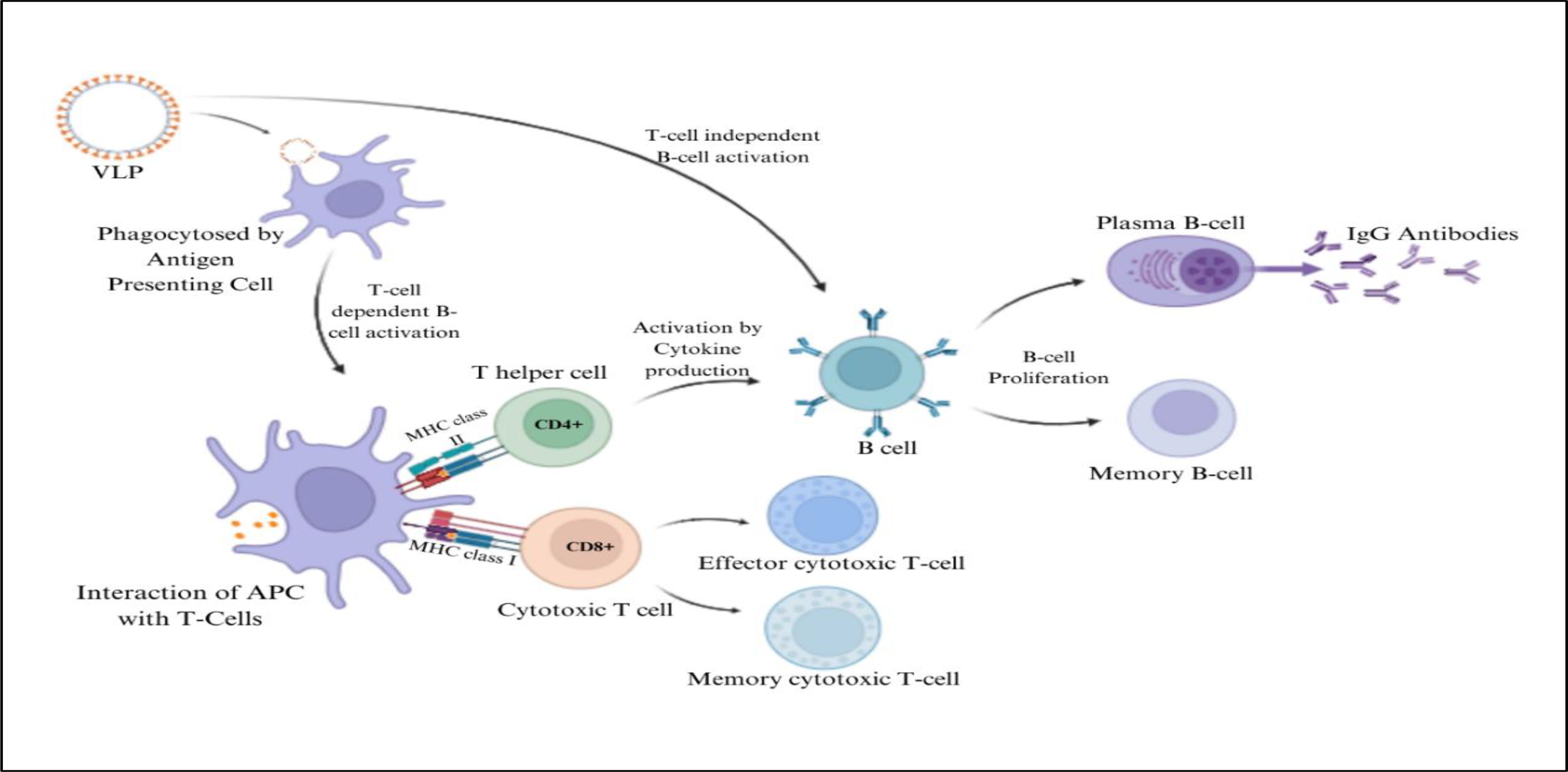
The most important attribute of VLP-based vaccines is their ability to act as good substrates that deliver antigenic epitopes in proper conformation to both arms of adaptive immunity, i.e., humoral and cellular immunity [24], [152]. Usually, VLPs comprise of repetitive protein structures that can trigger, stimulate and induce the production of neutralizing antibodies by B cells [153], [154]. For example, preclinical trials conducted with influenza virus M2 protein VLPs are capable of inducing both types of adaptive immune responses at low antigen dose. It showed a significant increase in antiviral antibody titer and, led to stimulation of CD8+ T-cells to reduce the severity of disease as well as offer protection against various strains of the virus [155], [156], [157]. Similarly, porcine parvovirus VLP vaccines firmly induce both humoral and cellular immunity through major histocompatibility complexes (MHC) I and II class pathways, and, zika virus envelope protein domain III (HBcAg-zDIII) induced both immunities with just two doses of VLP vaccine [157]. VLPs are taken up by professional APCs, like dendritic cells (DCs) by phagocytosis and clathrin-dependent macropinocytosis following which, they are processed in acidic lysosomal compartments and presented using MHC class II molecules [158]. The activation and maturation of DC occurs by up-regulation of co-stimulatory molecules, production of cytokines and CD4+ T-helper cells stimulation. It also involves TLR4, NF-kB and proteoglycan receptors that can otherwise be inhibited by heparin [24], [159]. In another mechanism, the VLP moieties enter DC cytoplasm where they are processed and presented using MHC class I molecules to cytotoxic T-lymphocytes (CTLs) by cross-presentation [160], [161]. Moreover, activation of B-cells mediated by VLPs is strong enough to induce production of T-cell independent IgM antibodies [26], [162]. DCs act as a bridge linking innate and adaptive immunity. They take up particles of about 100-500nm size via phagocytosis or macropinocytosis, allowing efficient uptake, processing and presentation VLPs in size range of 10-200nm [158], [163]. Upon administration of VLP-based vaccines, via mucosal or parenteral route, DCs interact through pattern recognition receptors (PRRs) like, C-type lectin receptors and TLRs, that mainly detect natural viruses and transfer to secondary lymphoid organs such as spleen [105], [164], [165]. Following recognition and uptake of VLPs, maturation of DCs stimulate the release of pro-inflammatory cytokines like IL-1β and TNF-α [166]. This in-turn leads to enhanced recruitment of APCs and increased lysosomal proteolysis in DCs. VLPs are then processed into small peptides and loaded onto MHC II class molecule to form MHC-peptide complex on DC surface. There is simultaneous appearance of co-stimulatory molecules like CD80 and CD86, on DC surface required to activate B- and T-cells [167], [168]. The MHC-peptide complex and co-stimulatory molecules activate CD4+ T-helper cells essential for proliferation and differentiation of both B- and T-cells. Under certain circumstances, B cells are capable of VLP detection and humoral immunity activation in a T-helper cell independent manner [167], [168]. Therefore, the process of Immunostimulation by VLPs in the host system has been illustrated in Figure 4.
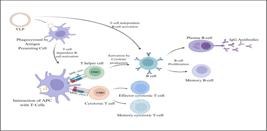
Immunostimulation involving induction of innate and adaptive immune responses. VLPs when phagocytosed by APCs interact with T cells to produce an immune response by activating B cells leading to production of antibodies during indirect stimulation. VLPs can also directly stimulate B cells to mount immune response by antibody production.
Reports showed that the type, direction and outcome of immune response to VLP-based vaccines administered were influenced by the expression systems used for VLP production [160]. For example, HPV VLPs displaying HPV L1 capsid produced in plant and insect systems exhibited immunogenicity of similar intensities. It was observed that half the mice fed with HPV VLP expressing transgenic potato developed antibodies specific to L1 protein [169]. Similarly, yeast derived HIV VLPs showed efficient and precise immunomodulatory effects compared to those from other systems. The HIV-1 Pr55Gag VLP can stimulate both humoral and cellular immunity [76]. Following their administration in HIV-infected individuals, DCs get loaded with VLPs and transform the Gag-specific memory CD8+ T-cells into effector cells, however, a few of memory T-cells showed no response [96]. Both DCs and Langerhans cells (LCs), internalize VLP using different routes like clathrin-coated pits and proteoglycans [170]. For example, nearly same amounts of HPV VLPs were taken up by DCs and LCs but by distinct mechanisms [171], [172]. The internalized VLPs result in the activation of DCs along with cross-presentation of peptide-MHC I to bring about T-cells co-stimulation, whereas, the VLPs results in cross-presentation without co-stimulation in-case of LCs. HPV VLPs are endocytosed by a non-clathrin, non-caveolae and actin-independent pathway by LCs, while, via clathrin-mediated mechanism and actin-dependent macropinocytosis by DCs [170], [171]. This can be overcome by the addition of CD40 ligand, where incubation of LCs with HPV VLPs produces good amount of pro-inflammatory cytokines (IL-12) and stimulate immune response against same after incubating it with T-cells [171]. A study conducted by [173], showed the ability of HPV16-based VLP vaccines to induce IFN-α and IL-6 production by plasmacytoid DCs resulting in antibody generation. Studies showed that chimeric HPV VLPs are capable of mounting a CTL immune response against HPV16 transformed tumors in mice, the mechanism of which is dubious. Usually, VLPs could interact using FccRIII with mature human MHC II positive DCs while, immature cells were activated following incubation. However, their binding and uptake by DC in FccRII, FccRIII and FccRII/III deficient mice showed at decline of about 50% than in wild-type mice. It also resulted in reduced murine DC maturation and antigen presentation [171].
A HBcAg VLP-based vaccine against Toxoplasma gondii, causing human toxoplasmosis, constituting of B cell, CD8+ cell and CD4+ cell epitopes of T. gondii stimulated humoral and cellular immune response thereby enhancing level of IgG and IFN-γ, respectively [174]. The administration of VLPs produced by recombinant baculovirus triggered the release of anti-inflammatory (IL-10), pro-inflammatory (IL-6, TNF- α), T-helper1 polarizing (IFN-c) cytokines, GM-CSF and MIP-1a by activated human peripheral blood mononuclear cells in a time and dose dependent fashion. An up-regulation in molecules essential for antigen presentation like, MHC II, CD86 and CD80, as well as, cell adhesion like, CD54, indicated VLP-induced monocyte activation. However, VLP’s exposure to serum deactivated its capability to trigger cytokine production. A study conducted using CD4+ T-cell knockout mice, vaccinated with Simian/Human immunodeficiency virus (SHIV) or chimeric influenza HA/SHIV VLP-based vaccine showed stimulation of Th-cell independent humoral immune response. Although, CD8+ cytotoxic T cells (Tc-cells) only detect intracellular pathogens presented as antigen-MHC I complex, this has been contravened by VLPs as they stimulate CD8+ cells without involving any extracellular antigens [163], [175], [176], [177]. In another study involving mannosylated rabbit haemorrhagic disease virus VLP, it was presented as exogenous antigen-MHC I enabling direct activation of Tc-cells [163]. Investigation of antigen cross presentation mechanism using p33-VLP model revealed that the VLPs were taken up by CD8- DCs and transferred to secondary lymphoid organ [178]. The antigen was presented as antigen-MHC I complex through two pathways, one, by an antigen processing-dependent pathways in conjunction with transporter, and another, TAP-independent pathway where a portion of antigen was presented by macrophages to adaptive immune system [177], [179]. The oral or intranasal administration of immunized mice with adjuvant-free Norwalk virus or Rotavirus VLPs, showed production of intestinal IgA and, the plasma cell precursors (derived from mucosal lymphoid tissues) that moved to genital tract did produce IgA antibodies [180], [181], [182]. The study also indicated that, immune responses mounted by systemic administration of VLPs were stronger than those observed on mucosal administration. A relatively higher amount of VLP-specific IgA was recorded in intestinal washes, followed by intrarectal than intravaginal immunization and, in vaginal washes followed by intramuscular than intrarectal or intravaginal immunization. As suggested by some studies, the immunogenicity of VLPs at mucosal surfaces can be attributed to the property of particulate antigens articulated as a multimer. Moreover, such VLP-specific immune responses were generated due to active VLP uptake by mucosal APCs via integrin receptors [182]. The poor immunogenicity of proteins administered mucosally acts as an obstacle for development of oral vaccines, which can be overcome using appropriate adjuvants. Instead, mucosal immunity can also be induced by delivering antigens in VLP form to mucosal surfaces [182]. It has been observed that the administration of VLPs with adjuvants like, poly (I:C) or CpG ODN1826, displayed a higher total specific IgG titer than the VLP alone. Therefore, on administering VLPs alone induced only a steady Th2 pattern, but those conjugated with adjuvant poly (I:C) elicited a Th1 biased IgG subclasses, IgG2a and IgG3, better than with CpG ODN1826 as seen in animal models [152]. Immunization of mice with chimeric SIV (Simian Immunodeficiency virus) VLPs containing GM-CSF stimulated production of SIV env-specific antibodies exhibiting enhanced neutralizing activity compared to those induced by either conventional SIV VLPs, those with CD40L or those mixed with soluble GM-CSF. The study showed that, addition of immunostimulatory molecules significantly raised CD4+ and CD8+ T-cells immune response to the SIV VLPs than observed in standard VLPs [152]. Besides these, many other studies focusing on enhancing the immunogenicity of currently available and future VLP-based vaccines without the aid of adjuvants in order to induce both arms of adaptive immunity are being carried out [12].
VLP-BASED VACCINES AGAINST VARIOUS CANCERS
The ability of VLPs to deliver antigens, activate MHC I class pathway and, induce strong CTL-mediated immune response essential to eradicate tumor cells make VLP an excellent tool for the development of therapeutic vaccines against cancer. The immune system is armed with three important elements to target and combat cancer cells that are as follows: First, DCs that must receive adequate signals needed for stimulation and maturation of adaptive immunity to ensure intolerance against antigens by activation of regulatory T cells (Tregs) and immune system suppression. Second, are the tumor antigens most of which are either related or identical to self-antigens. Finally, the T-cells must be capable of overcoming the immunosuppressive signals produced by tumor cells [183].
VLPs against Cervical Cancer
About 70% of the cases of cervical cancer have known to be caused by two main types of viruses, Human papillomavirus (HPV)16 and 18 [184]. However, it is also responsible for other cancers including hand, throat, anal and genital warts. The viral capsid contains two proteins called L1 and L2, forming the basis for vaccine development. Commercially available vaccines namely, GlaxoSmithKline’s Cervarix® and Merck’s Gardarsil®, aim at preventing cervical cancer. They act directly against genotypes 16 and 18, but can also protect against genotypes 6 and 11 causing benign genital warts only. The two commercial L1-based VLP vaccines against HPV have been expressed in insect and yeast cells expression platforms [24], [185]. But, these prophylactic VLP-based vaccines do not cure infected individuals [185]. L1 protein is largely used to produced VLPs, but, isn’t conserved amongst different HPV types. As a result, researchers are aiming at developing L2-based VLP vaccines. The L2 structural protein are produced during later stages of HPV replication, however, its inability to form VLP acts a challenge in VLP development [186], [187], [188], [189]. The administration of L2-based VLP produced cross-neutralizing antibodies and cross-protection in immunized animals despite a lower antibody titer produced compared to those stimulated by L1-based VLP [190], [191]. Hence, [192], designed a new vaccine to enhance VLP immunogenicity by incorporating L2 peptide into C-terminal region of L1 peptide. On expressing the L1/L2 fusion peptide in N. benthamiana, it was noted that the L2 component was exposed on the surface of chimeric VLPs. Immunization studies conducted by [192] demonstrated that the chimeric VLPs induced humoral immune responses specific to both L1 and L2 and offered protection against HPV genotypes 16 and 52 in immunized mice. In another study carried out by [193], the chimeric VLP 18L1-45RG1consisting of cross-neutralizing epitopes of L2 protein of HPV 45 and L1 of HPV 18 offered cross-protection against HPV-18, -39 -45 and -68 in mice that were passively immunized with the antisera obtained from rabbits immunized with chimeric VLPs [193]. All HPV infected cells and cervical tumor cells have shown to express two tumor-specific antigens, E6 and E7, while the expression in latter is in increased amounts [185]. They play role in repressing two crucial tumor suppressor proteins where, E6 binds to ubiquitin ligase E6AP inactivating p53 while, E7 degrades phosphorylated retinoblastoma tumor suppressor pRb [194]. The E7 HPV protein required to maintain the transformed phenotype of cancerous cells was inserted into infectious bursal disease virus VLPs (VLP E7), which when vaccinated into mice along with VLP E7, led to complete tumor rejection [195]. In order to improve CTL activity, an MHC II class restricted T-cell epitope was incorporated into the VLPs. A similar approach by [196] revealed that the incorporation of a potent T-cell inducer, pan HLA DR-binding epitope (PADRE) into RHDV VLP expressing E6 protein was required for malignant conversion. Moreover, the administration of E6-RHDV-VLP-PADRE vaccine in combination with antibody treatment reactivated T-cells, lowered TC-1 tumor outgrowth and, improved survival rate of immunized animals [196]. Therefore, the HPV VLP-based vaccines offer certain degree of protection against HPV related diseases [197].
VLPs against Breast Cancer
Breast cancer is a fatal disease majorly occurring in women [198]. About 20-30% of invasive breast cancer cases are attributed to overexpression of human epidermal growth factor receptor 2 (Her2). Her2 is required for the proliferation and inhibition of programmed cell death [199], [200]. Invasive mastectomy has been used as one of the most common treatments for breast cancer despite affecting the body self-esteem of patient due to lack of effective treatment methods [201]. Hence, the use of monoclonal antibodies to induce passive immunity has proven effective in prevention of metastasis and tumor growth, but their application has been limited due to high cost, undesirable side effects and need for multiple doses at regular intervals for long-term protection [202], [203]. However, evaluation of FVB mice transplanted with human Her2 positive breast cancer cells after active vaccination with VLP vaccine displaying Her2 of its surface, derived from Acinetobacter phage AP205 coat protein displayed its ability to inhibit tumor growth by overcoming strong immunotolerance and, inducing strong humoral immune response [204]. The incorporation of breast cancer TAA HER2/neu into murine polyomavirus VLPs created a chimeric Her21-683Py VLPs against breast cancer [205]. In-vivo immunization studies with subsequent tumor challenge showed that a single VLP dose elicited strong Her2-specific cellular immune response resulting in tumor rejection and extended survival. However, the immunization of transgenic mice expressing mutated Her2 oncogene with Her21-683Py VLPs provided complete protection by preventing tumor growth. But, the time of vaccination does affect vaccine efficacy as delay in VLP administration led to just postponed tumor outgrowth without any significant protection. However, humoral immunity wasn’t observed probably due to internal expression of Her2 in VLPs [205]. In an approach by [206], the breast cancer Her2 was modified into glycophosphatidylinositol (GPI)- anchored form, GPI-Her2, in order to incorporate it into enveloped influenza virus VLPs. This VLP was found capable of inducing both humoral and cellular immune responses, and, therefore increased survival rate of immunized mice up to 66%. The study showed that both GPI-Her2 VLP and GPIHer2, produced similar levels of Her2-specific antibodies. But, the treatment with GPIHer2 did not offer protection against tumor challenge suggesting the crucial role of cellular immunity. The observed results significantly correlate with the Th1-skewed immune response observed in a study conducted by [206]. Although Her2 is largely responsible of breast cancer, their overexpression can also cause, gastric cancer, endometrial carcinoma, lungs adenocarcinoma, salivary duct carcinoma and ovarian cancer [207], [208], [209], [210]. The potential of Her2 VLPs in preventing or combatting other cancer types are yet to be investigated [197].
VLPs against Pancreatic Cancer
Pancreatic cancer, ranked as one of the top five causes of cancer associated mortality, is a highly aggressive type of cancer caused by overexpression of mesothelin (MSLN), a cell surface glycoprotein acting as a biomarker for early cancer detection [211], [212], [213]. MSLN is required for cell adhesion and allows cancer cell mass to attach to mesothelial cells as a result of which, it is used as a potential anti-cancer drug target [212], [214]. Conventional therapies including chemotherapy, surgery and radiotherapy have been currently used to treat pancreatic cancer [214]. Studies involving immunization of mice harbouring pancreatic tumors, with VLP-based on SHIV VLPs displaying human MSLN on particle surface showed tumor growth inhibition and increased life expectancy of ~60% of treated mice [12], [212], [215]. The hMSLN (human MSLN) VLPs elicited both humoral and cellular immunity. It triggered strong CTL activity causing reduction in tumor mass and specific anti-tumor antibodies, and, also prevented suppression of self-antigen by inhibiting Treg cells [12], [212]. Presence of high IFN-γ producing T-cells indicated Th1-skewed immune response [215]. However, immunization of mice with mMSLN (mice MSLN) VLPs followed by tumor challenge, disrupted self-tolerance and induced specific CTL activity resulting in tumor mass reduction and increased survival rate of treated mice. A significant reduction in Treg cell levels was thought to be a crucial driver for cancer growth inhibition and prolonged survival of immunized mice [212]. The overexpression of Trop2, a tumor-associated antigen, is associated with tumorigenesis, metastasis, decreased survival rates and grade of developed tumor [214], [216]. The limited expression of Trop2 makes it a potential tool for cancer immunotherapy targeting. The immunization of C57BL/6 mice with chimeric mTrop2 VLPs, formed by incorporation of Trop2 into enveloped SIV VLPs, followed by tumor challenge caused tumor size reduction and extended life expectancy by approximately 36%. However, the treatment of mice immunized with mTrop2 VLPs along with gemcitabine resulted in higher survival rates of ~70%. This immunization procedure induced both humoral and cellular immunity by elevating levels of antigen specific tumor-infiltrating T-lymphocytes (CD4+ and CD8+) and natural killer cells (NK), while reducing immunosuppressive cytokines levels, such as IL-10 and TGF-β, MDSCs and Treg cells. Moreover, the enhance expression of IL-2, IL-3 and IFN-γ indicated Th1-skewed immune response essential for cancer cell destruction [214]. In addition to pancreatic cancer, the overexpression of transmembrane glycoprotein Trop2 can lead to other cancer types whose treatment with Trop2-based VLPs are yet to be studied [214], [216].
VLPs against Melanoma
Skin cancer has been reported as one of the most common cancers and, divided into melanomas and non-melanomas [217]. No vaccines against skin cancer have been developed till date. However, a bacteriophage Qβ VLP-based vaccine against melanoma cancer (MelQbG10) developed by Cytos Biotechnology AG, has cleared Phase 2 clinical trials and is expected to be the very first commercial vaccine against melanoma [24], [218]. [219] developed VLP-based melanoma vaccine by incorporating melanoma associated antigen 3, whose overexpression leads to poor prognosis and melanoma metastasis, into HBc VLPs. However, the immunogenicity of developed VLP vaccine was improved by packing single-stranded CpG oligonucleotides into the synthesized VLPs. Whilst their immunogenicity is yet to be studied in vivo, [219] expects it to strong elicit a cellular immune response. In another approach, the ability of empty CPMV VLP (eCPMV) to suppress tumor growth was studied, where, the treatment of B16F10 lung melanoma cells with eCPMV, resulted in alteration of the tumor microenvironment immune cell organization. An increase in tumor-infiltrating neutrophils and decrease in immune suppressing cells like CD11b-Ly6G+ neutrophils was observed. Null mutant mice devoid of neutrophils and cytokines, IL-12 and IFN-γ, were used to determine the mechanism of eCMPV VLP action, however, the VLP was ineffective and failed to exhibit any protective anti-tumor effects, indicating the crucial role played by various elements of immune system [220]. A study conducted by [221], involving incorporation of H-2Kb-restricted CTL epitope of melanoma differentiation antigen tyrosinase-related protein 2 (TRP2) and H-2Kb-restricted ovalbumin (OVA)257-264 epitope into VP1 of murine polyomavirus VLPs produced chimeric murine polyomavirus-like pentamers (VP1-TRP2180-192PP) and chimeric PLP (VP1-OVA252-270PLP), respectively. In vivo studies conducted in mice revealed that, VP1-OVA252-270PLP offered significant protection by inducing strong CTL activity against OVA-expressing melanoma cells. In case of VP1-TRP2180-192PP, even though it failed assembly into VLP, it offered partial protection from lethal melanoma challenge. Hence, these PLP-based vaccines serve as efficient antigen carrying immunotherapeutic agents against cancer by inducing CTL response [197], [221]. The RHDV VLPs could be an alternative to human-infecting viruses, like HBV or polyomavirus, in order to deliver tumor antigens as they don’t infect human hosts [222]. These RHDV VLPs expressed model antigen gp33 which was conjugated with an immunostimulatory adjuvant called α-galactosylceramide. Vaccination followed by tumor challenge in mice with B16 melanoma expressing gp33, induced activation of CTL and MKT activity manifested by significant increase in levels of IL-4 and IFN-γ [223], [224].
VLPs against Hepatocellular carcinoma
HBV core (HBc) particles were used as carriers of either single or multiple hepatocellular carcinoma (HCC) epitopes namely, MAGE-1 (278-286 aa), MAGE-3 (271-279 aa), AFP1 (158-166 aa), or AFP2 (542-550 aa). Therefore, administration of VLP-based vaccine enabled DCs to induce a strong CTL activity and cytokine IFN-γ secretion by T-cells allowing it to efficient respond against the antigen compared to that observed after administration of just antigenic peptide in HLA-A*0201/kb transgenic mice. Thus, the vaccination with VLPs helped inhibited the growth pf B16-pIR-HH tumors leading to enhanced survival rate of vaccinated mice [225]. [226] constructed a multiepitope VLP-based vaccine where HBc VLP was loaded with four HBx-dominant CTL epitopes including, HBx (115-123), HBx (92-100), HBx (140-148), or HBx (52-60). The chimeric VLPs on vaccination caused, the DCs in HLA-A*0201 transgenic mice and the peripheral blood lymphocytes from HLA-A2(+)/HBx (+) HBV infected HCC patients to exhibit strong CTL immune response against them. In addition to higher immunogenicity, it exhibited enhanced anti-tumor activity [226].
VLPs against Lung cancer
A VLP-based vaccine against lung cancer was constructed by incorporating TAA isoform 2 of Claudin-18 (CLDN18.2), the tight junction molecule, into HBc VLPs. In vivo studies involving immunization followed by tumor challenge in mice and CLDN18.2-expresing CT26 colon cancer cells, respectively, showed decrease in tumors around the lung region. Moreover, the protection obtained can be attributed to complement-dependent and antibody-dependent mediated cytotoxicity. However, antibodies produced as part of response were highly specific against CLDN18.2 but not cross-reactive to CLDN18.1 variant usually found on healthy lung tissues, regardless of their protein sequence homology [227].
VLPs against Epstein-Barr virus related cancer
Epstein-Barr virus infected individual may also develop conditions like Hodgkin lymphoma, Burkitt’s lymphoma, Nasopharyngeal carcinoma, B-cell lymphoma, gastric and sporadic carcinoma [228], [229]. The heterologous antigenic glycoprotein gp350/220 of EBV was incorporated into new castle disease virus (NDV) VLPs to form a chimeric EBVgp350/220F VLP. Vaccination of mice with VLPs, produced long-lasting gp350/220-specific antibodies capable of neutralizing EBV in vitro. The antibodies thus produced, were detected to be predominantly IgG1 subclass indicating a Th2-skewed immune response in immunized animals’ sera [230]. In addition to this, the VLPs also elicited cellular immune response involving CTLs, where, the EBV mRNA encompassed in the EBV VLP translates within infected cells followed by presentation via MHC I class molecule resulting in the activation of EBV-specific CD8+ T cells [231], [232]. Moreover, the absence of any viral DNA in the EBV VLPs employed indicated the non-infectious and non-replicative nature of VLPs [231].
VLP-BASED VACCINES AGAINST EMERGING INFECTIOUS DISEASES
Besides cancer, various emerging infectious diseases pose great threat to human health. Most of the viral disease breakouts act as risk factors especially in developing countries with poor health conditions as well as those having increased international travel and business. Although vaccination is thought to be the most effective preventive measure, they aren’t available for a large number of viral diseases. However, VLPs exhibiting antigenicity similar to that of native viruses could be used for prophylaxis as an attribute to their ability to stimulate both humoral and cellular immunity. Therefore, a number of VLP-based vaccines have been developed for extremely virulent emerging or re-emerging infectious diseases [233]. Moreover, the various commercially available and those yet to reach the market under clinical trials have been listed in Table 2.
VLP-based vaccines under clinical trials or are FDA approved against emerging infectious disease.
Arenavirus VLP-based vaccines
Lassa fever virus (LASV), a rodent-borne arenavirus belonging to Arenaviridae family, is the causative agent of severe haemorrhagic fever. The genome consists of 2 RNA segments, S and L, where, S codes for viral nucleocapsid protein (NP) and two glycoproteins (GP1 and GP2), while, L encodes viral polymerase (L) and zinc finger matrix finger protein (Z). The GP1 and GP2 proteins produced via post-translational cleavage of precursor glycoprotein act as receptor-binding proteins and transmembrane proteins, respectively [234]. Mammalian cells expressing the major immunological determinants, GP1, GP2, NP and Z proteins were used to produce LASV VLPs [48]. It was observed that the strongly membrane associated, Z protein alone enabled the release of lipid enveloped VLPs [48]. Immunization of mice with LASV VLPs triggered production of high IgG response level to individual proteins and, this observation was confirmed by reaction of LASV infected patient serum with LASV VLPs [48].
Bunyavirus VLP-based vaccines
Viruses belonging to Bunyaviridae family have been grouped into 5 genera including Phlebovirus, Orthobunyavirus, Hantavirus, Nairovirus, and Tospovirus. Their genome consists of 3 negative sense RNA segments including, large segment (L), middle segment (M), and, small segment (S). The surface glycoproteins embedded in the envelope consist of two heterodimers, GN and GC. The hantavirus VLPs expressed in CHO cells by co-expression of genes for GN, GC and NP, showed an increase in CD8+ T-cell activity and antibody response in comparison to response elicited by inactivated virus vaccines [140], [141]. It has been suggested that not all three proteins are needed for VLP formation. Therefore, co-expression of GC and GN protein in in mammalian cells to yield VLPs indicate that NP proteins unessential for VLP assembly [235]. Likewise, other members of the family also form VLPs by co-expression of GN and GC [179], [236]. [237] produced VLPs of Crimean-Congo hemorrhagic fever virus (CCHFV) via expression of NP in B/IC expression system whose safety, immunogenicity and performance are yet to be studied. A similar approach has been used for the production of VLPs against severe fever with thrombocytopenia-syndrome virus (SFTSV) [233], [237].
Coronavirus VLP-based vaccines
Coronaviruses such as SARS-CoV-1, MERS-CoV and SARS-CoV have led to global pandemics threatening mankind with deadly diseases. The Corona virus particles comprise of 4 main structural proteins that includes, spike protein (S), membrane protein (M), envelope protein (E), and nucleoprotein (N). In studies conducted on SARS-CoV-1, the VLPs produced by expression of only M and E proteins in baculovirus expression system were smooth and lacked spikes, while, those formed by the co-expression of M, E and S proteins yielded VLPs similar to the native SARS-CoV-1 virus [238]. The latter have been tested for their protective ability [12]. It was observed that the systemic or mucosal immunization with SARS VLPs expressed in baculovirus expression platforms resulted in strong IgG and IgA response. In addition, it also caused activation of DCs and elicited cellular immunity [239], [240].
Filovirus VLP-based vaccines
These are enveloped viruses containing the major structural protein, VP40 matrix protein essential for the structure, assembly and budding of filovirus VLPs. But, the sole expression on matrix protein yields poor VLP produce [241], [242]. The Ebola virus (EBOV) VLPs exhibiting morphological similarity with that of native viruses were produced by expression of Ebola VP40 and viral envelope glycoprotein in 293T cells [243]. The resultant VLPs were highly immunogenic both in vitro and in vivo and thus, elicited cytokines and chemokines maturation, activation and secretion. Vaccination of mice with EBOV VLPs resulted in activation of B cells, CD4+ and CD8+ T cells and, high levels of EBOV-specific antibodies. Hence, these VLPs were capable of protecting mice from lethal challenge [243]. Similarly, those produced in B/IC expression system showed effective protection of mice and also, resulted in high yield [241]. Similarly, VLPs for Lake Victoria Marburg virus were produced by the expression of a single protein, VP40, whose efficacy was found to be lower than those formed via co-expression of GP, NP and VP24 that can stimulate the formation and budding of virions [242]. Thus, co-expression of GP and VP40 in mammalian and insect cell lines, in presence or absence of NP, produced VLPs sharing morphological similarities with the native virus [241], [244]. Hence, vaccination of Marburg VLPs into guinea pigs mounted MARV-specific and neutralizing antibody response, and stimulated differentiation of DC, promoted T-cell proliferation and conferred complete protection to the animal against MARV infection [242]. However, the administration of MARV VLPs to monkeys resulted in an increased level of MARV-specific antibodies after first vaccination and peaked after third dose [242].
The Hepatitis B causing Hepatitis B virus (HBV), belongs to the family Hepadnaviridae. Moreover, infection with HBV can cause acute and chronic hepatitis resulting in increased complications and mortality [256]. Since vaccination is the only existing preventive measure, VLP-based vaccines based on self-assembly of HBV HBsAg into VLPs have been developed [257]. There is a total of three generations of VLP vaccines developed against Hepatitis-B virus. The Heptavax-B vaccine consisting of HBsAg (Hepatitis B surface antigen), is the first-generation of HBV hematogenous VLP-based vaccine commercially known as Heptavax vaccine. The second-generation vaccines include genetically engineered HBV VLP-based vaccine developed individually by Merck and GlaxoSmithKline using S. cerevisiae expression system. It resulted in the production of octagonal, symmetrical particles of about 20nm size which are safer and strongly immunogenic than first generation VLPs. Finally, the third-generation vaccine called SciB-Vac containing three antigens namely, S, PreS1 and Pre-S2, were expressed in mammalian CHO cells [8], [80].
HEV VLP-based vaccines
Hepatitis E virus (HEV) usually causes intestinal hepatitis and can also cause severe diseases like sporadic and epidemic acute hepatitis [258]. China is the producer of the very first VLP-based vaccine, Hecolin, with an aim to prevent HEV infections [78]. The HEV RNA genome consisting of three open reading frames is about 7.2kb in size. The structural protein pORF2 (containing 660 amino acids) encoded by ORF2 is shortened to generate Hecolin VLP of 20-30 nm size, also known as, p239 [259], [260]. Clinical trials with Hecolin VLPs have shown their ability to produce high HEV-specific antibody titer and thus, is the only currently available prophylactic vaccine against HEV infections. Moreover, production of VLPs in E. coli expression platform has led to significant reduction in production cost enabling its application in developing countries. Apart from Hecolin, two other HEV-VLP vaccines based on 112-607 amino acids, 439-617 amino acids, and p179 of pORF2 are under clinical phase studies with the objective to evaluate their efficacy, safety and performance [259].
HIV VLP-based vaccines
The expression of HIV VLPs has been tried out in various expression platforms. Those produced in S. cerevisiae consists of p17 and p24 structural proteins and, are now under clinical trials [261], [262]. An approach to produce HIV VLPs based on Gag and/or envelope (env) glycoproteins via transient or stable transfection of several mammalian lines have been carried out. B/IC expression systems have also been used to stably express Gag-env VLPs of HIV [263].
HPV VLP-based vaccines
A persistent infection of human papillomavirus (HPV) is the sole cause of diseases like genital warts and cervical cancer [11]. At present, four prophylactic L1-based self-assembled VLPs against HPV are available in the market namely, Cervarix (GSK), Gardarsil (Merck), Cecolin (Innovax) and Gardarsil-9 (Merck) [9], [264]. The L1 protein has been largely used due to its self-assembling ability and highly immunogenic nature enabling it to elicit type specific immune response. Compared to all the mentioned vaccines, Cervarix contains the least concentration of antigen while, exhibiting high immunogenicity and can provide long-term protection against HPV16 and HPV18. In addition to L1 protein, it contains AISo4 adjuvant and TLR4 MPL agonist enabling direct stimulation of APCs [249], [265]. Lately, a new recombinant HPV type 16/18 vaccine called Solulin, has been produced using E. coli expression platform and, has displayed acceptable levels of performance and safety in Phase 3 clinical trials [249].
Human Parvovirus VLP-based vaccines
Two main structural proteins called VP1 and VP2 are the major drug targets of Human Parvovirus B19 (HPVB19). HPVB19 VLPs have been produced by infection of Sf9 cell of B/IC expression system with two baculoviruses resulting in production and self-assembly of synthesized proteins into immunogenic VLPs. Thus, HPVB19 VLPs consisting of the two structural proteins have now reached clinical trial stage [266].
Influenza A virus VLP-based vaccines
Influenza A viruses belonging to Orthomyxoviridae family are enveloped virus [267]. Influenza A virus VLPs are largely based on three epitopes haemagglutinin (HA), neuraminidase (NA) and matrix proteins (M1) encoded by their corresponding genes. While HA and NA are major surface glycoproteins, M1 is involved budding of virion during viral replication. The matrix genes code for M2 protein, a homo-tetrameric transmembrane proton ion channel, responsible for uncoating of virus upon entry. The traditional vaccines available are egg-based inactive vaccine, however, their application has been limited due to the lethality exerted by HPAI virus against chick embryos. Hence, a new approach involves the development of VLP-based vaccines to treat HPAI as well Influenza A virus is being studied [233], [268], [269], [270]. Influenza VLPs have been produced via co-expression of HA and/or NA with M1 and M2 proteins [271]. The VLPs, thus produced, resulted in a broader immune response relative to inactivated virus or recombinant haemagglutinin protein vaccines alone. In addition, Influenza VLPs produced using transgenic plant technology showed promising results during preclinical stages [233], [268], [269], [270]. However, the HPAI VLPs have known to be produced in insect cells, plant cells and mammalian cells. B/IC expression systems consisting of Sf9 cells infected with three different baculoviruses containing genes coding for HA, NA and M1 protein, produced H5 VLPs that protected mice from lethal challenge and also, conferred cross-protection against heterogenous H5N1viruses. Thus, companies like Novavax (MD, USA) have produced H5N1 VLPs in insect cell lines that are currently under Phase I clinical study [233].
Norovirus VLP-based vaccines
The Norovirus is a member of the Caliciviridae family [272]. This virus codes for a large protein which is fragmented into structural proteins VP1 and VP2, and, NS1/2 to NS7 regulatory proteins [273]. In an effort to generate vaccines against norovirus, the lack of in vitro cultivation methods obstructs the development of traditional vaccines [274]. As of result, VLPs appear to be a promising candidate to prevent infections. The VLP-based vaccines composed of VP1 capsid protein, produced in B/IC system using Sf9 cells showed promising results in clinical trials. While, those expressed in transgenic plants are being studied in early clinical trials [273]. The results of various studies involving norovirus VLP (NoV VLPs) displayed the safety and immunogenicity of the VLPs administered via oral and intranasal routes. Moreover, the NoV VLPs expression achieved in insects, plants and mammalian cells displayed similarity in antigenicity and morphology with native virions [22], [275]. The ability of these VLPs to induce both humoral and cellular immune response was observed upon oral, intranasal or parenteral VLP administration in mice. Similarly, the safety, efficacy and immunogenicity of orally and intranasally delivered VLPs were assessed during Phase I clinical study [276]. In a study where bivalent and multivalent VLP vaccines were delivered to adult volunteers, it was observed that multivalent VLPs induced broad antibody response against the multiple epitopes of the vaccine and of non-vaccine norovirus strains [277]. The developed VLPs have effective results against infection in a proof-of-concept human experimental infectious model [64].
Paramyxovirus VLP-based vaccines
The VLPs of paramyxoviruses can be assembled upon simultaneous expression of viral matrix protein and glycoproteins. Thus, VLPs of Nipah virus (Niv), belonging to Paramyxoviridae family, were produced via co-transfection of HEK293T cells with plasmids encoding viral glycoprotein, fusion glycoprotein and matrix protein [253]. NDVs were used as an expression vector for the expression of NiV proteins in BHK-21 cells and vaccination of mice with NiV VLP during in-vivo studies produced high levels of NiV-specific antibodies and strong CD8+ T-cells response. However, studies conducted in pigs showed production of neutralizing antibodies but no CD8+ response [278]. VLPs developed from the proteins of other paramyxoviruses like respiratory syncytial virus (RSV) are undergoing initial pre-clinical studies [136]. However, no studies have yet been reported for the production of another paramyxovirus, Hendra virus [233].
VLP PARTICLES AS A TOOL FOR DRUG DELIVERY
The delivery of drugs in target tissue specific manner is one of the major challenges in pharmacology. The most viable approach would be to encapsulate drug components into other particles called nanocarriers, that would protect the drug from degradation in blood during circulation. This property of nanocarriers is achieved through modification of their outer surface to carry drug molecule to target sites. Nanocarriers including liposomes, dendrimers, micelles, polymersomes and VLPs displayed increased efficiency in drug delivery and tissue targeting. In addition, they make drug molecules more water soluble or colloidal, biocompatible, ensuring low toxicity and high uptake at the cellular level [279]. Besides their ability to display peptide/proteins of the surface, VLPs are capable of entrapping nucleic acids, proteins and other small molecules and, can thus be used as vehicles to deliver them in cell or tissue specific manner [280]. On administration, cells employ receptor-mediated endocytosis to take up VLPs during which, the host cell plasma membrane surrounds it and forms a vesicle within. The vesicle then, detaches from the plasma membrane and is released into the cytosol. The vesicles are transported along cytoskeleton in order to combine with primary endosomes following which, the vesicle separates and matures into final endosome. The final endosome combines with the pre-lysosomal vesicle composed of acidic hydrolases to yield lysosomes. Inside lysosomes, the foreign antigenic particles undergo degradation and are released. The lysosomal degradation of drug molecules alone has made about 40% of newly produced drugs unfit for application due to poor bioavailability, making encapsulation within nanocarriers a good strategy for enhanced drug delivery [280]. Hence amongst these nanocarriers, the ability of VLPs to escape lysosomal degradation within endosomes makes them highly suitable for drug delivery [280]. LPs from different sources including CPMV, CCMV, red clover necrotic mosaic virus (RCNMV), TMV, bacteriophage b, MS2 RNA-containing bacteriophage and M13 bacteriophage, have been used for drug delivery [281]. Drug delivery using VLPs can be achieved by loading the drug moieties through attachment of drugs or their analogs to particular reactive residues on the VLP capsid protein, resulting in targeted, intracellular delivery with improved accumulation and bioavailability of drug at specific sites [280]. The target-specific delivery by VLPs can be attributed to the natural tropism toward a particular tissue exhibited by native VLPs from which they were derived [280]. For example, HBV shows affinity to liver and thus, HBV-derived VLPs can be used to target liver. Since, rotavirus directly infect the intestine therefore, rotavirus-derived VLPs can be applied to target the intestine for efficient drug delivery [280]. In addition, target-specific delivery of molecules can be achieved through display of specific receptor binding domains on the surface of VLP particles. These target receptor domains can be attached to VLP surface either chemically or genetically allowing them to selectively bind to cancer cells expressing specific receptors thereby enhancing the therapeutic effects of the drug administered [77].
A wide range of cancer cell targeting ligands including small molecules, peptides and proteins, antibodies and DNA aptamers were linked to different types of VLP for therapeutic purposes. Folic acid used to target cancer cells were taken up by corresponding cells with the aid of folate receptor [282]. Hepatocytes or hepatoma cells bearing a sialoglycoprotein receptors were specifically targeted by rotavirus capsid VP6 on applying lactobionic acid [283]. The Human holo-transferrin (Tfn), highly essential for iron homeostasis, is recognized by Tfn receptors that are over-expressed on various tumor cell surfaces and, taken up by clathrin-mediated endocytosis [284], [285]. It can be conjugated with CPMV214 and bacteriophage Qb [286], [287]. The uptake of conjugated Qb-Tfn particles by respective cells were based on Tfn density and, their corresponding internalization was prevented by a certain concentration of free Tfn found in the external milieu. VLP particles could also be chemically tagged with antibodies containing other groups of targeting proteins such as, the single chain antibody recognizing carcinoembryonic antigen over-expressed in tumor cells linked to CPMV [288]. The efficient delivery and enhanced cellular uptake of therapeutic drugs can be achieved by linking VLPs with cell penetrating peptides like HIV-1 tat peptide. However, those of hydrophobic molecules like dyes or drugs, requires encapsulation of these molecules within VLP through cyclodextrins (CD) to obtain highly efficient intracellular delivery. In a study using CD, a model anti-cancer drug called Paclitaxel-CD complex enclosed in VLPs displayed a dose-dependent cytotoxic effect with 20-fold smaller than IC50 than that observed with free Paclitaxel dissolved in DMSO [289]. VLPs could also be used to deliver nucleic acids. In an in-vivo study, the systematic delivery of a gene silencer, miR-146a, was achieved using bacteriophage MS2-derived VLPs as an effective treatment to systemic lupus erythematosus in order to reduce inflammatory cytokines [77].
OTHER APPLICATIONS OF VLP PARTICLES
As Delivery systems
Besides drug delivery, VLP particles have been used as carriers to deliver other components including protein/peptide, DNA and siRNA [76].
Protein/peptide Delivery
The application of numerous VLPs in delivery of protein/peptide can be attributed to their unique characteristic features often absent in the native viruses from which they have been derived. It was reported that, the fusion of peptides of self-antigen or pathogens to either N-terminal or C-terminal region of RNA bacteriophage SP205 coat protein resulted in chimeric AP205-derived VLPs that were highly immunogenic during in vivo studies with mice. Similarly, immunization of mice with Influenza M2 VLPs mounted a strong M2-specific antibody response and offered complete protection against lethal virus challenge [290]. A chimeric VLP, consisting of potato virus X displaying H-2Db restricted epitope, ASNENMETM of Influenza A virus nucleoprotein, activated CD8+ T cells specific to ASNENMETM without any aid of adjuvants [291]. Likewise, chimeric VLPs obtained by the expression of highly conserved ELDKWA epitope of HIV-1 glycoprotein 41 as an N-terminal translational fusion protein with PVX coat protein, produced in plant expression system triggered a high antibody titers of HIV-1 specific IgG and IgA antibodies in mice [292]. In another study, the hybrid VLPs produced by fusion of C-terminal region of Gag with T cell epitopes from human cytomegalovirus pp65 was observed to activate antigen-specific CD8+ memory T cells under ex-vivo conditions [293].
DNA Delivery
The Delivery of foreign genes to digestive tract mucosa through oral administration of non-replicating gene transfer vectors can serves as a useful method for gene therapy and vaccination [294]. A study showed that plasmid DNA could be packed in vitro into VLP consisting of ORF2 of HEV, an orally transmissible virus. The resultant VLPs were capable of delivering the foreign gene to intestinal mucosa in vivo thereby inducing an enhance mucosal and systemic immune response in mice in the absence of adjuvants. Thus, the oral administration of HEV VLPs harbouring HIV DNA induced mucosal and systemic humoral and cellular immunity [294]. Similarly, HPV pseudoviruses created by disrupting HPV VLP followed by mixing with plasmid DNA and reassembly, induced better immune responses compared to DNA vaccines [24]. These VLPs were examined to mediate delivery and expression of plasmid DNAs both in vivo and in vitro [295]. Pseudoviruses can thus, be used in gene therapy by direct transfer of therapeutic genes into human lymphoid tissues [24]. In another study, recombinant HPV16 L1 VLPs, expressed in insect cell lines, were capable of encapsulating a plasmid bearing either gene for green fluorescent protein (GFP) or βgalactosidase during the in-vitro disassembly-reassembly process of VLPs [296]. It was found that the delivery of GFP reporter in VLP-mediated manner required the presence of a full-length L2 protein within the VLPs. In addition, it was observed that the co-administration of L1/L2 VLPs enhanced the expression of GFP and luciferase reporter plasmids in vivo. Similarly, co-administration of VLPs along with HPV16 E6 VLPs plasmid expressing enhanced E6-specific cellular immune responses [296].
siRNA Delivery
The poor stability and low cell-penetrating ability of small interfering RNA (siRNA) highly restricts its systemic delivery, but this has been overcome by designing an efficient delivery system by utilizing polyethyleneimine (PEI)-coated VLPs derived from adeno-associated virus type 2 (PEI-AAV2-VLPs). The main strategy employed to integrate siRNA into nanoparticles include, coating them with positively charged polymers (like PEI), poly L-lysine or poly b-amino ester, because electrostatic coating could ensure efficient systemic delivery of siRNA attributed to its improved cellular uptake and protective effects. The AAV2-VLPs have been expressed in B/IC expression systems and, the resulting PEI-AAV2-VLPs were tested for their ability to condense siRNA, offer protection from enzymatic degradation, exhibit efficient transfer and induce cell death in MCF-7 breast cancer cells. Hence, PEI-AAV2-VLPs could be used as a potential therapeutic agent for breast cancer [297].
Cell Targeting
Cell targeting has been largely employed for effective uptake of diagnostic and/or therapeutic agents at specific locations in patient’s body [298]. A large variety of ligands including proteins/peptides, antibodies, nucleic acids, aptamers, vitamins, small molecules and carbohydrates could be tagged to various VLP particles displaying site specificity. Such ligand-VLP complexes are often taken up by cells via receptor-mediated endocytosis. For example, VLPs of bacteriophage MS2 were chemically conjugated with SP94, a targeting peptide, to achieve selective delivery of siRNA cocktails, chemotherapeutic drugs, protein toxins and nanoparticles to human HCC [299], [300], [301]. Similarly, the simian virus 40 VLPs were chemically conjugated with human epidermal growth factor (EGF). The resultant VLPs has gained attention in their application of gene therapy due to the low toxicity and high stability of VLPs in blood [76], [302].
Bioimaging
The unique characteristics such as biocompatibility and ability to undergo chemical or genetic modifications allows application of VLPs in bioimaging upon bioconjugation of fluorescent dyes or other probes to desired VLPs. Quantum dots and GFP were used as alternatives to conventional labeling for in vitro and in vivo bioimaging [303]. For example, fluorescent chimeric VLPs of canine parvovirus were created by genetically engineering GFP onto the N-terminal region of viral VP2 protein and expressed in insect cell lines [303], [304], [305]. The GFP were also used to generate fluorescent chimeric HIV VLPs aiming at tracking proteins during assembly and transmission using live-cell imaging [304], [305].
MAJOR CHALLENGES WITH VLP PRODUCTION
Although, VLPs are quite effective and offer additional benefits in comparison to conventional vaccines, there are several obstacles involved in the development and production of VLP-based vaccines as below [4], [306].
Stability of Enveloped VLPs
According to [306], VLPs are considered to be more stable than existing subunit vaccines against viral diseases. But the lack of corresponding viral genome makes then unstable when the environmental conditions change during downstream processing [10], [18]. Among the two major types, enveloped VLPs are highly susceptible to variation in external conditions such as dissolved oxygen, temperature, fluid dynamics, shear stress, agitation rate and chemical treatment as it may affect the particle integrity and stability [306], [307], [308]. In addition, structural breakdown of VLPs may reduce their immunogenicity and interfere with the cell growth and production of metabolic proteins [306]. The thermostability of VLP particles could be improved by making some modifications using two techniques. First, by insertion of stabilizing mutations, for example, poliovirus type-3 VLPs having stabilizing mutations within coat protein displayed retention of native antigenic conformation and repetitive structure of the native virus with improved stability relative to wild type VLPs [306], [308]. In addition, the mutations induced modifications in the viral capsid precursor and proteases without hindering the antigenic epitopes and structural conformation of VLPs. Second involves the addition of a C-terminal linker hexa-histidine peptide to chimeric VLPs like HBcAg VLP [309].
High Production Cost
With increase in complexity of VLP particles synthesized, the cost of production is expected to increase. Thus, their large-scale production and purification requires various processes like chromatography to produce the final product. Hence, these complex processes are expensive, tedious and time-consuming [310]. In addition, the complexity of VLPs makes large-scale production difficult due to requirement of several quality control measures during downstream processing thereby leading to poor quality of VLPs produced [311]. Hence, better and efficient analytical techniques are required to ensure better quality and quantity of VLPs produced to allow the clinical and pharmaceutical applications [312]. Thus, analytical techniques like high throughput screening, clone screening, bioreactor engineering, filtration, material/matrix screening, flow-through or size-exclusion chromatography and polishing have been used during upstream and downstream processing enabling feasible and scalable industrial scale VLP production [10], [313].
Difficulty in Assembly
The genetic of series of antigenic epitopes into VLPs derived from viral capsid proteins may hinder the self-assembly property of VLPs or lead to their misfolding making production a laborious process [174]. Hence, time intensive planning and optimization of production processes are required for testing each antigen incorporated into VLPs [3].
Expression levels in different expression platforms
Although VLPs can be produced in various platforms ranging from bacteria to cell-free systems, the secretory expression of glycoproteins is a difficult process. As formation of enveloped VLPs require budding of VLP precursors from the cell membrane, cell lysis or other suitable extraction methods could be used to release the entrapped particles if not secreted by expression system, but such situations may increase the complexity of downstream processing. Therefore, the expression level of transmembrane glycoproteins could be improved via deletion or replacement of the transmembrane region anchoring proteins in membrane [101]. For example, co-expression of prM and E proteins leads to failure in secretion of VLPs from expressing cells [314]. Hence, the expression and extra-cellular secretion of DENV VLP can be improved by replacing stem-transmembrane domain of DENV2 E protein, containing strong ER retention signal, with its corresponding region derived from Japanese encephalitis virus [314], [315].
Impurities during Downstream processing
Various process-related impurities such as host cell debris, host cell protein, host cell DNA and lipids get co-purified as part of downstream processing of VLP particles [316]. These impurities have undesirable side effects on individuals receiving the contaminated VLP-based vaccines if not eliminated properly [316]. However, in-case of enveloped baculovirus particles produced in baculovirus expression systems, the similarity in biophysical properties like architecture, size, electrostatic, etc., exhibited by both enveloped baculovirus particles and enveloped VLPs make downstream processing difficult. As baculovirus particles show adjuvant activity and hence, their presence in VLP formulation may induce undesirable synergistic effect with VLPs [316]. Therefore, to ensure safety purified enveloped VLPs are inactivated to eradicate the pathogenicity of baculovirus. However, this may also affect the antigenicity of resulting VLPs. Under such circumstances, techniques including clarification, intermediate purification (like precipitation, centrifugation, chromatography, and ultra-centrifugation) and polishing without hampering the immunogenicity of VLPs have been employed to improve product purity during downstream processing [317].
FUTURE PROSPECTS
The VLP-based vaccines are protein structures that cannot be amplified within the recipient body unlike viral vector or mRNA vaccines. Besides, they do not require freezing conditions of about -20˚C to -80˚C for transportation and storage. In addition, unlike mRNA and DNA vaccines, VLP particles are readily processed by APCs upon administration [1]. Despite possessing such advantageous features, it is necessary to conduct further studies in the aspects of cost-effective production and their applications in terms of their dosage, specificity, immunogenicity, stability and so on [76].
VLP particles have lately been used as vehicles to display antigenic peptides due their ability to enhance the immunogenicity of peptides obtained from either infectious-, like bacteria and viruses, or non-infectious-, like cancer, agents. Such enhanced immunogenicity offered by VLPs helps in conferring protection to recipient against these agents [318], [319], [320], [321], [322], [323]. Nevertheless, presence of preexisting antibodies, acquired either naturally or via vaccination, against human pathogenic viruses amongst the human population may act as an obstacle to the use of human viruses as display platforms of various foreign peptides [324]. For example, administration of poliovirus VLPs to children harbouring circulating preexisting maternal antibodies against the same antigen has shown decrease in vaccine efficacy by up to 28% [325]. Hence to overcome this limitation, VLPs derived from animal viruses (not causing zoonosis), plant viruses and, even bacterial viruses could be used as display platforms [1]. The application of VLP particles as genetic antigen displaying platforms for SARS-CoV-2 has not yet begun. However, the use of chemical antigen displaying platforms have exposed the existing competition in vaccine research and development as, many vaccine developers have designed SARS-CoV-2 VLP vaccines that are already under phase-1 or -2 of clinical trials [326]. Although it has been observed in various studies that VLPs are immunogenic at lower doses, we are unsure if the lesser doses administered could offer a long-term protection against viruses of different genotypes like HBV with ~9 genotypes, HPV with >20 genotypes associated with cancer or, HEV with ~7 genotypes [327], [328], [329]. Hence, it was reported that the VLP-based vaccines offered cross-protection against different genotypes of HPV, HBV and HEV, yet, offers less protection against other genotypes or escape mutants [259], [330], [331], [332].
Usually, VLP-based vaccines are administered at doses of nearly 10 μg/immunization [333]. However, it has been dubious if reduction in dose/immunization could reduce the cross-protection offered by VLP vaccines. Hence, studies have been conducted by increasing the concentration of VLPs or number of booster doses aiming at enhancing cross-protection against different genotypes. For example, an in-vivo study conducted revealed that an increase the dose of antigen injected, enhanced the cross-protection against an influenza virus subtype followed by increased survival [334]. In a similar study, immunization of mice with higher dose (of 106 plaque forming units) of ASFV vaccine provided complete cross-protection against a strain of ASFV in comparison with the partial cross-protection offered upon immunization with half the former dose [335]. Moreover, the cross-protectivity of VLPs against all genotypes of the virus of interest, whose distribution largely varies from one geographical location to another, is essential to ensure global efficacy. For example, various genotypes of HBV can be found more prevalent in different parts of the world such as, genotype A in continents including Africa, Europe, North America and South America; genotype B and C in East and Southeast Asia; and, genotype D in Europe and Western/Central/Southern Asia [336]. Such cases, provokes researchers to design HBV-derived VLP vaccines with broad spectrum of activity that would offer protection against all the prevalent HBV genotypes globally [327]. Thus, efforts have been made to create Gardarsil-9 vaccine offering broad spectrum protection against the commonly occurring oncogenic HPV genotypes namely, 16, 18, 31, 33, 45, 52 and 58. However, other genotypes including 35, 39, 51, 59, etc., remain untouched and un-approached by the existing vaccines [327]. Currently, Gardarsil-9 vaccine has shown to offer protection against ~90% of the cervical cancer cases caused by oncogenic HPVs in Europe, Africa, North America, Latin America and the Caribbean, while, it offers protection against 86.5% and 87.5% of cases in Australia and Asia, respectively [327]. Unfortunately, Gardarsil-9 doesn’t protect infections by HPV35 causing 1.4%, 1.6%, 1.8%, 2.3% and 3.4% cases in Europe; Asia; Australia; Latin America and the Caribbean; and Africa, respectively. Similarly, no protection is offered by Gardarsil-9 vaccine against nearly 1.4% and 1.0% of cervical cancer cases occurring in Europe and Africa; and, North America, respectively [327]. Hence, the development of a 14-valent HPV-derived VLP vaccine that would offer protection against other less common HPV genotypes is need of the hour [1].
Likewise, in order to be globally effective VLP-based vaccines designed against HEV like Hecolin must offer better, integrated protection against all 4 genotypes of HEV, from HEV1 - HEV4, prevailing amongst human population inhabiting different parts of the globe. As Hecolin is known to be derived from HEV1, it offers cross-protection against HEV2 and HEV4 and is expected to provide certain degree of protection against HEV3 [259], [331], [337]. Although expression of VLPs in bacterial expression system is cheaper, it doesn’t provide PTMs like glycosylation leading to poor efficacy in some vaccines [259]. However, in case of Hecolin vaccine expressed in bacterial systems, this does not seem to be the case and also, it conferred complete protection in vaccinated monkeys against HEV1 [259]. In addition, VLPs could be used as platforms for the development of hybrid/mosaic vaccines against viruses with segmented genomes like African horse sickness virus (AHSV) transmitted by insects [1]. The incapability of AHSV to replicate in insect host reduces the risk of particle reassortment that would otherwise occur with live-attenuated vaccines derived from viruses with segmented genomes. In addition, AHSV derived VLPs helps in differentiating between vaccinated and infected horses, that would be difficult with live-attenuated vaccine [1]. Hence, it is highly essential to shed more light onto the development and large-scale production of VLP-based vaccines to aid in combatting a wide range of ailments associated with humans, animals or plants in a safe and feasible manner that would ensure that reliable and reproduceable results are obtained.
CONCLUSION
The rise of antimicrobial resistance amongst human pathogens against commercially available drugs has resulted due to the misuse and overuse of drugs, genetic adaptations and over-the counter sale of antimicrobial drugs. In addition to this, administration of conventional vaccines may be suspected to result in toxicity. Thus, the rapidly advancing VLP technology has been widely used to generate products that could be employed in vaccination, as nanocarriers and in molecular diagnostics. Virus-Like Particles (VLPs) are safe, flexible and stable molecules, possessing special immunogenic properties allowing rapid recognition of particles by recipient’s immune system. Hence, these can be used as an alternative to existing traditional vaccines against a numerous infectious and non-infectious diseases. An important feature is, their natural affinity towards different body sites that can attributed to target-specificity exhibited by native viruses from which VLPs were derived. Finally, the lack of viral genome renders VLP them incapable of replication and infection in recipient body after administration. Hence, VLP-based vaccines expressed in different expression platforms (either prokaryotic or eukaryotic or cell-free systems) can be used for either prophylaxis or therapy. Despite such applications, numerous challenges have been faced in their production and purification which are yet to be addressed. However, more evidence is required to assess the spectrum of activity, efficacy, benefits, side-effects and challenges involved in the application of VLP-based vaccine in prevention or treatment of human ailments.
-
Funding:
This research received no external funding.
Acknowledgment:
The authors extend their sincere gratitude to Vellore Institute of Technology for the excellent facilities and support provided.
Data availability statement:
Research data are available in the body of the manuscript.
REFERENCES
- 1 Kheirvari M, Liu H, Tumban E. Virus-like Particle Vaccines and Platforms for Vaccine Development. Viruses. 2023;15.
- 2 Sarkar B, Islam SS, Zohora US, Ullah MA. Virus like particles-A recent advancement in vaccine development. Korean J. Microbiol. 2019;55.
- 3 Lampinen V, Heinimäki S, Laitinen OH, Pesu M, Hankaniemi MM, Blazevic V, et al. Modular vaccine platform based on the norovirus-like particle. J Nanobiotechnology. 2021 Dec 19;19(1):25.
- 4 Tariq H, Batool S, Asif S, Ali M, Abbasi BH. Virus-Like Particles: Revolutionary Platforms for Developing Vaccines Against Emerging Infectious Diseases. Front. Microbiol. 2022;12.
- 5 Pumpens P, Grens E. HBV core particles as a carrier for B cell/T cell epitopes. Intervirology. 2001;44(2-3).
- 6 Yan D, Wei YQ, Guo HC, Sun SQ. The application of virus-like particles as vaccines and biological vehicles. Appl Microbiol Biotechnol. 2015 Dec 10;99(24):10415-32.
- 7 Rodrigues AF, Soares HR, Guerreiro MR, Alves PM, Coroadinha AS. Viral vaccines and their manufacturing cell substrates: New trends and designs in modern vaccinology. Biotechnol. J. 2015;10.
- 8 Qian C, Liu X, Xu Q, Wang Z, Chen J, Li T, et al. Recent progress on the versatility of virus-like particles. Vaccines. 2020;8.
- 9 Mohsen MO, Gomes AC, Vogel M, Bachmann MF. Interaction of Viral Capsid-Derived Virus-Like Particles (VLPs) with the Innate Immune System. Vaccines (Basel). 2018 Jul 2;6(3):37.
- 10 Vicente T, Roldão A, Peixoto C, Carrondo MJT, Alves PM. Large-scale production and purification of VLP-based vaccines. J. Invertebr. Pathol. 2011;107.
- 11 Wei M, Wang D, Li Z, Song S, Kong X, Mo X, et al. N-terminal truncations on L1 proteins of human papillomaviruses promote their soluble expression in Escherichia coli and self-assembly in vitro. Emerg Microbes Infect. 2018 Dec 26;7(1):1-12.
- 12 Nooraei S, Bahrulolum H, Hoseini ZS, Katalani C, Hajizade A, Easton AJ, et al. Virus-like particles: preparation, immunogenicity and their roles as nanovaccines and drug nanocarriers. J. Nanobiotechnology. 2021;19.
- 13 Steven AC, Trus BL, Booy FP, Cheng N, Zlotnick A, Caston JR, et al. The making and breaking of symmetry in virus capsid assembly: glimpses of capsid biology from cryoelectron microscopy. FASEB J. 1997 Aug;11(10):733-42.
- 14 Zlotnick A, Mukhopadhyay S. Virus assembly, allostery and antivirals. Trends Microbiol. 2011;19.
- 15 Keikha R, Daliri K, Jebali A. The use of nanobiotechnology in immunology and vaccination. Vaccines (Basel). 2021;9(2).
- 16 Bundy BC, Swartz JR. Efficient disulfide bond formation in virus-like particles. J Biotechnol. 2011;154(4).
- 17 Bundy BC, Franciszkowicz MJ, Swartz JR. Escherichia coli -based cell-free synthesis of virus-like particles. Biotechnol Bioeng. 2008 May 19;100(1):28-37.
- 18 Mohsen MO, Zha L, Cabral-Miranda G, Bachmann MF. Major findings and recent advances in virus-like particle (VLP)-based vaccines. Semin. Immunol. 2017;34.
- 19 Rodríguez-Limas WA, Tyo KEJ, Nielsen J, Ramírez OT, Palomares LA. Molecular and process design for rotavirus-like particle production in Saccharomyces cerevisiae. Microb Cell Factories. 2011;10.
- 20 Fernandes F, Teixeira AP, Carinhas N, Carrondo MJ, Alves PM. Insect cells as a production platform of complex virus-like particles. Expert Rev Vaccines. 2013 Feb 9;12(2):225-36.
- 21 Li HY, Han JF, Qin CF, Chen R. Virus-like particles for enterovirus 71 produced from Saccharomyces cerevisiae potently elicits protective immune responses in mice. Vaccine. 2013 Jul;31(32):3281-7.
- 22 Scotti N, Rybicki EP. Virus-like particles produced in plants as potential vaccines. Expert Rev Vaccines. 2013 Feb 9;12(2):211-24.
- 23 Lua LHL, Connors NK, Sainsbury F, Chuan YP, Wibowo N, Middelberg APJ. Bioengineering virus-like particles as vaccines. Biotechnol. Bioeng. 2014;111.
- 24 Kushnir N, Streatfield SJ, Yusibov V. Virus-like particles as a highly efficient vaccine platform: Diversity of targets and production systems and advances in clinical development. Vaccine. 2012;31.
- 25 Hillebrandt N, Vormittag P, Bluthardt N, Dietrich A, Hubbuch J. Integrated Process for Capture and Purification of Virus-Like Particles: Enhancing Process Performance by Cross-Flow Filtration. Front Bioeng Biotechnol. 2020;8.
- 26 Zeltins A. Construction and characterization of virus-like particles: A review. Mol. Biotechnol. 2013;53.
- 27 Roldão A, Mellado MCM, Castilho LR, Carrondo MJ, Alves PM. Virus-like particles in vaccine development. Expert Rev Vaccines. 2010 Oct 9;9(10):1149-76.
- 28 David R. O’Reilly, Lois K. Miller, Verne A. Luckow. Baculovirus Expression Vectors: A Laboratory Manual . Oxford University Press; 1994.
- 29 Roldão A, Vieira HLA, Charpilienne A, Poncet D, Roy P, Carrondo MJT, et al. Modeling rotavirus-like particles production in a baculovirus expression vector system: Infection kinetics, baculovirus DNA replication, mRNA synthesis and protein production. J Biotechnol. 2007;128(4).
- 30 Robinson JM. An Alternative to the Scale-up and Distribution of Pandemic Influenza Vaccine. BioPharm Int. 2009;
- 31 Vicente T, Mota JPB, Peixoto C, Alves PM, Carrondo MJT. Rational design and optimization of downstream processes of virus particles for biopharmaceutical applications: Current advances. Biotechnol Adv. 2011 Nov;29(6):869-78.
- 32 Teixeira AP, Carinhas N, Dias JML, Cruz P, Alves PM, Carrondo MJT, et al. Hybrid semi-parametric mathematical systems: Bridging the gap between systems biology and process engineering. J Biotechnol. 2007;132(4).
- 33 Teixeira AP, Oliveira R, Alves PM, Carrondo MJT. Advances in on-line monitoring and control of mammalian cell cultures: Supporting the PAT initiative. Biotechnol Adv. 2009 Nov;27(6):726-32.
- 34 Mellado MCM, Mena JA, Lopes A, Ramírez OT, Carrondo MJT, Palomares LA, et al. Impact of physicochemical parameters on in vitro assembly and disassembly kinetics of recombinant triple-layered rotavirus-like particles. Biotechnol Bioeng. 2009 Nov 21;104(4):674-86.
- 35 Fontana D, Kratje R, Etcheverrigaray M, Prieto C. Immunogenic virus-like particles continuously expressed in mammalian cells as a veterinary rabies vaccine candidate. Vaccine. 2015;33(35).
- 36 Paulova L, Chmelik J, Branska B, Patakova P, Drahokoupil M, Melzoch K. Comparison of lactic acid production by L. casei in batch, fed-batch and continuous cultivation, testing the use of feather hydrolysate as a complex nitrogen source. Braz Arch Biol Technol. 2020;63.
- 37 Weiss K, Gerstenberger J, Salzig D, Mühlebach MD, Cichutek K, Pörtner R, et al. Oncolytic measles viruses produced at different scales under serum-free conditions. Eng Life Sci. 2015;15(4).
- 38 Cull M, McHenry CS. [12] Preparation of extracts from prokaryotes. Methods Enzymol. 1990;
- 39 Steppert P, Burgstaller D, Klausberger M, Berger E, Aguilar PP, Schneider TA, et al. Purification of HIV-1 gag virus-like particles and separation of other extracellular particles. J Chromatogr A. 2016;1455.
- 40 Lai H, He J, Engle M, Diamond MS, Chen Q. Robust production of virus-like particles and monoclonal antibodies with geminiviral replicon vectors in lettuce. Plant Biotechnol J. 2012 Jan 26;10(1):95-104.
- 41 Lewis GD, Metcalf TG. Polyethylene glycol precipitation for recovery of pathogenic viruses, including hepatitis A virus and human rotavirus, from oyster, water, and sediment samples. Appl Environ Microbiol. 1988 Aug;54(8):1983-8.
- 42 Birnbaum F, Nassal M. Hepatitis B virus nucleocapsid assembly: primary structure requirements in the core protein. J Virol. 1990;64(7).
- 43 PHELPS J, DAO P, JIN H, RASOCHOVA L. Expression and self-assembly of cowpea chlorotic mottle virus-like particles in Pseudomonas fluorescens. J Biotechnol. 2007 Feb 1;128(2):290-6.
- 44 Wróbel B, Yosef Y, Oppenheim AB, Oppenheim A. Production and purification of SV40 major capsid protein (VP1) in Escherichia coli strains deficient for the GroELS chaperone machine. J Biotechnol. 2000 Dec;84(3):285-9.
- 45 White LJ, Hardy ME, Estes MK. Biochemical characterization of a smaller form of recombinant Norwalk virus capsids assembled in insect cells. J Virol. 1997 Oct;71(10):8066-72.
- 46 Kalnciema I, Skrastina D, Ose V, Pumpens P, Zeltins A. Potato Virus Y-Like Particles as a New Carrier for the Presentation of Foreign Protein Stretches. Mol Biotechnol. 2012 Oct 14;52(2):129-39.
- 47 Freivalds J, Dislers A, Ose V, Pumpens P, Tars K, Kazaks A. Highly efficient production of phosphorylated hepatitis B core particles in yeast Pichia pastoris. Protein Expr Purif. 2011 Feb;75(2):218-24.
- 48 Branco LM, Grove JN, Geske FJ, Boisen ML, Muncy IJ, Magliato SA, et al. Lassa virus-like particles displaying all major immunological determinants as a vaccine candidate for Lassa hemorrhagic fever. Virol J. 2010 Dec 20;7(1):279.
- 49 Goodridge L, Goodridge C, Wu J, Griffiths M, Pawliszyn J. Isoelectric Point Determination of Norovirus Virus-like Particles by Capillary Isoelectric Focusing with Whole Column Imaging Detection. Anal Chem. 2004 Jan 1;76(1):48-52.
- 50 Hewat EA, Booth TF, Loudon PT, Roy P. Three-dimensional reconstruction of baculovirus expressed bluetongue virus core-like particles by cryo-electron microscopy. Virol. 1992;189(1).
- 51 Fuller SD, Wilk T, Gowen BE, Kräusslich HG, Vogt VM. Cryo-electron microscopy reveals ordered domains in the immature HIV-1 particle. Curr Biol. 1997;7(10).
- 52 Yu X, Qiao M, Atanasov I, Hu Z, Kato T, Liang TJ, et al. Cryo-electron microscopy and three-dimensional reconstructions of hepatitis C virus particles. Virol. 2007;367(1).
- 53 Voss JE, Vaney MC, Duquerroy S, Vonrhein C, Girard-Blanc C, Crublet E, et al. Glycoprotein organization of Chikungunya virus particles revealed by X-ray crystallography. Nature. 2010 Dec 1;468(7324):709-12.
- 54 Kumar S, Ochoa W, Singh P, Hsu C, Schneemann A, Manchester M, et al. Tomato bushy stunt virus (TBSV), a versatile platform for polyvalent display of antigenic epitopes and vaccine design. Virol. 2009 May;388(1):185-90.
- 55 Goldsmith CS, Miller SE. Modern Uses of Electron Microscopy for Detection of Viruses. Clin Microbiol Rev. 2009 Oct;22(4):552-63.
- 56 Persson M, Tars K, Liljas L. The Capsid of the Small RNA Phage PRR1 Is Stabilized by Metal Ions. J Mol Biol. 2008;383(4).
- 57 Plevka P, Kazaks A, Voronkova T, Kotelovica S, Dishlers A, Liljas L, et al. The Structure of Bacteriophage φCb5 Reveals a Role of the RNA Genome and Metal Ions in Particle Stability and Assembly. J Mol Biol. 2009;391(3).
- 58 Donaldson B, Lateef Z, Walker GF, Young SL, Ward VK. Virus-like particle vaccines: immunology and formulation for clinical translation. Expert Rev. Vaccines. 2018;17.
- 59 Hutchins B, Sajjadi N, Seaver S, Shepherd A, Bauer SR, Simek S, et al. Working toward an adenoviral vector testing standard. Mol. Ther. 2000;2.
- 60 Glass PJ, White LJ, Ball JM, Leparc-Goffart I, Hardy ME, Estes MK. Norwalk Virus Open Reading Frame 3 Encodes a Minor Structural Protein. J Virol. 2000 Jul 15;74(14):6581-91.
- 61 Cimica V, Galarza JM. Adjuvant formulations for virus-like particle (VLP) based vaccines. Clin Immunol. 2017 Oct;183:99-108.
- 62 Martins KAO, Cooper CL, Stronsky SM, Norris SLW, Kwilas SA, Steffens JT, et al. Adjuvant-enhanced CD4 T Cell Responses are Critical to Durable Vaccine Immunity. EBioMedicine. 2016 Jan;3:67-78.
- 63 Temizoz B, Kuroda E, Ishii KJ. Vaccine adjuvants as potential cancer immunotherapeutics. Int Immunol. 2016 Jul 1;28(7):329-38.
- 64 Atmar RL, Bernstein DI, Harro CD, Al-Ibrahim MS, Chen WH, Ferreira J, et al. Norovirus Vaccine against Experimental Human Norwalk Virus Illness. N Engl J Med. 2011;365(23).
- 65 Galarza JM, Latham T, Cupo A. Virus-like particle (VLP) vaccine conferred complete protection against a lethal influenza virus challenge. Viral Immunol. 2005;18(1).
- 66 Quan FS, Ko EJ, Kwon YM, Joo KH, Compans RW, Kang SM. Mucosal adjuvants for influenza virus-like particle vaccine. Viral Immunol. 2013;26(6).
- 67 Thiam F, Charpilienne A, Poncet D, Kohli E, Basset C. B subunits of cholera toxin and thermolabile enterotoxin of Escherichia coli have similar adjuvant effect as whole molecules on rotavirus 2/6-VLP specific antibody responses and induce a Th17-like response after intrarectal immunization. Microb Pathog. 2015 Dec;89:27-34.
- 68 Latham T, Galarza JM. Formation of Wild-Type and Chimeric Influenza Virus-Like Particles following Simultaneous Expression of Only Four Structural Proteins. J Virol. 2001;75(13).
- 69 Caldeira JC, Perrine M, Pericle F, Cavallo F. Virus-like particles as an immunogenic platform for cancer vaccines. Viruses. 2020;12.
- 70 Huang X, Wang X, Zhang J, Xia N, Zhao Q. Escherichia coli-derived virus-like particles in vaccine development. NPJ Vaccines. 2017;2.
- 71 Lünsdorf H, Gurramkonda C, Adnan A, Khanna N, Rinas U. Virus-like particle production with yeast: Ultrastructural and immunocytochemical insights into Pichia pastoris producing high levels of the Hepatitis B surface antigen. Microb Cell Fact. 2011;10.
- 72 Syomin B V., Ilyin Y V. Virus-Like Particles as an Instrument of Vaccine Production. Mol Biol. 2019 May 17;53(3):323-34.
- 73 Donaldson B, Al-Barwani F, Young V, Scullion S, Ward V, Young S. Virus-like particles, a versatile subunit vaccine platform. Adv Deliv Sci Technol. 2015;2014.
- 74 Fuenmayor J, Gòdia F, Cervera L. Production of virus-like particles for vaccines. New Biotechnol. 2017 Oct;39:174-80.
- 75 Masavuli MG, Wijesundara DK, Torresi J, Gowans EJ, Grubor-Bauk B. Preclinical Development and Production of Virus-Like Particles As Vaccine Candidates for Hepatitis C. Front Microbiol. 2017 Dec 5;8.
- 76 Shirbaghaee Z, Bolhassani A. Different applications of virus-like particles in biology and medicine: Vaccination and delivery systems. Biopolymers. 2016.
- 77 Naskalska A, Pyrć K. Virus like particles as immunogens and universal nanocarriers. Pol. J. Microbiol. 2015;64.
- 78 Proffitt A. First HEV vaccine approved. Nat Biotechnol. 2012;30(4).
- 79 Birkett A, Lyons K, Schmidt A, Boyd D, Oliveira GA, Siddique A, et al. A Modified Hepatitis B Virus Core Particle Containing Multiple Epitopes of the Plasmodium falciparum Circumsporozoite Protein Provides a Highly Immunogenic Malaria Vaccine in Preclinical Analyses in Rodent and Primate Hosts. Infect Immun. 2002 Dec;70(12):6860-70.
- 80 Nardin EH, Oliveira GA, Calvo-Calle JM, Wetzel K, Maier C, Birkett AJ, et al. Phase I testing of a malaria vaccine composed of hepatitis B virus core particles expressing Plasmodium falciparum circumsporozoite epitopes. Infect Immun. 2004;72(11).
- 81 Lacson E, Teng M, Ong J, Vienneau L, Ofsthun N, Lazarus JM. Antibody response to Engerix-B® and Recombivax-HB® hepatitis B vaccination in end-stage renal disease. Hemodial Int. 2005;9(4).
- 82 Duffy PE, Patrick Gorres J. Malaria vaccines since 2000: progress, priorities, products. NPJ Vaccines. 2020 Jun 9;5(1):48.
- 83 Aires KA, Cianciarullo AM, Carneiro SM, Villa LL, Boccardo E, Pérez-Martinez C, et al. Production of human papillomavirus type 16 L1 virus-like particles by recombinant Lactobacillus casei cells. Appl Environ Microbiol. 2006;72(1).
- 84 Cortes-Perez NG, Kharrat P, Langella P, Bermúdez-Humarán LG. Heterologous production of human papillomavirus type-16 L1 protein by a lactic acid bacterium. BMC Res Notes. 2009;2(1):167.
- 85 Chen XS, Garcea RL, Goldberg I, Casini G, Harrison SC. Structure of small virus-like particles assembled from the L1 protein of human papillomavirus 16. Mol Cell. 2000;5(3).
- 86 Brown SD, Fiedler JD, Finn MG. Assembly of hybrid bacteriophage Qβ virus-like particles. Biochem J. 2009;48(47).
- 87 Mett V, Farrance CE, Green BJ, Yusibov V. Plants as biofactories. Biol. 2008 Nov;36(6):354-8.
- 88 Yusibov V, Rabindran S. Recent progress in the development of plant-derived vaccines. Expert Rev. Vaccines. 2008;7.
- 89 Liu W, Jiang H, Zhou J, Yang X, Tang Y, Fang D, et al. Recombinant dengue virus-like particles from Pichia pastoris: Efficient production and immunological properties. Virus Genes. 2010;40(1).
- 90 Keating GM, Noble S, Averhoff FM, Belloni C, Duval B, Goldwater PN, et al. Recombinant hepatitis B vaccine (Engerix-B®): A review of its immunogenicity and protective efficacy against hepatitis B. Drugs. 2003;63.
- 91 Saraswat S, Athmaram TN, Parida M, Agarwal A, Saha A, Dash PK. Expression and Characterization of Yeast Derived Chikungunya Virus Like Particles (CHIK-VLPs) and Its Evaluation as a Potential Vaccine Candidate. PLoS Negl Trop Dis. 2016 Jul 11;10(7):e0004782.
- 92 Freivalds J, Dislers A, Ose V, Skrastina D, Cielens I, Pumpens P, et al. Assembly of bacteriophage Qβ virus-like particles in yeast Saccharomyces cerevisiae and Pichia pastoris. J Biotechnol. 2006 May;123(3):297-303.
- 93 Legendre D, Fastrez J. Production in Saccharomyces cerevisiae of MS2 virus-like particles packaging functional heterologous mRNAs. J Biotechnol. 2005 May;117(2):183-94.
- 94 Brumfield S, Willits D, Tang L, Johnson JE, Douglas T, Young M. Heterologous expression of the modified coat protein of Cowpea chlorotic mottle bromovirus results in the assembly of protein cages with altered architectures and function. J Gen Virol. 2004 Apr 1;85(4):1049-53.
- 95 Morikawa Y, Goto T, Yasuoka D, Momose F, Matano T. Defect of Human Immunodeficiency Virus Type 2 Gag Assembly in Saccharomyces cerevisiae J Virol. 2007 Sep 15;81(18):9911-21.
- 96 Tsunetsugu-Yokota Y, Morikawa Y, Isogai M, Kawana-Tachikawa A, Odawara T, Nakamura T, et al. Yeast-Derived Human Immunodeficiency Virus Type 1 p55 gag Virus-Like Particles Activate Dendritic Cells (DCs) and Induce Perforin Expression in Gag-Specific CD8 + T Cells by Cross-Presentation of DCs. J Virol. 2003 Oct;77(19):10250-9.
- 97 Sherry L, Grehan K, Snowden JS, Knight ML, Adeyemi OO, Rowlands DJ, et al. Comparative Molecular Biology Approaches for the Production of Poliovirus Virus-Like Particles Using Pichia pastoris . mSphere. 2020;5(2).
- 98 Chaves LCS, Ribeiro BM, Blissard GW. Production of GP64-free virus-like particles from baculovirus-infected insect cells. J Gen Virol. 2018 Feb 1;99(2):265-74.
- 99 Strobl F, Ghorbanpour SM, Palmberger D, Striedner G. Evaluation of screening platforms for virus-like particle production with the baculovirus expression vector system in insect cells. Sci Rep. 2020;10(1).
- 100 Gupta K, Tölzer C, Sari-Ak D, Fitzgerald DJ, Schaffitzel C, Berger I. MultiBac: Baculovirus-Mediated Multigene DNA Cargo Delivery in Insect and Mammalian Cells. Viruses. 2019 Feb 26;11(3):198.
- 101 Chang GD, Chen CJ, Lin CY, Chen HC, Chen H. Improvement of glycosylation in insect cells with mammalian glycosyltransferases. J Biotechnol. 2003;102(1).
- 102 Baumert TF, Ito S, Wong DT, Liang TJ. Hepatitis C Virus Structural Proteins Assemble into Viruslike Particles in Insect Cells. J Virol. 1998 May;72(5):3827-36.
- 103 Gheysen D, Jacobs E, de Foresta F, Thiriart C, Francotte M, Thines D, et al. Assembly and release of HIV-1 precursor Pr55gag virus-like particles from recombinant baculovirus-infected insect cells. Cell. 1989;59(1).
- 104 Overton HA, Fujii Y, Price IR, Jones IM. The protease and gag gene products of the human immunodeficiency virus: Authentic cleavage and post-translational modification in an insect cell expression system. Virol. 1989 May;170(1):107-16.
- 105 Buonaguro L, Tornesello ML, Tagliamonte M, Gallo RC, Wang LX, Kamin-Lewis R, et al. Baculovirus-Derived Human Immunodeficiency Virus Type 1 Virus-Like Particles Activate Dendritic Cells and Induce Ex Vivo T-Cell Responses. J Virol. 2006;80(18).
- 106 Kirnbauer R, Taub J, Greenstone H, Roden R, Dürst M, Gissmann L, et al. Efficient self-assembly of human papillomavirus type 16 L1 and L1-L2 into virus-like particles. J Virol. 1993;67(12).
- 107 Jiang X, Wang M, Graham DY, Estes MK. Expression, self-assembly, and antigenicity of the Norwalk virus capsid protein. J Virol. 1992;66(11).
- 108 Sari-Ak D, Bahrami S, Laska MJ, Drncova P, Fitzgerald DJ, Schaffitzel C, et al. High-throughput production of influenza virus-like particle (VLP) array by using VLP-factoryTM, a MultiBac baculoviral genome customized for enveloped VLP expression. In: Methods in Molecular Biology. 2019.
- 109 Sequeira DP, Correia R, Carrondo MJT, Roldão A, Teixeira AP, Alves PM. Combining stable insect cell lines with baculovirus-mediated expression for multi-HA influenza VLP production. Vaccine. 2018;36(22).
- 110 Pastor AR, González-Domínguez G, Díaz-Salinas MA, Ramírez OT, Palomares LA. Defining the multiplicity and time of infection for the production of Zaire Ebola virus-like particles in the insect cell-baculovirus expression system. Vaccine. 2019;37(47).
- 111 Vipin Kumar Deo TK. Functional Virus-Like Particles Production Using Silkworm and Their Application in Life Science. J Biotechnol Biomater. 2012;s9(01).
- 112 Molinari P, Peralta A, Taboga O. Production of rotavirus-like particles in Spodoptera frugiperda larvae. J Virol Methods. 2008 Feb;147(2):364-7.
- 113 Yao L, Wang S, Su S, Yao N, He J, Peng L, et al. Construction of a baculovirus-silkworm multigene expression system and its application on producing virus-like particles. PLoS One. 2012;7(3).
- 114 Rybicki EP. Plant molecular farming of virus-like nanoparticles as vaccines and reagents. Wiley Interdiscip Rev: Nanomed Nanobiotechnology. 2020;12(2).
- 115 Yusibov V, Modelska A, Steplewski K, Agadjanyan M, Weiner D, Hooper DC, et al. Antigens produced in plants by infection with chimeric plant viruses immunize against rabies virus and HIV-1. Proc Natl Acad Sci U S A. 1997;94(11).
- 116 Gleba Y, Klimyuk V, Marillonnet S. Magnifection - A new platform for expressing recombinant vaccines in plants. In: Vaccine. 2005.
- 117 D’Aoust MA, Couture MMJ, Charland N, Trépanier S, Landry N, Ors F, et al. The production of hemagglutinin-based virus-like particles in plants: A rapid, efficient and safe response to pandemic influenza. Plant Biotechnol. J. 2010;8.
- 118 Chen Q, Lai H. Plant-derived virus-like particles as vaccines. Hum. Vaccines Immunother. 2013;9.
- 119 Kim HS, Jeon JH, Lee KJ, Ko K. N-glycosylation modification of plant-derived virus-like particles: An application in vaccines. Biomed Res Int. 2014.
- 120 Santi L, Batchelor L, Huang Z, Hjelm B, Kilbourne J, Arntzen CJ, et al. An efficient plant viral expression system generating orally immunogenic Norwalk virus-like particles. Vaccine. 2008;26(15).
- 121 Huang Z, Chen Q, Hjelm B, Arntzen C, Mason H. A DNA replicon system for rapid high-level production of virus-like particles in plants. Biotechnol Bioeng. 2009 Jul 23;103(4):706-14.
- 122 Young KR, Arthus-Cartier G, Yam KK, Lavoie PO, Landry N, D’Aoust MA, et al. Generation and characterization of a trackable plant-made influenza H5 virus-like particle (VLP) containing enhanced green fluorescent protein (eGFP). FASEB J. 2015 Sep;29(9):3817-27.
- 123 van Zyl AR, Meyers AE, Rybicki EP. Transient Bluetongue virus serotype 8 capsid protein expression in Nicotiana benthamiana. Biotechnol Rep. 2016 Mar;9:15-24.
- 124 Xiao Y, Chen HY, Wang Y, Yin B, Lv C, Mo X, et al. Large-scale production of foot-and-mouth disease virus (serotype Asia1) VLP vaccine in Escherichia coli and protection potency evaluation in cattle. BMC Biotechnol. 2016;16(1).
- 125 Veerapen VP, van Zyl AR, Rybicki EP, Meyers AE. Transient expression of heatand acid-resistant foot-and-mouth disease virus P1-2A mutants in Nicotiana benthamiana. Virus Res. 2018 Sep;256:45-9.
- 126 Diamos AG, Mason HS. High-level expression and enrichment of norovirus virus-like particles in plants using modified geminiviral vectors. Protein Expr Purif. 2018;151.
- 127 Scotti N, Alagna F, Ferraiolo E, Formisano G, Sannino L, Buonaguro L, et al. High-level expression of the HIV-1 Pr55gag polyprotein in transgenic tobacco chloroplasts. Planta. 2009;229(5).
- 128 Shchelkunov SN, Salyaev RK, Pozdnyakov SG, Rekoslavskaya NI, Nesterov AE, Ryzhova TS, et al. Immunogenicity of a novel, bivalent, plant-based oral vaccine against hepatitis B and human immunodeficiency viruses. Biotechnol Lett. 2006;28(13).
- 129 Varsani A, Williamson AL, Rose RC, Jaffer M, Rybicki EP. Expression of Human papillomavirus type 16 major capsid protein in transgenic Nicotiana tabacum cv. Xanthi. Arch Virol. 2003 Jan 1;148(9):1771-86.
- 130 Musivchuk K, Mett V, Casta L, Farrance CE, Jones RM, Chichester JA, et al. Plant-produced transmission blocking Plasmodium falciparum Pfs25 subunit and VLP based vaccine candidates. Malar J. 2012;11(S1).
- 131 Chichester JA, Green BJ, Jones RM, Shoji Y, Miura K, Long CA, et al. Safety and immunogenicity of a plant-produced Pfs25 virus-like particle as a transmission blocking vaccine against malaria: A Phase 1 dose-escalation study in healthy adults. Vaccine. 2018;36(39).
- 132 Zhang T, Breitbart M, Lee WH, Run JQ, Wei CL, Soh SWL, et al. RNA viral community in human feces: Prevalence of plant pathogenic viruses. PLoS Biol. 2006;4(1).
- 133 Wang C, Beiss V, Steinmetz NF. Cowpea Mosaic Virus Nanoparticles and Empty Virus-Like Particles Show Distinct but Overlapping Immunostimulatory Properties. J Virol. 2019;93(21).
- 134 Dumont J, Euwart D, Mei B, Estes S, Kshirsagar R. Human cell lines for biopharmaceutical manufacturing: history, status, and future perspectives. Crit. Rev. Biotechnol. 2016;36.
- 135 Grillberger L, Kreil TR, Nasr S, Reiter M. Emerging trends in plasma-free manufacturing of recombinant protein therapeutics expressed in mammalian cells. Biotechnol J. 2009 Feb 18;4(2):186-201.
- 136 McGinnes LW, Gravel KA, Finberg RW, Kurt-Jones EA, Massare MJ, Smith G, et al. Assembly and Immunological Properties of Newcastle Disease Virus-Like Particles Containing the Respiratory Syncytial Virus F and G Proteins. J Virol. 2011;85(1).
- 137 Buffin S, Peubez I, Barrière F, Nicolaï MC, Tapia T, Dhir V, et al. Influenza A and B virus-like particles produced in mammalian cells are highly immunogenic and induce functional antibodies. Vaccine. 2019;37(46).
- 138 Patzer EJ, Nakamura GR, Simonsen CC, Levinson AD, Brands R. Intracellular assembly and packaging of hepatitis B surface antigen particles occur in the endoplasmic reticulum. J Virol. 1986 Jun;58(3):884-92.
- 139 Purdy DE, Chang GJJ. Secretion of noninfectious dengue virus-like particles and identification of amino acids in the stem region involved in intracellular retention of envelope protein. Virol. 2005 Mar;333(2):239-50.
- 140 Li C, Liu F, Liang M, Zhang Q, Wang X, Wang T, et al. Hantavirus-like particles generated in CHO cells induce specific immune responses in C57BL/6 mice. Vaccine. 2010;28(26).
- 141 Gutiérrez-Granados S, Cervera L, Segura M de las M, Wölfel J, Gòdia F. Optimized production of HIV-1 virus-like particles by transient transfection in CAP-T cells. Appl Microbiol Biotechnol. 2016;100(9).
- 142 Xu R, Shi M, Li J, Song P, Li N. Construction of SARS-CoV-2 Virus-Like Particles by Mammalian Expression System. Front Bioeng Biotechnol. 2020;8.
-
143 Hsin WC, Chang CH, Chang CY, Peng WH, Chien CL, Chang MF, et al. Nucleocapsid protein-dependent assembly of the RNA packaging signal of Middle East respiratory syndrome coronavirus. J Biomed Sci [Internet]. 2018 May 24 [cited 2024 Oct 2];25(1):1-12. Available from: https://link.springer.com/articles/10.1186/s12929-018-0449-x
» https://link.springer.com/articles/10.1186/s12929-018-0449-x - 144 Wu CY, Yeh YC, Yang YC, Chou C, Liu MT, Wu HS, et al. Mammalian expression of virus-like particles for advanced mimicry of authentic influenza virus. PLoS One. 2010;5(3).
-
145 Mena I, Vivo A, Rez EP, Agusti´ A, Portela A, Mena S, et al. Rescue of a synthetic chloramphenicol acetyltransferase RNA into influenza virus-like particles obtained from recombinant plasmids. J Virol [Internet]. 1996 Aug [cited 2024 Oct 2];70(8):5016-24. Available from: https://journals.asm.org/doi/10.1128/jvi.70.8.5016-5024.1996
» https://journals.asm.org/doi/10.1128/jvi.70.8.5016-5024.1996 - 146 Venereo-Sanchez A, Simoneau M, Lanthier S, Chahal P, Bourget L, Ansorge S, et al. Process intensification for high yield production of influenza H1N1 Gag virus-like particles using an inducible HEK-293 stable cell line. Vaccine. 2017;35(33).
- 147 Cervera L, Gutiérrez-Granados S, Martínez M, Blanco J, Gòdia F, Segura MM. Generation of HIV-1 Gag VLPs by transient transfection of HEK 293 suspension cell cultures using an optimized animal-derived component free medium. J Biotechnol. 2013;166(4).
- 148 Akahata W, Yang ZY, Andersen H, Sun S, Holdaway HA, Kong WP, et al. A virus-like particle vaccine for epidemic Chikungunya virus protects nonhuman primates against infection. Nat Med. 2010 Mar 28;16(3):334-8.
- 149 Carlson ED, Gan R, Hodgman CE, Jewett MC. Cell-free protein synthesis: Applications come of age. Biotechnol. adv. 2012;30.
- 150 Smith MT, Varner CT, Bush DB, Bundy BC. The incorporation of the A2 protein to produce novel Qβ virus-like particles using cell-free protein synthesis. Biotechnol Prog. 2012 Mar 28;28(2):549-55.
- 151 Patel KG, Swartz JR. Surface Functionalization of Virus-Like Particles by Direct Conjugation Using Azide-Alkyne Click Chemistry. Bioconjug Chem. 2011 Mar 16;22(3):376-87.
- 152 Visciano ML, Tagliamonte M, Tornesello ML, Buonaguro FM, Buonaguro L. Effects of adjuvants on IgG subclasses elicited by virus-like Particles. J Transl Med. 2012;10(1).
- 153 Zabel F, Kündig TM, Bachmann MF. Virus-induced humoral immunity: on how B cell responses are initiated. Curr Opin Virol. 2013 Jun;3(3):357-62.
- 154 Link A, Zabel F, Schnetzler Y, Titz A, Brombacher F, Bachmann MF. Innate Immunity Mediates Follicular Transport of Particulate but Not Soluble Protein Antigen. J Immunol. 2012 Apr 15;188(8):3724-33.
- 155 Ross TM, Mahmood K, Crevar CJ, Schneider-Ohrum K, Heaton PM, Bright RA. A Trivalent Virus-Like Particle Vaccine Elicits Protective Immune Responses against Seasonal Influenza Strains in Mice and Ferrets. PLoS One. 2009 Jun 24;4(6):e6032.
-
156 Song JM, Wang BZ, Park KM, van Rooijen N, Quan FS, Kim MC, et al. Influenza Virus-Like Particles Containing M2 Induce Broadly Cross Protective Immunity. PLoS One [Internet]. 2011 [cited 2024 Oct 2];6(1):e14538. Available from: https://journals.plos.org/plosone/article?id=10.1371/journal.pone.0014538
» https://journals.plos.org/plosone/article?id=10.1371/journal.pone.0014538 - 157 Serradell MC, Rupil LL, Martino RA, Prucca CG, Carranza PG, Saura A, et al. Efficient oral vaccination by bioengineering virus-like particles with protozoan surface proteins. Nat Commun. 2019;10(1).
- 158 Win SJ, Ward VK, Dunbar PR, Young SL, Baird MA. Cross-presentation of epitopes on virus-like particles via the MHC i receptor recycling pathway. Immunol Cell Biol. 2011;89(6).
- 159 Yan M, Peng J, Jabbar IA, Liu X, Filgueira L, Frazer IH, et al. Activation of dendritic cells by human papillomavirus-like particles through TLR4 and NF-κB-mediated signalling, moderated by TGF-β. Immunol Cell Biol. 2005 Feb 22;83(1):83-91.
- 160 Ludwig C, Wagner R. Virus-like particles-universal molecular toolboxes. Curr. Opin. Biotechnol. 2007;18.
- 161 Powilleit F, Breinig T, Schmitt MJ. Exploiting the yeast L-A viral capsid for the in vivo assembly of chimeric VLPs as platform in vaccine development and foreign protein expression. PLoS One. 2007;2(5).
- 162 Liu Q, Yan K, Feng Y, Huang X, Ku Z, Cai Y, et al. A virus-like particle vaccine for coxsackievirus A16 potently elicits neutralizing antibodies that protect mice against lethal challenge. Vaccine. 2012 Oct;30(47):6642-8.
- 163 Al-Barwani F, Young SL, Baird MA, Larsen DS, Ward VK. Mannosylation of virus-like particles enhances internalization by antigen presenting cells. PLoS One. 2014;9(8).
- 164 Bournazos S, Wang TT, Ravetch J V. The Role and Function of Fcγ Receptors on Myeloid Cells. Microbiol Spectr. 2016;4(6).
- 165 Yang Y, Su Z, Ma G, Zhang S. Characterization and stabilization in process development and product formulation for super large proteinaceous particles. Eng. Life Sci. 2020;20.
- 166 Fiebiger E, Meraner P, Weber E, Fang IF, Stingl G, Ploegh H, et al. Cytokines Regulate Proteolysis in Major Histocompatibility Complex Class II-Dependent Antigen Presentation by Dendritic Cells. J Exp Med. 2001 Apr 16;193(8):881-92.
- 167 Sallusto F, Lanzavecchia A. Understanding dendritic cell and T-lymphocyte traffic through the analysis of chemokine receptor expression. Immunol. Rev. 2000;177.
- 168 Döring M, Blees H, Koller N, Tischer-Zimmermann S, Müsken M, Henrich F, et al. Modulation of TAP-dependent antigen compartmentalization during human monocyte-to-DC differentiation. Blood Adv. 2019;3(6).
- 169 Hefferon K. Clinical Trials Fuel the Promise of Plant-Derived Vaccines. Am J Clin Med. 2010;7(1).
- 170 Yan M, Peng J, Jabbar IA, Liu X, Filgueira L, Frazer IH, et al. Despite differences between dendritic cells and Langerhans cells in the mechanism of papillomavirus-like particle antigen uptake, both cells cross-prime T cells. Virol. 2004;324(2).
- 171 Da Silva DM, Fausch SC, Verbeek JS, Kast WM. Uptake of Human Papillomavirus Virus-Like Particles by Dendritic Cells Is Mediated by Fcγ Receptors and Contributes to Acquisition of T Cell Immunity. J Immunol. 2007 Jun 15;178(12):7587-97.
- 172 Bousarghin L, Hubert P, Franzen E, Jacobs N, Boniver J, Delvenne P. Human papillomavirus 16 virus-like particles use heparan sulfates to bind dendritic cells and colocalize with langerin in Langerhans cells. J Gen Virol. 2005;86(5).
- 173 Lenz P, Lowy DR, Schiller JT. Papillomavirus virus-like particles induce cytokines characteristic of innate immune responses in plasmacytoid dendritic cells. Eur J Immunol. 2005;35(5).
- 174 Guo J, Zhou A, Sun X, Sha W, Ai K, Pan G, et al. Immunogenicity of a Virus-Like-Particle Vaccine Containing Multiple Antigenic Epitopes of Toxoplasma gondii Against Acute and Chronic Toxoplasmosis in Mice. Front Immunol. 2019 Mar 29;10.
- 175 Janeway CA, Travers P, Walport M, Shlomchik MJ. Immunobiology : the immune system in health and disease. 5th ed. NewYork: Garland Science; 2011.
- 176 Heath WR, Carbone FR. Cross-presentation in viral immunity and self-tolerance. Nat Rev Immunol. 2001 Nov 1;1(2):126-34.
- 177 Morón VG, Rueda P, Sedlik C, Leclerc C. In Vivo, Dendritic Cells Can Cross-Present Virus-Like Particles Using an Endosome-to-Cytosol Pathway. J Immunol. 2003 Sep 1;171(5):2242-50.
- 178 Ruedl C, Storni T, Lechner F, Bächi T, Bachmann MF. Cross-presentation of virus-like particles by skin-derived CD8(-) dendritic cells: a dispensable role for TAP. Eur J Immunol. 2002 Mar;32(3):818-25.
- 179 Mandell RB, Koukuntla R, Mogler LJK, Carzoli AK, Freiberg AN, Holbrook MR, et al. A replication-incompetent Rift Valley fever vaccine: Chimeric virus-like particles protect mice and rats against lethal challenge. Virol. 2010 Feb;397(1):187-98.
- 180 Balmelli C, Roden R, Potts A, Schiller J, De Grandi P, Nardelli-Haefliger D. Nasal Immunization of Mice with Human Papillomavirus Type 16 Virus-Like Particles Elicits Neutralizing Antibodies in Mucosal Secretions. J Virol. 1998 Oct;72(10):8220-9.
- 181 Liu F, Ge S, Li L, Wu X, Liu Z, Wang Z. Virus-like particles: Potential veterinary vaccine immunogens. Res. Vet. Sci. 2012;93.
- 182 Dale CJ, Liu XS, De Rose R, Purcell DFJ, Anderson J, Xu Y, et al. Chimeric Human Papilloma Virus-Simian/Human Immunodeficiency Virus Virus-like-Particle Vaccines: Immunogenicity and Protective Efficacy in Macaques. Virol. 2002 Sep 15;301(1):176-87.
- 183 Muñoz, Khan MJ, Herrero R, Schiffman M. Re: Muñoz et al., “Against which human papillomavirus types shall we vaccinate and screen? The international perspective.” Int J Cancer 2004;111:278-85. Int J Cancer. 2005 Jul 18;115(4):670-670.
- 184 Smith JS, Lindsay L, Hoots B, Keys J, Franceschi S, Winer R, et al. Human papillomavirus type distribution in invasive cervical cancer and high-grade cervical lesions: A meta-analysis update. Int J Cancer. 2007 Aug 25;121(3):621-32.
- 185 Yim EK, Park JS. The Role of HPV E6 and E7 Oncoproteins in HPV-associated Cervical Carcinogenesis. Cancer Res Treat. 2005;37(6):319.
- 186 Graham S V. Human Papillomavirus: Gene Expression, Regulation and Prospects For Novel Diagnostic Methods and Antiviral Therapies. Future Microbiol. 2010 Oct 12;5(10):1493-506.
- 187 Pouyanfard S, Spagnoli G, Bulli L, Balz K, Yang F, Odenwald C, et al. Minor Capsid Protein L2 Polytope Induces Broad Protection against Oncogenic and Mucosal Human Papillomaviruses. J Virol. 2018 Feb 15;92(4).
- 188 Zhang T, Chen X, Liu H, Bao Q, Wang Z, Liao G, et al. A rationally designed flagellin-L2 fusion protein induced serum and mucosal neutralizing antibodies against multiple HPV types. Vaccine. 2019 Jul;37(30):4022-30.
- 189 Yadav R, Zhai L, Tumban E. Virus-like Particle-Based L2 Vaccines against HPVs: Where Are We Today? Viruses. 2019 Dec 23;12(1):18.
- 190 Pastrana D V., Gambhira R, Buck CB, Pang YYS, Thompson CD, Culp TD, et al. Cross-neutralization of cutaneous and mucosal Papillomavirus types with anti-sera to the amino terminus of L2. Virol. 2005;337(2).
- 191 Gambhira R, Jagu S, Karanam B, Gravitt PE, Culp TD, Christensen ND, et al. Protection of Rabbits against Challenge with Rabbit Papillomaviruses by Immunization with the N Terminus of Human Papillomavirus Type 16 Minor Capsid Antigen L2. J Virol. 2007 Nov;81(21):11585-92.
- 192 Pineo CB, Hitzeroth II, Rybicki EP. Immunogenic assessment of plant-produced human papillomavirus type 16 L1/L2 chimaeras. Plant Biotechnol J. 2013 Oct 7;11(8):964-75.
- 193 Huber B, Schellenbacher C, Jindra C, Fink D, Shafti-Keramat S, Kirnbauer R. A Chimeric 18L1-45RG1 Virus-Like Particle Vaccine Cross-Protects against Oncogenic Alpha-7 Human Papillomavirus Types. PLoS One. 2015 Mar 19;10(3):e0120152.
- 194 Huibregtse JM, Scheffner M. Mechanisms of tumor suppressor protein inactivation by the human papillomavirus e6 and e7 oncoproteins. Semin Virol. 1994;5(5).
- 195 Martin Caballero J, Garzón A, González-Cintado L, Kowalczyk W, Jimenez Torres I, Calderita G, et al. Chimeric Infectious Bursal Disease Virus-Like Particles as Potent Vaccines for Eradication of Established HPV-16 E7-Dependent Tumors. PLoS One. 2012 Dec 31;7(12):e52976.
- 196 Jemon K, Young V, Wilson M, McKee S, Ward V, Baird M, et al. An Enhanced Heterologous Virus-Like Particle for Human Papillomavirus Type 16 Tumour Immunotherapy. PLoS One. 2013;8(6).
- 197 Ong HK, Tan WS, Ho KL. Virus like particles as a platform for cancer vaccine development. PeerJ. 2017;2017(11).
- 198 Torre LA, Bray F, Siegel RL, Ferlay J, Lortet-Tieulent J, Jemal A. Global cancer statistics, 2012. CA Cancer J Clin. 2015 Mar 4;65(2):87-108.
- 199 Slamon DJ, Clark GM, Wong SG, Levin WJ, Ullrich A, McGuire WL. Human Breast Cancer: Correlation of Relapse and Survival with Amplification of the HER-2/ neu Oncogene. Science (1979). 1987 Jan 9;235(4785):177-82.
- 200 Bianchini G, Gianni L. The immune system and response to HER2-targeted treatment in breast cancer. Lancet Oncol. 2014;15.
- 201 Markopoulos C, Tsaroucha AK, Kouskos E, Mantas D, Antonopoulou Z, Karvelis S. Impact of breast cancer surgery on the self-esteem and sexual life of female patients. Int J Med Res. 2009;37(1).
- 202 Leyland-Jones B. Dose scheduling - Herceptin®. In: Oncol. 2001.
- 203 Garg A, Quartino A, Li J, Jin J, Wada DR, Li H, et al. Population pharmacokinetic and covariate analysis of pertuzumab, a HER2-targeted monoclonal antibody, and evaluation of a fixed, non-weight-based dose in patients with a variety of solid tumors. Cancer Chemother Pharmacol. 2014 Oct 14;74(4):819-29.
- 204 Palladini A, Thrane S, Janitzek CM, Pihl J, Clemmensen SB, de Jongh WA, et al. Virus-like particle display of HER2 induces potent anti-cancer responses. Oncoimmunology. 2018 Mar 4;7(3):e1408749.
- 205 Tegerstedt K, Lindencrona JA, Curcio C, Andreasson K, Tullus C, Forni G, et al. A Single Vaccination with Polyomavirus VP1/VP2Her2 Virus-Like Particles Prevents Outgrowth of HER-2/ neu -Expressing Tumors. Cancer Res. 2005 Jul 1;65(13):5953-7.
- 206 Patel JM, Vartabedian VF, Kim M, He S, Kang S, Selvaraj P. Influenza virus-like particles engineered by protein transfer with tumor-associated antigens induces protective antitumor immunity. Biotechnol Bioeng. 2015 Jun 17;112(6):1102-10.
- 207 Santin AD, Bellone S, Roman JJ, McKenney JK, Pecorelli S. Trastuzumab treatment in patients with advanced or recurrent endometrial carcinoma overexpressing HER2/neu. Int J Gynecol Obstet. 2008 Aug;102(2):128-31.
- 208 Meza-Junco J, Au HJ, Sawyer MB. Critical appraisal of trastuzumab in treatment of advanced stomach cancer. Cancer Manag. Res. 2011;3.
- 209 Rüschoff J, Hanna W, Bilous M, Hofmann M, Osamura RY, Penault-Llorca F, et al. HER2 testing in gastric cancer: A practical approach. Mod. Pathol. 2012;25.
- 210 Chiosea SI, Williams L, Griffith CC, Thompson LDR, Weinreb I, Bauman JE, et al. Molecular characterization of apocrine salivary duct carcinoma. Am J Surg Pathol. 2015;39(6).
- 211 Laheru D, Jaffee EM. Immunotherapy for pancreatic cancer - science driving clinical progress. Nat Rev Cancer. 2005 Jun 20;5(6):459-67.
- 212 Zhang S, Yong LK, Li D, Cubas R, Chen C, Yao Q. Mesothelin Virus-Like Particle Immunization Controls Pancreatic Cancer Growth through CD8+ T Cell Induction and Reduction in the Frequency of CD4+foxp3+ICOS- Regulatory T Cells. PLoS One. 2013 Jul 9;8(7):e68303.
- 213 Gostimir M, Bennett S, Moyana T, Sekhon H, Martel G. Complete pathological response following neoadjuvant FOLFIRINOX in borderline resectable pancreatic cancer - a case report and review. BMC Cancer. 2016 Dec 10;16(1):786.
- 214 Cubas R, Li M, Chen C, Yao Q. Trop2: A possible therapeutic target for late stage epithelial carcinomas. Biochim Biophys Acta - Rev Cancer. 2009 Dec;1796(2):309-14.
- 215 Li M, Bharadwaj U, Zhang R, Zhang S, Mu H, Fisher WE, et al. Mesothelin is a malignant factor and therapeutic vaccine target for pancreatic cancer. Mol Cancer Ther. 2008 Feb 1;7(2):286-96.
- 216 Fong D, Moser P, Krammel C, Gostner JM, Margreiter R, Mitterer M, et al. High expression of TROP2 correlates with poor prognosis in pancreatic cancer. Br J Cancer. 2008 Oct 23;99(8):1290-5.
- 217 Simões MCF, Sousa JJS, Pais AACC. Skin cancer and new treatment perspectives: A review. Cancer Lett. 2015;357.
- 218 Goldinger SM, Dummer R, Baumgaertner P, Mihic-Probst D, Schwarz K, Hammann-Haenni A, et al. Nano-particle vaccination combined with <scp>TLR</scp> -7 and -9 ligands triggers memory and effector <scp>CD</scp> 8 + <scp>T</scp> -cell responses in melanoma patients. Eur J Immunol. 2012 Nov 28;42(11):3049-61.
- 219 Kazaks A, Balmaks R, Voronkova T, Ose V, Pumpens P. Melanoma vaccine candidates from chimeric hepatitis B core virus-like particles carrying a tumor-associated MAGE-3 epitope. Biotechnol J. 2008 Nov 14;3(11):1429-36.
- 220 Lizotte PH, Wen AM, Sheen MR, Fields J, Rojanasopondist P, Steinmetz NF, et al. In situ vaccination with cowpea mosaic virus nanoparticles suppresses metastatic cancer. Nat Nanotechnol. 2016 Mar 21;11(3):295-303.
- 221 Brinkman M, Walter J, Grein S, Thies MJW, Schulz TW, Herrmann M, et al. Beneficial therapeutic effects with different particulate structures of murine polyomavirus VP1-coat protein carrying self or non-self CD8 T cell epitopes against murine melanoma. Cancer Immunol Immunother. 2005 Jun 1;54(6):611-22.
- 222 Steinmetz NF, Mertens ME, Taurog RE, Johnson JE, Commandeur U, Fischer R, et al. Potato Virus X as a Novel Platform for Potential Biomedical Applications. Nano Lett. 2010 Jan 13;10(1):305-12.
- 223 Tomura M, Yu WG, Ahn HJ, Yamashita M, Yang YF, Ono S, et al. A Novel Function of Vα14+CD4+NKT Cells: Stimulation of IL-12 Production by Antigen-Presenting Cells in the Innate Immune System. J Immunol. 1999 Jul 1;163(1):93-101.
- 224 McKee SJ, Young VL, Clow F, Hayman CM, Baird MA, Hermans IF, et al. Virus-like particles and α-galactosylceramide form a self-adjuvanting composite particle that elicits anti-tumor responses. J Control Release. 2012 May;159(3):338-45.
- 225 Zhang Y, Song S, Liu C, Wang Y, Xian X, He Y, et al. Generation of chimeric HBc proteins with epitopes in E.coli: Formation of virus-like particles and a potent inducer of antigen-specific cytotoxic immune response and anti-tumor effect in vivo. Cell Immunol. 2007;247(1).
- 226 Ding FX, Wang F, Lu YM, Li K, Wang KH, He XW, et al. Multiepitope peptide-loaded virus-like particles as a vaccine against hepatitis B virus-related hepatocellular carcinoma. Hepatol. 2009;49(5).
- 227 Klamp T, Schumacher J, Huber G, Kühne C, Meissner U, Selmi A, et al. Highly Specific Auto-Antibodies against Claudin-18 Isoform 2 Induced by a Chimeric HBcAg Virus-Like Particle Vaccine Kill Tumor Cells and Inhibit the Growth of Lung Metastases. Cancer Res. 2011 Jan 15;71(2):516-27.
- 228 Thompson MP, Kurzrock R. Epstein-Barr Virus and Cancer. Clin Cancer Res. 2004 Feb 1;10(3):803-21.
- 229 Hjalgrim H, Friborg J, Melbye M. The epidemiology of EBV and its association with malignant disease. 2007.
- 230 Ogembo JG, Muraswki MR, McGinnes LW, Parcharidou A, Sutiwisesak R, Tison T, et al. A chimeric EBV gp350/220-based VLP replicates the virion B-cell attachment mechanism and elicits long-lasting neutralizing antibodies in mice. J Transl Med. 2015 Dec 6;13(1):50.
- 231 Ruiss R, Jochum S, Wanner G, Reisbach G, Hammerschmidt W, Zeidler R. A Virus-Like Particle-Based Epstein-Barr Virus Vaccine. J Virol. 2011 Dec 15;85(24):13105-13.
- 232 Jochum S, Ruiss R, Moosmann A, Hammerschmidt W, Zeidler R. RNAs in Epstein-Barr virions control early steps of infection. Proc Natl Acad Sci USA. 2012 May 22;109(21).
- 233 Liu J, Dai S, Wang M, Hu Z, Wang H, Deng F. Virus like particle-based vaccines against emerging infectious disease viruses. Virol. Sin. 2016;31.
- 234 Oldstone MBA. Arenaviruses. I. The epidemiology molecular and cell biology of arenaviruses. Introduction. Curr Top Microbiol Immunol. 2002;262:V-XII.
- 235 Acuña R, Cifuentes-Muñoz N, Márquez CL, Bulling M, Klingström J, Mancini R, et al. Hantavirus Gn and Gc Glycoproteins Self-Assemble into Virus-Like Particles. J Virol. 2014;88(4).
- 236 Överby AK, Popov V, Neve EPA, Pettersson RF. Generation and Analysis of Infectious Virus-Like Particles of Uukuniemi Virus ( Bunyaviridae ): a Useful System for Studying Bunyaviral Packaging and Budding. J Virol. 2006 Nov;80(21):10428-35.
- 237 Zhou ZR, Wang ML, Deng F, Li TX, Hu ZH, Wang HL. Production of CCHF virus-like particle by a baculovirus-insect cell expression system. Virol Sin. 2011;26(5).
- 238 Mortola E, Roy P. Efficient assembly and release of SARS coronavirus-like particles by a heterologous expression system. FEBS Lett. 2004;576(1-2).
- 239 Bai B, Hu Q, Hu H, Zhou P, Shi Z, Meng J, et al. Virus-Like Particles of SARS-Like Coronavirus Formed by Membrane Proteins from Different Origins Demonstrate Stimulating Activity in Human Dendritic Cells. PLoS One. 2008 Jul 16;3(7):e2685.
- 240 Lu B, Huang Y, Huang L, Li B, Zheng Z, Chen Z, et al. Effect of mucosal and systemic immunization with virus-like particles of severe acute respiratory syndrome coronavirus in mice. Immunol. 2010;130(2).
- 241 Warfield KL, Posten NA, Swenson DL, Olinger GG, Esposito D, Gillette WK, et al. Filovirus-like particles produced in insect cells: Immunogenicity and protection in rodents. In: J Infect Dis. 2007.
- 242 Warfield KL, Aman MJ. Advances in Virus-Like Particle Vaccines for Filoviruses. J Infect Dis. 2011 Nov;204(suppl_3):S1053-9.
- 243 Warfield KL, Bosio CM, Welcher BC, Deal EM, Mohamadzadeh M, Schmaljohn A, et al. Ebola virus-like particles protect from lethal Ebola virus infection. Proc Natl Acad Sci U S A. 2003;100(26).
- 244 Swenson DL, Warfield KL, Kuehl K, Larsen T, Hevey MC, Schmaljohn A, et al. Generation of Marburg virus-like particles by co-expression of glycoprotein and matrix protein. FEMS Immunol Med Microbiol. 2004;40(1).
- 245 Splawn LM, Bailey CA, Medina JP, Cho JC. Heplisav-B vaccination for the prevention of hepatitis B virus infection in adults in the United States. Drugs Today. 2018;54(7).
- 246 Netter HJ, Chang SF, Bruns M. Host-Range and Pathogenicity of Hepatitis B Viruses. Future Virol. 2008 Jan 17;3(1):83-94.
- 247 Wu T, Li SW, Zhang J, Ng MH, Xia NS, Zhao Q. Hepatitis E vaccine development. Hum Vaccin Immunother. 2012 Jun 12;8(6):823-7.
- 248 Li SW, Zhang J, Li YM, Ou SH, Huang GY, He ZQ, et al. A bacterially expressed particulate hepatitis E vaccine: Antigenicity, immunogenicity and protectivity on primates. Vaccine. 2005;23(22).
- 249 Cai W, Tang ZM, Wen GP, Wang SL, Ji WF, Yang M, et al. A high-throughput neutralizing assay for antibodies and sera against hepatitis E virus. Sci Rep. 2016 Apr 28;6(1):25141.
- 250 Weber J. Immunogenicity of the yeast recombinant p17/p24:Ty virus-like particles (p24-VLP) in healthy volunteers. Vaccine. 1995 Jun;13(9):831-4.
- 251 Doan LX, Li M, Chen C, Yao Q. Virus-like particles as HIV-1 vaccines. Rev Med Virol. 2005 Mar;15(2):75-88.
- 252 Pulcini C, Massin S, Launay O, Verger P. Factors associated with vaccination for hepatitis B, pertussis, seasonal and pandemic influenza among French general practitioners: A 2010 survey. Vaccine. 2013;31(37).
- 253 Kondo K. Development of an HPV vaccine--remaining issues and perspective. Nippon Rinsho. 2009;67.
- 254 Beran J. Safety and immunogenicity of a new hepatitis B vaccine for the protection of patients with renal insufficiency including pre-haemodialysis and haemodialysis patients. Expert Opin Biol Ther. 2008 Feb 14;8(2):235-47.
- 255 Deschuyteneer M, Elouahabi A, Plainchamp D, Plisnier M, Soete D, Corazza Y, et al. Molecular and structural characterization of the L1 virus-like particles that are used as vaccine antigens in Cervarix TM, the AS04-adjuvanted HPV-16 and -18 cervical cancer vaccine. Hum Vaccines Immunother. 2010 May 27;6(5):407-19.
- 256 Havlíková S, Licková M, Klempa B. Non-viraemic transmission of tick-borne viruses. Acta Virol. 2013;57(2).
- 257 Lvov DK, Alkhovsky S V., Shchelkanov MY, Shchetinin AM, Deryabin PG, Aristova VA, et al. Taxonomic status of the Tyulek virus (TLKV) (Orthomyxoviridae, Quaranjavirus, Quaranfil group) isolated from the ticks Argas vulgaris Filippova, 1961 (Argasidae) from the birds burrow nest biotopes in the Kyrgyzstan. Vopr Virusol. 2014;59(2).
- 258 Kang SM, Song JM, Quan FS, Compans RW. Influenza vaccines based on virus-like particles. Virus Res. 2009;143.
- 259 Marsian J, Lomonossoff GP. Molecular pharming-VLPs made in plants. Curr. Opin. Biotechnol. 2016;37.
- 260 Yuen CYS, Dodgson JE, Tarrant M. Perceptions of Hong Kong Chinese women toward influenza vaccination during pregnancy. Vaccine. 2016;34(1).
- 261 Buonaguro L, Tagliamonte M, Tornesello ML, Buonaguro FM. Developments in virus-like particle-based vaccines for infectious diseases and cancer. Expert Rev Vaccines. 2011 Nov 9;10(11):1569-83.
- 262 Patel MM, Hall AJ, Vinjé J, Parashar UD. Noroviruses: A comprehensive review. J. Clin. Virol. 2009;44.
- 263 Herbst-Kralovetz M, Mason HS, Chen Q. Norwalk virus-like particles as vaccines. Expert Rev. Vaccines. 2010;9.
- 264 Moore MD, Goulter RM, Jaykus LA. Human norovirus as a foodborne pathogen: Challenges and developments. Annu. rev. food sci. technol. 2015;6.
- 265 Harrington PR, Vinjé J, Moe CL, Baric RS. Norovirus Capture with Histo-Blood Group Antigens Reveals Novel Virus-Ligand Interactions. J Virol. 2004 Mar 15;78(6):3035-45.
- 266 Ball JM, Estes MK, Hardy ME, Conner ME, Opekun AR, Graham DY. Recombinant Norwalk virus-like particles as an oral vaccine. Arch Virol Suppl. 1996;1996(12).
- 267 Lindesmith LC, Ferris MT, Mullan CW, Ferreira J, Debbink K, Swanstrom J, et al. Broad Blockade Antibody Responses in Human Volunteers after Immunization with a Multivalent Norovirus VLP Candidate Vaccine: Immunological Analyses from a Phase I Clinical Trial. PLoS Med. 2015;12(3).
- 268 Walpita P, Barr J, Sherman M, Basler CF, Wang L. Vaccine Potential of Nipah Virus-Like Particles. PLoS One. 2011;6(4).
- 269 Kong D, Wen Z, Su H, Ge J, Chen W, Wang X, et al. Newcastle disease virus-vectored Nipah encephalitis vaccines induce B and T cell responses in mice and long-lasting neutralizing antibodies in pigs. Virol. 2012 Oct;432(2):327-35.
- 270 Principi N, Esposito S. Development of Vaccines against Emerging Mosquito-Vectored Arbovirus Infections. Vaccines. 2024;12.
- 271 Zhao H, Li HY, Han JF, Deng YQ, Li YX, Zhu SY, et al. Virus-like particles produced in Saccharomyces cerevisiae elicit protective immunity against Coxsackievirus A16 in mice. Appl Microbiol Biotechnol. 2013;97(24).
- 272 O’Donnell K, Marzi A. The Ebola virus glycoprotein and its immune responses across multiple vaccine platforms. Expert Rev Vaccines. 2020 Mar 3;19(3):267-77.
- 273 Pillay S, Meyers A, Williamson AL, Rybicki EP. Optimization of chimeric HIV-1 virus-like particle production in a baculovirus-insect cell expression system. In: Biotechnol Prog. 2009.
- 274 Schädler J, Sigrist B, Meier SM, Albini S, Wolfrum N. Virus-like particles in a new vaccination approach against infectious laryngotracheitis. Journal of General Virology. 2019 Jun 1;100(6):1013-26.
- 275 López-Macías C. Virus-like particle (VLP)-based vaccines for pandemic influenza. Hum Vaccines Immunother. 2012;8(3).
- 276 Villa LL, Costa RLR, Petta CA, Andrade RP, Ault KA, Giuliano AR, et al. Prophylactic quadrivalent human papillomavirus (types 6, 11, 16, and 18) L1 virus-like particle vaccine in young women: A randomised double-blind placebo-controlled multicentre phase II efficacy trial. Lancet Oncol. 2005;6(5).
- 277 Jares Baglivo S, Polack FP. The long road to protect infants against severe RSV lower respiratory tract illness. F1000Research. 2019;8.
- 278 Garg H, Mehmetoglu-Gurbuz T, Joshi A. Virus Like Particles (VLP) as multivalent vaccine candidate against Chikungunya, Japanese Encephalitis, Yellow Fever and Zika Virus. Sci Rep. 2020;10(1).
- 279 Ma Y, Nolte RJM, Cornelissen JJLM. Virus-based nanocarriers for drug delivery. Adv. Drug Deliv. Rev. 2012;64.
- 280 Zdanowicz M, Chroboczek J. Virus-like particles as drug delivery vectors. Acta Biochim. Pol. 2016;63.
- 281 Langer R. Drug delivery and targeting. Nature. 1998 Apr 30;392(6679 Suppl):5-10.
- 282 Rijnboutt S, Jansen G, Posthuma G, Hynes JB, Schornagel JH, Strous GJ. Endocytosis of GPI-linked membrane folate receptor-alpha. J Cell Biol. 1996 Jan 1;132(1):35-47.
- 283 Zhao Q, Chen W, Chen Y, Zhang L, Zhang J, Zhang Z. Self-assembled virus-like particles from rotavirus structural protein VP6 for targeted drug delivery. Bioconjug Chem. 2011;22(3).
- 284 Daniels TR, Delgado T, Helguera G, Penichet ML. The transferrin receptor part II: Targeted delivery of therapeutic agents into cancer cells. Clin. Immunol. 2006;121.
- 285 Daniels TR, Delgado T, Rodriguez JA, Helguera G, Penichet ML. The transferrin receptor part I: Biology and targeting with cytotoxic antibodies for the treatment of cancer. Clin. Immunol. 2006;121.
- 286 Gupta S Sen, Kuzelka J, Singh P, Lewis WG, Manchester M, Finn MG. Accelerated Bioorthogonal Conjugation: A Practical Method for the Ligation of Diverse Functional Molecules to a Polyvalent Virus Scaffold. Bioconjug Chem. 2005 Nov 1;16(6):1572-9.
- 287 Banerjee D, Liu AP, Voss NR, Schmid SL, Finn MG. Multivalent display and receptor-mediated endocytosis of transferrin on virus-like particles. ChemBioChem. 2010;11(9).
- 288 Destito G, Schneemann A, Manchester M. Biomedical nanotechnology using virus-based nanoparticles. Curr. Top. Microbiol. Immunol. 2009;327.
- 289 Frankel AD, Pabo CO. Cellular uptake of the tat protein from human immunodeficiency virus. Cell. 1988;55(6).
- 290 Fawell S, Seery J, Daikh Y, Moore C, Chen LL, Pepinsky B, et al. Tat-mediated delivery of heterologous proteins into cells. Proc. Natl. Acad. Sci. 1994 Jan 18;91(2):664-8.
- 291 Heitz F, Morris MC, Divita G. Twenty years of cell-penetrating peptides: From molecular mechanisms to therapeutics. Br. J. Pharmacol. 2009;157(2).
- 292 Niikura K, Sugimura N, Musashi Y, Mikuni S, Matsuo Y, Kobayashi S, et al. Virus-like particles with removable cyclodextrins enable glutathione-triggered drug release in cells. Mol Biosyst. 2013;9(3):501.
- 293 Tissot AC, Renhofa R, Schmitz N, Cielens I, Meijerink E, Ose V, et al. Versatile Virus-Like Particle Carrier for Epitope Based Vaccines. PLoS One. 2010 Mar 23;5(3):e9809.
- 294 Lico C, Mancini C, Italiani P, Betti C, Boraschi D, Benvenuto E, et al. Plant-produced potato virus X chimeric particles displaying an influenza virus-derived peptide activate specific CD8+ T cells in mice. Vaccine. 2009 Aug;27(37):5069-76.
- 295 Marusic C, Rizza P, Lattanzi L, Mancini C, Spada M, Belardelli F, et al. Chimeric Plant Virus Particles as Immunogens for Inducing Murine and Human Immune Responses against Human Immunodeficiency Virus Type 1. J Virol. 2001 Sep 15;75(18):8434-9.
- 296 Schiller JT, Lowy DR. Papillomavirus-Like Particle Vaccines. JNCI Monogr. 2000 Dec 1;2000(28):50-4.
- 297 Takamura S, Niikura M, Li TC, Takeda N, Kusagawa S, Takebe Y, et al. DNA vaccine-encapsulated virus-like particles derived from an orally transmissible virus stimulate mucosal and systemic immune responses by oral administration. Gene Ther. 2004;11(7).
- 298 Touze A. In vitro gene transfer using human papillomavirus-like particles. Nucleic Acids Res. 1998 Mar 1;26(5):1317-23.
- 299 Prakash S, Wei Shao M, Paul A, Abbasi S, Chahal P, Mena JA, et al. A novel polyethyleneimine-coated adeno-associated virus-like particle formulation for efficient siRNA delivery in breast cancer therapy: preparation and in vitro analysis. Int J Nanomed. 2012 Mar;1575.
- 300 Petros RA, Desimone JM. Strategies in the design of nanoparticles for therapeutic applications. Nat. Rev. Drug Discov. 2010;9.
- 301 Ashley CE, Carnes EC, Phillips GK, Durfee PN, Buley MD, Lino CA, et al. Cell-specific delivery of diverse cargos by bacteriophage MS2 virus-like particles. In: ACS Nano. 2011.
- 302 Smith AE, Lilie H, Helenius A. Ganglioside-dependent cell attachment and endocytosis of murine polyomavirus-like particles. FEBS Lett. 2003 Dec 4;555(2):199-203.
- 303 Caruso M, Belloni L, Sthandier O, Amati P, Garcia MI. α4β1 Integrin Acts as a Cell Receptor for Murine Polyomavirus at the Postattachment Level. J Virol. 2003 Apr;77(7):3913-21.
- 304 Doll TAPF, Raman S, Dey R, Burkhard P. Nanoscale assemblies and their biomedical applications. J R Soc Interface. 2013 Mar 6;10(80):20120740.
- 305 Gilbert L, Toivola J, Lehtomäki E, Donaldson L, Käpylä P, Vuento M, et al. Assembly of fluorescent chimeric virus-like particles of canine parvovirus in insect cells. Biochem Biophys Res Commun. 2004 Jan;313(4):878-87.
- 306 Medintz IL, Uyeda HT, Goldman ER, Mattoussi H. Quantum dot bioconjugates for imaging, labelling and sensing. Nat Mater. 2005 Jun 1;4(6):435-46.
- 307 Dale BM, McNerney GP, Hübner W, Huser TR, Chen BK. Tracking and quantitation of fluorescent HIV during cell-to-cell transmission. Methods. 2011;53.
- 308 Deng F. Advances and challenges in enveloped virus-like particle (VLP)-based vaccines. J Immunol Sci. 2018 Mar 1;2(2):36-41.
- 309 Roldão A, Silva AC, Mellado MCM, Alves PM, Carrondo MJT. Viruses and Virus-Like Particles in Biotechnology. In: Comprehensive Biotechnology. Elsevier; 2011. p. 625-49.
- 310 Marsian J, Fox H, Bahar MW, Kotecha A, Fry EE, Stuart DI, et al. Plant-made polio type 3 stabilized VLPs-a candidate synthetic polio vaccine. Nat Commun. 2017 Aug 15;8(1):245.
- 311 Schumacher J, Bacic T, Staritzbichler R, Daneschdar M, Klamp T, Arnold P, et al. Enhanced stability of a chimeric hepatitis B core antigen virus-like-particle (HBcAg-VLP) by a C-terminal linker-hexahistidine-peptide. J Nanobiotechnology. 2018 Apr 13;16(1):39.
- 312 Wetzel D, Barbian A, Jenzelewski V, Schembecker G, Merz J, Piontek M. Bioprocess optimization for purification of chimeric VLP displaying BVDV E2 antigens produced in yeast Hansenula polymorpha. J Biotechnol. 2019 Dec;306:203-12.
- 313 Diamos AG, Larios D, Brown L, Kilbourne J, Kim HS, Saxena D, et al. Vaccine synergy with virus-like particle and immune complex platforms for delivery of human papillomavirus L2 antigen. Vaccine. 2019;37(1).
- 314 Moleirinho MG, Silva RJS, Alves PM, Carrondo MJT, Peixoto C. Current challenges in biotherapeutic particles manufacturing. Expert Opin Biol Ther. 2020 May 3;20(5):451-65.
- 315 Lagoutte P, Mignon C, Donnat S, Stadthagen G, Mast J, Sodoyer R, et al. Scalable chromatography-based purification of virus-like particle carrier for epitope based influenza A vaccine produced in Escherichia coli. J Virol Methods. 2016 Jun;232:8-11.
- 316 Zhang S, Liang M, Gu W, Li C, Miao F, Wang X, et al. Vaccination with dengue virus-like particles induces humoral and cellular immune responses in mice. Virol J. 2011 Dec 30;8(1):333.
- 317 Chang GJJ, Hunt AR, Holmes DA, Springfield T, Chiueh TS, Roehrig JT, et al. Enhancing biosynthesis and secretion of premembrane and envelope proteins by the chimeric plasmid of dengue virus type 2 and Japanese encephalitis virus. Virol. 2003;306(1).
- 318 van Oers MM. Opportunities and challenges for the baculovirus expression system. J. Invertebr. Pathol. 2011;107.
- 319 Peixoto C, Sousa MFQ, Silva AC, Carrondo MJT, Alves PM. Downstream processing of triple layered rotavirus like particles. J Biotechnol. 2007;127(3).
- 320 Varsani A, Williamson AL, de Villiers D, Becker I, Christensen ND, Rybicki EP. Chimeric Human Papillomavirus Type 16 (HPV-16) L1 Particles Presenting the Common Neutralizing Epitope for the L2 Minor Capsid Protein of HPV-6 and HPV-16. J Virol. 2003;77(15).
- 321 Kondo K, Ochi H, Matsumoto T, Yoshikawa H, Kanda T. Modification of human papillomavirus-like particle vaccine by insertion of the cross-reactive L2-epitopes. J Med Virol. 2008;80(5).
- 322 Kotiw M, Johnson M, Pandey M, Fry S, Hazell SL, Netter HJ, et al. Immunological response to parenteral vaccination with recombinant hepatitis B virus surface antigen virus-like particles expressing Helicobacter pylori KatA epitopes in a murine H. pylori challenge model. Clin Vaccine Immunol. 2012;19(2).
- 323 Chu X, Li Y, Long Q, Xia Y, Yao Y, Sun W, et al. Chimeric HBcAg virus-like particles presenting a HPV 16 E7 epitope significantly suppressed tumor progression through preventive or therapeutic immunization in a TC-1-grafted mouse model. Int J Nanomed. 2016;11.
- 324 Schellenbacher C, Kwak K, Fink D, Shafti-Keramat S, Huber B, Jindra C, et al. Efficacy of RG1-VLP vaccination against infections with genital and cutaneous human papillomaviruses. J Investig Dermatol. 2013;133(12).
- 325 Chen CW, Saubi N, Kilpeläinen A, Joseph-Munné J. Chimeric Human Papillomavirus-16 Virus-like Particles Presenting P18I10 and T20 Peptides from HIV-1 Envelope Induce HPV16 and HIV-1-Specific Humoral and T Cell-Mediated Immunity in BALB/c Mice. Vaccines (Basel). 2023;11(1).
- 326 Mok DZL, Chan KR. The effects of pre-existing antibodies on live-attenuated viral vaccines. Viruses. 2020;12.
- 327 Voysey M, Kelly DF, Fanshawe TR, Sadarangani M, O’Brien KL, Perera R, et al. The Influence of Maternally Derived Antibody and Infant Age at Vaccination on Infant Vaccine Responses. JAMA Pediatr. 2017 Jul 1;171(7):637.
- 328 Sharifzadeh M, Mottaghi-Dastjerdi N, Soltany Rezae Raad M. A Review of Virus-Like Particle-Based SARS-CoV-2 Vaccines in Clinical Trial Phases. Iran J Pharm Res. 2022 May 9;21(1).
- 329 Zhai L, Tumban E. Gardasil-9: A global survey of projected efficacy. Antivir. Res. 2016;130.
- 330 Bruni R, Villano U, Equestre M, Chionne P, Madonna E, Trandeva-Bankova D, et al. Hepatitis E virus genotypes and subgenotypes causing acute hepatitis, Bulgaria, 2013-2015. PLoS ONE. 2018;13.
- 331 Elizalde MM, Tadey L, Mammana L, Quarleri JF, Campos RH, Flichman DM. Biological Characterization of Hepatitis B virus Genotypes: Their Role in Viral Replication and Antigen Expression. Front Microbiol. 2021;12.
- 332 Hamada-Tsutsumi S, Iio E, Watanabe T, Murakami S, Isogawa M, Iijima S, et al. Validation of cross-genotype neutralization by hepatitis B virus-specific monoclonal antibodies by in vitro and in vivo infection. PLoS One. 2015;10(2).
- 333 Wen J, Behloul N, Dai X, Dong C, Liang J, Zhang M, et al. Immunogenicity difference between two hepatitis e vaccines derived from genotype 1 and 4. Antivir Res. 2016;128.
- 334 Inoue T, Tanaka Y. Cross-protection of hepatitis b vaccination among different genotypes. Vaccines (Basel). 2020;8(3).
-
335 U.S. Food and Drug Administration [Internet]. 1989 [cited 2024 Oct 3]. Available from: https://www.fda.gov/
» https://www.fda.gov/ - 336 Huang B, Wang W, Li R, Wang X, Jiang T, Qi X, et al. Influenza A virus nucleoprotein derived from Escherichia coli or recombinant vaccinia (Tiantan) virus elicits robust cross-protection in mice. Virol J. 2012;9.
- 337 Bosch-Camós L, Alonso U, Esteve-Codina A, Chang CY, Martín-Mur B, Accensi F, et al. Cross-protection against African swine fever virus upon intranasal vaccination is associated with an adaptive-innate immune crosstalk. PLoS Pathog. 2022 Nov 9;18(11):e1010931.
-
Editor-in-Chief:
Paulo Vitor Farago
-
Associate Editor:
Paulo Vitor Farago