First Comparative Study of the Microbiota of Cheese and Associated Cheese Mites
Abstract
Mites are arachnids that can infest animals, plants, and stored foods, and in many cases act as vectors of diseases. These small acarids are naturally present in many sites, including cheese-ripening environments. When mycophagous, mites have fungi as their main food source and are therefore attracted to the fungi that develop on cheese surfaces during the ripening. Mites can impart a desirable sensory quality to cheeses, and although arthropod-associated microorganisms play a significant role in host fitness, protecting them from pathogens, little is known about the relationship between the microbiota of mites and cheese. Using metataxonomic analysis, this study aimed at ripening cheeses with autochthonous mites for six months to compare the microbiota of mites with the cheese (rind and core) microbiota. Although mites are seemingly carriers of food pathogens, the results obtained show that Burkholderia-Caballeronia-Paraburkholderia contaminans and Lactobacillus delbrueckii were the bacterial species shared among the intestinal samples of mites, the cheese rind, and the cheese inner. Streptococcus salivarius was the bacterial species shared among the intestinal samples of mites and cheese rind. No potential food pathogens were identified in the mite microbiota interacting with the cheese microbiota. This is the first report of a comparative metataxonomic study of the microbiota of mites and cheese. However, further exploratory analysis may contribute to the findings in demonstrating that mites have the potential to improve the sensory attributes of cheeses without harmful effects.
Keywords:
food safety; cheese ripening; Tyrophagus putrescentiae; metataxonomic analysis.
HIGHLIGHTS
Burkholderia-Caballeronia-Paraburkholderia contaminant and Lactobacillus delbrueckii were the bacterial species shared among the intestinal samples of mites, the cheese rind, and the cheese inner.
Streptococcus salivarius was the bacterial species shared among the intestinal samples of mites and cheese rind.
Potential food pathogens were not identified in the mite microbiota interacting with cheese microbiota.
INTRODUCTION
Mites belong to the Phylum Arthropoda and have some characteristics common to insects, such as the presence of a chitinous exoskeleton and articulated legs [1]. Mites are naturally present in many sites, including cheese-ripening environments. Mites, when mycophagous, have fungi as their main food source and are therefore attracted to the fungi that develop on cheese surfaces during the ripening [2].
Carvalho and coauthors [3] found that mites present in Brazilian cheeses differ from those found in other countries (e.g. France; Germany) identified in the literature as manufacturing coadjuvants [4]. It demonstrates the need for a better understanding of the presence of these species in the cheese-making process.
The presence of these arachnids during ripening, together with the existing microbial ecosystem in the cheeses, can benefit the final product from a sensory point of view [5]. Through Nuclear Magnetic Resonance (NMR) and Gas Chromatography (GC) analytical techniques, it was verified in two types of Brazilian cheeses ripened in the presence of mites, a higher quantity of compounds that conferred to the product a fruity and sweet flavor, characteristic of caramel. No compounds potentially harmful to human health were identified.
Cheeses made from raw milk have a complex microbiota composed of bacteria, with many non-starter lactic acid bacteria (NSLAB), as well as phages and yeasts. Since this complex network of interactions can influence the scent and flavor of cheese, studies have focused on describing the microbiota in different types of cheese and at various stages of ripening [6,7]. Various approaches, such as metagenomics, metatranscriptomics, metaproteomics, and metabolomics, are being used to identify the species present in the cheese microbiota as well as to better understand this complex network of interactions and how they influence cheese quality [8].
However, food consumption that uses unusual organisms in its technological processes requires further elucidation, especially regarding microbiological safety [2]. Considering these aspects, this work aimed to investigate the relationship between the microbiota present in the gut of mites and the microbiota present in the cheese, both on the surface (rind) and in the inner part (middle), to assess the diversity of microorganisms and infer their potential risks or benefits in cheeses ripened with these organisms.
MATERIALS AND METHODS
Cheese ripening with mites and identification of mite species
Two samples of Minas artisanal cheese produced in the region of Cerrado Mineiro (Minas Gerais State, Brazil) were ripened with autochthonous mites for six months, as described by Carvalho and coauthors [5]. The mite species in the ripened cheeses were identified as described by Carvalho and coauthors [3] using a taxonomic identification key.
DNA extraction from bacterial microbiota present in ripened cheese
From each of the two mite-ripened cheeses, a 1 g sample was collected from surface (rind) scrapings (C1: cheese rind 1; C2: cheese rind 2), and similarly from the inner part (middle) (M1: middle of cheese 1; M2: middle of cheese 2) and each sample was dissolved separately in 15 mL of 2% (w/v) sodium citrate. These homogenates were sent to Neoprospecta company (Florianópolis, SC, Brazil), where the total DNA was extracted using a method based on magnetic beads and a guanidine thiocyanate-based lysis buffer [9], and further steps of amplification of the hypervariable region (V3-V4) of 16S ribosomal RNA gene, library construction and sequencing were performed.
DNA extraction from bacterial microbiota present in the digestive system of mites
From each ripened cheese, a sample containing a pool of mites (around 500 individuals) that were present on the surface was collected (A1: mite pool cheese 1; A2: mite pool cheese 2) and used for total DNA extraction using the QIAmp® DNA Micro Kit (Qiagen, Hilden, Germany), following the manufacturer's instructions for tissue DNA extraction protocol. Before DNA extraction, each mite pool was washed five times in PBS-T buffer [Phosphate buffered saline plus 0.5% (v/v) Tween 20] for external antisepsis of the organisms [10]. The extracted DNA contains mainly the bacterial microbiota DNA present in the digestive system of the mites. Total DNA from each mite pool was checked in a Nanodrop spectrophotometer (Picodrop Microliter UV/Vis Spectrophotometer) and sent to the Neoprospecta company for the next steps.
16S ribosomal RNA amplification, library construction, and sequencing
The hypervariable region V3-V4 of the 16S ribosomal RNA gene was amplified, from each DNA sample of mite pools and cheese homogenates, using primers 341F and 806R [11]. Library preparation of each sample was carried out in a two-step PCR protocol, where the first PCR primers contain the Illumina sequences based on TruSeq structure adapter (Illumina, San Diego, CA), allowing the second PCR with indexing sequences. The final PCR reaction was cleaned up using AMPureXP beads (Beckman Coulter, Brea, CA), and samples were pooled in the sequencing libraries for quantification. The pool amplicon estimations were performed with Picogreen dsDNA assays (Invitrogen, USA). The pooled libraries were diluted for accurate qPCR quantification using the KAPA Library Quantification Kit for Illumina platforms (KAPA Biosystems, Woburn, MA). The libraries were sequenced in a MiSeq system, using the standard Illumina primers provided in the kit, and a 300nt run was performed [9].
Metataxonomic analysis
The quality of raw sequences was analyzed using the Trimmomatic software [12]. Samples A1, C2, M1, and M2 were analyzed in paired-end. Samples A2 and C1 were analyzed in single-end. The phred value adopted was 33. The Illuminaclip was used to remove the classical sequence of barcodes, leading to removal the first three and the last three bases of each sequence. The reads with a phred lower than 30 was removed. After that, the samples were processed using the DADA2 (version 3.11) in RStudio (version 4.0.0), following the strategy described by Callahan and coauthors [13] . Reads were truncated to a length of 150 bp, and using machine learning, errors in the reads were identified and then removed low-quality reads. The R1 and R2 reads were merged using the DADA2 :: mergePairs. After quality filtering, the sample A1 showed 74.83% of sequences recovered, the sample C2 74.88%, the sample M1 72.84%, and the sample M2 75.73% of good quality sequences.
As for the samples analyzed in single-end, the results were: recovery of 95.08% of good quality sequences for sample A2 and 95.86% for sample C1. The chimeras were removed using the DADA2 : removeBimeraDenov, using the consensus method. Taxonomic assignment was performed using the SILVA v132 database at six taxonomic levels [14]. The microbial community analyses of the samples were then performed using Phyloseq 1.24.2 [15], also in RStudio. The taxa with assigned taxonomic classification as “Mitochondria” and “Chloroplast” were removed.
Microbial diversity and statistical analyses
To evaluate the microbiota, the Phyloseq package and Microbiome Analyst online server (https://www.microbiomeanalyst.ca/) [8] were used to normalize the data and generate the graphs. The graphs, multivariate analyses, and statistics were constructed using the RStudio software, with package ggplot2 [16] and Microbiome Analyst. Initially, data distribution analysis was performed using the Kolmogorov-Smirnov test. The data does not follow the Gaussian distribution profile.
Alpha Diversity was estimated for each sample by calculating the Chao, Simpson, and Shannon diversity indices and was compared using the Mann-Whitney test (α = 0.05%). For microbiome comparison between samples, Beta Diversity was performed by PCoA (Principal Coordinate Analysis) multidimensional analysis using the Bray-Curtis distance and compared by PERMANOVA. The composition of the bacterial communities was analyzed at the level of phylum, family, and genus, and described in percentage terms, and the bar graphs were constructed to show the most abundant taxa. Venn diagrams were drawn at species level, considering as present in a treatment the ASV that presented an abundance value greater than or equal to one in at least half plus one of the number of repetitions.
RESULTS
The only mite species identified in the ripened cheese samples by morphological analysis was Tyrophagus putrescentiae (Figure 1a). Despite non-significant differences, the Bray-Curtis distance analysis shows that the cheese rind replicates seem to be the most distant from each other in the graph (least similar) and the contrary for the inner part cheese replicates, that are most similar from each other (Figure 1b).The number of species-level taxa identified, that are exclusive and shared among each sample type is shown in Table 1, being the two OTUs shared between the gut bacterial microbiota of mites, cheese rind and middle identified as Burkholderia-Caballeronia-Paraburkholderia contaminans and Lactobacillus delbrueckii and one OTU shared between the gut bacterial microbiota of mites and cheese rind identified as Streptococcus salivarius (Figure 1c, Table 1)
Bacterial taxa identified to specie level exclusive to mite gut, in the cheese inner part, and in the cheese rind samples.

a) Tyrophagus putrescentiae identified in samples of ripened cheese; b) Projection of samples onto multi-dimensional scaling (NMDS) graphs calculated using Jaccard's distance. The colors represent the different samples analyzed, cheese inner part/middle (blue), cheese rind (green), and mite gut microbiota (red); c) Venn diagram showing all species-level shared and specific taxa among samples analyzed (mite gut, cheese rind, and cheese inner part/middle); d) Analysis of Chao, Shanon, and Simpson diversity indices of experimental groups. The T-test compared the values of each sample within each index.
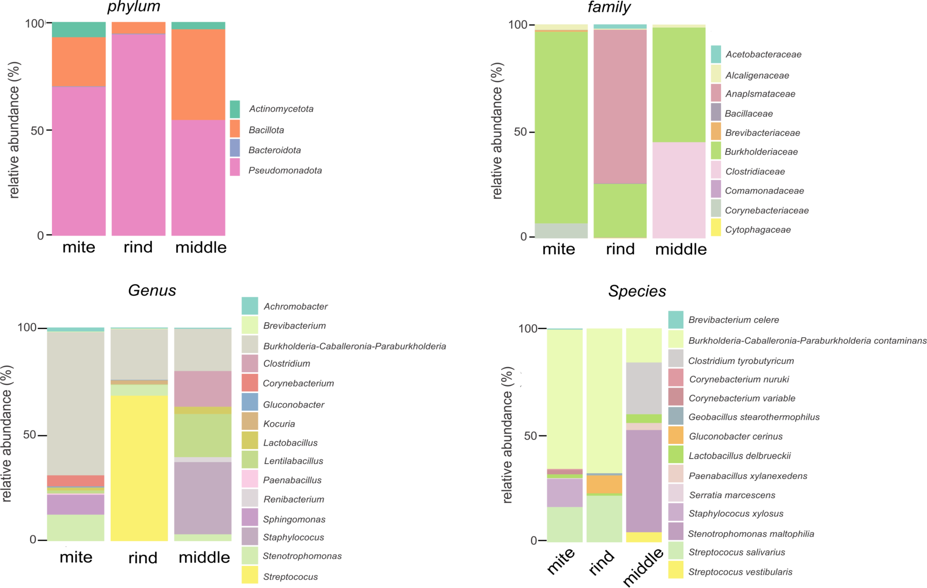
About Chao, Simpson, and Shannon’s indices, no significant difference among the samples was observed (Mann-Whitney test, p > 0.05). The distribution of the most abundant taxa classified at the taxonomic level of phylum, family, genus, and species is presented in Figure 2. On a phylum level, the microbiota of the mites, cheese rind and inner was predominantly composed by Pseudomonadota, followed by Bacillota. Regarding the family level, the microbiota of mites was composed predominantly of Burkolderiaceae, the cheese rind by Anaplasmataceae, and the cheese inner part by Burkolderiaceae and Clostridiaceae. The dominant genus in the mite microbiota was composed of Burkholderia-Caballeronia-Paraburkholderia, the cheese rind by Streptococcus, and the cheese inner part by Staphylococcus. On the species level, the microbiota of mites and cheese rind was composed predominantly of Burkholderia-Caballeronia-Paraburkholderia contaminans and the cheese inner part by Stenotrophomonas maltophilia.

Taxonomic distribution of Operational Taxonomic Units (OTUs) at phylum, family, genus, and species level of bacteria and archaebacteria identified in the experimental groups.
Figures and Tables
DISCUSSION
The presence of the cosmopolitan mite species T. putrescentiae demonstrates its predominance in Brazilian ripened cheeses and the importance and interest of this work for other countries [2,3,5]. T. putrescentiae is a species found in different types of habitats, being very common in the dust of human-created environments such as farms and food industry [2,17] and in stored products such as grains and dry pet food [18-20]. This mite species explores food resources rich in proteins and fats, and its success in colonizing different habitats and food is probably mediated by different bacterial communities found associated with their populations [21].
Analyses of Chao, Shannon, and Simpson diversity indices, while revealing no significant differences among the experimental groups, showed a greater similarity between the mite and cheese rind microbiota. This indicates that these experimental groups are more similar in terms of species presence/absence and their relative abundance (Figure 1). However, the Jaccard index showed a different similarity relationship, likely because this index only takes into account the presence/absence of species between the compared sets.
The analysis of 16S rRNA reads revealed that the Pseudomonadota phylum (formerly Proteobacteria) was predominant in all samples, especially in the rind. The Bacillota phylum (formerly Firmicutes) was the second most abundant. This finding was similar to results for some fermented dairy products from Northeast Asia [22]. In a study of Stela cheeses (a Greek cheese made from ovine, caprine, or a mixture of both types of milk) using 16S rRNA sequencing and Matrix-Assisted Laser Desorption/Ionization Time-of-Flight Mass Spectrometry (MALDI-TOF MS) approaches to analyze artisanal and industrial samples, the Bacillota phylum was predominant [23]. It is worth noting that the authors of this study did not report the presence of mites in the artisanal cheeses.
Burkholderia-Caballeronia-Paraburkholderia, shared between the microbiota of the mites, the rind, and the cheese middle, is a new taxonomic grouping that includes many species of environmental and beneficial bacteria [24] and is not associated with pathogenic microorganisms [25]. Bacteria from this group were the most abundant in the mite midgut, and one of the most abundant in the cheese rind and middle (Figure 2). This bacteria group also were found, relatively abundant (>1%) across most fermentation stages of ogi (a fermented cereal beverage), produced from millet varieties helping to reduce mycotoxins along with other lactic acid bacteria (LAB) [26]. During the rice wine production, mainly at the beginning of fermentation, bacteria from this group also were present [27]. The presence in this beverage was related to decreased formation of lactic, citric, and succinic acids and the presence of free amino acids. However, it showed a positive correlation with the presence of ethyl pentanoate, suggesting that these bacteria may enhance volatile flavor compounds during fermentation [27].
Lb. delbrueckii is another species present in the microbiota of mites, cheese rind and inner part. This species is abundant in cheeses, particularly those made from raw milk [28]. The species found in both the mite midgut and cheese rind was St. salivarius, a LAB. This bacterium has also been identified in other cheeses, such as a Brazilian cheese from Serra da Canastra [6,29]. This bacterium can contribute to the fermentation and ripening of cheese, positively affecting its flavor and aroma, particularly in artisanal varieties [7].
Despite being generally considered unpalatable, cheeses ripened with mites can have interesting sensory and flavor characteristics. Some studies show that the presence of these organisms in certain cheeses, such as Mimolette and Milbenkäse, can add a unique profile, including a volatile flavor component reminiscent of lemon oil [30,31].
In a previous work of our research group, analyzing two types of Brazilian cheeses (Colonial and Minas) ripened with T. putrescentiae mites, was identified in common, an increase in the concentrations of volatile compounds isoamyl butanoate and 2-nonanol compared to the same cheeses ripened without mites [5]. Additionally, also was identified decreased lactic acid, as reported by Liu and coauthors [27], decreased fatty acids, and increased glycerol, alanine, and citric acid.
Autochthonous microbiota display a vast interaction network that can give cheese specific, attractive properties for consumers. Different approaches have the potential to provide information about how to create an autochthonous microbiome starter culture capable of maintaining the desired characteristics of traditional products, such as the flavor and scent of artisanal cheeses. This study presents pioneering results in the analysis of cheeses ripened with mites.
CONCLUSION
Burkholderia-Caballeronia-Paraburkholderia contaminans and Lb. delbrueckii were the bacterial species shared among the intestinal samples of mites, the cheese rind, and the cheese’s inner part. St. salivarius was the bacterial species shared among the intestinal samples of mites and cheese rind. No potential food pathogens were identified in the mite microbiota interacting with the cheese microbiota. This is the first report of a comparative metataxonomic study of the microbiota of mites and cheese. However, further exploratory analysis may contribute to the findings in demonstrating that mites have the potential to improve the sensory attributes of cheeses without harmful effects.
-
Funding:
This research was funded by Foundation for Research Support and Innovation of Santa Catarina State (FAPESC), grant number 001, Minas Gerais State Foundation of Support to the Research (FAPEMIG), and Brazilian Council for Scientific and Technological Development (CNPq).
Acknowledgments:
None
Data Availability Statement:
Research data are only available upon request for corresponding author.
REFERENCES
- 1 Flechtmann CWH. [Mites in stored products and household dust]. Piracicaba. FEALQ. Escola Superior de Agricultura Luiz de Queiroz. 97:1986.
-
2 Carvalho MM, Oliveira EE, Matioli AL, Ferreira CLL, Machado da Silva N, De Dea Lindner J. Stored products mites in cheese ripening: Health aspects, technological and regulatory challenges in Brazil. J Stored Prod Res. 2018;76:116-21. Available from: https://doi.org/10.1016/j.jspr.2018.01.010
» https://doi.org/10.1016/j.jspr.2018.01.010 -
3 Carvalho MM, Machado da Silva N, Matioli AL, Oliveira EE, Ferreira CLL, De Dea Lindner J. Morphological and molecular characteristics of stored-product mites found on Brazilian ripened cheeses. J Stored Prod Res. 2018 79:79-82. Available from: https://doi.org/10.1016/j.jspr.2018.09.003
» https://doi.org/10.1016/j.jspr.2018.09.003 -
4 Melnyk JP, Smith A, Scott-Dupree C, Marcone MF, Hill A. Identification of cheese mite species inoculated on Mimolette and Milbenkase cheese through cryogenic scanning electron microscopy. J Dairy Sci. 2010;93:3461-8. Available from: https://doi.org/10.3168/jds.2009-2937
» https://doi.org/10.3168/jds.2009-2937 -
5 Carvalho MM, Alves Filho EG, Silva LMA, Martins FICC, Matioli AL, Oliveira EE, et al. Chemometric evaluation of the metabolites and volatile profiles of mite-ripened cheeses. Int Dairy J. 2020;110. Available from: https://doi.org/10.1016/j.idairyj.2020.104806
» https://doi.org/10.1016/j.idairyj.2020.104806 -
6 Queiroz LL, Lacorte GA, Isidorio WR, Landgraf M, de Melo Franco BDG, Pinto UM, et al. High Level of Interaction between Phages and Bacteria in an Artisanal Raw Milk Cheese Microbial Community. MSystems. 2023;8:1-16. Available from: https://doi.org/10.1128/msystems.00564-22
» https://doi.org/10.1128/msystems.00564-22 -
7 Coelho MC, Malcata FX, Silva CCG. Lactic Acid Bacteria in Raw-Milk Cheeses: From Starter Cultures to Probiotic Functions. Foods. 2022;11:1-32. Available from: https://doi.org/10.3390/foods11152276
» https://doi.org/10.3390/foods11152276 -
8 Dhariwal A, Chong J, Habib S, King IL, Agellon LB, Xia J. MicrobiomeAnalyst: A web-based tool for comprehensive statistical, visual and meta-analysis of microbiome data. Nucleic Acids Res. 2017;45:W180-8. Available from: https://doi.org/10.1093/nar/gkx295
» https://doi.org/10.1093/nar/gkx295 - 9 Christoff AP, Fernanda A, Sereia R, Dellyana, Boberg R, Lucio R, et al. Sequencing Neoprospecta Microbiome Technologies Bacterial identification through accurate library preparation and high-throughput sequencing. White Pap Bact NGS. 2017.
-
10 Hubert J, Kopecký J, Perotti MA, Nesvorná M, Braig HR, Ságová-Marečková M, et al. Detection and Identification of Species-Specific Bacteria Associated with Synanthropic Mites. Microb Ecol. 2012;63:919-28. Available from: https://doi.org/10.1007/s00248-011-9969-6
» https://doi.org/10.1007/s00248-011-9969-6 -
11 Wang Y, Qian PY. Conservative fragments in bacterial 16S rRNA genes and primer design for 16S ribosomal DNA amplicons in metagenomic studies. PLoS One. 2009;4. Available from: https://doi.org/10.1371/journal.pone.0007401
» https://doi.org/10.1371/journal.pone.0007401 -
12 Bolger AM, Lohse M, Usadel B. Trimmomatic: A flexible trimmer for Illumina sequence data. Bioinformatics. 2014;30:2114-20. Available from: https://doi.org/10.1093/bioinformatics/btu170
» https://doi.org/10.1093/bioinformatics/btu170 -
13 Callahan BJ, McMurdie PJ, Rosen MJ, Han AW, Johnson AJA, Holmes SP. DADA2: High-resolution sample inference from Illumina amplicon data. Nat Methods. 2016;13:581-3. Available from: https://doi.org/10.1038/nmeth.3869
» https://doi.org/10.1038/nmeth.3869 -
14 Quast C, Pruesse E, Yilmaz P, Gerken J, Schweer T, Yarza P, et al. The SILVA ribosomal RNA gene database project: Improved data processing and web-based tools. Nucleic Acids Res. 2013;41:590-6. Available from: https://doi.org/10.1093/nar/gks1219
» https://doi.org/10.1093/nar/gks1219 -
15 McMurdie PJ, Holmes S. Phyloseq: An R Package for Reproducible Interactive Analysis and Graphics of Microbiome Census Data. PLoS One. 2013;8. Available from: https://doi.org/10.1371/journal.pone.0061217
» https://doi.org/10.1371/journal.pone.0061217 - 16 Wickham H, Winston C. ggplot2: An implementation of the Grammar of Graphics. 2008.
-
17 Zhao Y, Abbar S, Phillips TW, Williams JB, Smith BS, Schilling MW. Developing food-grade coatings for dry-cured hams to protect against ham mite infestation. Meat Sci. 2016;113:73-9. Available from: https://doi.org/10.1016/j.meatsci.2015.11.014
» https://doi.org/10.1016/j.meatsci.2015.11.014 -
18 Olivry T, Mueller RS. Critically Appraised Topic on Adverse Food Reactions of Companion Animals (8): Storage Mites in Commercial Pet foods. BMC Vet Res. 2019;15:1-5. Available from: https://doi.org/10.1186/s12917-019-2102-7
» https://doi.org/10.1186/s12917-019-2102-7 -
19 Zou Z, Yang Y, Chen Y, Sun W, Xi J, Zhu P, et al. Chronic lead exposure prolongs the immature stages of brown-legged grain mite, Aleuroglyphus ovatus, in a long-term population study. Environ Pollut. 2023;337:122339. Available from: https://doi.org/10.1016/j.envpol.2023.122339
» https://doi.org/10.1016/j.envpol.2023.122339 -
20 Collins DA. A review on the factors affecting mite growth in stored grain commodities. Exp Appl Acarol. 2012;56:191-208. Available from: https://doi.org/10.1007/s10493-012-9512-6
» https://doi.org/10.1007/s10493-012-9512-6 -
21 Erban T, Klimov PB, Smrz J, Phillips TW, Nesvorna M, Kopecky J, et al. Populations of stored product mite tyrophagus putrescentiae differ in their bacterial communities. Front Microbiol. 2016;7:1-19. Available from: https://doi.org/10.3389/fmicb.2016.01046
» https://doi.org/10.3389/fmicb.2016.01046 -
22 Yu Z, Peng C, Kwok LY, Zhang H. The bacterial diversity of spontaneously fermented dairy products collected in northeast asia. Foods. 2021;10. Available from: https://doi.org/10.3390/foods10102321
» https://doi.org/10.3390/foods10102321 -
23 Tsouggou N, Slavko A, Tsipidou O, Georgoulis A, Dimov SG, Yin J, et al. Investigation of the Microbiome of Industrial PDO Sfela Cheese and Its Artisanal Variants Using 16S rDNA Amplicon Sequencing and Shotgun Metagenomics. Foods. 2024;13. Available from: https://doi.org/10.3390/foods13071023
» https://doi.org/10.3390/foods13071023 -
24 Qin L, Qi J, Shen G, Qin D, Wu J, Song Y, et al. Effects of Microbial Transfer during Food-Gut-Feces Circulation on the Health of Bombyx mori. Microbiol Spectr. 2022;10. Available from: https://doi.org/10.1128/spectrum.02357-22
» https://doi.org/10.1128/spectrum.02357-22 -
25 Estrada-de los Santos P, Palmer M, Chávez-Ramírez B, Beukes C, Steenkamp ET, Briscoe L, et al. Whole genome analyses suggests that Burkholderia sensu lato contains two additional novel genera (Mycetohabitans gen. nov., and Trinickia gen. nov.): Implications for the evolution of diazotrophy and nodulation in the Burkholderiaceae. Genes (Basel). 2018;9. Available from: https://doi.org/10.3390/genes9080389
» https://doi.org/10.3390/genes9080389 -
26 Chibuzor-Onyema IE, Ezeokoli OT, Sulyok M, Notununu I, Petchkongkaew A, Elliott CT, et al. Metataxonomic analysis of bacterial communities and mycotoxin reduction during processing of three millet varieties into ogi, a fermented cereal beverage. Food Res Int. 2021;143:110241. Available from: https://doi.org/10.1016/j.foodres.2021.110241
» https://doi.org/10.1016/j.foodres.2021.110241 -
27 Liu A, Yang X, Guo Q, Li B, Zheng Y, Shi Y, et al. Microbial Communities and Flavor Compounds during the Fermentation of Traditional Hong Qu Glutinous Rice Wine. Foods. 2022;11. Available from: https://doi.org/10.3390/foods11081097
» https://doi.org/10.3390/foods11081097 -
28 Stachelska, M.A.; Ekielski, A.; Karpiński, P.; Żelaziński T. K. New Genetic Determinants for qPCR Identification and the. Molecules. 2024;29:1533. Available from: https://doi.org/https://doi.org/10.3390/molecules29071533
» https://doi.org/https://doi.org/10.3390/molecules29071533 -
29 Imek B, Tuncer Y. Some properties of fresh and ripened traditional akcakatik cheese. Korean J Food Sci Anim Resour. 2018;38:110-22. Available from: https://doi.org/10.5851/kosfa.2018.38.1.110
» https://doi.org/10.5851/kosfa.2018.38.1.110 -
30 Shimizu N, OConnor BM, Hiruta SF, Hagino W, Shimano S. Mite secretions from three traditional mite-ripened cheese types: are ripened French cheeses flavored by the mites (Acari: Astigmata)? Exp Appl Acarol. 2022;87:309-23. Available from: https://doi.org/10.1007/s10493-022-00734-7
» https://doi.org/10.1007/s10493-022-00734-7 -
31 Bruckner A, Heethoff M. Scent of a mite: origin and chemical characterization of the lemon-like flavor of mite-ripened cheeses. Exp Appl Acarol. 2016;69:249-61. Available from: https://doi.org/10.1007/s10493-016-0040-7
» https://doi.org/10.1007/s10493-016-0040-7
-
Editor-in-Chief:
Bill Jorge Costa
-
Associate Editor:
Paulo Vitor Farago