Hypocholesterolemic, Antiaterogenic, Antimutagenic Potential and Non-Toxic in vivo of Sorghum (Sorghum bicolor (l.) Moench) Flour and its Protein Hydrolysate
Abstract
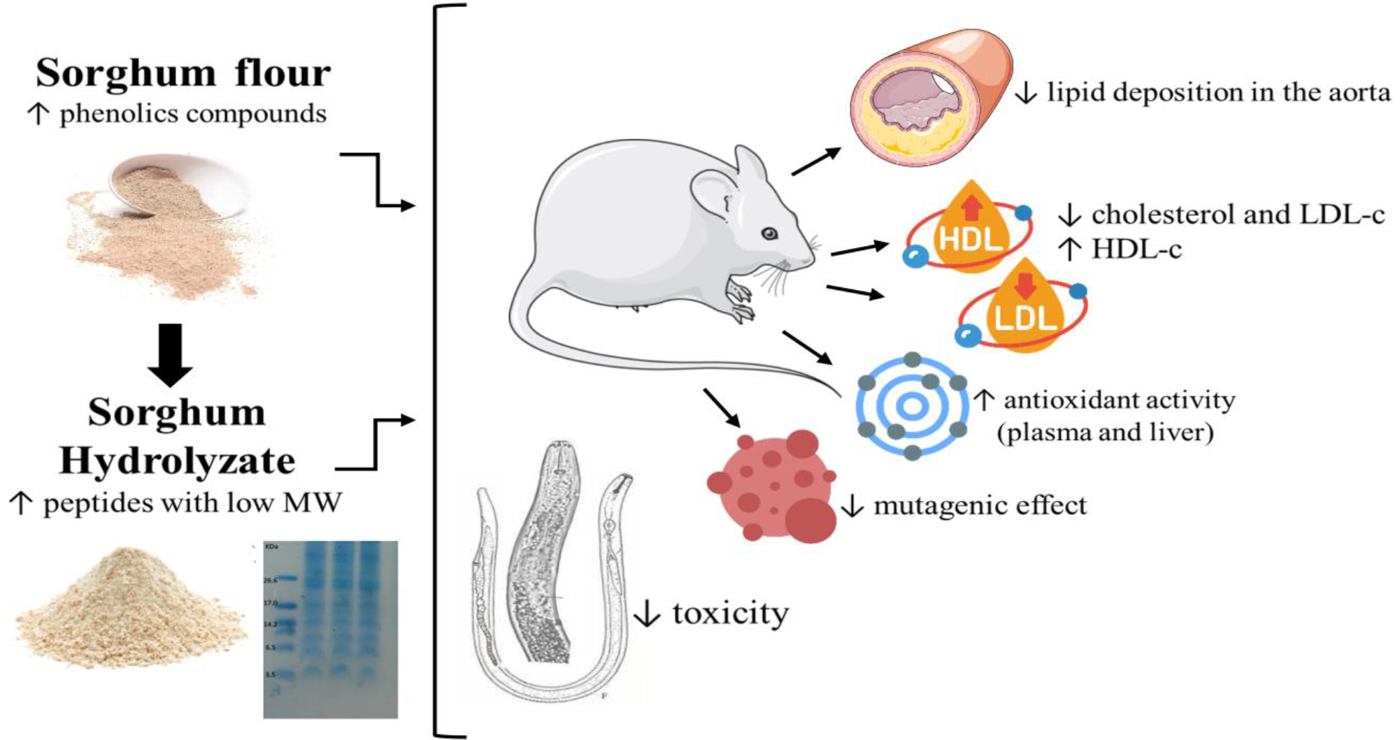
Sorghum (Sorghum bicolor (L.) Moench) is an alternative to the consumption of conventional cereals, with high technological, nutritional and functional potential. The peptides formed during the enzymatic digestion of their proteins have shown a modulating effect on variables related to chronic noncommunicable diseases, including cardiovascular ones. The aim of this study was to identify and characterize the bioactive compounds of sorghum and its hydrolysate in their toxicity, hypocholesterolemic, antiatherogenic, and antioxidant properties in vivo. For the toxicity test, nematodes Caenorhabditis elegans were used. For the experimental study, wild-type C57BL/6 and LDL-cholesterol receptor knockout (LDL-/-) mice were maintained on a normal or atherogenic diet and received whole sorghum flour or its hydrolysate by intragastric oral gavage (800 mg/kg) for 8 weeks. The protein hydrolysate showed the highest concentration of phenolic compounds and high antioxidant activity; the enzymatic hydrolysis reached 42.5% of the proteins, with low molecular weight peptides, in addition to not showing toxicity at the dose used. The in vivo results demonstrated that the flour and hydrolysate reduced the levels of total and LDL-c cholesterol and the lipid deposition in aorta of animals with atherogenic diet, and increased the antioxidant activity in plasma and liver, in addition to plasmatic HDL-c. The hydrolysate also showed an antimutagenic effect by micronucleus test. Therefore, whole sorghum flour and its hydrolysate are a source of antioxidant nutritional compounds that act against the development of hypercholesterolemia and atherogenesis, in addition to an antimutagenic effect and absence of toxicity.
Keywords:
Sorghum hydrolyzed; Peptides; Atherosclerosis; Phenolic compounds; Caenorhabditis elegans.
HIGHLIGHTS
Sorghum has high content of phenolic compounds, dietary fiber and it’s gluten-free.
Its proteins during enzymatic digestion can form peptides with bioactive potential.
hydrolysate hasn’t genotoxicity by the micronucleus assay and antimutagenic effect.
Sorghum flour and hydrolysate improve lipid and antioxidant profile.
INTRODUCTION
The increase in overweight and obesity rates, associated with the greater aging of the population, are leading to an increase in non-communicable chronic diseases incidence, especially cardiovascular diseases. The primary cause in triggering cardiovascular disease is the atherosclerotic process development and progression [1,2]. In this condition, the plasma lipid rates increase, particularly the low-density lipoproteins (LDL), which can enter the endothelial layer of arteries and be phagocytosed by macrophages that end up dying and turning into foam cells that driving to the atherosclerotic plaques formation [3].
In general, the prevention or risk reduction and even the cardiovascular diseases treatment must be started by lifestyle changes, which requires exploring alternative sources for the acquisition of healthy eating habits. In this context, sorghum (Sorghum bicolor (L.) Moench) has emerged as a possibility for consumption over conventional cereals. Sorghum stands out for being the cereal with the highest content of phenolic compounds, such as phenolic acids, anthocyanins and tannins, in addition to having a relevant content of dietary fiber and being gluten-free [4]. Its composition also points out to the presence of an important amount of proteins (from 7 to 15%) that during enzymatic digestion can form peptides, which may reflect in bioactive potential, depending on their amino acid composition [5].
However, despite the importance of vegetal origin compounds, include peptides and phenolic coumponds, in human health care, the toxicological and/or genotoxic potential of these therapeutic resources needs to be further studied [6]. In this context, obtaining sorghum protein hydrolysate through enzymatic hydrolysis and its evaluation in cholesterolemia and atherosclerosis control, compared to the whole sorghum flour, with toxicological safety and absent genotoxic potential, turns to be an innovative study for development of new functional products, which can contribute to prevention and reduction of chronic non-communicable diseases risks in the human population. Then, the objective of this study was to identify and characterize the bioactive compounds of sorghum protein hydrolysate and to evaluate the toxicity and hypocholesterolemic, antiatherogenic, and antioxidant properties in vivo.
MATERIAL AND METHODS
Material
The BRS 310 whole sorghum flour of red pericarp and with no tannins was kindly donated by Embrapa Sorghum and Corn (Sete Lagoas, Minas Gerais, Brazil). The flour was thermally treated using dry heat in a combined oven, at 121 ºC for 25 min. Subsequently, it was vacuum-conditioned and frozen at -20 ºC until the analysis was performed.
Sorghum protein hydrolysate
The protein hydrolysate was obtained through simulated enzymatic digestion, according to [7]. Briefly, the sorghum flour was suspended in deionized water at a 1:10 (w/v) ratio followed by pepsin enzymatic digestion (Sigma-Aldrich®, EC 3.4.23.1), substrate 1:10 (w/w) at pH 2.0 for 2 hours. Then, pancreatin (Sigma-Aldrich®, EC 232-468-9) was added to the suspension, substrate 1:10 (w/w) at pH 7.5 for another 2 hours, at 37 °C, under constant agitation (Fisatom® magnetic stirrer). The process was interrupted by heating at 75°C for 20 minutes.
Then, the samples were centrifuged (Excelsa 2206 - Fanem) at speed of 3.500 rpm for 25 minutes and the isolated hydrolyzed protein, in supernatant, was dialyzed to remove salts by means of cellulose membrane filtration (Spectra/Por®, Biotech), under agitation, with cutoff point of 500 Da. Afterwards the samples were lyophilized (Lyophilizer Enterprise I, Terroni) and stored at -20 ºC until the analysis.
Nutritional and protein profile
The determination of the moisture content was carried out by drying method in oven at 105 °C, ashes by muffle incineration at 550 °C, the total protein by the Kjeldahl method (using 5.75 factor), total lipids by Goldfish extraction method, and the carbohydrate content by difference [8].
The protein profile of the sample was obtained by tricine-SDS gel electrophoresis, according to the method described by [9]. The separation gel was prepared at a concentration of 16.4% acrylamide/bis-acrylamide, and the stacking gel at 3.9%. The gels were assembled using the Mini-PROTEAN® Tetra System (Bio-Rad). A total of 100 µg of the sample was loaded onto the gel.
For sample preparation, 20 mg of the hydrolysate was weighed and diluted in 200 µL of Tris-HCl buffer, pH 8.0, 125 mM. Electrophoresis was carried out at a constant voltage of 18 V for approximately 15 hours, using a cathode buffer (0.1 M Tricine; 0.1 M Tris-HCl, pH 8.31; 0.1% SDS) and an anode buffer (0.2 M Tris-HCl, pH 8.9; 0.2% SDS). To estimate the molecular weight of the peptides, a marker with the following molecular weights was used: 26,600; 17,000; 14,200; 6,500; 3,496; 1,060 Da (M3546 - Ultra Low Range Molecular Weight Marker, Sigma).
After the run was completed, the gel was carefully removed from the glass plates and placed in a fixing solution (40 mL ethanol, 10 mL acetic acid, and 50 mL water) for 30 minutes. It was then transferred to a staining solution (colloidal Coomassie Blue G), according to the modified method of [10], under constant agitation until the protein bands were visible (approximately 48 hours). Subsequently, the gel was placed in water to remove excess dye.
The determination of protein concentration in the hydrolysate (identified as total protein) and in the protein fractions of sorghum flour was performed in triplicate using the Bradford method [11], according to the manufacturer’s instructions. The results were obtained using a standard curve of bovine serum albumin (BSA), y = 0.0003x - 0.0074, R2 = 0.9923, and expressed in mg of soluble protein/mL of sample.
Protein quantification of the samples was also carried out using the Kjeldahl method, applying a conversion factor of 5.75 [8].
Protein hydrolysis degree
The hydrolysis degree of the sorghum protein hydrolysate in relation to the flour was calculated considering the equation [12,13]: HD = (h / hhot) *100, which HD is the hydrolysis degree obtained; h is the number of hydrolyzed peptide bonds; hhot is the total number of peptide bonds present in the native protein, considering the protein content of the flour.
The results obtained from the protein determination using the Bradford method [11] were used for this calculation.
Determination of the total phenolic compounds (TPC) content and total antioxidant activity (TAA)
One gram of each sample (flour and hydrolysate sorghum) and 10 mL methanol (Neon®) 60% were added in a tube covered with aluminum foil and shaken until complete solubilization. This mixture was taken to the ultrasound bath (Elmasonic P - Elma) for 25 min, at 40 ºC, 37 khz and 50% amplitude. After that, the tube was taken to centrifuge (Excelsa 2206 - Fanem) at 3500 rpm for 10 min, and the filtered and collected supernatant had its volume completed to 15 ml with deionized water [14].
TPC: Aliquots of 20 µL from each extract were pipetted into a microplate, followed by 80 µL of 10% Folin-Ciocalteu reagent. After 4 minutes, 100 µL of 7.5% sodium carbonate (w/v - Sigma®) was added. After 2 hours, the absorbance was read at 765 nm using a microplate reader (SpectraMax® 190). The results for TPC were expressed in milligrams of gallic acid equivalents per gram of sample [11], using the equation , with
.
ABTS: Thirty microliters of each extract were pipetted into a microplate, and 270 µL of ABTS radical solution was added. After 6 minutes, absorbance was measured at 734 nm (SpectraMax® 190). The blank analysis was performed using concentrated methanol mixed with the ABTS radical solution [15].
DPPH: Twenty microliters of each extract and 280 µL of DPPH radical solution were pipetted into a microplate. Absorbance was measured at 517 nm (SpectraMax® 190) after 60 minutes of incubation. The blank analysis was performed using concentrated methanol mixed with the DPPH radical solution [16].
For the ABTS and DPPH assays, a gallic acid calibration curve was generated with concentrations ranging from 0.005 to 0.200 µg/mL, producing the regression equation , with , to express results in milligrams of gallic acid equivalents per gram of sample. Results were also expressed as the antioxidant activity index (AAI), using the equation: AAI (%) = [(Blank Abs - Sample Abs) × 100] / Blank Abs [17].
Toxicity test in nematodes Caenorhabditis elegans
The toxicological safety of sorghum flour and its protein hydrolysate was tested on nematodes Caenorhabditis elegans (C. elegans), wild-type strain N2. They were stored in nematode growth medium and inoculated with the bacterium Escherichia coli OP50 at 20 ºC. After worms underwent complete synchronization in the L1 larval stage, they were exposed to 50 µl Levamisole (L), positive control anthelmintic, 50 µl sorghum flour (F), 50 µl sorghum hydrolysate (H), or 50 µl saline, for the negative control group (C), at 20 °C for 30 minutes (acute treatment) in BOD incubator (TECNAL, TE-371, São Paulo, BR) under continuous agitation in a homogenizer in 0.5% NaCl liquid medium.
The C. elegans mortality evaluation (DL50 estimation) had 2,500 larvae in stage L1. Furthermore, 48 h after treatment, the body surface area of the adult worm (µm2) was measured using a stereomicroscope (Olympus IX71) to assess the worms’ development. The worms were photographed to have their body contour measured (10 measurements) using the AxioVision Rel. 4.8 software [18].
In vivo assessment of cholesterolemia and oxidative stress
Thirty male mice were used, being 6 C57BL/6 and 24 knockout for LDL cholesterol receptor, all from the Experimental Monitoring Laboratory of the Complex BioPractices - UVV. The mice weighed between 20 and 25 g and eight weeks old, having been fed during this period on a standard normocaloric diet, and kept in mini-isolators, with humidity and temperature control,12 h light-dark cycle and access to water and food ad libitum.
All procedures followed the ethical principles for animal experimentation, according to the NIH Guide for the Care and Use of Laboratory Animals [19], and were approved by the Ethics, Bioethics, and Animal Welfare Commission of Vila Velha University (CEUA-UVV; protocol number 515/2018), and were conducted in accordance with [7, 20].
The mice were randomly divided into 5 experimental groups, 6 animals per group:
-
• C57NL: wild control with standard diet and water by gavage.
-
• CNL: LDL-/- with standard diet and water by gavage,
-
• CHL: LDL -/- with high-fat diet and water by gavage,
-
• HHL: LDL-/- with a high-fat diet and 800 mg/kg body weight of protein hydrolysate by gavage,
-
• FHL: LDL-/- with a high-fat diet and 800 mg/kg body weight of sorghum flour by gavage,
The concentration of 800 mg of flour or hydrolysate per kilogram of body weight (BW) was defined based on a higher amount than that used in the studies by [7], with black bean protein hydrolysate (700 mg/kg BW), and by [20], with chia protein hydrolysate (400 mg/kg BW). Considering that sorghum flour has a lower protein content (between 8% and 10%) than the foods used in the aforementioned studies (approximately 20% to 30%), and that there are no previous studies using sorghum protein hydrolysate in an in vivo model, it was decided to use a slightly higher dose. This was done without overloading the animals’ food intake, taking into account their gastric capacity and the use of gavage to administer the flour or hydrolysate in a single daily dose.
The high-fat diet provided by PragSoluções® (2% cholesterol, 37.45% carbohydrates, 20% protein and 30% fat) and normolipidic (50.9% carbohydrates, 23% protein and 4% fat) by Alinutri®.
The HHL and FHL groups received daily, for 60 days, sorghum flour and protein hydrolysate diluted in water, by intragastric oral gavage, a total volume of 1 ml/100 g of Body Weight. The control group animals received only water daily, by gavage.
At the end of experiment, the animals were fasted for 8 h and then anesthetized with Ketamine and Xylazine (11.5 and 0.1 mg/100 g BW, respectively). Blood samples were collected through cardiac puncture to obtain the plasma. The aorta, liver, adipose tissues, and femur of all animals were removed and kept in buffers, until analysis.
Assessment of adiposity, body weight, and food consumption indicators
The animals' body weight and food consumption were weekly monitored. Obesity was measured by the Lee index, which is the ratio between the cube root of body weight (g) and nasal-anal length (cm). Food Efficiency Coefficient was calculated by weight gain (g)/ total diet consumption (g).
To calculate the adiposity index, the sum of the abdominal and epididymal adipose tissues was divided by the body mass and multiplied by 100, and the hepatosomatic index was calculated by the liver weight (g)/ body mass (g) x 100 [21].
Total antioxidant activity
For the liver homogenate preparation, a 100 mg fragment was weighed and added with 140 mM phosphate buffer pH 7.4. The mixture was ground in Ultra Turrax Metabo GE700 followed by homogenization and centrifuged (Revan, 14000A, SP, Brazil) at 6500 rpm for 20 min. The total antioxidant activity was evaluated in the animals’ plasma and liver using ABTS and DPPH radicals, as described in topic “Determination of the total phenolic compounds (TPC) content and total antioxidant activity (TAA)”.
Lipid profile
Total Cholesterol (TC), High Density Lipoprotein (HDL-c), Low Density Lipoprotein (LDL-c) and Triglycerides (TG) were determined in animal’s plasm, using commercial kits (BIOCLIN, Rio de Janeiro, Brazil), following the manufacturer's instructions.
Determination of lipid peroxidation
Briefly, 200 mg liver and 250 μl of 1% thiobarbituric acid solution (TBA) were homogeneited and 125 μl of this sample were used. The determination of thiobarbituric acid reactive metabolites (TBARS) was obtained in a spectrophotometer at 532 nm (SpectraMax 190). The result was expressed in malondialdehyde nmol (MDA)/mg of protein from the MDA standard curve (10 to 500 µM MDA/mg of proteins - y = 0,057x+0,0086R2 = 0,9828) [22].
Superoxide Dismutase Assay (SOD)
About 200 mg of aorta was diluted 1:10 in phosphate buffered saline (PBS) [23] and then mixed with 1.0 ml carbonate buffer (0.2 M, pH 10.2) and 0.8 ml KCl (0.015 M). The reaction was initiated by the 0.2 ml addition of epinephrine (0.025 M). The change in absorbance was recorded at 480 nm (Kasuaki spectrophotometer), at 15-second intervals, for one minute. Enzyme activity was defined as the amount of enzyme that causes 50% epinephrine auto-oxidation inhibition. Results were expressed as Unit SOD/mg protein.
Catalase Assay (CAT)
The CAT enzyme activity was carried out in the aortas’ cellular homogenate, as described by [24] and reaction starts with the 40 μl action of hydrogen peroxide (H2O2) (0.066 M in phosphate buffer). Absorbances at 240 nm (Kasuaki spectrophotometer) were recorded every 15 seconds, for a 1-min interval. The enzyme activity was defined as the enzyme amount which consumes half of the H2O2 in one minute. The results were expressed as peroxide extinction coefficient per minute (∆E.min/mg protein).
Analysis of vascular lipid deposition en face
The en face analysis was standardized as previously described by [25]. After cutting to open the aortic arch, the aortas were pinned onto an ethylene-vinyl acetate (EVA) surface. Lipid deposition was assessed by staining with the lipid-specific dye Oil Red O (Sigma-Aldrich®). All samples were immersed in an alcoholic solution of Oil Red for one minute and then rinsed in a water bath to remove any excess red dye. Lipid deposition analysis was based on the intensity and extent of red staining. Images were captured using a high-resolution digital camera attached to a microscope (Nikon, Eclipse 200) with a 100x objective. Morphometric analysis was performed using the ImageJ software (public domain - National Institutes of Health, USA).
Evaluation of genotoxicity by the Micronucleus test
For this analysis were used only the CHL and HHL groups. The HHL group was divided into a control group and a group that received cyclophosphamide, totaling 3 groups, containing 5 animals each, as follows:
• HHL-: high-fat diet, sorghum hydrolyzed by gavage (800 mg/kg BW) for 60 days, without damage-inducing agents’ application,
• HHL+: high-fat diet and sorghum protein hydrolysate (800 mg/kg BW), via gavage, for 60 days, and application of a single cyclophosphamide dose (50 mg/kg BW) 24 hours before euthanasia,
• CHL+: high-fat diet and water by gavage, with application of a single cyclophosphamide dose (50 mg/kg of weight) 24 hours before euthanasia.
After the animals were euthanized, the right femur was removed by two cuts, the first one on the hardest part and the second one on the opposite side, being the bone marrow collection performed with the aid of an insulin syringe, along the bone marrow's path. Afterwards they were fixed, being immersed in methanol (Neon®) for 10 min and stained for 7 min in dye (Leishman, Sigma-Aldrich) and washed with distilled water.
The micronucleus count was performed in 2000 bone marrow cells from each animal’s femur, totaling 10000 cells per group. The slides were visualized in immersion oil, with a 100X objective and eyepiece (Nikon, Eclipse e200) [18]. The counting started with 200 polychromatic (PCE) and normochromatic (NCE) erythrocytes, to perform the correlation and the number of micronuclei in PCE, later the counting went on only with PCE and micronuclei up to a total of 2000 cells.
Statistical analysis
The data were normal by the Shapiro-Wilk test, thus, the 2 independent variables analysis (flour and hydrolysate) was compared by the T-test and the groups analysis using Analysis of Variance (ANOVA) and, subsequently, for significant differences at 5% probability level, Duncan's comparison test was performed at the same probability. The results were analyzed by the SAS software and expressed as mean and standard deviation. The graphics were performed with the GraphPad program, version 9.0.
RESULTS
Nutritional, protein and bioactive compound profile of sorghum flour and its hydrolysate
All analyzed compounds showed a difference (p≤0.05) between flour and protein hydrolysate. The flour was characterized by having higher moisture content, lipids, and total carbohydrates. While the protein hydrolysate, higher ash content, phenolic compounds and antioxidant activity by the two evaluated methods (Table 1).
Nutritional composition, total phenolic compounds, and antioxidant profile of sorghum flour and protein hydrolysate.
The protein hydrolysate had 76% higher protein content by Kjeldahl method. When compared to the result obtained for the hydrolysate by the Bradford method, there is a reduction in protein content compared to flour, contrary to what was observed in the determination by Kjeldahl (Table 1).
The degree of hydrolysis (DH) was calculated based on the original protein content provided by the flour and the protein content after enzymatic hydrolysis.
The enzymatic hydrolysis process carried out in the flour has led to reduction of the high-molecular weight protein content in 42%, formatting peptides and free amino acids with molecular weight of 3.5 and up 26.6 kDa. The samples showed bands between 3.5 and up to 26.6 KDa, however sorghum hydrolizate had mainly proteins with 6.5-14.2 kDa and sorghum-flour higher 17 kDa (Figure 1).
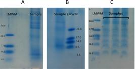
(A) Electrophoresis of sorghum hydrolysate performed with a molecular weight marker up to 6.5 kDa; (B) electrophoresis of hydrolysate performed with a marker up to 3.5 kDa; and (C) electrophoresis of heat-treated flour performed with a marker up to 3.5 kDa. LMWM = Low Molecular Weight Marker.
Evaluation of the in vivo toxicity of sorghum flour and its hydrolysate
The percentage survival values of the worms that were fed with flour and sorghum hydrolysate were equal to those without intervention (control) and higher than those of the Levamisole group (p≤0.05) (Figure 2A). Likewise, C. elegans exposed to flour had no reduction in body area, as their values were similar to the control (p>0.05), however, higher than those of the Levamisole group (p≤0.05). It is noteworthy that the worms fed with sorghum hydrolysate had a better development than the control and flour groups (p≤0.05) (Figure 2B).
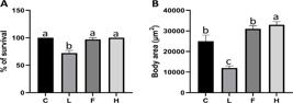
Survival and development of Caenorhabditis elegans exposed to sorghum flour and protein hydrolysate. (A) Percentage of C. elegans survival; (B) body area of C. elegans. Groups: C: Control group; L: Levamisole; F: sorghum flour group; H: sorghum hydrolyzed group. Analyzes performed with 2,500 C.elegans larvae, in stage L1, during 24 h (% of survival) or 48h (body area).
Different letters indicate a significant difference by ANOVA and post hoc of the Duncan test at 5%.
In vivo effect of flour and protein hydrolysate in experimental groups
The animals those received the flour (FHL) and hydrolysate (HHL) presented a reduction (p≤0.05) in the adiposity index and hepatosomatic index related to the CHL, and also achieved similar results to the C57NL and CNL groups (p>0.05). There was no statistical difference (p>0.05) in relation to the food efficiency ratio (FER) and the obesity index (Lee index) between the groups. It was observed a higher weight gain in C57NL control group compared to others groups, which were similar between then. Furthermore, there was a lower (p≤0.05) feed intake from the animals fed with high-fat diet (CHL, FHL, HHL), compared to the control groups (C57NL, CNL) (Table 2).
Effect of sorghum whole flour and its hydrolysate on biometric measurements and biochemical analyzes of plasma, liver, and aorta animals in the experimental groups.
Values expressed as mean ± standard deviation. Different letters on the same line indicate a significant difference by ANOVA and post hoc of the Duncan Test at 5%. C57NL: normal control group, with normal chow (n=6); CNL: LDL-/- knockout group with normal chow (n=6); CHL the LDL-/- knockout group with high-fat diet (n=6); FHL: LDL-/- knockout group with high-fat diet and 800mg of flour/kg Body Weight (n=6); HHL: LDL-/- knockout group with high fat diet and 800mg of protein hydrolysate/Kg Body Weight (n=6). FER: Food efficiency ratio; ABTS: 2,2-azinobis-(3-ethylbenzothiazoline-6-sulfonate; DPPH: 2,2-diphenylpicrylhydrazyl; AAI%: antioxidant activity index; SOD: Superoxide Dismutase; CAT: catalase; TBARS: thiobarbituric acid reactive metabolites.
The antioxidant capacity of the animals' plasma and liver was evaluated through the antioxidant activity index (AAI%) of the ATBS and DPPH radicals. The groups fed with sorghum flour and protein hydrolysate showed greater capacity to inhibit the ABTS and DPPH radicals in both plasma and liver, especially compared to the CHL group (p≤0.05) and, in some cases, with CNL (p≤0.05). There was no difference (p>0.05) between the experimental groups to liver TBARS. However, plasma TBARS was minor to HHL groups compared to the CNL, CHL and FHL groups (p≤0.05). There was no difference to the levels of the antioxidant enzymes superoxide dismutase (SOD) and catalase (CAT) between the goups (p>0.05).
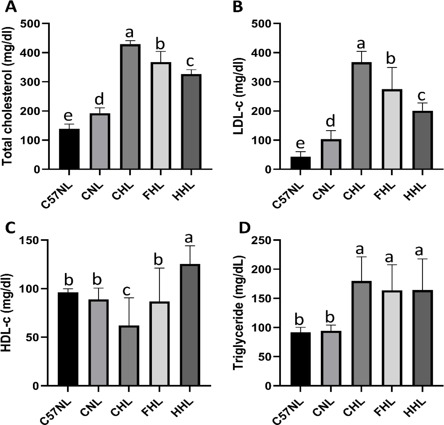
Regarding the lipid profile, the highest mean of total cholesterol and LDL cholesterol was found in the CHL group, which showed a significant difference (p≤0.05) in relation to the other experimental groups. The FHL and HHL groups showed reduced rates, indicating that the intervention was able to control total cholesterol rates even under predisposition conditions of the animals (Figure 3A). In fact, the HHL group managed to reduce LDL to values similar to the CNL control (p>0.05), also with predisposition, but receiving a normolipidic diet (Figure 3B). In addition to these results, it was observed that higher mean HDL (p≤0.05) also for the HHL group and similar means (p>0.05) for the FHL groups and their normal controls - C57NL and CNL (Figure 3C). Triglyceride dosages were similar between the groups that received high-fat diet and different (p≤0.05) from the CNL and C57NL control groups, showing no intervention effects on triglyceride levels in the FHL and HHL groups (Figure 3D).
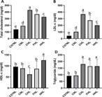
Effect of sorghum whole flour and its hydrolysate on plasm lipid profile in animals of the experimental groups. (A) levels of total cholesterol; (B) levels of LDL-c; (C) levels of HDL-c; (D) levels of triglyceride. Values expressed as mean ± standard deviation. Different letters on the same line indicate a significant difference by ANOVA and post hoc of the Duncan test at 5%. Groups: C57NL: normal control group, with normal chow (n=6); CNL: LDL-/- knockout group with normal chow (n=6); CHL the LDL-/- knockout group with high-fat diet (n=6); FHL: LDL-/- knockout group with high-fat diet and 800mg of flour/kg Body Weight (n=6); HHL: LDL-/- knockout group with high fat diet and 800mg of protein hydrolysate/Kg Body Weight (n=6). HDL: High-density lipoprotein; LDL: low-density lipoprotein.
Effect of sorghum flour and protein hydrolysate on animal atherosclerosis
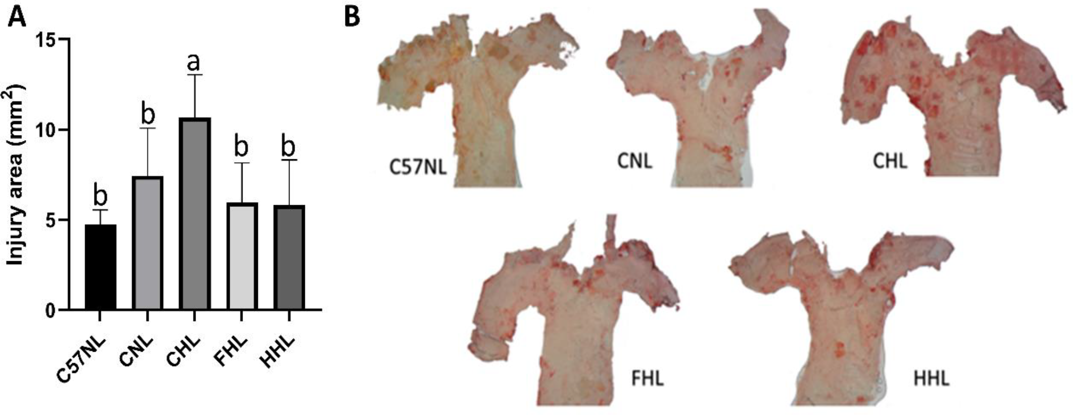
The lesion area was smaller in the HHL and FHL groups when compared to the CHL group (p≤0.05), and similar to the CNL group (p>0.05) (Figure 4A). Therefore, the intervention for 60 days with sorghum flour and hydrolysate decreased lipid deposition in aortas from LDL receptor knockout mice fed by high-fat diet (Figure 4B).
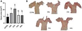
Area of injury (mm2) and lipid deposition (en face) of the aortas of mice receiving high-fat diet and treated with sorghum whole flour and its hydrolysate. (A) injury area of aortas; (B) photographs of the analysis of lipid deposition (en face) in the aorta. Groups: C57NL: normal control group, with normal chow (n=6); CNL: LDL-/- knockout group with normal chow (n=6); CHL the LDL-/- knockout group with high-fat diet (n=6); FHL: LDL-/- knockout group with high-fat diet and 800mg of flour/kg Body Weight (n=6); HHL: LDL-/- knockout group with high fat diet and 800mg of protein hydrolysate/Kg Body Weight (n=6). Values expressed as mean ± standard deviation. Different letters indicate statistical difference at ANOVA and post hoc of the Duncan at 5%. Samples stained with Oil-Red lipid marker.
Evaluation of genetic damage by micronucleus test
In the present study, the hydrolysate had the ability to protect cells against damage induced by cyclophosphamide (antimutagenic effect) and its ability to cause genetic damage (genotoxicity) after administration for 60 days were evaluated. The data from the bone marrow bioassay of the mice are presented in Table 3 and Figure 5.
Number of polychromatic (PCE) and normochromatic (NCE) erythrocytes, micronucleated polychromatic erythrocytes (MN-PCE), and the PCE / NCE ratio, in bone marrow of LDL-/- knockout mice
Results presented as mean ± standard deviation. Different letters in the same column indicate a significant difference (p≤0.05) by ANOVA and post hoc of the Duncan at 5%. HHL-: high-fat diet and 800 mg/kg body weigh of sorghum protein hydrolysate; HHL+: high-fat diet and 800 mg/kg body weight of protein hydrolysate with application of Cyclophosphamide 24 h before euthanasia; CHL+: high-fat diet and application of cyclophosphamide 24 h before euthanasia.

Photographic record of a slide with a bone marrow smear from an animal in the CHL+ group, showing: NCE = normochromatic erythrocyte; MN-NCE = micronucleated normochromatic erythrocyte; PCE = polychromatic erythrocyte; and MN-PCE = micronucleated polychromatic erythrocyte.
The CHL+ group which received cyclophosphamide and didn’t receive sorghum hydrolysate showed a high micronuclei frequency and a low PCE/NCE ratio. In groups receiving sorghum protein hydrolysate, with or without cyclophosphamide damage induction (HHL+ and HHL-, respectively), MN rates were significantly (p≤0.05) reduced and PCE/NCE ratios increased. The MN-PCE fraction didn’t increase during the intervention. The PCE/NCE ratio for the HHL- group indicated the absence of any cytotoxicity under these experimental conditions. The MN-PCE fraction didn’t increase during the intervention, therefore, there was no evidence of mutagenic activity by the hydrolysate.
The MN-PCE rate and the PCE/NCE ratio for the group that received hydrolysate and cyclophosphamide (HHL+) indicate that the hydrolysate administration to the animals for 60 days can have an antimutagenic and chemoprotective effect. The number of micronuclei reduced from 7.0 (CHL+) to 3.0 (HHL+) when the hydrolysate was administered before cyclophosphamide.
DISCUSSION
Sorghum flour and its hydrolysate, obtained by enzymatic digestion, show differences in all its nutritional constituents and antioxidant action. Protein hydrolysates have aroused great industry’s interest and are produced for several purposes. Regarding protein quantification by the Kjeldahl method, the protein hydrolysate had a protein content 56% higher than found in flour, which may be related to the higher content of free nitrogen compounds, such as peptides and amino acids, obtained after enzymatic hydrolysis, since this method quantifies nitrogen in general, without distinguishing the proteins from peptides and amino acids or other non-protein nitrogenous compounds [8]. However, when compared to the result obtained by the Bradford method, there is a 57% reduction in the hydrolysate protein content. This result can be attributed to the fact that this method quantifies whole proteins, of high molecular weight, disregarding peptides and amino acids. Thus, the result indicates that enzymatic hydrolysis reduced the protein amount in the sample, as expected, once it generated free peptides and amino acids, which was confirmed by the degree of hydrolysis of 42.5%.
The enzymatic hydrolysis showed, according to the hydrolysate electrophoretic profile, that there was an increase in the prevalence of peptides with molecular weight close to 6.5 kDa, however, it didn’t show the presence of 3.5 kDa peptides, suggesting a possible degradation of these compounds or their decomposition into amino acids, during the enzymatic hydrolysis process, as they were present in the flour that originated the hydrolysate. Some authors report the difficulty of electrophoresis in detecting small peptides (<2.0 kDa), since these can be removed during washing and gel development procedures, and it is also difficult to separate peptides with similar molecular masses or that have electrical charges [26, 27].
The high levels of phenolic compounds can be associate with the antioxidant activity observed in sorghum flour and hydrolysate. However, the molecular mass, amino acid composition, and hydrophobicity of peptides also can be relatede antioxidant activity of sorghum [28]. Previous studies have already demonstrated higher antioxidant activity and phenolic compounds concentration in peptide fractions with molecular weight between 3 and 10 kDa, obtained from enzymatic hydrolysis of sorghum proteins [29, 30], making the results found in the present study promising, since there was a large peptides concentration in the bands around 6.5 kDa, as shown by our results.
For this work, as a new product was developed, without reference in the literature so far of the analysis, it was considered relevant to evaluate, in addition to the potential benefits obtained from sorghum hydrolysate consumption, whether there would be any possibility of it causing toxicity in vivo. Thus, the protein hydrolysate was evaluated using the nematode Caenorhabditis elegans. In the present study, the sorghum hydrolysate have no toxic effect when evaluated in the experimental model of C. elegans nematodes, since it didn’t change the survival rate and favored the animals' development. It is believed the bioactive compounds presents in sorghum hydrolisate reduced the oxidative stress in worms, which had protective effect, leading to a higher development and survival rate. Associated with this, the higher permeability of peptides and amino acids, present in greater amounts in the hydrolysate, may have contributed to the increased development of worms exposed to it.
In the in vivo experiment, the results indicate that both the flour and the hydrolysate were able to inhibit fat accumulation in the liver and adipose tissue. This effect may be related to the action of bioactive compounds such as anthocyanins and phenolic acids, as well as the dietary fiber present in red sorghum, whose cholesterol-lowering properties and ability to reverse hepatic steatosis have already been reported [31].
The results also demonstrate a considerable capacity to inhibit ABTS and DPPH free radicals evaluated in plasma and liver, especially for the hydrolysate, which may be related to the peptides action, obtained both by heat treatment applied to flour and by the enzymatic hydrolysis process to obtain protein hydrolysate, as well as the higher phenolic compounds concentration found in the hydrolysate. These high antioxidant effect of sorghum and its hydrolisate reduced the need for activation of endogenous enzymes, such as SOD and catalase, a fact that explains no change in the values of these antioxidant enzymes between the groups.
Besides the antioxidant and oxidative stress reduction effects, interesting results were observed regarding the levels of triglycerides, total cholesterol, and LDL-c, while there was an increase in HDL-c. Sterols, such as sitosterol, present in sorghum, are able to reduce the concentration of cholesterol and LDL-cholesterol in human and animals. Furthermore, peptides present in the thermally treated flour and in the hydrolysate may have a hypocholesterolemic effect, through the inhibition, modulation or regulation of some transporter genes or enzymes related to the endogenous synthesis inhibition and intestinal cholesterol absorption [32].
These reduction in plasmatic lipids levels and high antioxidant ability can controlled the inflammatory process development and lipid deposition in the aortas’ wall, even in presence of an atherogenic diet, in animals fed with flour and sorghum protein hydrolysate [21, 30, 33]. It is important to mention that, to date, no previous studies have been found evaluating lipid deposition (en face) in the aorta of animals fed with sorghum and/or its derivatives. This highlights the pioneering nature of the present study, which should therefore be further investigated, considering the high functional potential of sorghum flour and hydrolysate in reducing the risk of atherosclerosis.
Sorghum protein hydrolysate appears as a new product with great functional potential, having its genetic safety evaluated through the micronucleus test, carried out to assess both the hydrolysate's ability to protect cells against damage induced by cyclophosphamide (antimutagenic effect) as to its ability to cause genetic damage (genotoxicity), because the micronucleus count provides a sensitive indicator of chromosomal damage.
The PCE/NCE ratio for the HHL- group indicated the absence of any cytotoxicity under these experimental conditions. The MN-PCE fraction didn’t increase during the intervention, being within the acceptable limit of MN frequency of up to 3 MN/1000 PCEs [34, 35].
The hydrolysate showed a possible mechanism to protect against DNA damage by controlling the cell cycle through antioxidant and anticarcinogenic action, already attributed to sorghum peptides in previous studies [30, 33, 35], since the micronuclei presence indicates that mutations occur along the cell cycle [35, 36]. These results may justify the possible antimutagenic effect shown by the hydrolysate in the present study, since the mechanism of cyclophosphamide action, used as a damage-inducing agent, interferes with the normal DNA function by alkylation and cross-linking of DNA strands, and by possible proteins modification [35].
Some studies have evaluated the effect of kafirin protein hydrolysate, which effectively reduced the growth of hepatocellular carcinoma cells through non-toxic mechanisms, indicating its anticancer potential and demonstrating promising antioxidant activity [29], as well as the increased production of antioxidant peptides from sorghum kafirin (3 to 10 kDa), which exhibit strong antioxidant properties in terms of free radical scavenging, metal ion chelation, reducing power, and oxygen radical absorbance capacity [30].
The action of bioactive peptides and phenolic compounds contributes to maintaining the integrity of the cell membrane and DNA by reducing and neutralizing reactive oxygen species (ROS), such as hydroxyl radicals, superoxide anions, nitric oxide, and peroxyl radicals [37].
These findings are particularly important; however, no previous studies have been found evaluating the antimutagenic activity of sorghum flour and/or hydrolysate. Therefore, further investigation is needed to confirm their effects and mechanisms of action.
CONCLUSION
The protein hydrolysate showed high nutritional value, with higher phenolic compounds and antioxidant activity levels, in addition to not showing toxicity in the C. elegans animal model, nor genotoxicity by the micronucleus assay, with additional antimutagenic effect. In the in vivo evaluation, both flour and hydrolysate reduced the hiperlipemia induced by high-fat diet and improve the antioxidant activity in plasma and liver. Then, besides encouraging the consumption of sorghum as a functional food and as a vegetable protein source for clinical or special purpose products, protein hydrolysate appears as a possible ingredient in nutraceutical products, being an accessible alternative for disease prevention and controlling chronic diseases, such as dyslipidemia and atherosclerosis.
-
Funding:
This research was funded by Espírito Santo Research Support Fund (FAPES), grant number 21/2018.
Acknowledgments:
To EMBRAPA Millho e Sorghum for the partnership and donation of sorghum flour and Professor Dr. Solange Cristina Garcia from the Federal University of Rio Grande do Sul for kindly provided the nematodes Caenorhabditis elegans (C. elegans).
Data Availability Statement:
Research data are only available upon request for corresponding author.
REFERENCES
-
1 Grancieri M, Martino HSD, Mejia EG. [Chia (Salvia hispanica L.) Seed Total Protein and Protein Fractions Digests Reduce Biomarkers of Inflammation and Atherosclerosis in Macrophages In Vitro]. Mol. Nutr. Food Res. 2019A; 63, 1900021. https://doi.org/10.1002/mnfr.201900021
» https://doi.org/10.1002/mnfr.201900021 -
2 Grancieri M, Verediano TA, Sant’Ana CT, Assis A, Toledo RL, Mejia EG, Martino HSD. [Digested protein from chia seed (Salvia hispanica L) prevents obesity and associated inflammation of adipose tissue in mice fed a high-fat diet]. PharmaNutrition. 2022; 21, 100298. https://doi.org/10.1016/j.phanu.2022.100298
» https://doi.org/10.1016/j.phanu.2022.100298 - 3 Francisco A. [Helicobacter Pylori Infection Induces Intestinal Dysbiosis That Could Be Related to the Onset of Atherosclerosis]. Biomed Res. Int. 2022; 2022, 1-16.
-
4 Martinez ODM, Theodoro JMV, Grancieri M, Toledo RCL, Barros FAR, Tako E, et al. [Dry heated sorghum BRS 305 hybrid flour as a source of resistant starch and tannins improves inflammation and oxidative stress in Wistar rats fed with a high-fat high-fructose diet]. Food Funct. 2021. https://doi.org/10.1039/d1fo00802a
» https://doi.org/10.1039/d1fo00802a -
5 Chauhan V, Kanwar SS. [Bioactive peptides: Synthesis, functions and biotechnological applications, Biotechnological Production of Bioactive Compounds]. Elsevier B.V. 2019. https://doi.org/10.1016/B978-0-444-64323-0.00004-7
» https://doi.org/10.1016/B978-0-444-64323-0.00004-7 -
6 Turolla MSR, Nascimento ES.[Toxicological information on some herbal medicines used in Brazil] . Braz. J. Pharm. Sci. 2006; 42, 289-06. https://doi.org/10.1590/s1516-93322006000200015
» https://doi.org/10.1590/s1516-93322006000200015 -
7 Alves NEG, Mejía EG, Vasconcelos CM, Bassinello PZ, Martino HSD. [Postharvest storage of Carioca bean (Phaseolus vulgaris L.) did not impair inhibition of inflammation in lipopolysaccharide-induced human THP-1 macrophage-like cells]. J. Funct. Foods. 2016; 23, 154-66. https://doi.org/10.1016/j.jff.2016.02.029
» https://doi.org/10.1016/j.jff.2016.02.029 - 8 AOAC - Association of Official Analytical Chemistry. Official methods of analysis. Gaithersburg. 2012.
-
9 Schägger H, Von Jagow G. [Tricine-sodium dodecyl sulfate-polyacrylamide gel electrophoresis for the separation of proteins in the range from 1 to 100 kDa]. Anal. Biochem. 1987; 166: 368-79. https://doi.org/10.1016/0003-2697(87)90587-2
» https://doi.org/10.1016/0003-2697(87)90587-2 - 10 Neuhoff V, Arold N, Taube D, Ehrhardt W. [Improved staining of proteins in polyacrylamide gels including isoelectric focusing gels with clear background at nanogram sensitivity using Coomassie Brilliant Blue G-250 and R-250]. Electrophoresis. 1988; 9: 255-62.
- 11 Bradford MM. [A rapid and sensitive method for the quantitation of microgram quantities of protein utilizing the principle of protein-dye binding]. Anal. Biochem. 1976; 72, 248-54.
- 12 Benítez R, Ibarz A, Pagan J. [Protein hydrolysates: processes and applications]. Acta Bioquím Clin Latinoam. 2008; 42(2):227-36.
- 13 Rutherfurd SM. [Methodology for determining degree of hydrolysis of proteins in hydrolysates: a review]. Food Comp Additives 2010; 93(5): 1515-22.
- 14 Krepsky PB, Isidório RG, de Souza Filho JD, Côrtes SF, Braga FC. [Chemical composition and vasodilatation induced by Cuphea carthagenensis preparations]. Phytomedicine. 2012; 19(11), 953-57.
-
15 Awika JM, Rooney LW, Wu X, Prior RL, Cisneros-Zevallos L. [Screening Methods to Measure Antioxidant Activity of Sorghum (Sorghum bicolor) and Sorghum Products]. J. Agric. Food Chem. 2003; 51, 6657-62. https://doi.org/10.1021/jf034790i
» https://doi.org/10.1021/jf034790i -
16 Luo X, Cui J, Zhang H, Duan Y, Zhang D, Cai M, Chen G. [Ultrasound assisted extraction of polyphenolic compounds from red sorghum (Sorghum bicolor L.) bran and their biological activities and polyphenolic compositions]. Ind. Crops Prod. 2018; 112, 296-04. https://doi.org/10.1016/j.indcrop.2017.12.019
» https://doi.org/10.1016/j.indcrop.2017.12.019 - 17 Scherer R, Godoy HT. [Antioxidant activity index (AAI) by the 2,2-diphenyl-1-picrylhydrazyl method]. Food Chem. 2009:654-58.
-
18 Charão MF, Souto C, Brucker N, Barth A, Jornada DS, Fagundez D, et al. [Caenorhabditis elegans as an alternative in vivo model to determine oral uptake, nanotoxicity, and efficacy of melatonin-loaded lipid-core nanocapsules on paraquat damage]. Int. J. Nanomedicine 2015; 10, 5093-06. https://doi.org/10.2147/IJN.S84909
» https://doi.org/10.2147/IJN.S84909 - 19 Garber JC, Barbee RW, Bielitzki JT, Clayton LA, Donovan JC, Hendriksen CFM, et al. [Guide for the care and use of laboratory animals]. Eighth Edition. The National Academics Press. Washington, D.C., 2011.
-
20 Grancieri M, Martino HSD, Mejia EG. [Digested total protein and protein fractions from chia seed (Salvia hispanica L.) had high scavenging capacity and inhibited 5-LOX, COX-1-2, and iNOS enzymes]. Food Chem. 2019B; 289, 204-14. https://doi.org/.1037//0033-2909.I26.1.78
» https://doi.org/.1037//0033-2909.I26.1.78 -
21 Arbex PM, Moreira MEC, Toledo RCL, Cardoso LM, Pinheiro-Sant’ana HM, Benjamin LA, et al. [Extruded sorghum flour (Sorghum bicolor L.) modulate adiposity and inflammation in high fat diet-induced obese rats]. J. Funct. Foods. 2018; 42, 346-55. https://doi.org/10.1016/j.jff.2018.01.010
» https://doi.org/10.1016/j.jff.2018.01.010 - 22 Patočková J, Kršiak M, Marhol P, Tůmová E. [Cerebrolysin inhibits lipid peroxidation induced by insulin hypoglycemia in the brain and heart of mice]. Physiol. Res. 2003; 52, 455-60.
- 23 Misra HP, Fridovich I. [The univalent reduction of oxygen by reduced flavins and quinones]. J. Biol. Chem. 1972; 247, 188-92.
-
24 Aebi H. [Catalase in Vitro Methods]. Enzymol. 1984;105,121-26. https://doi.org/10.1016/S0076-6879(84)05016-3
» https://doi.org/10.1016/S0076-6879(84)05016-3 -
25 Paigen B, Morrow A, Holmes PA, Mitchell D, Williams RA. [Quantitative assessment of atherosclerotic lesions in mice]. Atherosclerosis. 1987 Dec;68(3):231-40. https://doi.org/10.1016/0021-9150(87)90202-4
» https://doi.org/10.1016/0021-9150(87)90202-4 - 26 Kanekanian A, Gallagher J, Evans EP. [Casein hydrolysis and peptide mapping]. Int J Dairy Technol 2000;53(1):1-5.
- 27 Farvin KH, Baron CP, Nielsen SN, Otte J, Jacobsen C. [Antioxidant activity of yoghurt peptides: part 2-characterisation of peptide fractions]. Food Chem 2010; 123(4):1090-7.
-
28 Sarmadi BH, Ismail A. [Antioxidative peptides from food proteins: a review]. Peptides. 2010 Oct;31(10):1949-56. https://doi.org/10.1016/j.peptides.2010.06.020 Epub 2010 Jun 30.
» https://doi.org/10.1016/j.peptides.2010.06.020 -
29 Xu J, Liu T, Li Y, Liu W, Ding Z, Ma H, et al. [Jamun (Eugenia jambolana Lam.) Fruit Extract Prevents Obesity by Modulating the Gut Microbiome in High-Fat-Diet-Fed Mice]. Mol. Nutr. Food Res. 2019; 9. https://doi.org/10.1002/mnfr.201801307
» https://doi.org/10.1002/mnfr.201801307 -
30 Xu S, Shen Y, Li Y. [Antioxidant activities of sorghum kafirin alcalase hydrolysates and membrane/gel filtrated fractions]. Antioxidants 2019; 8. https://doi.org/10.3390/antiox8050131
» https://doi.org/10.3390/antiox8050131 - 31 Cruz AB, Pitz HS, Veber B, Bini LA, Maraschin M, Zeni ALB. [Assessment of bioactive metabolites and hypolipidemic effect of polyphenolic-rich red cabbage extract]. Pharm Biol. 2016; 54(12):3033-39.
- 32 Katan MB, Grundy SM, Jones P, Law M, Miettinen T, Paoletti R [Efficacy and safety of plant stanols and sterols in the management of blood cholesterol levels]. Mayo Clin Proct. 2003; 78 (8): 965-78.
-
33 Agrawal H, Joshi R, Gupta M. [Isolation and characterisation of enzymatic hydrolysed peptides with antioxidant activities from green tender sorghum]. Lwt 2017; 84, 608-16. https://doi.org/10.1016/j.lwt.2017.06.036
» https://doi.org/10.1016/j.lwt.2017.06.036 -
34 Achel DG, Alcaraz-Saura M, Castillo J, Olivares A, Alcaraz M. [Radioprotective and antimutagenic effects of Pycnanthus angolensis warb seed extract against damage induced by x rays]. J. Clin. Med. 2020; 9. https://doi.org/10.3390/jcm9010006
» https://doi.org/10.3390/jcm9010006 -
35 Alcaraz M, Olivares A, Achel DG, García-Gamuz JA, Castillo J, Alcaraz-Saura M. [Genoprotective effect of some flavonoids against genotoxic damage induced by x-rays in vivo: Relationship between structure and activity]. Antioxidants. 2022; 11. https://doi.org/10.3390/antiox11010094
» https://doi.org/10.3390/antiox11010094 - 36 Ribeiro LR, Salvadori DMF, Marques EK. [Test of micronucleus in bone marrow of rodents in vivo] Environ. Mutagen. 2003; 173-200.
- 37 Sousa AR. [Effect of extruded sorghum flour (Sorghum bicolor L.) on hepatic steatosis, intestinal microbiota, inflammation and oxidative stress in obese rats]. Viçosa, MG, 2017. J. Funct Foods , Elsevier, 58: 282-91.
-
Editor-in-Chief:
Bill Jorge Costa
-
Associate Editor:
Paulo Vitor Farago