In Vitro Evaluation of Anticancer Activity and Phenolic Compounds of Hypericum olympicum in Human Lung Cancer Cells
Abstract
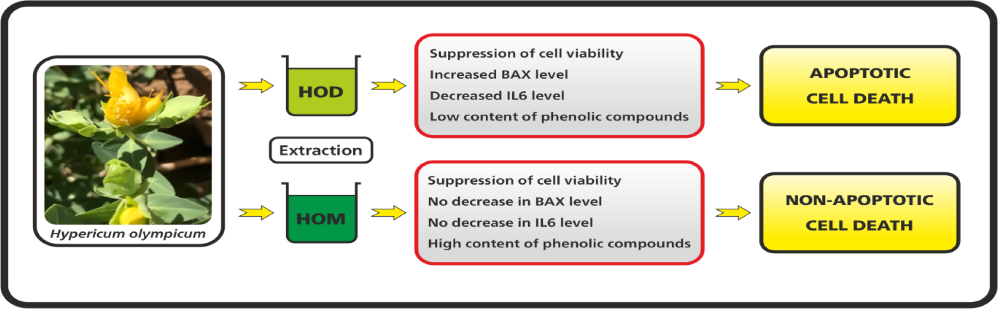
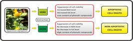
Lung cancer is the leading cause of cancer-related death and it is crucial to identify new sources for its treatment and suppression. Medicinal plants represent one of the most important resources in challenge against the cancer. Hypericum species have been shown in previous studies to suppress viability of various cancer cells. However, studies investigating the anticancer activities of Hypericum olympicum specifically on lung cancer cells remain quite limited. In this study, the anticancer activity of sequential dichloromethane (HOD) and methanol (HOM) extracts of H. olympicum on lung cancer cells (A549) was investigated, focusing on the reduction of cell viability, induction of apoptotic cell death, and suppression of proinflammatory cytokines. In this context, the effects of HOD and HOM on A549 cell viability were assessed using MTT method. The percentage of apoptotic and non-apoptotic cells was determined by AnnexinV/PI staining. The levels of Interleukin 6 (IL6) and 8 (IL8), as well as the apoptotic marker caspase-3, were measured using ELISA method. The mRNA levels of proapoptotic BAX and antiapoptotic BCL-2 were measured by Real-Time PCR method. The viability of A549 cells decreased following treatment with both HOD and HOM. In the HOD group, BAX level increased, while pro-caspase-3 and IL6 levels decreased. Additionally, percentage of apoptotic cells increased in HOD group, whereas the percentage of non-apoptotic cells increased in HOM group. Consequently, it is suggested that the anticancer activity of H. olympicum is associated with apoptotic pathway in the case of HOD, and a non-apoptotic pathway in the case of HOM.
Keywords:
Apoptosis; Hypericum olympicum; Lung cancer; Proinflammatory cytokines; Phenolics.
HIGHLIGHTS
Dichloromethane extract of H. olympicum (HOD) has a cytotoxic effect on A549 cells
HOD increases BAX level and reduces pro-Caspase 3 level in A549 cells
HOD reduces the level of IL6, which can suppress apoptosis
H. olympicum exhibited anticancer activity on lung cancer cells in vitro
INTRODUCTION
The cancer is the second leading cause of death worldwide following cardiovascular disease. Factors such as stress, malnutrition, occupational exposure, air pollution, heavy metals and especially smoking significantly increase the risk of developing cancer. Although incidence rates vary by countries, the most commonly diagnosed types of cancer are generally breast, lung, colon, prostate, cervix, skin and stomach. Among these, the lung cancer is the most prevalent in both men and women. Every year, the hundreds of thousands of people pass away from the lung cancer [1]. The lung cancer remains the leading cause of cancer-related death in the developed country such as United States and worldwide [2, 3]. It has been estimated that in 2023, approximately 1,950,000 new cases will be diagnosed in the United States alone, with around 610,000 cancer-related deaths [1].
The cancer has a process that generally occurs with an initial mutation. During this process, cancer cells accumulate many mutations that enable them to survive and proliferate. Under normal conditions, apoptosis (programmed cell death pathway) is activated to eliminate such abnormal cells. However, cancer cells often developed mechanisms to evade the apoptosis [4]. Disruptions in the apoptotic mechanism can lead to the transformation of cells into a malignant state, enable their spread, and contribute to the development of drug resistance [5]. When the apoptosis mechanism is impaired, various diseases may arise [6]. Apoptosis generally occurs through two distinct processes: the extrinsic and intrinsic pathways. At the onset of apoptosis, the balance between pro-apoptotic and anti-apoptotic proteins is altered. Alterations that lead to in the upregulation of apoptotic proteins promote the progression of the programmed cell death pathway. In the intrinsic apoptotic pathway, anti-apoptotic proteins such as BCL2, BCL-XL and MCL1, located in the mitochondrial membrane, are suppressed by increased levels of the proapoptotic protein BAX [5]. As Bax level rise, pores form in the mitochondrial membrane, allowing the release of cytochrome c, caspase inhibitor suppressors (SMAC/DIABLO). Cytochrome-c then binds with APAF1 (Apoptotic protease activating factor 1) to activate caspase 9, which subsequently cleaves procaspase-3 into active caspase-3. The caspase 9 is involved in the cleavage of procaspase 3 to form active caspase 3 that cleaves ICAD, leading to CAD (Caspase-activated DNase) formation. Subsequently, CAD initiates fragmentation of cellular DNA, a characteristic feature of apoptosis [7]. Therefore, many studies have aimed to activate the apoptotic pathway to eliminate of cancer cells using both synthetic and natural agents. [8-10]. However, due to the side effects associated with synthetic drugs, there is growing interest in herbal compounds that are believed to have fewer or no side effects. Medicinal plants play an important role among natural resources due to their unique phytochemicals, many of which have demonstrated the ability to induce apoptosis and suppress various types of cancer cells [8-11].
The genus Hypericum L. contain a total of 484 taxa that are naturally distributed across the world [12]. Bioactive compounds derived from Hypericum species exhibit a wide range biological activities including antitumor, anti-inflammatory, antimicrobial, antidepressant and antioxidant effects [13-16]. Especially, Hypericum species have demonstrated significant anti-cancer activities, such as anti-proliferative effects and inhibition of cancer cell migration and invasion [17, 18]. Moreover, several Hypericum species have been reported to exert anticancer effect by inducing apoptosis in cancer cells [17, 19]. On the other hand, other studies have indicated that cancer cell death is not solely due to apoptosis [20] and that non-apoptotic pathways, such as necrosis or autophagy, may also be involved [21].
H. olympicum is a deciduous shrub species native to West Asia and Southeast Europe [22]. Previous studies have reported that various extracts and compounds derived from H. olympicum exhibit cytotoxic effects against different cancer cell lines [20, 23]. However, studies investigating the apoptotic and non-apoptotic effects of sequential dichloromethane and methanol extracts of H. olympicum on A549 lung cancer cells are quite limited. In particular, the dichloromethane extract of H. olympicum has not been thoroughly examined in relation to apoptosis in cancer cells. Therefore, this study was designed to evaluate the anticancer potential of the dichloromethane (HOD) and methanol (HOM) extracts of H. olympicum on A549 lung cancer cells. Specifically, the study investigated the effects of HOD and HOM on cell proliferation inhibition, suppression of inflammatory cytokines, and induction of apoptosis in A549 cells.
MATERIAL AND METHODS
Collection of H. olympicum and preparation of dichloromethane and methanol extracts
H. olympicum was collected from its natural habitat in Huzurlu Plateau (Gaziantep, Turkiye) on 02.07.2023 and identified by Dr. Mustafa PEHLIVAN (individually herbarium voucher number: MPH2023-2). H. olympicum was cleaned and dried on blotting paper away from sunlight, then ground into a fine powder using a morter. For extraction, 200 mL of dichloromethane was first applied to the powdered plant material using a Soxhlet apparatus at 40 °C for 6 hours. After the dichloromethane extraction was completed, the solvent was removed, and the extraction process was continued with 200 mL of methanol at 50 °C for another 6 hours. The resulting dichloromethane (HOD) and methanol (HOM) extracts were filtered and concentrated using a rotary evaporator under reduced pressure. The HOD and HOM extracts were then stored at +4 °C until further use in assays.
Maintenance and Growth of Lung cancer cells (A549) and BEAS2B cells
A549 cells (ATCC, human, epithelial non-small-cell lung cancer) and human lung epithelial cell (BEAS-2B as control cells) were obtained from Advanced Cell Analysis Laboratory of Gaziantep University-Turkiye. A549 and BEAS-2B cells were grown in RPMI supplemented with 10% fetal bovine serum (FBS; Gibco, USA) and 1% antibiotic (Gibco, USA). A549 and BEAS-2B cells were cultured in an incubator at 37 °C in 5% CO2 medium. The cells were seeded into 96-well plates at a density of 5×10⁶ cells/mL and maintained in RPMI medium for 24 hours. Following this incubation period, the cells were treated with various concentrations (25, 50, 100, 200 µg/mL) of HOD and HOM for 24 hours. The extracts were prepared by dissolving in 10% dimethyl sulfoxide (DMSO).
Determination of cell viability of A549 and BEAS-2B cells following HOD and HOM treatment
The cells in 96-well plates, at 70-80% confluency, were treated with varying concentrations (25, 50, 100, and 200 µg/mL) of HOD and HOM extracts for 24 hours. A positive control group consisted of cells cultured in RPMI-1640 medium supplemented with 10% fetal calf serum (FCS) without extract treatment. The viability of cells was assessed using the MTT [3-(4,5-dimethylthiazol-2-yl)-2,5-diphenyltetrazolium bromide] assay. After treatment, the culture medium was removed, and cells were incubated with MTT solution (1 mg/mL in saline) at 37 °C for 60 minutes. Following incubation, the MTT solution was discarded, and the resulting formazan crystals were dissolved in DMSO. Absorbance was measured at 550 nm using a microplate spectrophotometer. Cell viability was expressed as a percentage of the absorbance relative to untreated control cells.
Determination of mRNA expressions of proapoptotic BAX and antiapoptotic BCL2 in A549 cells
The A549 cells (5×10⁶ cells/mL) were treated with 25 and 200 µg/mL concentrations of HOD and HOM for 24 hours. After incubation, the supernatant was discarded, and 700 μl of QIAzol Lysis Reagent was added to lyse the cells. Total RNA was extracted using TRIpure Total RNA Extraction Reagent (ELK Biotechnology, Wuhan, China) according to the manufacturer's instructions. The extracted RNA was then reverse transcribed into complementary (cDNA) by using reverse transcriptase kit following the manufacturer's protocol. Quantitative real-time PCR (RT-PCR) was performed using a Rotor-Gene Q system (Qiagen, Germany). cDNA template for each primer with SYBR green master mix reacted at 95 °C for 15 min, 40 cycles 95 °C for 20 s,60 °C for 30s, 72 °C for 30 s. For the following PCR reaction was prepared; 10 µL SYBR Green Master Mix, 1 µL forward primer, 1 µL reverse Primer, 5 µL RNase-free water and 3 µL cDNA. Reactions were carried out on a Rotor-Gene Q instrument using the following cycling conditions: initial denaturation at 95 °C for 15 minutes, followed by 40 cycles of 95 °C for 15 seconds, 60 °C for 30 seconds, and 72 °C for 30 seconds. After the reaction, Ct (cycling threshold) values were taken at an appropriate threshold. Relative gene expression for BAX (forward: GCCCTTTTGCTTCAGGGTTT, reverse: TCCAATGTCCAGCCTTTG) and BCL-2 (F: CGGAGGCTGGGATGCCTTTG, reverse: TCACTTGTGGCCCAGATAGG) genes was determined with normalization to GAPDH (forward: GATCATCAGCAATGCCTCCT, reverse: TGTGGTCATGAGTCCTTCCA). The mRNA expression levels of the target genes were calculated using the 2-∆∆CT method.
Determination of Apoptotic and Non-Apoptotic Cell Percentages by Annexin V and Propidium Iodide (PI) Staining
To evaluate the effects of HOD and HOM on apoptosis and cell viability, A549 cells were seeded at a density of 5×10⁶ cells/mL. The cells were treated with the minimum (25 µg/mL) and maximum (200 µg/mL) concentrations of the extracts for 24 hours. Following treatment, the percentages of apoptotic and non-apoptotic cells were determined using flow cytometry (Becton-Dickinson) with an Annexin V/PI staining kit, according to the manufacturer’s instructions.
Determination of Pro-Caspase-3 Levels
After treatment with HOD and HOM, the supernatants were discarded to assess apoptosis in A549 cells (5×10⁶ cells/mL). The A549 cells were then washed with PBS and lysed using a lysis solution. Pro-caspase-3 level in the cell lysates were measured using a ELISA kit following the manufacturer’s instructions. Absorbance for pro-caspase-3 levels were read at 450 nm using a ELISA reader (BioTek instrument, USA).
Determination of interleukin 6 (IL6) and interleukin 8 (IL8) levels
After 24 hours of treatment with HOD and HOM, the supernatants from A549 lung cancer cells (5×10⁶ cells/mL) were collected. To determine the levels of IL-6 and IL-8 in the supernatants were used a ELISA kits, according to the manufacturer’s instructions. Absorbance for IL-6 and IL-8 was recorded at 450 nm using an ELISA reader (BioTek Instruments, USA).
Screening of phenolic compounds in HOD and HOM by LC-MS-MS
For the screening of phenolic compounds by LC-MS-MS, HOD and HOM were dissolved in methanol and subsequently filtered through a 0.22 μM filter. LC-20AD two-pump Nexera Ultra High Performance Liquid Chromatography (UHPLC) (Shimadzu) Liquid chromatography-mass spectrometry (LC-MS-MS) instrument, DGU-20A3R degasser, CTO-10ASVP column oven and SIL-20AC autosampler were utilized. For this screening, the analysis was run with a C18 Intersil ODS-4 analytical column (3.0mm x 100mm, 2µm). Injection volume of the samples was 2 μL and flow rate was 0.3 ml/min. In linear gradient flow, it was used mobile phase A (Water and 0.1% Formic acid) and mobile phase B (Methanol and 0.1% Formic acid). The column temperature was initially set to 40 °C.
Statistical Analysis
To evaluate statistically the results of MTT, apoptosis, BAX, BCL2, IL6, IL8 and pro-caspase3 assay, Dunnett’s test of one-way ANOVA were used. The results of all the assays were presented as mean ± SD, and the level of significance was accepted as "*" p<0.05, "**" p<0.01 and "***" p<0.001.
RESULTS
Effects of HOD and HOM on viability of A549 and BEAS2B cells
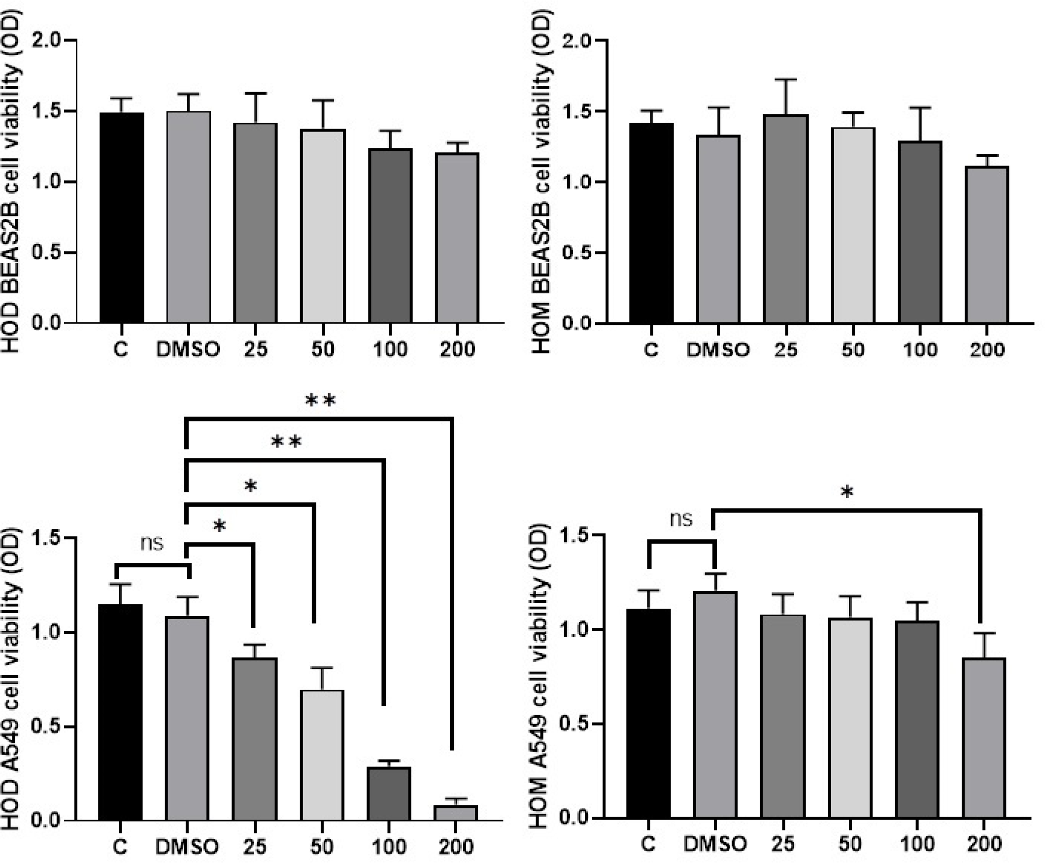
To determine the effect of HOD and HOM on the viability of A549 and BEAS-2B cells, MTT assay was used. As shown in Figure 1, the viability of A549 cells decreased in concentration-dependent manner at concentrations of 25, 50, 100, and 200 µg/mL of HOD. All concentrations of HOD significantly reduced cell viability compared to the control group (p < 0.05 for 25 and 50 µg/mL; p < 0.01 for 100 and 200 µg/mL). On the other hand, the viability of A549 cells was significantly reduced only at a 200 µg/ml concentration of HOM, whereas no change was observed at the other concentrations when compared to the controls. In addition, HOD and HOM were also tested on the viability of BEAS2B cells for 24 hours. Although there was a decrease in the viability of the cells, especially at concentrations of 100 and 200 µg/ml, these changes were not statistically significant. Therefore, further anticancer activity assays were conducted exclusively with A549 cells.
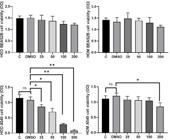
Cytotoxic effect of HOD and HOM on viability of lung cancer cells. Statistical analyzes were performed using the One-way Anova Dunnett’ test. C: Control, OD: optic density, HOD: Dichloromethane extract of H. olympicum, HOM: Methanol extract of H. olympicum, DMSO: dimethyl sulfoxide. "*" and "**" mean p<0.05 and p<0.01, respectively
Determination of apoptotic cells percentages by Annexin-V/PI staining
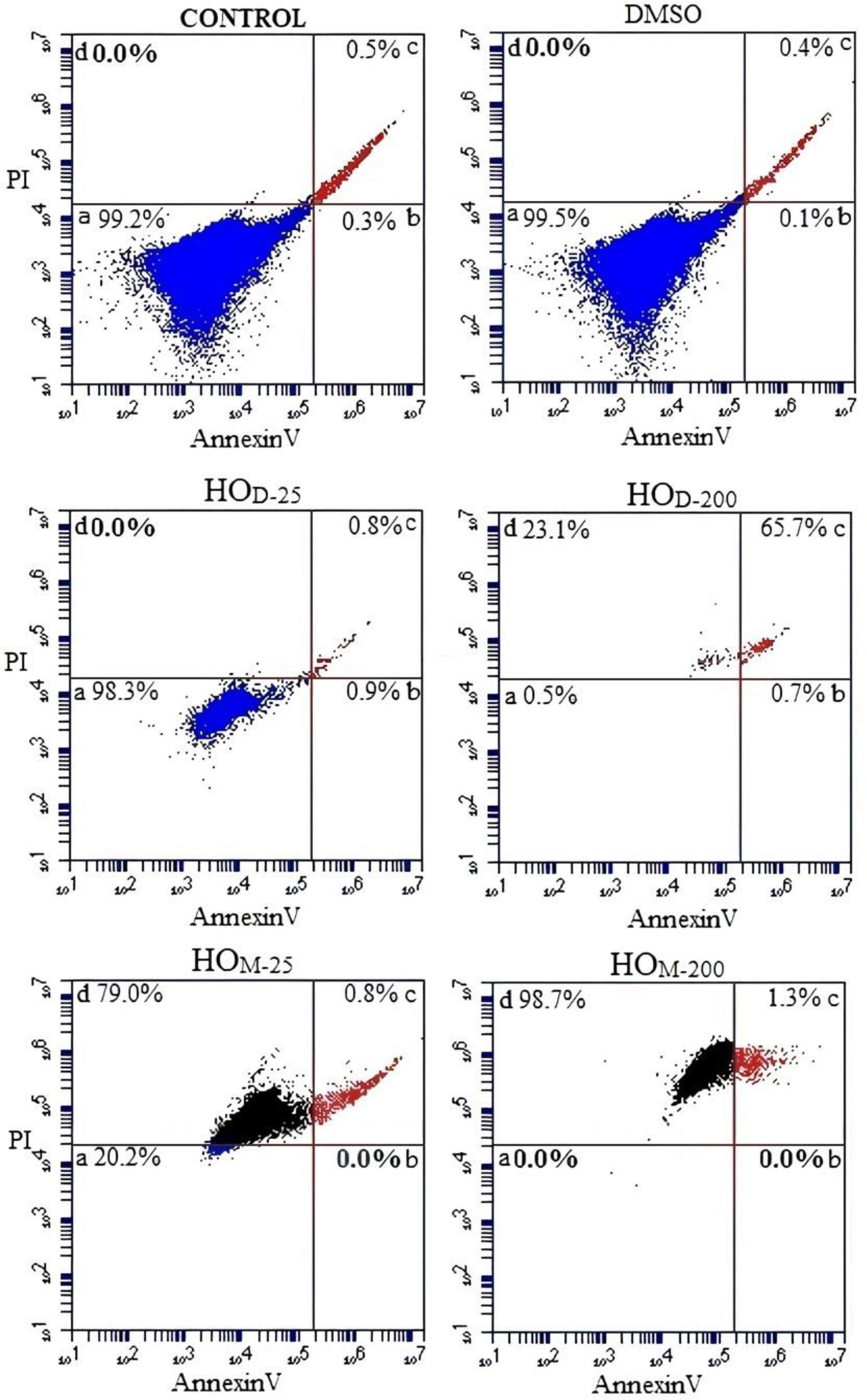
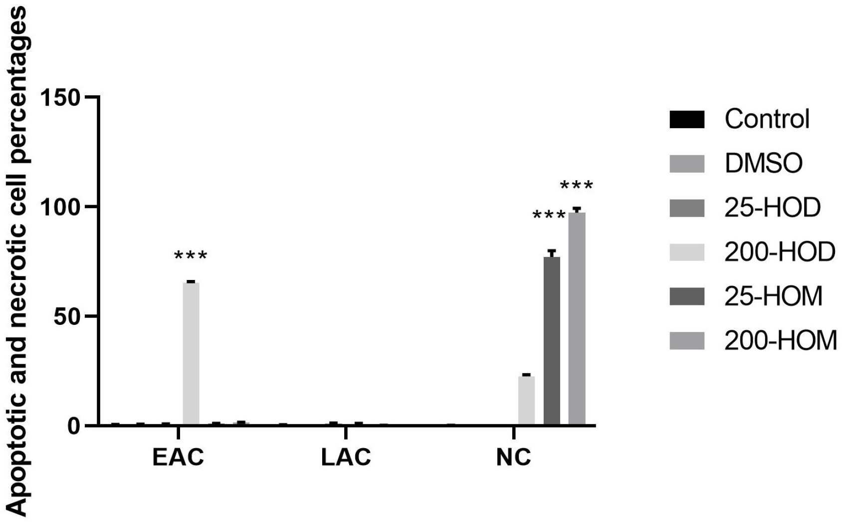
In this study, apoptosis assay was performed using the lowest and highest concentrations of HOD and HOM. To assess the percentage of early apoptotic, late apoptotic and non-apoptotic, A549 cells were treated with HOD and HOM for 24 hours. Apoptotic cell populations were then quantified using flow cytometry (FACS, fluorescence-activated cell sorting). The apoptotic effects of HOD and HOM were shown in Figure 2a. At 200 µg/ml concentration of HOD, 65.7% of A549 cells were found to be in the late apoptotic stage. This increase in late apoptotic cells was statistically significant compared to the control group (p < 0.001) (Figure 2b). Additionally, as shown in Figure 2b, the percentages of non-apoptotic cells following HOM treatment were 79% and 98.7% at 25 and 200 µg/mL concentrations, respectively (p < 0.001). On the other hand, no significant change was observed in the percentage of non-apoptotic cells after HOD treatment.
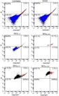
Percentages of the apoptotic and non-apoptotic A549 cells after HOD and HOM treatment. HOD: Dichloromethane extract of H. olympicum, HOM: Methanol extract of H. olympicum, DMSO: dimethyl sulfoxide.
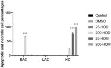
Statistical evaluation of percentages of the apoptotic and non-apoptotic A549 cells. HOD: Dichloromethane extract of H. olympicum, HOM: Methanol extract of H. olympicum, DMSO: dimethyl sulfoxide, EAC: Early apoptotic cells , LAC: Late apoptotic cells , NC: Non apoptotic cells.
Determination of mRNA levels of BAX and BCL-2 in A549 cells
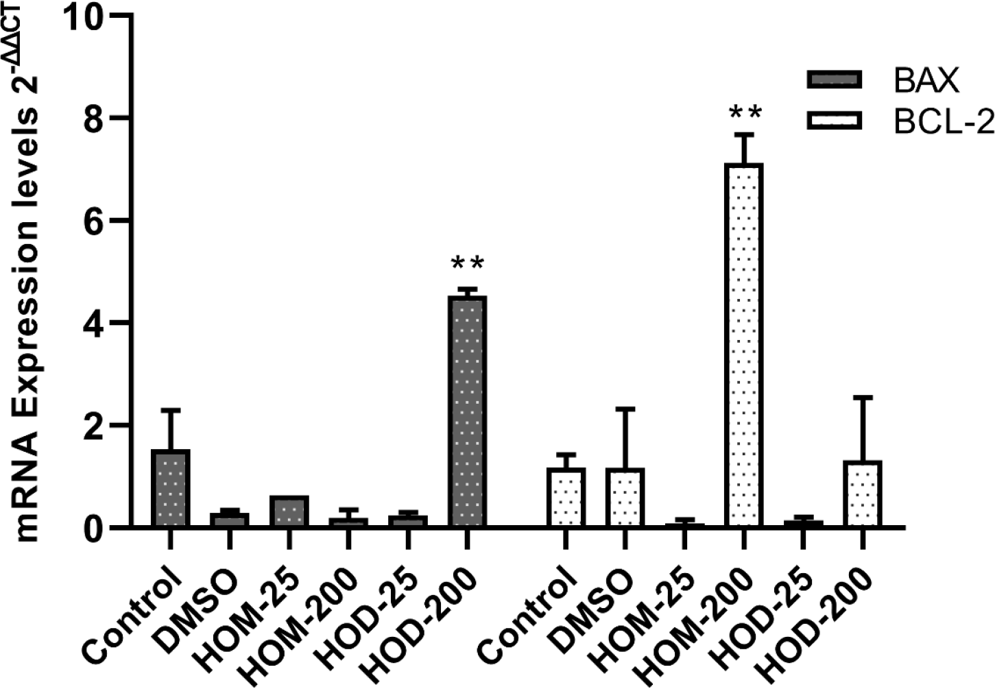
Proapoptotic BAX and antiapoptotic BCL-2 mRNA expressions were determined in A549 cells after treatment of HOD and HOM, and the results were shown in Figure 3. As shown in Figure 3, BAX expression increased approximately 4.53 fold at 200 µg/ml HOD concentration compared to the control (p<0.05). Furthermore, BCL-2 level increased approximately 7.12-folt at 200 µg/ml HOM concentration (p<0.05), while BAX mRNA level did not change.
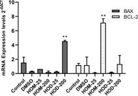
BAX and BCL2 expressions in A549 cells after treatment of HOD and HOM. Statistical analyzes were performed using the One-way Anova Dunnett’s test. C: Control, HOD: Dichloromethane extract of H. olympicum, HOM: Methanol extract of H. olympicum, DMSO: dimethyl sulfoxide, BAX: BCL2 Associated X, BCL2: B-Cell Lymphoma 2 "*" and "***" mean p<0.05 and p<0.001, respectively.
Effects of HOD and HOM on IL6 and IL8 levels in A549 cells
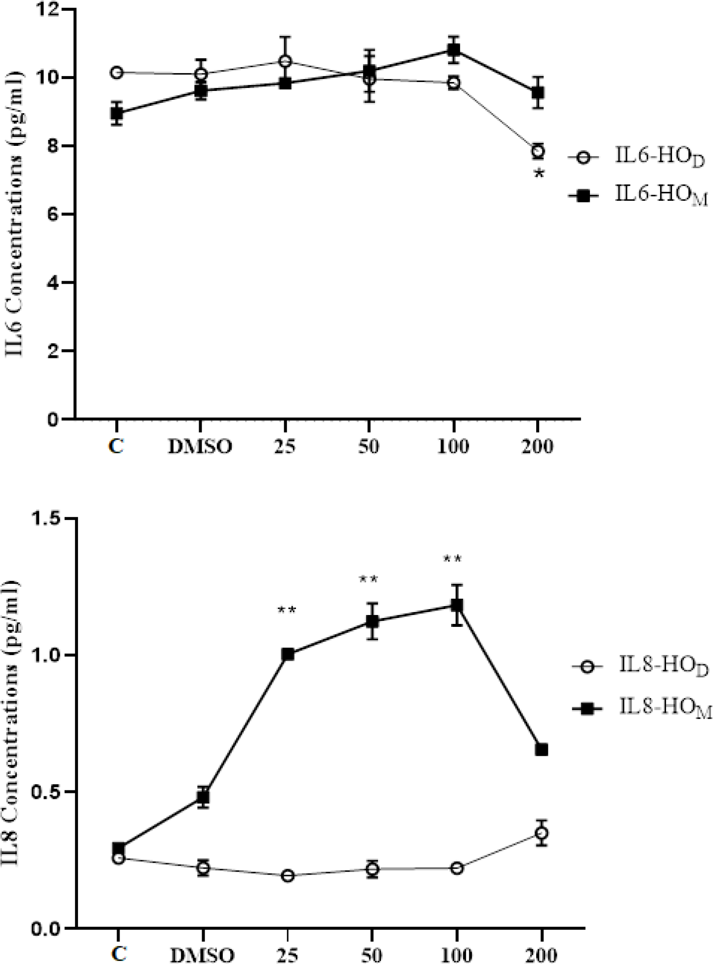
After A549 cells were treated with HOD and HOM for 24 hours, the culture medium was collected, and IL6 and IL8 levels were measured from the supernatants. As shown in Figure 4, IL6 level did not change significantly at concentrations of 25 and 50 µg/ml of HOD, but decreased at 100 and 200 µg/mL, with only the decrease at 200 µg/mL being statistically significant (p < 0.05). IL-8 levels, also presented in Figure 4, were not affected by any concentration of HOD. In contrast, IL-8 levels increased at 25, 50, and 100 µg/mL concentrations of HOM, while the 200 µg/mL concentration of HOM caused a strong decrease in IL-8 levels.
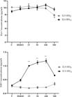
Change of IL6 and IL8 levels in A549 cells after HOD and HOM treatment. Statistical analyzes were performed using the One-way Anova Dunnett’s test. C: Control, HOD: Dichloromethane extract of H. olympicum, HOM: Methanol extract of H. olympicum, DMSO: dimethyl sulfoxide, "**" mean p<0.01.
Determination of levels of pro-caspase 3
Figure 5 illustrates the change in pro-caspase 3 levels in A549 cells after exposure to various concentrations of HOD and HOM for 24 hours. As shown in Figure 5, pro-caspase-3 levels in A549 cells decreased at all concentrations of HOD, with the most significant reductions observed at 100 and 200 µg/mL (p < 0.001). On the other hand, none of HOM concentrations caused a significant change in pro-caspase-3 levels (p>0.05).
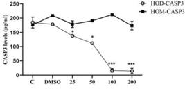
Change of pro-caspase 3 level in A549 cells after treatment of HOD and HOM. HOD: Dichloromethane extract of H. olympicum, HOM: Methanol extract of H. olympicum, C: Control, DMSO: dimethyl sulfoxide, CASP3: Caspase 3
Determination of phenolic acid and flavonoids of HOD and HOM by LC-MS-MS
The phenolic acid and flavonoids of HOD and HOM was screened for 22 compounds by LC-MS-MS and the results were given in Table 1. As a result of LC-MS-MS analysis, while 15 compounds in HOM were found, only five phenolic compounds were determined in HOD. As a result of the screening performed by LC-MS-MS, phenolic acids ferulic acid and protocatechuic acid were determined in HOD, while flavonoids myricetin and phenolic acids fumaric, gallic and protocatechuic acid were the most abundant phenolic compounds in HOM.
DISCUSSION
Medicinal plants have been used since ancient times for the treatment of various diseases. Moreover, the extracts and compounds derived from certain medicinal plants have been reported to exhibit anticancer activity in previous studies [19, 22, 23]. These natural products may influence multiple anticancer mechanisms, including suppression of cancer cell viability [23], induction of cell death pathways such as apoptosis and necrosis [22], cell cycle arrest [24], inhibition of migration and invasion [25], modulation of multidrug resistance [26] and suppression of epithelial-mesenchymal transition [27]. The ability of extracts containing diverse phytocompounds to target several of these mechanisms simultaneously may result in stronger anticancer effects compared to single isolated compounds [28]. Additionally, cytotoxic studies on H. olympicum have generally focused on the methanol extract. Sequential extractions with solvents of different polarity such as dichloromethane and methanol, may yield fractions containing distinct compounds. This approach can help clarify the relationship between the observed anticancer activity and the specific extract fraction. Therefore, this study investigated the potential anticancer effects of HOD and HOM, including suppression of cell viability, induction of apoptosis, and inhibition of proinflammatory cytokines. The cytotoxic effects of HOD and HOM on viability of lung cancer cells was evaluated using the MTT assay. It was determined that HOD extract exhibited cytotoxic activity a concentration-dependent manner on A549 cells, while HOM reduced only at higher concentrations. Previous studies have reported the cytotoxic activities of Hypericum species against several cancer cell lines, including lung, prostate, breast, liver, glioma. It has reported that methanol extracts of H. adenotrichum and H. olympicum had cytotoxic effects on A549 and PC3 (prostate cancer) cells in a dose-dependent manner [20]. Additionally, H. olympicum subsp. olympicum has demonstrated strong cytotoxic effects on breast cancer cell lines MCF7 and MDA-MB-231 [29]. Methanol extracts of H. olympicum and H. adenotrichum also reduced viability in human Hep3B (heap tocarcinoma) and C6 (rat glioma) cell lines [21]. Considering these previous findings alongside our study results, it can be concluded that both HOD and HOM possess in vitro cytotoxic activity against lung cancer cells.
In addition to cytotoxic effect, identifying the cell death pathway responsible for this effect is important for determining anticancer activity. Generally, one of the most desired outcomes in suppressing of cancer cells in vitro is the induction of apoptosis. In order to determine the relationship between cytotoxic effect and apoptosis induction, Real Time PCR was performed to evaluate the mRNA levels of BAX and BCL2. Treatment of cancer cells with HOD at a concentration of 200 µg/ml caused an increase in level of BAX, while HOM at a concentration of 200 µg/ml had no effect in BAX expression, while HOM at the same concentration had no effect on BAX levels. Conversely, BCL2 expression significantly increased after HOM treatment but remained unchanged following HOD treatment. Next, the effects of HOD and HOM on induction of apoptosis in lung cancer cells were evaluated using AnnexinV/PI staining. Phosphotidylserine is normally located in the inner layer of the cell membrane, but during apoptosis, it translocates to the outer leaflet, serving as an “eat me” signal for phagocytes [30, 31]. The phosphotidylserine in the outer layer of cell membrane could be stained with AnnexinV, allowing the percentage of apoptotic cells to be determined. Following AnnexinV/PI staining of A549 cells treated with the lowest and highest HOD concentrations, a high percentage of lung cancer cells were found to be in the late apoptotic stage. When the BAX and BCL-2 mRNA expression levels were evaluated alongside the Annexin V/PI staining results, consistent evidence for apoptosis induction was observed. The increase in BAX mRNA levels, together with the elevated percentage of cells in the late apoptotic phase, suggests that HOD has apoptosis-inducing activity. On the other hand, an increase in the percentage of non-apoptotic dead A549 cells was observed following HOM treatment. There are a few reports on the cell death pathways activated by Hypericum species in cancer cells, and one of them is the necrotic pathway [21]. Previous studies have demonstrated that the methanol extract of H. olympicum reduced the viability of Hep3B and C6 cell lines, and this decrease attributed to the necrotic pathway rather than apoptosis [21]. Furthermore, the level of caspase 3 was determined as an indicator of apoptosis. Activation of caspase-cascade is a key step in the execution of apoptosis. Caspase 3, which is generated through the cleavage of procaspase3, functions as an effector caspase and plays a central role in both the intrinsic and extrinsic apoptosis pathways [32]. The present study showed that level of pro-caspase 3 decreased by HOD treatment, whereas no change was observed with HOM. Considering the reduction in cell viability, the increase in the percentage of cells in late apoptotic phase, the up-regulation of BAX mRNA level, and the decrease in pro-caspase-3 level, it can be concluded that HOD exhibits its the anticancer activity on lung cancer cells by activating the apoptotic pathway. However, previous studies have reported no effect of HOM on caspase 3 levels in various cancer cells such as breast and lung [20, 29]. Therefore, the unchanged levels of BAX and caspase 3, along with the increase in non-apoptotic cell death suggest that HOM may triggers a death pathway other than apoptosis such as necrosis in lung cancer cells. However, a limitation of our study is that necrotic markers such as RIPK1, RIPK3 (Receptor-interacting serine/threonine-protein kinases 1 and 3), and MLKL (Mixed Lineage Kinase Domain-Like protein) were not evaluated. Thus, it is recommended that future studies investigate the mRNA and protein expression levels of these necrotic markers to allow a clearer interpretation of the underlying cell death mechanisms.
It has been reported the up-regulation of IL6 and IL8 enhances proliferation, invasion and metastasis in many cancer cells [33-35], and also contributes to the suppression of the apoptosis process [36]. In this study, HOD treatment led to a decrease in IL6 level, while IL8 levels remained unchanged. The IL6 promotes cancer cells proliferation and inhibits apoptosis by activating the STAT3 (Signal transducer and activator of transcription 3) signaling pathway, which frequently activated in cancer pathogenesis [37, 38]. Therefore, the reduction in IL6 levels following HOD treatment is thought to contribute to decreased cell viability by promoting apoptosis in lung cancer cells. In contrast, no significant changes were observed in IL6 or IL8 levels after HOM treatment.
In previous studies, the phytochemicals present in HOD and HOM have been identified using various methods, with hypericin being the most well-known among them [39]. In addition, (E)-anethole and β-farnesene were identified as the main compounds of H. olympicum essential oil using Gas Chromatography-Mass Spectrometry (GC-MS) [40]. Additionaly, Ilieva and coauthors [41] reported that the presence of olympiforin A and olympiforin B in H. olympicum. In a previous study, rutin, hyperoside, isoquercitrin and chlorogenic acid were determined as the major phenolic compounds in H. olympicum using HPTLC [42]. Ferulic acid and protocatechuic acid were the most abundant phenolic compounds in HOD, while fumaric acid, gallic acid, protocatechuic acid, and myricetin were predominant in HOM. Additionally, kaempferol, luteolin, and quercetin were also detected in HOM. Saddiqe Ilieva and coauthors (2011) determined various amounts of quercetin, myricetin, luteolin, apigenin, rhamnetin, isorhamnetin and kaempferol in HPLC analysis of polar extracts of H. androsaemum, H. ericoides, H. calycinum, H. patulum and H. olympicum species. Moreover, the high amounts of rhamnetin and isorhamnetin were determined in H. olympicum, but myricetin was not detected [43]. In our study, the lack of apoptosis induction activity in HOM, contrasted with the clear apoptosis induction by HOD suggests that the apoptosis inducing effect of H. olympicum may not be related to the phenolic compounds identified.
CONCLUSION
This study aimed to determine the potential cytotoxic effects, apoptosis-inducing activity, and pro-inflammatory cytokine suppression of HOD and HOM extracts-with differing polarities-on A549 lung cancer cells, as well as to analyze their phenolic compositions. Based on the overall findings, it is suggested that both HOD and HOM exhibit cytotoxic effects on lung cancer cells, with the effect of HOD likely associated with apoptosis and that of HOM potentially linked to necrosis. Although HOM contained a greater variety and quantity of phenolic compounds compared to HOD, HOD demonstrated stronger apoptotic activity. This suggests that the phenolic compounds identified may not be responsible for the apoptosis-related anticancer activity of H. olympicum.
Lastly, limitations of our study are that protein levels of apoptotic markers and necrotic markers such as RIPK1, RIPK3 and MLKL1 were not determined. Future studies should investigate the mRNA and protein levels of these markers to better understand the necrotic cell death observed following HOM treatment in lung cancer cells. In addition, a single type of lung cancer cells was used in our study. It is recommended to perform apoptotic and necrotic cell death experiments with different lung cancer cells.
-
Funding:
This research received no external funding.
Acknowledgments:
The authors would like to thank Adiyaman, Gaziantep, and Gaziantep Islam Science and Technology Universities for their support.
Data Availability Statement:
Research data are available in the body of the manuscript.
REFERENCES
- 1 Siegel RL, Miller KD, Wagle NS, Jemal A. Cancer statistics, 2023. CA Cancer J Clin. 2023;73(1):17-48.
- 2 Siegel RL, Giaquinto AN, Jemal A. Cancer statistics, 2024. CA Cancer J Clin. 2024;74(1).
- 3 Maconachie R, Mercer T, Navani N, McVeigh G. Lung cancer: diagnosis and management: summary of updated NICE guidance. Bmj. 2019;364.
- 4 Fernald K, Kurokawa M. Evading apoptosis in cancer. Trends Cell Biol. 2013;23(12):620-33.
- 5 Wong RS. Apoptosis in cancer: from pathogenesis to treatment. J Exp Clin Cancer Res. 2011;30(1):1-14.
- 6 Akşit H. Bildik A. [Apoptosis]. Van Vet J. 2008;19(1):55-63.
- 7 Sakahira H, Enari M, Nagata S. Cleavage of CAD inhibitor in CAD activation and DNA degradation during apoptosis. Nature. 1998;391(6662):96-9.
- 8 Russo A, Graziano A, Bruno M, Cardile V, Rigano D. Apoptosis induction of essential oils from Artemisia arborescens L. in human prostate cancer cells. J Ethnopharmacol. 2023;303:115929.
- 9 Zhao F, Zhao H, Wu W, Wang W, Li W. Research on anthocyanins from Rubus “Shuofeng” as potential antiproliferative and apoptosis-inducing agents. Foods. 2023;12(6):1216.
- 10 Cocelli G, Pehlivan M, Yumrutas O, Bonfante R, Parlar A. Sideritis perfoliata inhibits cell proliferation, induces apoptosis and exhibits cellular antioxidant activity in cervical cancer cells. Bol. latinoam. Caribe Plantas Med. Aromát. 2021;20(4).
- 11 Yumrutaş Ö, Pehlivan M, Güven C, Bozgeyik I, Bozgeyik E, Yumrutaş P, et al. Investigation of cytotoxic effect of salvia pilifera extracts and synthetic chlorogenic and caffeic acids on DU145 prostate cancer cells line. KSU J Agric Nat. 2018;21(2):141-7.
- 12 Crockett SL, Robson NK. Taxonomy and chemotaxonomy of the genus Hypericum. Med Aromat Plant Sci Biotechnol. 2011;5(Special Issue 1):1.
- 13 Bender O, Llorent-Martínez EJ, Zengin G, Mollica A, Ceylan R, Molina-Garcia L, et al. Integration of in vitro and in silico perspectives to explain chemical characterization, biological potential and anticancer effects of Hypericum salsugineum: A pharmacologically active source for functional drug formulations. PloS one. 2018;13(6):e0197815.
- 14 Ozkan EE, Ozsoy N, Ozden TY, Ozhan G, Mat A. Evaluation of chemical composition and in-vitro biological activities of three endemic Hypericum species from Anatolia (H. thymbrifolium, H. spectabile and H. pseudolaeve). I Iran J Pharm Res. 2018;17(3):1036.
- 15 Tian J, Zhang F, Cheng J, Guo S, Liu P, Wang H. Antidepressant-like activity of adhyperforin, a novel constituent of Hypericum perforatum L. Sci Rep. 2014;4(1):5632.
- 16 Zorzetto C, Sánchez-Mateo CC, Rabanal RM, Lupidi G, Petrelli D, Vitali LA, et al. Phytochemical analysis and in vitro biological activity of three Hypericum species from the Canary Islands (Hypericum reflexum, Hypericum canariense and Hypericum grandifolium). Fitoterapia. 2015;100:95-109.
- 17 Hamzeloo-Moghadam M, Khalaj A, Malekmohammadi M. Cytotoxic activity and apoptosis induction of Hypericum scabrum L. Iran Red Crescent Med J. 2015;17(10).
- 18 Qiu D, Zhou M, Chen J, Wang G, Lin T, Huang Y, et al. Hyperelodiones AC, monoterpenoid polyprenylated acylphoroglucinols from Hypericum elodeoides, induce cancer cells apoptosis by targeting RXRα. Phytochemistry. 2020;170:112216.
- 19 Yumrutas O, Bozgeyik I. Anticancer activity of Inula graveolens by induction of ROS-independent apoptosis and suppression of IL6-IL8 in cervical cancer cells. Bol. latinoam. Caribe plantas med. aromát. 2023;22(3).
- 20 Aztopal N, Erkisa M, Celikler S, Ulukaya E, Ari F. Antigrowth and apoptosis inducing effects of Hypericum olympicum L. and Hypericum adenotrichum Spach. on lung cancer cells in vitro: Involvement of DNA damage. J Food Biochem. 2016;40(4):559-66.
- 21 Ferda A, Aztopal N, Erkisa M, Celikler S, Sahin S, Ulukaya E. Nonapoptotic cell death induced by Hypericum species on cancer cells. Eur Res J. 2017;3(1):1-10.
- 22 Yumrutaş Ö, Pehlivan M, Yumrutaş P, Kahraman D. Possible induction of apoptotic and necrotic death pathways in lung cancer cells by Clinopodium vulgare L. and phenolic acids and flavonoids detection by LC-MS-MS. Bol. latinoam. Caribe plantas med. aromát. 2024;23(5):740-8.
- 23 Yumrutaş Ö, Yumrutas P, Pehlivan M, Kahraman D, Korkmaz M, Doğan M. Anticancer activity of Origanum vulgare on lung cancer: Antiproliferative, morphological, and apoptotic effects. Indian J Nat Prod Resour. 2024;15(3):357-63.
- 24 Lam M, Carmichael AR, Griffiths HR. An aqueous extract of Fagonia cretica induces DNA damage, cell cycle arrest and apoptosis in breast cancer cells via FOXO3a and p53 expression. PloS one. 2012;7(6):e40152.
- 25 Du Y, Feng J, Wang R, Zhang H, Liu J. Effects of flavonoids from Potamogeton crispus L. on proliferation, migration, and invasion of human ovarian cancer cells. PLoS One. 2015;10(6):e0130685.
- 26 Costea T, Vlad OC, Miclea L-C, Ganea C, Szöllősi J, Mocanu M-M. Alleviation of multidrug resistance by flavonoid and non-flavonoid compounds in breast, lung, colorectal and prostate cancer. Int J Mol Sci. 2020;21(2):401.
- 27 Ang HL, Mohan CD, Shanmugam MK, Leong HC, Makvandi P, Rangappa KS, et al. Mechanism of epithelial-mesenchymal transition in cancer and its regulation by natural compounds. Med Res Rev. 2023;43(4):1141-200.
- 28 Zhao Y, Zhao L, Luo Y, Li X, Zhang Y, Liu X, et al. Synergistic effect of radiation and traditional Chinese medicine Rhizomatyphonii ethanol extracts depends on P53 expression in treatment of Lewis mouse lung cancer cells. Afr J Food Agric Nutr Dev. 2015;12(1):109-14.
- 29 Balıkçı N, Sarımahmut M, Arı F, Aztopal N, Özel MZ, Ulukaya E, et al. Toxicity assessment of Hypericum olympicum subsp. olympicum L. on human lymphocytes and breast cancer cell lines. J Appl Biomed. 2020; 18(1): 18-25.
- 30 Fadok VA, Bratton DL, Frasch SC, Warner ML, Henson PM. The role of phosphatidylserine in recognition of apoptotic cells by phagocytes. Cell Death Differ. 1998;5(7):551-62.
- 31 Segawa K, Nagata S. An apoptotic ‘eat me’signal: phosphatidylserine exposure. Trends Cell Biol. 2015;25(11):639-50.
- 32 Asadi M, Taghizadeh S, Kaviani E, Vakili O, Taheri-Anganeh M, Tahamtan M, et al. Caspase-3: structure, function, and biotechnological aspects. Biotechnol Appl Biochem. 2022;69(4):1633-45.
- 33 Yuan A, Chen JJ, Yao P-L, Yang P-C. The role of interleukin-8 in cancer cells and microenvironment interaction. Front Biosci. 2005;10(1):853-65.
- 34 Kumari N, Dwarakanath B, Das A, Bhatt AN. Role of interleukin-6 in cancer progression and therapeutic resistance. Tumour Biol. 2016;37:11553-72.
- 35 Maynard JP, Ertunc O, Kulac I, Baena-Del Valle JA, De Marzo AM, Sfanos KS. IL8 expression is associated with prostate cancer aggressiveness and androgen receptor loss in primary and metastatic prostate cancer. Mol Cancer Res. 2020;18(1):153-65.
- 36 Guo Y, Xu F, Lu T, Duan Z, Zhang Z. Interleukin-6 signaling pathway in targeted therapy for cancer. Cancer Treat Rev. 2012;38(7):904-10.
- 37 Menegazzi M, Masiello P, Novelli M. Anti-tumor activity of Hypericum perforatum L. and hyperforin through modulation of inflammatory signaling, ROS generation and proton dynamics. Antioxidants. 2020;10(1):18.
- 38 Fathi N, Rashidi G, Khodadadi A, Shahi S, Sharifi S. STAT3 and apoptosis challenges in cancer. Int J Biol Macromol. 2018;117:993-1001.
- 39 Ayan AK, Çirak C. Hypericin and Pseudohypericin contents in some Hypericum. species growing in Turkey. Pharm Biol. 2008;46(4):288-91.
- 40 Gudžić B, Dordević S, Palić R, Stojanović G. Essential oils of Hypericum olympicum L. and Hypericum perforatum L. Flavour Fragr J. 2001;16(3):201-3.
- 41 Ilieva Y, Momekov G, Zaharieva MM, Marinov T, Kokanova-Nedialkova Z, Najdenski H, et al. Cytotoxic and Antibacterial Prenylated Acylphloroglucinols from Hypericum olympicum L. Plants. 2023;12(7):1500.
- 42 Akyüz S, Kurt-Celep İ, İnan Y, Özdemir OE, Celep E, Yesilada E. In vitro evaluation of the bioactivity and bioaccessibility of Hypericum olympicum L. S Afr J Bot. 2021;142:316-24.
- 43 Saddiqe Z, Naeem I, Maimoona A, Sautreau A, Hellio C. Assay of flavonoid aglycones with HPLC in four species of genus Hypericum. J Med Plants Stud. 2011;5(9):1526-30.
-
Editor-in-Chief:
Paulo Vitor Farago
-
Associate Editor:
Jane Manfron