Molecular Docking and Dynamic Diterpenoid-Stevia Compounds from Water Maceration-Sonification Extract as Amylase and Glucosidase Enzyme Inhibits
Abstract
Stevia (Stevia rebaudiana), with its intense sweetness, therapeutic properties, and safety profile, serves not only as a sugar substitute but also promotes holistic metabolic health. This study aimed to investigate secondary metabolite compounds in stevia, particularly diterpenoids, and their potential as alpha-amylase and alpha-glucosidase inhibitors through molecular docking and dynamics simulations for antidiabetic therapy. The methodology included: extraction, maceration-sonification of stevia simplicia water extract; compound analysis, qualitative and quantitative profiling of stevia water extract via LC-MS; and in silico studies: molecular docking and dynamics simulations to evaluate diterpenoid interactions with alpha-amylase and alpha-glucosidase enzymes. Analysis identified 33 diterpenoid compounds, including, including ent-Kaurane diterpenoid steviol glycoside rebaudioside (34%); labdane diterpenoid (22%), tetracyclic diterpenoid (19%), ent-Kaurene diterpenoid stevioside (11%), diterpenoid glucoside (8%), and others (6%). Enzyme Inhibition, all 33 diterpenoids exhibited inhibitory activity against alpha-glucosidase and alpha-amylase. Binding Energy (MMGBSA): stevioside-alpha-glucosidase complex (∆G: -45.6658 ± 4.0256 kcal/mol) and rebaudioside-S-alpha-amylase complex (∆G: -3.1122 ± 4.3667 kcal/mol). The aqueous stevia extract contains 33 diterpenoids with dual inhibitory potential against alpha-amylase and alpha-glucosidase. Molecular dynamics simulations highlighted stevioside as the most potent alpha-glucosidase inhibitor and rebaudioside-S as the optimal alpha-amylase inhibitor, underscoring their therapeutic relevance in diabetes management.
Keywords:
ditepenoids; stevioside; rebaudioside S; alpha-glucosidase; alpha-amylase.
HIGHLIGHTS
Stevia water extract contain thirty-three diterpenoid, mainly rebaudioside, labdane, tetracyclic, stevioside, glucosides, and others.
Molecular docking thirty-three diterpenoid compounds of stevia water extract have the ability to inhibit alpha glucosidase and alpha-amylase enzymes.
The best and most stable of thirty-three compounds in molecular dynamic as antidiabetics are the complexes of stevioside-alpha-glucosidase complex and rebudioside-S-alpha amylase.
This extracts acts as an alternative sweetener, and also has potential as an antidiabetic agent through the mechanism of inhibiting alpha-glucosidase and alpha-amylase.
INTRODUCTION
In recent decades, the prevalence of diabetes mellitus, particularly type 2 diabetes, has increased significantly worldwide. Contributing factors include shifts in modern lifestyles, such as diets high in sugar and fat, insufficient physical activity, and a rise in obesity rates. As a result, individuals with diabetes are often advised to reduce their consumption of sugar and simple carbohydrates. This recommendation drives demand for non-nutritive sweeteners (also known as non-caloric sweeteners), which offer a sweet taste without adding calories or significantly raising blood sugar levels, thereby helping to manage blood glucose. Non-nutritive sweeteners like stevia, sucralose, and aspartame serve as alternatives to sugar, meeting the desire for sweetness without the associated risks of increased blood glucose. Consequently, these sweeteners are in high demand among individuals with diabetes and health-conscious consumers. The increasing prevalence of diabetes mellitus, coupled with greater awareness of the risks associated with excessive carbohydrate consumption, has significantly boosted the demand for non-nutritive sweeteners. These sweeteners provide an alternative solution, satisfying the need for a sweet taste without the negative effects on blood sugar levels and body weight. This makes them highly relevant in efforts to prevent and manage both diabetes and obesity [1,2]. Excessive intake of carbohydrate enhances the possibility of glucose conversion into fats [3]. Gillespie and coauthors [4], reviewed a negative effect of excessive sugar intake on health and wellbeing, specifically diabetes, obesity, cardiovascular diseases, mood, and cognition. There is many evidence supporting the hypothesis that sucrose intake results in adverse pathophysiological effects such as altered emotional expression, morphological neuronal changes, and altered behavior in experimental animal and human models [5].
Nowadays, people are increasingly aware of the excessive sugar consumption risk. Individuals widely use a non-nutritive sweetener to lower their overall sugar and calorie intake, have a healthy diet, and lose weight [6]. Thus, the demand for non-nutritive sweeteners is increasing sharply. The global market of non-nutritive sweeteners reached USD 2.7 billion in 2023 and is estimated to reached USD 3.56 billion by 2032, with a CAGR of 7.25%. The most commonly consumed non-nutritive sweeteners are artificial substances, mainly aspartame, acesulfame potassium, cyclamic acid, sucralose, cyclamic acid salts, aspartame-acesulfame salt, saccharin and its salts (saccharins), neotame, and neohesperidin dihydrochalcone [7,8]. Liauchonak and coauthors [6], summarized some studies that revealed artificial non-nutritive sweeteners induce gut microbiota dysbiosis and promote glucose intolerance in healthy individuals, leading to type 2 DM development.
Natural non-calorie and non-nutritive sweeteners are the alternatives to combat artificial sweeteners disadvantages. At this moment, individuals are increasingly aware and showing more interest in natural foods. The commercial and commonly used natural sweeteners are having good taste, high stability, and high solubility, also are safe with economical cost-on-use, including steviol glycosides, tagatose, erythritol glycyrrhizin, and thaumatin. Each natural sweetener has disadvantages besides some exhibit beneficial effects on health. Tagatose has slightly less sweetening power than sucrose, sucrose-like taste, no cooling effect but also produces lower calorie and is used as a sugar replacer in reduced calorie food formulation [9]. Glycyrrhizin is from Glycyrrhiza glabra roots and rhizome, exhibiting anti-inflammatory, anticancer, antiviral, antioxidant, and hepatoprotective properties. However, it has an intense aftertaste and potential to increase blood tension [10]. Thaumatin is a protein and naturally derived from the fruit arils of a Thaumatococcus daniellii (Benth). Its sweetness is 100,000 times higher than that of sucrose, but its thermal stability is quite low due to protein denaturation [11]. Stevia offers various health benefits, functioning as a low-calorie agent that helps lower blood sugar levels. It also has antioxidant properties and contributes to protecting organs, while aiding in the regulation of glucose metabolism. Additionally, stevia is regarded as safe and practical for consumption. Its secondary metabolites further contribute importantly by serving as natural defense compounds [14].
Steviol glycosides, commonly known as stevia, are derived from Stevia rebaudiana and possess non-cariogenic, non-caloric, and non-fermentative properties. Importantly, no adverse effects have been reported for this natural sweetener. Bioactive compounds from Stevia rebaudiana have demonstrated a range of beneficial activities, including antihypertensive, antidiabetic, anti-inflammatory, anticancer, antioxidant, and antidiarrheal effects. Clinical studies have highlighted the antidiabetic potential of stevia in humans. Earlier, Orellana-Paucar reviewed the antidiabetic effects of steviol glycosides in experimental animal models [12]. The mechanisms underlying these antidiabetic properties include stimulation of insulin secretion and enhancement of insulin sensitivity. Specifically, rebaudioside A increases insulin production, stevioside promotes insulin-mediated glucose transport into skeletal muscle, and both stevioside and rebaudioside A enhance channel activity in pancreatic β-cells. The bioactive profile of Stevia rebaudiana includes several compounds such as stevioside and rebaudioside [13]. Therefore, due to its combination of intense sweetness, therapeutic effects, and safety, stevia not only serves as a sugar substitute but also holistically supports metabolic health.
Metabolism of carbohydrate into glucose and increasing blood glucose level after absorption involves several enzymes. Some medicines for DM management are based on the inhibition of involved enzymes in carbohydrate digestion such as alpha-glucosidase inhibitors (AGIs) [15] and alpha amylase [16]. Hanh and coauthors (2024) reported an in vitro alpha glucosidase inhibition activity of Ent-kaurane glycosides from Stevia rebaudiana [17]. In vitro antidiabetic potential activity had been reported by Zaidan and coauthors [18] from the phenolic compounds and steviol glycoside extracts from Stevia rebaudiana. However, the molecular interaction of bioactive compounds from Stevia rebaudiana with alpha glucosidase and alpha amylase by in silico study has not been elucidated yet. This study aims to prove the molecular interaction of steviosides and rebaudiosides with alpha glucosidase and alpha amylase. The intermolecular dynamics of these interactions are also evaluated to establish the affecting factors for these interactions.
MATERIAL AND METHODS
Stevia Simplisa Maceration-Sonification Extraction
Fresh Stevia rebudiana leaves are harvested when the stevioside content reaches optimal levels, typically before flowering or at the beginning of flowering. The harvested leaves are rinsed with potable water to remove dirt, dust, and any foreign materials.The stevia leaves are then dried using the air-drying method by spreading them in a thin layer in a well-ventilated, shaded area for several days. This process reduces the moisture content to approximately 6-10%, meeting the standards for simplicia. The quality of the simplicia obtained is ensured by being free from mold, discoloration, or other defects. The dried leaves are ground using a grinder into a fine powder with a particle size between 40 and 60 mesh, suitable for the extraction process. Finally, the stevia leaf powder is stored in an airtight container in a cool, dry, and dark place to prevent degradation of the active compounds [19]. Extraction of the stevia simplicia was performed using a maceration-sonication method with water as the solvent. Following a modified procedure 25 grams of stevia powder were weighed and macerated in 100 mL of water for 3 hours [20]. This was followed by sonication at 200 W and 40 kHz for 30 minutes at 70°C. The extract was then filtered, and the residue subjected to a second maceration using the same solvent and conditions. The filtrates from both macerations were combined and concentrated to a volume of 50 mL using a rotary evaporator under vacuum at 55°C. Subsequently, the concentrated extract was centrifuged for 10 minutes to remove any remaining solids. The resulting 40-50 mL clear filtrate was stored in a freezer until fully frozen, after which it underwent freeze-drying for 62 hours to obtain a dry powdered extract.
Quantitative and qualitative analysis of stevia water extract compounds with LC-MS/MS Shimadzu LCMS 8040.
The Shimadzu LCMS 8040 LC/MS was used to conduct high-resolution MS/MS analysis. The chemical profile by liquid chromatography-tandem mass spectrometry (LC-MS/MS) analysis was obtained on the basis of the research, sample preparation was carried out by weighing 1 mg of stevia resulting from freezer drying, poured into 20 mL of distilled water in a 25 mL volumetric flask, dissolved and added distilled water to the mark. Quantitative and qualitative analysis of stevia sample with LC-MS/MS instruments was based on a triple mass spectrometer model Shimadzu LCMS 8040. Liquid chromatography with a gradient pump model LC-30 AD, degasser model DGU20A3R, column oven model CTO-10Asvp, and automatic model equipment autosampler from Shimadzu (SIL) was performed. The column was separated by chromatography on Shimadzu Shim Pack FC-ODS (2 mm x 150 mm, 3 µm), injection volume 1 µL, capillary voltage 3.0 kV, column temperature 350°C, mobile phase mode isocratic, flow rate 0.5 mL/min, sampling cone 23.0 V, eluent methanol 90%, MS focused ion mode Io type [M]+, collison energy 5.0 V, desolvation gas flow 60 mL/hr, desolvation temperature 350°C, fragmentation method, low energy CID Ionization, ESI Scanning 0.6 sec/scan (mz: 10-1000), source temperature 100 °C, and run time 60 minutes. LC-MS was identified by comparing the mass-to-charge (m/z) data and fragmentation patterns obtained from the sample with data in compound databases or with standard compounds. This process involves matching the mass spectrum of the sample with a reference spectrum to identify the compounds contained therein. Databases used include the National Institute of Standards and Technology (NIST) Mass Spectral Library, Wiley Registry of Mass Spectral Data, MassBank, ChemSpider, PubChem, and other specific databases such as the Human Metabolome Database (HMDB).
In silico analysis
Design
This in silico method used a molecular docking analysis method between the active diterpenoid stevia compounds against the human alpha amylase (PDB ID: 4w93) and human alpha glucosidase (PDB ID: 5kzw). In addition, a molecular docking comparison was also carried out the 33 diterpenoids compounds from Stevia rebaudiana maceration-sonfication with water solvent.
Searching for amino acid sequence
Amino acid sequences that make up the human alpha amylase and human alpha glucosidase receptor were obtained from The Research Collaboratory for Structural Bioinformatics Protein Data Bank database (https://www.rcsb.org). The three-dimension structure of protein was downloaded in the human alpha amylase (PDB ID:4w93) and human alpha glucosidase (PDB ID:5kzw).
Preparation of Ligand Compounds
The ligands in this study were compounds found in 33 ditepenoid stevia compounds obtained from LCMS results. The ligands were then interacted with a receptor, and their chemical structures were obtained by accessing PubChem. The three-dimensional structure of the 33 diterpenoid stevia compounds was obtained from the PubChem Open Chemistry Database. The three-structure of various compounds in the smile format was then converted into *.pdb files using LigParGen is a web-based service that provides force field (FF) parameters for organic molecules or ligands [23-25]
Molecular Docking of Ligand with Protein Target
Protein preparation
The three-dimensional structure of alpha-amylase receptor (PDB ID:4w93) and alpha-glucosidase receptor (PDB ID:5kzw) was prepared separately by removing water molecules, ions and cofactors present in the protein. Furthermore, the protein is edited by adding hydrogen atoms and given a charge by computing the Gasteiger. The grid box is set by focusing on the active site residues of the protein with dimensions (40×40x40) centered on (-1.354;-1.171;0.822) [26]. Protein preparation was carried out using AutoDock Tools 1.5.7 software. Next, the file is saved in *pdbqt format for use in molecular docking.
Ligand preparation
Preparation of the active compounds of 33 compounds of ditepenoid stevia form LC-MS was carried out using the AutoDock Tools 1.5.7 software. Ligand files are saved in *pdbqt format for use in molecular docking.
Molecular docking using AutoDock Vina
Docking simulations between the 33-diterpenoid stevia compounds form LC-MS and target proteins were carried out using AutoDock Vina v.1.2.3 software. After receptor and ligand preparation, the docking process was started using the command prompt. Docking results were then visualized with the Discovery Studio 4.1 software [26].
Visualization and Analysis of Docking Results
Protein and ligand interactions from the docking results were then analyzed and visualized using BIOVIA Discovery Studio software. The results were analyzed by determining the ligand conformation that had the best binding affinity value and analyzing binding interactions based on amino acid residues in 2D and 3D form. The binding affinity value was determined based on the most negative value and compare to native ligand.
Molecular Dynamic of Ligand Stevioside and Rebaudioside S with Protein Target
Molecular dynamics simulations were carried out to study the inhibitor binding interactions of compound ligands with the best docking score models (stevioside and (CID442089) and rebaudioside- S (CID132566506)) with alpha amylase (PDBID:4w93) and alpha glucosidase (PDB ID:5kwz) receptors. The interaction uses OpenMM in Google Colab. Ligand preparation Three-dimensional structures of stevioside and rebaudioside-S were obtained from the PubChem Open Chemistry Database. Three structures of various compounds in smile format are then converted into *.pdb files using LigParGen is a web-based service that provides force field (FF) parameters for organic molecules or ligands [23-25]. Setting the environment for MD calculation, we need to install all necessary libraries and packages for our simulation. Ligand and protein *.pdf files are uploaded and diversified on g-drive and g-colab. Molecular dynamic simulations are carried out systematically following the steps set out by Pablo R. Arantes and coauthors. Parameters to generate the protein topology. ie: force field: FF19sb; water type: TIP3P; the concentration in Molar units, AMBER tleap will neutralize your system ion NaCl, concentration 0.15M; parameters to generate the ligand topology ligand Force field: GAFF2. Parameters for MD Equilibration protocol, ie: Minimization steps 1,000; simulation time 5 nanoseconds and integration time 2 femtoseconds, temperature 3100K, pressure 1 bar, frequency to write the trajectory file (10 picoseconds), and frequency to write the log file (10 picoseconds). Running a Production MD simulation with simulation time (10 nanoseconds), number of strides (1 integers) and integration timestep (2 femtoseconds), temperature (3100Kelvin) and Pressure (1 bar), frequency to write the trajectory file (10 picoseconds) and frequency to write the log file (10 picoseconds). Calculate the interaction energy and solvation free energy for the complex, receptor and ligand and average the results to obtain an estimate of the binding free energy. The binding energy calculation using both the MM-GBSA method and the MM-PBSA method for comparison. GB/SA input parameters, the OBC models, igb=2 and salt concentration 0.15. Apart from that, you will also get a picture of LigPlot before and after simulation, interaction energy, compute distance between the ligand and catalytic site residues, compute distance between the ligand and specific residues, compute RMSD of protein's CA atoms, plot RMSD as a distribution, compute RMSF of protein's CA atoms, and other analyses [27].
RESULTS
Maceration-sonification extraction and LC-MS analysis
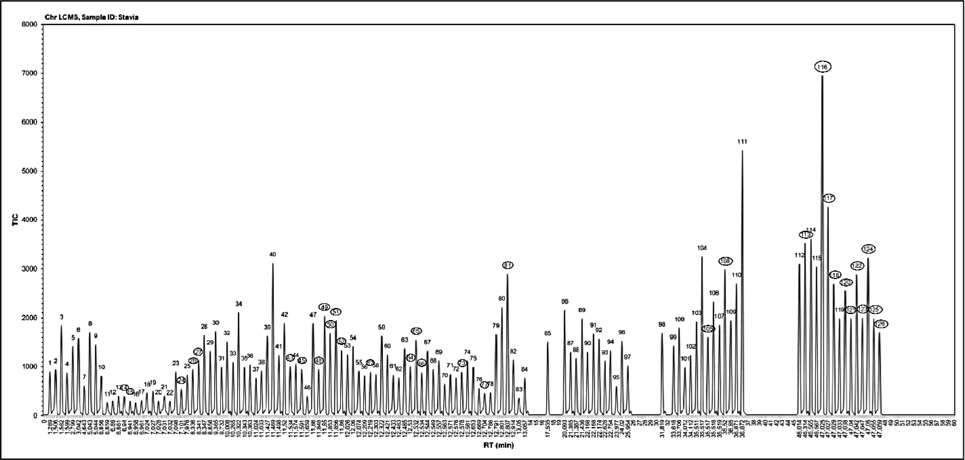
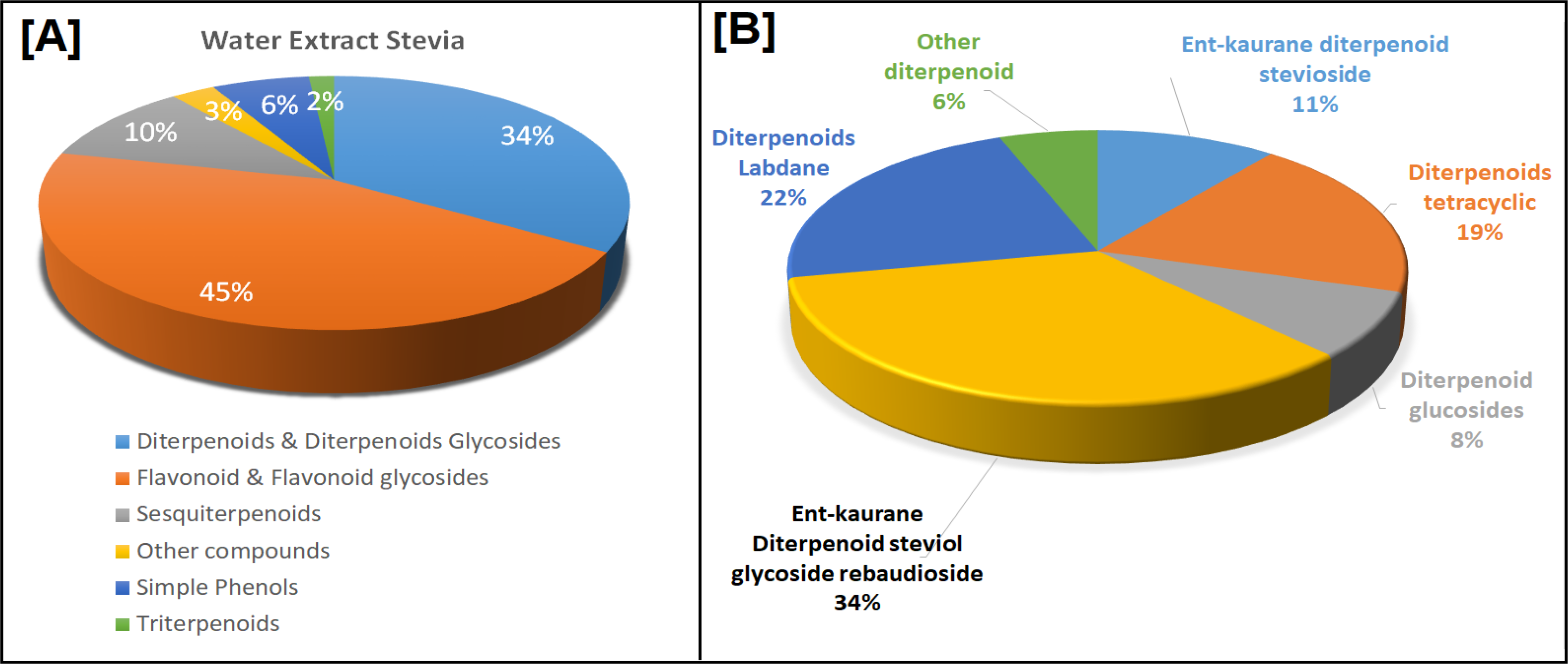
The quality of the stevia simplicia powder used for extraction by the sonication-maceration method is characterized as green simplicia powder with a particle size of 40-60 mesh, moisture content of 6-10%, and free from contaminants. The extract obtained from the sonication-maceration process was then dried using a fresh drying method, resulting in dry stevia extract powder. Subsequently, the extract was analyzed using Shimadzu LCMS 8040 LC-MS and the results are presented in Figure 1 and Figure 2.
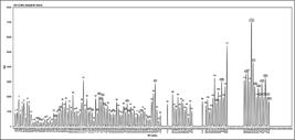
TIC of LCMS Diterpenoid-Stevia Compounds Extract with Water Extract using LCMS Shimadzu LCMS-8040 LC/MS Column Shimadzu Shim Pack FC-ODS (2 mm x 150 mm, 3 µm), eluent methanol 90%.
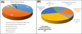
[A] Metabolic secondary group compounds composition in water stevia exctract by maceration-sonfication [B] Diterpenoids groups composition of water stevia extract
Molecular Docking and Dynamic of thirty-three Diterpenoids compounds with alpha-glucosidase and alpha-amylase
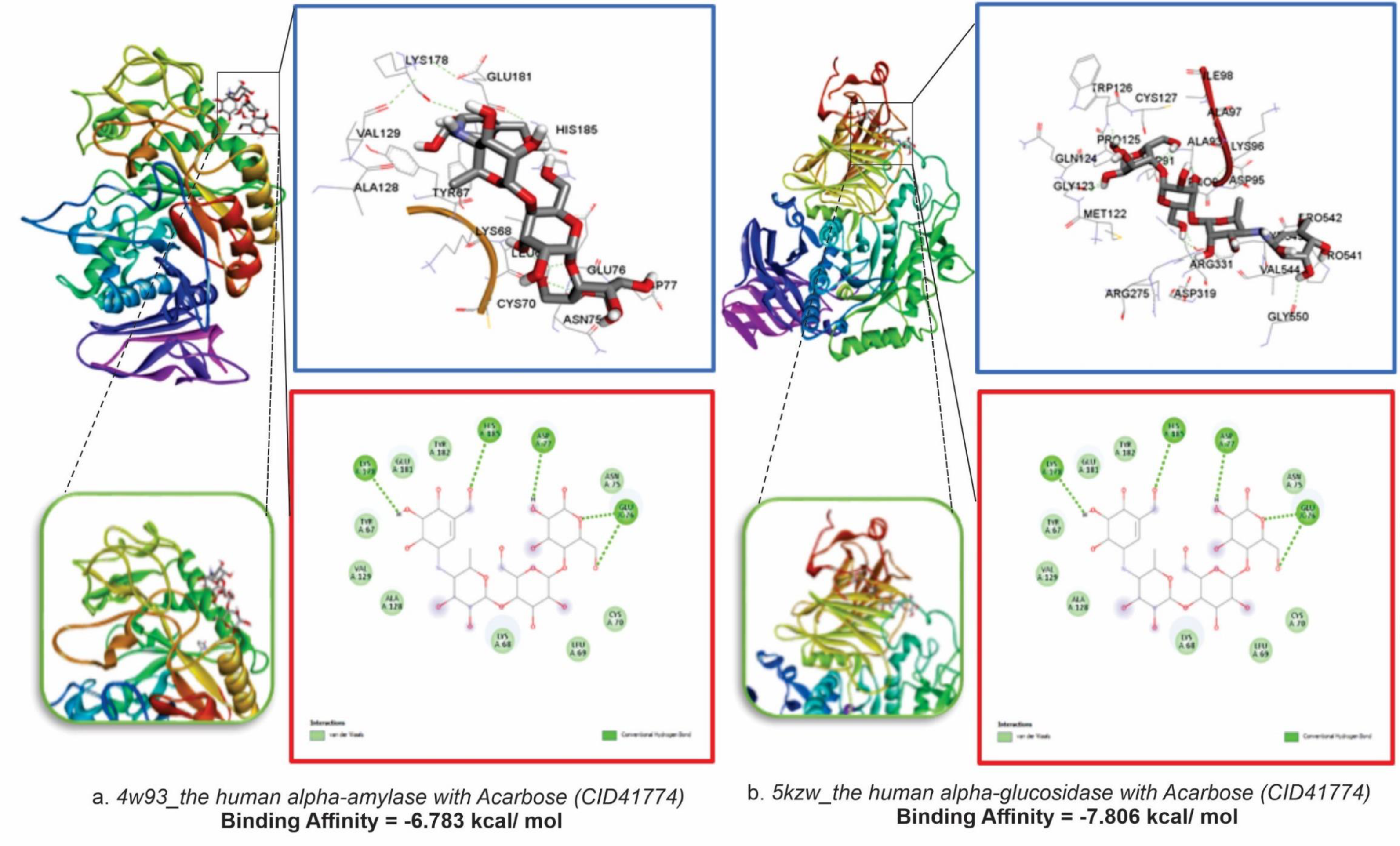
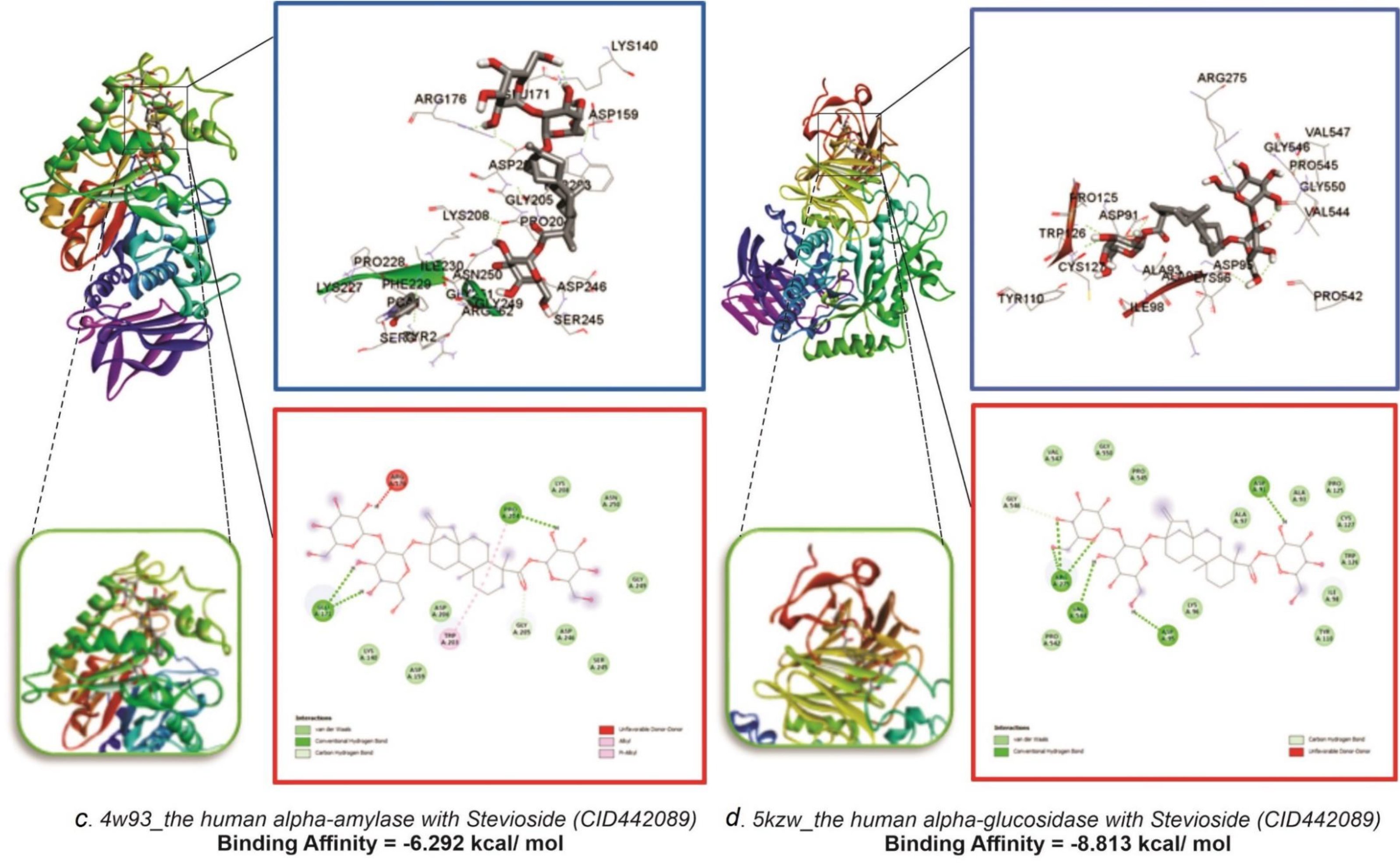
Molecular docking analysis of 33 diterpenoid stevia compounds from LC-MS against human alpha-amylase (PDB ID: 4w93) and human alpha-glucosidase (PDB ID: 5kzw) was performed using AutoDock Tools 1.5.7 software. The screening results were obtained through binding affinity scores. The binding of these compounds was compared with that of the native ligand acarbose, which served as a control. Stevia bioactive compounds were found to bind to the receptors of human alpha-amylase (PDB ID: 4w93) and human alpha-glucosidase (PDB ID: 5kzw), as presented in Table 1, supplementary-1, and Figure 3.
Binding Affinity of Diterpenoid-Stevia Compounds Maceration-Sonification Extract with Water Extract as Amylase and Glucosidase Enzyme Inhibits
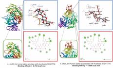
3D Visualization of Binding Poses and 2D Interactions of acarbose-alpha-amylase complexes and acarbose-alpha-glucosidase complexes
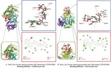
3D Visualization of Binding Poses and 2D Interactions of stevioside-alpha-amylase and stevioside-alpha-glucosidase
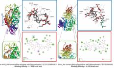
3D Visualization of Binding Poses and 2D Interactions of rebaudioside S-alpha-amylase and rebaudioside S-alpha-glucosidase.
DISCUSSION
Maceration-sonification extraction and LC-MS analysis
Stevia simplicia was extracted using a combined maceration-sonication method with water as the solvent. This combination of maceration and sonication is an innovative approach that synergistically merges the advantages of both techniques to significantly enhance the efficiency and quality of bioactive compound extraction. This innovative method improves extraction efficiency and reduces extraction time, while preserving heat-sensitive bioactive compounds. It also enhances the quality of the extracted compounds, reduces microbial activity during extraction, and optimizes solvent usage. Sonication extraction employs ultrasonic waves to generate cavitation bubbles that violently collapse, disrupting plant cell walls and increasing solvent penetration. This process accelerates mass transfer and releases bioactive compounds much faster than maceration alone, which depends on slow diffusion over extended periods. Traditional maceration can take several days to fully extract compounds, whereas sonication can achieve equal or better results within minutes. Combining sonication with maceration drastically shortens the overall extraction time while maintaining or improving yield. Maceration is a cold, non-thermal process that preserves delicate and volatile phytochemicals. Sonication is also a gentle mechanical method that does not involve high temperatures, thus protecting heat-sensitive compounds from degradation. Together, this combination ensures a high-quality extract with an intact bioactive profile [26-28]. In addition to increasing yield, sonication enhances the antioxidant and polyphenol content of the extract. Studies have shown that ultrasonic maceration produces extracts with richer flavor, more intense color, and greater stability compared to conventional maceration, which can suffer oxidative degradation during prolonged extraction. Furthermore, sonication’s cavitation effect can disrupt and inactivate microbial cells, reducing the risk of spoilage during extraction. This is especially important when using fresh plant material, which is prone to microbial growth during long maceration periods. This combined method also uses less solvent and energy compared to extended maceration processes, making it more sustainable and cost-effective for industrial applications. The sonication-maceration combination accelerates plant cell wall penetration during sonication and enhances mass transfer within the gentle, cool extraction environment of maceration. This integrated approach results in faster, more efficient extraction of high-quality bioactive compounds, increases antioxidant content, minimizes microbial contamination risk, and lowers energy and solvent consumption-making it a superior method for producing potent and stable extracts [28]. In our study, we investigated the bioactive potential of aqueous stevia extracts obtained through maceration and ultrasonic-assisted extraction (UAE). LC-MS analysis identified several secondary metabolites, including 34% diterpenoids and diterpenoid glycosides, flavonoids, 45% flavonoid glycosides, 10% sesquiterpenoids, 6% simple phenols, 2% triterpenoids, and 10% other compounds. Among the diterpenoids and diterpenoid glycosides, the following were detected ent-Kaurane diterpenoid steviol glycoside rebaudioside (34%), including: rebaudioside-B (2.29%), -A (1.73%), -C (1.55%), -G (1.45%), -F (1.37%), -S (1.08%), -R (1.07%), -E (1.07%), and -O (0.51%); Labdane diterpenoids (22%), such as: Sterebin-F (1.05%), -E (0.91%), -I (1.09%), -G (0.83%), -B (0.74%), and -M (0.72%); Tetracyclic diterpenoids (19%), including: steviolbioside (1.75%), rubusoside (1.60%), and steviolmonoside (1.56%); ent-Kaurene diterpenoid stevioside (11%), with stevioside at 3.74%; diterpenoid glucosides (8%), such as dulcoside A (1.90%) and 13-[(2-O-beta-D-glucopyranosyl-3-O-beta-D-fructofuranosyl-beta-D-glucopyranosyl)oxy]kaur-16-en-18-oic acid beta-D-glucopyranosyl ester (0.91%); and other diterpenoids (6%) as illustrated in Figures 1 and 2.
The results of LC-MS analysis show that the secondary metabolite compounds extracted in water solvent using the maceration-sonification method are more polar compounds, such as flavonoid glycosides, free flavonoids (flavonoids aglicon) and diterpenoids. Flavonoid glycosides have optical activity, and most of them are left-handed. Only free flavonoids with chiral carbon atoms in the molecule have optical activity. Free flavonoids are generally soluble in methanol, ethanol, ethyl acetate, chloroform, ether, and other organic solvents and dilute lye, but insoluble or insoluble in water [31]. Since the presence of a sugar moiety usually increases the solubility of flavonoids in water, the flavonoid glycosides are generally soluble in high polar solvents such as water, methanol and ethanol, but insoluble in lipophilic organic solvents such as benzene, chloroform, and ether [32].
Molecular docking
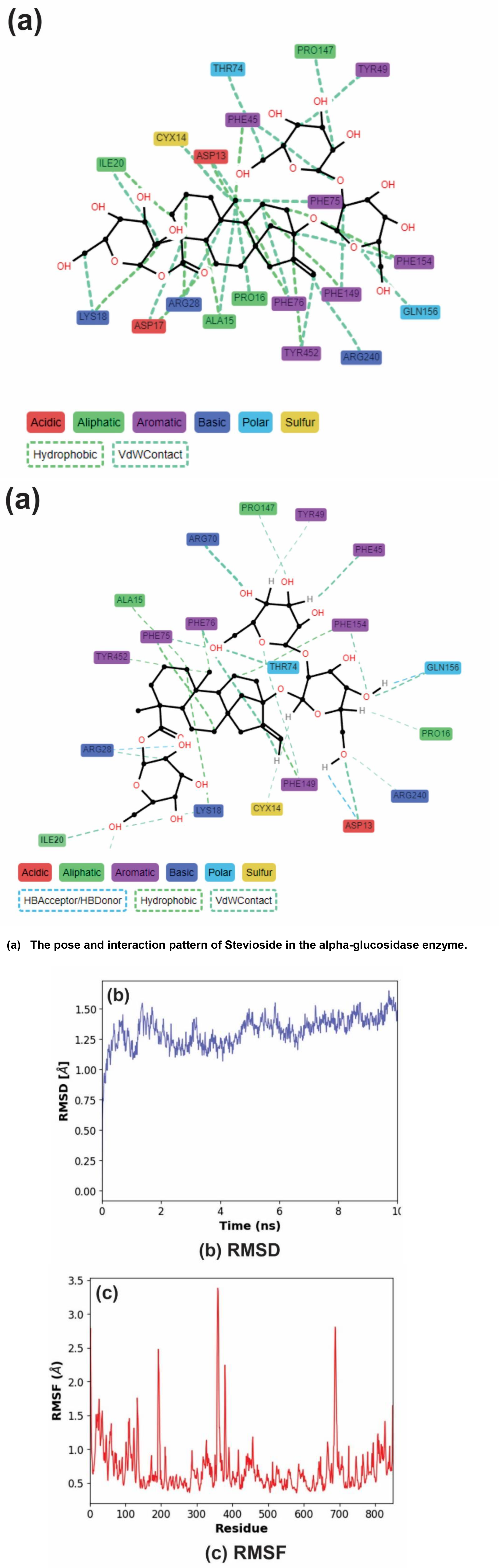
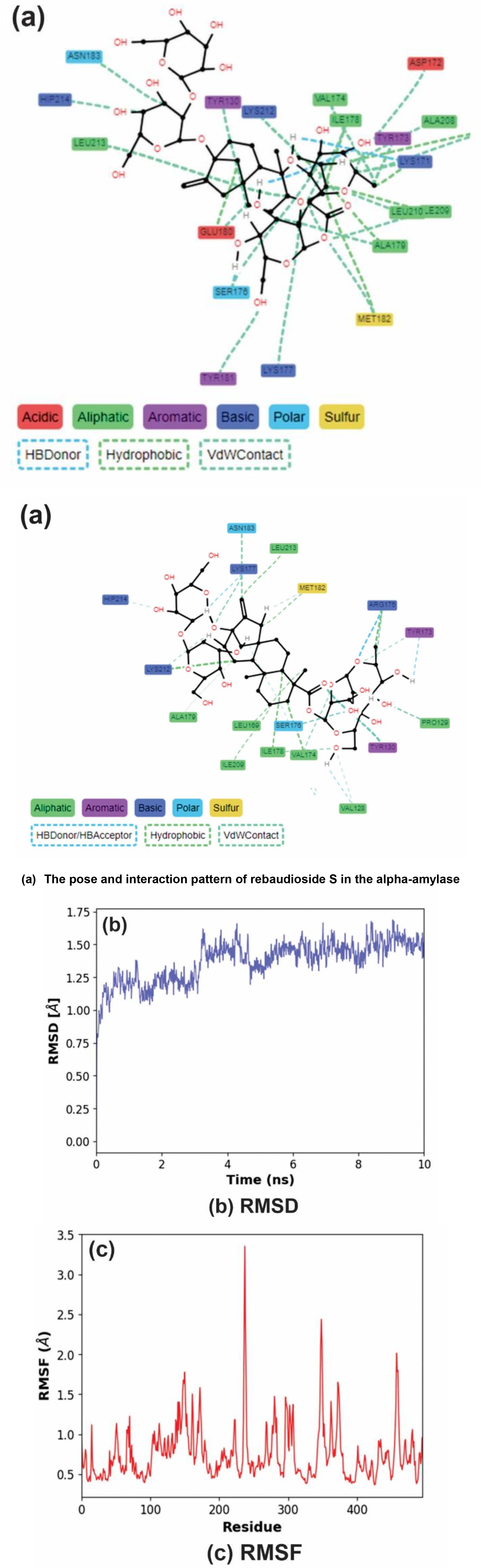
Identification of potential alpha-glucosidase and alpha-amylase inhibitors was conducted using in silico analysis by comparing the molecular docking activities of diterpenoid compound groups with acarbose on alpha-glucosidase and alpha-amylase enzymes. Virtual in silico analysis, including docking and molecular dynamics screening, is widely used for discovering new therapeutic agents [33].The potential of stevia bioactive compounds, particularly against the human alpha-glucosidase receptor (PDB ID: 5kzw) and human alpha-amylase receptor (PDB ID: 4w93), was evaluated based on binding affinity and interaction at the active site [26,34-36]. Molecular docking results revealed that stevioside (CID442089) exhibited the highest binding activity among stevia diterpenoids and acarbose when binding to the human alpha-glucosidase (5kzw). Similarly, rebaudioside S (CID132566506) showed superior binding compared to other diterpenoids and acarbose for the human alpha-amylase (4w93), as detailed in Table 1, Supplementary 1, and Figure 3. Table 1, presents molecular docking analysis showing that the binding affinity of the acarbose-alpha-amylase complex (-6.783 kcal/ mol) is higher than that of the stevioside-alpha-amylase (-6.292 kcal/ mol), but rebaudioside S-alpha-amylase complexes (-7.550 kcal/mol) is higher than acarbose-alpha-amylase complex. Figure 3(a), (c), and (e) illustrate these differences: the acarbose-alpha-amylase interaction includes only five conventional hydrogen bonds and van der Waals interactions, whereas the stevioside-alpha-amylase complex forms three conventional hydrogen bonds, carbon-hydrogen bonds, unfavorable donor interactions, alkyl, Pi-alkyl, and van der Waals interactions. The rebaudioside S-alpha-amylase complex exhibits six conventional hydrogen bonds, pi-sigma, and van der Waals interactions. Similarly, molecular docking analysis shows that the binding affinity of the acarbose-alpha-glucosidase complex (-7.806 kcal/mol) is lower than that of the stevioside-alpha-glucosidase (-8.813 kcal/mol) and rebaudioside S-alpha-glucosidase (-9.153 kcal/mol) complexes. Figures 3(b), (d), and (f) depict these interactions: acarbose-alpha-glucosidase forms five conventional hydrogen bonds, carbon-hydrogen bonds, unfavorable donor-donor and acceptor-acceptor interactions, and van der Waals forces. In contrast, stevioside-alpha-glucosidase forms six conventional hydrogen bonds, carbon-hydrogen bonds, unfavorable donors, and van der Waals interactions. The rebaudioside S-alpha-glucosidase complex forms nine conventional hydrogen bonds, unfavorable acceptor interactions, and van der Waals forces. These two compounds, stevioside and rebaudioside-S, demonstrated higher binding affinities compared to other stevia diterpenoid compounds, as shown in Supplementary 1. All stevia diterpenoid compounds also appear to be promising inhibitors of alpha-glucosidase and alpha-amylase. These include ent-kaurane diterpenoids such as steviol glycoside rebaudioside variants (rebaudioside-B, -A, -C, etc.), labdane diterpenoids (sterebin -F, -E, -I), tetracyclic diterpenoids (steviolbioside, rubusoside), diterpenoid glucosides, and other diterpenoids. This aligns with reported IC50 values where acarbose exhibits an IC50 of 83.33 ± 0.34 μg/mL against alpha-amylase at 100 μg/mL concentration [37], and an IC50 of 2,154 μg/mL against alpha-glucosidase at the same concentration [38]. In comparison, Stevia rebaudiana extract showed alpha-amylase inhibition with an IC50 of 198.40 μg/mL and alpha-glucosidase inhibition with an IC50 of 596.77 μg/mL [39]. The relationship between binding affinity and active site interactions within the complexes-particularly the stevioside-alpha-glucosidase complex and the rebaudioside-S-alpha-glucosidase complex-requires molecular dynamics simulation analysis to investigate the stability of these interactions. This analysis is crucial for advancing the study of diterpenoid compounds from Stevia rebaudiana as potential antidiabetic agents.
Molecular Dynamic Simulation
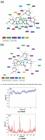
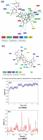
Molecular dynamics simulations were conducted only on the stevioside-alpha-glucosidase and rebaudioside-S-alpha-amylase complexes from the aqueous stevia extract, as these exhibited the best binding affinities in the molecular docking analysis. These simulations were performed using the MMPBSA and MMGBSA solvent models [34] to investigate the binding interactions and stability of the most potent compounds with the active sites of alpha-glucosidase and alpha-amylase, respectively [40]. The molecular dynamics simulation results for the stevioside-alpha-glucosidase complex are shown in Figure 4 and Supplementary 2, while those for the rebaudioside-S-alpha-amylase complex are presented in Figure 5 and Supplementary 2.
The stability of the protein-ligand complexes was assessed by calculating the Root Mean Square Deviation (RMSD) of the protein backbone from its initial to final conformation. The RMSD analysis showed that the stevioside-alpha-glucosidase complex achieved overall stability after two nanosecond of simulation time, with the RMSD stabilizing at an average of 1.5 Å (Fig. 4b). Similarly, the rebaudioside S-alpha-amylase complex reached stability after 4 ns, with an average RMSD of 1.60 Å (Fig. 5b) [40]. These results indicate that the rebaudioside-S-alpha-amylase complex was more stable throughout the simulation compared to the stevioside-alpha-glucosidase complex.
The Root Mean Square Fluctuation (RMSF) measures the flexibility of protein residues by quantifying their deviation from average positions during the simulation. Based on the timeline results, Stevioside more effectively engaged in interactions with the alpha-glucosidase binding site (Fig. 4c) compared to the interactions of rebaudioside S with alpha-amylase (Fig. 5c). Stevioside interacted with residues in both the active site and the B domain, reducing fluctuations in these regions. Similarly, rebaudioside-S also interacted with residues in the active site and B domain of alpha-amylase, leading to reduced fluctuations in those areas. Furthermore, Figure 4a illustrates the timeline of interactions between the stevioside moiety and the active sites of alpha-glucosidase during the simulation, while Figure 5a depicts the timeline of interactions between the rebaudioside-S moiety and the active sites of alpha-amylase over the simulation period.
In this study, the binding energies of the stevioside-alpha-glucosidase and rebaudioside S-alpha-amylase complexes were calculated using MMPBSA and MMGBSA solvent models via molecular dynamics simulations. Molecular Mechanics Generalized Born Surface Area (MMGBSA) and Molecular Mechanics Poisson-Boltzmann Surface Area (MMPBSA) are computational methods used to estimate binding free energies for macromolecules. These methods are favored for predicting binding free energy because they offer greater accuracy compared to molecular docking scoring functions, while requiring less computational resources than alchemical free energy methods [41]. The MMPBSA binding energy for the stevioside-alpha-glucosidase complex (∆G) was 10.6628 ± 6.4999 kcal/mol. For the rebaudioside S-alpha-amylase complex (∆G) -59.4471 ± 2.4727 kcal/mol. Using MMGBSA, the binding energy for stevioside-alpha-glucosidase (∆G) was -45.6658 ± 4.0256 kcal/mol, and rebaudioside-S-alpha-amylase (∆G) -3.1122 ± 4.3667 kcal/mol. The difference in binding energy values between MMPBSA and MMGBSA arises from the parameters included in each method. MMGBSA incorporates polar and nonpolar contributions to ∆G, while MMPBSA includes bond, angle, and dihedral energies, electrostatic energy, van der Waals energy, polar and nonpolar contributions to ∆G, and a ∆T∆S [42-44]. Furthermore, the binding energy value of the rebaudioside-S-alpha-amylase complex indicates greater stability than the stevioside-alpha-glucosidase complex, which aligns with the RMSD and RMSF values obtained in our analyses.
The results of molecular docking and molecular dynamics provide predictions of the interactions and stability of active compounds with target enzymes, which are further confirmed by IC50 values demonstrating the effectiveness of inhibiting glucosidase and amylase enzymes in preclinical studies. The synergy of these data is crucial for the development of active compound extracts as functional food ingredients and supplements for diabetes management, aiding in the creation of effective, safe, and standardized natural products.
The need for further analysis of stevia diterpenoid compounds remains, including in vitro and in vivo studies as well as clinical trials. Therefore, additional research is necessary to fully evaluate their potential.
CONCLUSION
In this study, molecular docking and molecular dynamics simulations were performed on thirty-three diterpenoid compounds extracted from stevia simplicia using the water maceration-sonication method. The results demonstrated their potential as inhibitors of alpha-amylase and alpha-glucosidase. Among these compounds, stevioside showed the greatest potential as an alpha-glucosidase inhibitor, while rebaudioside-S exhibited the strongest inhibition of alpha-amylase, along with the most stable interactions compared to other diterpenoids in the stevia water extract. This extract not only serves as an alternative sweetener but also holds promise as an antidiabetic agent through the mechanism of inhibiting alpha-glucosidase and alpha-amylase.
-
Funding:
Partially funded by Partially funded by the Directorate of Research, Technology and Community Service, Directorate General of Vocational Studies, Republic of Indonesia, No. 104/SPK/D.D4/PPK.01.APTV/III/2024, dated 19 March 2024 and partially funded by the Directorate of Research and Community Service, Directorate General of Research and Development, Ministry of Higher Education, Science and Technology, Republic of Indonesia, No. DIPA: SP-DIPA-139.04.1.693320/2026, revision 06, dated 14 July 2025.
Acknowledgments:
The authors are grateful for support of the Directorate of Research, Technology and Community Service, Directorate General of Vocational Studies, Republic of Indonesia and Directorate of Research and Community Service, Directorate General of Research and Development, Ministry of Higher Education, Science and Technology, Republic of Indonesia.
Data Availability Statement:
Research data are available in the body of the manuscript
Supplementary Material
Supplementary 1
Supplementary 2
REFERENCES
- 1 Sami W, Ansari T, Butt NS, Rashid M, Hamid A. Effect Of Diet Counseling on Type 2 Diabetes Mellitus: A Review. Int. J. Health Sci. (Qassim). 2017;(11)2:65-71.
-
2 Wan Y, Tobias DK, Dennis KK, Guasch-Ferré M, Sun Q, Rimm E B, et al. Association between changes in carbohydrate intake and long term weight changes: prospective cohort study. BMJ. 2023; 382: p. e073939. https://doi.org/10.1136/bmj-2022-073939
» https://doi.org/10.1136/bmj-2022-073939 -
3 Tappy L. Metabolism of sugars: A window to the regulation of glucose and lipid homeostasis by splanchnic organs. Clin. Nutr. 2021;(40)4:1691-8. https://doi.org/10.1016/j.clnu.2020.12.022
» https://doi.org/10.1016/j.clnu.2020.12.022 -
4 Gillespie KM, Kemps E, White MJ, Bartlett SE, The Impact of Free Sugar on Human Health-A Narrative Review. Nutrients. 15, 2023; 4:1-25. https://doi.org/10.3390/nu15040889
» https://doi.org/10.3390/nu15040889 -
5 Jacques A, Chaaya N, Beecher K, Ali SA, Belmer A, Bartlett S. The impact of sugar consumption on stress driven, emotional and addictive behaviors. Neurosci. Biobehav. Rev. 2019;103:178-99. https://doi.org/10.1016/j.neubiorev.2019.05.021
» https://doi.org/10.1016/j.neubiorev.2019.05.021 -
6 Liauchonak I, Qorri B, Dawoud F, Riat Y, Szewczuk M R. Non-nutritive sweeteners and their implications on the development of metabolic syndrome. Nutrients. 2019;(11)3:1-19. https://doi.org/10.3390/nu11030644
» https://doi.org/10.3390/nu11030644 -
7 Garcia K, Ferreira G, Reis F, Viana S. Impact of Dietary Sugars on Gut Microbiota and Metabolic Health. Diabetology. 2022;(3)4:549-60. https://doi.org/10.3390/diabetology3040042
» https://doi.org/10.3390/diabetology3040042 -
8 Blekas G A. Food Additives: Classification, Uses and Regulation. Encyclopedia of Food and Health 2016: 731-736, Elsevier Ltd. https://doi.org/10.1016/B978-0-12-384947-2.00304-4
» https://doi.org/10.1016/B978-0-12-384947-2.00304-4 -
9 Saraiva A, Carrascosa C, Raheem D, Ramos F, Raposo A. Natural sweeteners: The relevance of food naturalness for consumers, food security aspects, sustainability and health impacts. Int. J. Environ. Res. Public Health. 2020; (17)17:1-22. https://doi.org/10.3390/ijerph17176285
» https://doi.org/10.3390/ijerph17176285 -
10 Ruiz-Ojeda FJ, Plaza-Díaz J, Sáez-Lara MJ, Gil A. Effects of Sweeteners on the Gut Microbiota: A Review of Experimental Studies and Clinical Trials. Adv. Nutr., 2019;10:S31-S48 https://doi.org/10.1093/advances/nmy037
» https://doi.org/10.1093/advances/nmy037 -
11 Fry JC. Natural low-calorie sweeteners. Natural Food Additives, Ingredients and Flavourings Woodhead Publishing Series in Food Science, Technology and Nutrition. 2012:41-75 https://doi.org/10.1533/9780857095725.1.41
» https://doi.org/10.1533/9780857095725.1.41 -
12 Orellana-Paucar AM, Steviol Glycosides from Stevia rebaudiana: An Updated Overview of Their Sweetening Activity, Pharmacological Properties, and Safety Aspects. Molecules. 2023; (28)3: https://doi.org/10.3390/molecules28031258
» https://doi.org/10.3390/molecules28031258 -
13 Peteliuk V, Rybchuk L, Bayliak M, Storey KB, Lushchak O. Natural sweetener stevia rebaudiana: Functionalities, health benefits and potential risks. EXCLI J. 2021;20:1412-30, https://doi.org/10.17179/excli2021-4211
» https://doi.org/10.17179/excli2021-4211 -
14 Priscilla S, Keith Ayoob, Bernadene M, Ursula WR, Bendix JP, Peter R, et al. Stevia Leaf to Stevia Sweetener: Exploring Its Science, Benefits, and Future Potential. J. Nutr. 2018;(148)7:1186S-1205S. https://doi.org/10.1093/jn/nxy102
» https://doi.org/10.1093/jn/nxy102 -
15 Akmal M, Patel P, Wadhwa R. Alpha Glucosidase Inhibitors. StatPearls. 2024. https://www.ncbi.nlm.nih.gov/books/NBK557848/
» https://www.ncbi.nlm.nih.gov/books/NBK557848/ -
16 Arnal M, Gallego M, Talens P, Mora L. Impact of thermal treatments and simulated gastrointestinal digestion on the α-amylase inhibitory activity of different legumes. Food Chem. 2023; 418: p. 135884. https://doi.org/10.1016/j.foodchem.2023.135884
» https://doi.org/10.1016/j.foodchem.2023.135884 -
17 Hanh TTH, Huong PTM, Cham PT, Anh DH, Quang TH, Cuong NX, et al. Ent-kaurane glycosides from Stevia rebaudiana and their α-glucosidase inhibition. Tetrahedron. 2024;158:1-7. https://doi.org/10.1016/j.tet.2024.133993
» https://doi.org/10.1016/j.tet.2024.133993 -
18 Zaidan UH, Zen NIM, Amran NA, Gani SSA, Biochemical evaluation of phenolic compounds and steviol glycoside from Stevia rebaudiana extracts associated with in vitro antidiabetic potential, Biocatal. Agric. Biotechnol. 2019;(18)21:23-25, http://doi.org/10.1016/j.bcab.2019.101049
» http://doi.org/10.1016/j.bcab.2019.101049 - 19 Verma N K, Panda P, A study on Stevia Rebaudiana : A review. Int. J. Chem. Sci. 2018; (2)2:1-6.
-
20 Fu X, Wang D, Belwal T, Xu Y, Li L, Luo Z. Sonication-synergistic natural deep eutectic solvent as a green and efficient approach for extraction of phenolic compounds from peels of Carya cathayensis Sarg. Food Chem. 2021; 355. https://doi.org/10.1016/j.foodchem.2021.129577
» https://doi.org/10.1016/j.foodchem.2021.129577 -
21 Benouchenne D, Bellil I, Akkal S, Bensouici C, Khelifi D. LC-MS/MS analysis, antioxidant and antibacterial activities of Algerian fir (Abies numidica de Lannoy ex Carriere) ethylacetate fraction extracted from needles. J. King Saud Univ.-Sci. 2020;(32)8: 3321-7. https://doi.org/10.1016/j.jksus.2020.09.017
» https://doi.org/10.1016/j.jksus.2020.09.017 -
22 Juliana D, Aisyah SI, Priosoeryanto BP, Nurcholis W. Optimization of cardamom (Amomum compactum) fruit extraction using the Box-Behnken design focused on polyphenol extraction with antioxidant activity. J. Appl. Pharm. Sci. 2022;(12)6:194-209. https://doi.org/10.7324/JAPS.2022.120619
» https://doi.org/10.7324/JAPS.2022.120619 -
23 Dodda LS, Vilseck JZ, Tirado-Rives J, Jorgensen WL. 1.14∗CM1A-LBCC: Localized Bond-Charge Corrected CM1A Charges for Condensed-Phase Simulations. J. Phys. Chem. B, 2017; (121)15:3864-70. https://doi.org/10.1021/acs.jpcb.7b00272
» https://doi.org/10.1021/acs.jpcb.7b00272 -
24 Jorgensen WL, Tirado-Rives J. Potential energy functions for atomic-level simulations of water and organic and biomolecular systems. Proc. Natl. Acad. Sci. 2005;102(19):6665-70. https://doi.org/10.1073/pnas.0408037102
» https://doi.org/10.1073/pnas.0408037102 -
25 Dodda LS, De Vaca IC, Tirado-Rives J, Jorgensen WL. LigParGen web server: An automatic OPLS-AA parameter generator for organic ligands. Nucleic Acids Res. 2017;(45):W331-W336. https://doi.org/10.1093/nar/gkx312
» https://doi.org/10.1093/nar/gkx312 -
26 Ananda AN, Triawanti T, Setiawan B, Makati A C, Putri JA, Raharjo SJ. In silico study of the flavonoid compound of Sauropus androgynus leaves ON RNA-Dependent RNA polymerase (RdRp) SARS-CoV-2. Asp. Mol. Med. 2024;(3)128:100032. https://doi.org/10.1016/j.amolm.2023.100032
» https://doi.org/10.1016/j.amolm.2023.100032 -
27 Arantes PR, Polêto MD, Pedebos C, Ligabue-Braun R. Making it Rain: Cloud-Based Molecular Simulations for Everyone. J. Chem. Inf. Model. 2021;(61)10:4852-56. https://doi.org/10.1021/acs.jcim.1c00998
» https://doi.org/10.1021/acs.jcim.1c00998 -
28 Bitwell C, Sen Indra S, Luke C, Kakoma M K. A review of modern and conventional extraction techniques and their applications for extracting phytochemicals from plants. Scientific African. 2023; 19: e015852023. https://doi.org/10.1016/j.sciaf.2023.e01585
» https://doi.org/10.1016/j.sciaf.2023.e01585 -
29 Shafeeqa I, Ranjha MMAN, Nadeem M, Safdar MN, Jabbar S, Mahmood S, et al. Antioxidant Activity and Phenolic Content of Sonicationand Maceration-Assisted Ethanol and Acetone Extracts of Cymbopogon citratus Leaves. Separations, 2022; (9)9:1-9. https://doi.org/10.3390/separations9090244
» https://doi.org/10.3390/separations9090244 -
30 Ranjha MMAN, Irfan S, Lorenzo JM, Shafique B, Kanwal R, Pateiro M, et al. Sonication, a potential technique for extraction of phytoconstituents: A systematic review. Separations. 2022;9:244. https://doi.org/10.3390/separations9090244
» https://doi.org/10.3390/separations9090244 - 31 Prabakaran R, Arivoli S, Hema A, Kamatchi C. Isolation and characterization of Flavonoids from Chloroxylon Swietenia. J. Chem. Pharm. Res. 2011; (3)3:805-13.
-
32 Soczewiński E, Wójciak-Kosior M, Matysik G. Analysis of glycosides and aglycones of flavonoid compounds by double-development thin-layer chromatography. J. Planar Chromatogr. - Mod. TLC. 2004; (17)4:261-3. https://doi.org/10.1556/JPC.17.2004.4.4
» https://doi.org/10.1556/JPC.17.2004.4.4 -
33 Raharjo SJ, Kikuchi T. Molecular dynamic screening sesquiterpenoid pogostemon herba as suggested cyclooxygenase inhibitor. Acta Inform. Medica. 2016;(24)5:332-7, https://doi.org/10.5455/aim.2016.24.332-337
» https://doi.org/10.5455/aim.2016.24.332-337 -
34 Raharjo SJ, Mahdi C, Nurdiana N, Kikuchi T, Fatchiyah F. Binding energy calculation of patchouli alcohol isomer cyclooxygenase complexes suggested as COX-1/COX-2 selective inhibitor. Adv. Bioinformatics. 2014. https://doi.org/10.1155/2014/850628
» https://doi.org/10.1155/2014/850628 -
35 Raharjo SJ. Virtual Screening of Sesquiterpenoid Pogostemon herba as Predicted Cyclooxygenase Inhibitor. in Intech. 2016; 225-40. http://dx.doi.org/10.5772/intechopen.85319
» http://dx.doi.org/10.5772/intechopen.85319 -
36 Raharjo SJ, Mahdi C, Nurdiana N, Kikuchi T, Fatchiyah F. In vitro and In silico: Selectivities of Seychellene Compound as Candidate Cyclooxygenase Isoenzyme Inhibitor on Pre-Osteoblast Cells. Curr. Enzym. Inhib. 2015;13(1):2-10. https://doi.org/10.2174/1573408012666151202211035
» https://doi.org/10.2174/1573408012666151202211035 - 37 Narkhede A, Nirmal P, Tupe R, Kulkarni O, Harsulkar A, Jagtap S. In vitro Antioxidant , Antiglycation and α - amylase inhibitory potential of Eulophia ochreata L. J. Pharm. Res. 2012; (5)5:2532-37.
-
38 Sulistiyani, Safithri M, Sari Y P. Inhibition of α-glucosidase activity by ethanolic extract of Melia azedarach L. leaves. in IOP Conference Series: Earth and Environmental Science. 2016. http://dx.doi.org/10.1088/1755-1315/31/1/012025
» http://dx.doi.org/10.1088/1755-1315/31/1/012025 -
39 Ruiz-Ruiz JC, Moguel-Ordoñez YB, Matus-Basto AJ, Segura-Campos MR. Antidiabetic and antioxidant activity of Stevia rebaudiana extracts (Var. Morita) and their incorporation into a potential functional bread. J. Food Sci. Technol. 2015; 52(12): 7894-03. http://dx.doi.org/10.1007/s13197-015-1883-3
» http://dx.doi.org/10.1007/s13197-015-1883-3 -
40 Shayegan N, Haghipour S, Tanideh N, Moazzam A, Mojtabavi S, Faramarzi M A, et al. Synthesis, in vitro α-glucosidase inhibitory activities, and molecular dynamic simulations of novel 4-hydroxyquinolinone-hydrazones as potential antidiabetic agents. Sci. Rep. 2023;(13)1:1-13. http://dx.doi.org/10.1038/s41598-023-32889-7
» http://dx.doi.org/10.1038/s41598-023-32889-7 -
41 Wang E, Sun H, Wang J, Wang Z, Liu H, Zhang JZH. End-Point Binding Free Energy Calculation with MM/PBSA and MM/GBSA: Strategies and Applications in Drug Design. Chemical Reviews ACS Publication 2019; (119)16: 9361-80. http://dx.doi.org/10.1021/acs.chemrev.9b00055
» http://dx.doi.org/10.1021/acs.chemrev.9b00055 -
42 Sun H, Duan L, Chen F, Liu H, Wang Z, Pan P, et al. Assessing the performance of MM/PBSA and MM/GBSA methods. 7. Entropy effects on the performance of end-point binding free energy calculation approaches Phys. Chem. Chem. Phys. 2018; 20: p.14450. http://dx.doi.org/10.1021/ci100275a
» http://dx.doi.org/10.1021/ci100275a -
43 Weng G, Wang E, Chen F, Sun H, Wang Z, Hou T. Assessing the performance of MM/PBSA and MM/GBSA methods. 9. Prediction reliability of binding affinities and binding poses for protein-peptide complexes. Phys. Chem. Chem. Phys 2019; (21)19:10135-45. http://dx.doi.org/10.1039/c9cp01674k
» http://dx.doi.org/10.1039/c9cp01674k -
44 Hou T, Wang J, Li Y, Wang W. Assessing the Performance of the MM/PBSA and MM/GBSA Methods. 1. The Accuracy of Binding Free Energy Calculations Based on Molecular Dynamics Simulations. Journal of Chemical Information and Modeling. 2015;(51)1:449-61. http://dx.doi.org/10.1021/ci100275a
» http://dx.doi.org/10.1021/ci100275a
-
Editor-in-Chief:
Paulo Vitor Farago
-
Associate Editor:
Jane Manfron