In silico Functional and Structural Annotation of the qseB/qseC-encoded Quorum Sensing System in a Clinical Isolate of Klebsiella aerogenes
Abstract
Quorum sensing (QS) is a microbial communication mechanism that can modulate the expression of specific genes related to virulence, biofilm formation, antimicrobial resistance, enzyme production, adaptability to adverse conditions, among others. Various QS systems have been described, including the QseB/QseC system. In Klebsiella aerogenes, a nosocomial pathogen, this system remains poorly understood. This study aimed to identify the qseB and qseC genes in the genome of a clinical isolate of K. aerogenes, as well as to characterize the functions and structures of the QseB and QseC proteins. We utilized a suite of bioinformatics tools to assess the physicochemical and structural properties of these proteins, predict signal peptide presence, secretion pathways, subcellular localization, antigenic sites, and functional roles. Additionally, we modeled the three-dimensional structures and determined the active sites of QseB and QseC. Our results confirm the presence of qseB and qseC genes in the K. aerogenes. In silico analyses revealed that both proteins are stable, hydrophilic, and exhibit similar instability indices. Neither protein contains signal peptides for secretion; QseB is localized in the cytoplasm, while QseC is situated in the cellular membrane. Both proteins possess antigenic determinants. Functionally, QseB acts as a DNA-binding response regulator, whereas QseC functions as a histidine kinase. Structural models validated exhibit high-quality predictions for both proteins. Active site analysis pinpointed crucial amino acid residues involved in the biochemical activities of both proteins. These data show that the isolate has a complete QseB/QseC system that recognizes AI-3 and catecholamines and may modulate genes essential to pathogenicity.
Keywords:
bioinformatics analyses; bacterial communication mechanisms; QseB and QseC proteins; function prediction; structural modeling.
HIGHLIGHTS
Klebsiella aerogenes contains only a single copy of the qseB and qseC genes.
QseB is a cytoplasmic protein with DNA-binding domains.
QseC is a membrane-localized protein with histidine kinase functionality.
Both proteins are not secreted, lack signal peptides, and are stable.

INTRODUCTION
Quorum sensing (QS) is a cell-to-cell communication mechanism dependent on population density that regulates multiple genes critical for the survival and adaptation of various bacterial species in their environments [1,2]. For this mechanism to function, bacteria secrete signaling molecules known as autoinducers (AIs) into the extracellular environment. Once these AIs reach a threshold concentration, they are internalized and bind to regulatory proteins that control the expression of target genes involved in numerous physiological processes, including biofilm formation, motility, enzyme secretion, metabolism, virulence, and antibiotic resistance [3-5]. Moreover, the QS system can function in a peculiar manner, where the bacteria do not produce the AI but can detect, for example, eukaryotic hormones produced by other organisms and regulate specific phenotypes [6,7].
In this way, QS allows groups of bacteria to synchronously alter their behavior in response to changes in bacterial density. QS systems can typically be grouped into four well-defined classes based on the types of signaling molecules involved. The first class comprises the N-acyl homoserine lactones (AHLs), generically called autoinducer-1 (AI-1), which are produced and utilized by Gram-negative bacteria. The LuxI/LuxR system is responsible for the synthesis and recognition of these molecules [8].
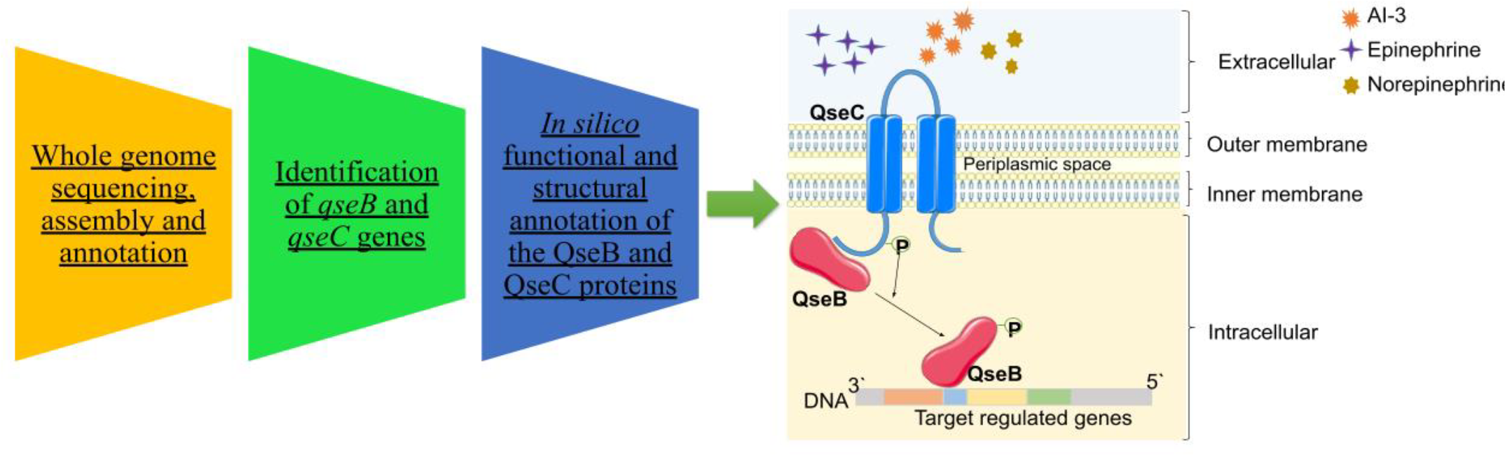
The second class includes autoinducer-2 (AI-2), a furanosyl borate diester. Various pathways, such as the LsrACDBFGE, LuxM/LuxN, and CqsA/CqsS systems and LuxS protein, have been identified for the production and recognition of these molecules, facilitating communication between both Gram-positive and Gram-negative bacteria [9,10]. The third class, extensively studied in enterohemorrhagic Escherichia coli (EHEC), is mediated by the detection of catecholamines produced by mammals, such as epinephrine and norepinephrine, playing a role in inter-kingdom communication, as well as by the bacteria-derived QS molecule autoinducer-3 (AI-3) via the QseB/QseC system [11-13]. Finally, there is the peptide-mediated system employed by Gram-positive bacteria, also known as autoinducing peptides (AIPs), which do not freely cross the cell membrane and require specialized export systems and two-component transmembrane sensors [14].
Thus, bacterial communication is closely linked to multiple pathogenic phenotypes and the development of antibiotic resistance. Therefore, disrupting, inhibiting, or attenuating this mechanism, commonly known as quorum quenching, emerges as a promising alternative for pathogen control [15]. Moreover, because the strategy of inhibiting QS relies on attenuating bacterial pathogenicity without affecting growth, the selective pressure and emergence of resistance are minimized. Overall, studies characterizing QS components are essential for developing anti-virulence drugs and controlling the emergence of multidrug-resistant strains [16,17].
In Klebsiella aerogenes, formerly known as Enterobacter aerogenes and highly capable of acquiring resistance to multiple antibiotics, the identification and function of QS systems remain poorly understood. This pathogen can be found in various environments, including water, soil, food, and colonizing the human intestine [18,19]. Although not yet fully elucidated, it is known that epinephrine and norepinephrine, which are involved in host-bacteria communication, are present in the intestine. These molecules are recognized by pathogens via the QseB/QseC system, thereby controlling the transcription of virulence genes [12,20]. To our knowledge, the role of the QseB/QseC system in the virulence of K. aerogenes has not yet been described. Therefore, this study aims to investigate the presence of genes associated with the QseB/QseC system and elucidate the physicochemical and functional characteristics of the QseB and QseC proteins using in silico analysis tools.
MATERIAL AND METHODS
Bacterial strain
The clinical isolate of K. aerogenes Ea5A from human rectum was donated to the laboratory by a public hospital in Recife, Pernambuco, Brazil [21].
Whole genome sequencing, assembly and annotation
Total DNA was extracted using the Wizard Genomic DNA Purification Kit (Promega) and quantified utilizing the Qubit fluorometric platform (ThermoFisher Scientific). Subsequently, the genetic material was used for the assembly of DNA libraries employing the TruSeq DNA PCR-Free Kit from Illumina and genomic sequencing was performed using the MiSeq system (Illumina) [21]. The quality of the generated fragments was evaluated using the FASTQC tool (http://www.bioinformatics.babraham.ac.uk/projects/fastqc/) and the Trimmomatic tool (http://www.usadellab.org/cms/index.php?page=trimmomatic) was utilized to clean low-quality data and remove interfering sequences. Contigs were assembled using the velveth and velvetg programs from the Velvet package. The alignment and ordering of the contigs relative to the reference sequences were performed using the blastall and Mauve programs. Gene prediction and annotation were performed using the RASTtk tool [21]. The genome of the isolate was deposited in GenBank under the accession codes: PRJNA310664 (Bioproject) and SAMN04461808 (Sample Accession).
Identification of qseB and qseC genes
The qseB and qseC genes were selected through a manual and accurate analysis of all annotated genes, using the MySQL database (https://dev.mysql.com/) and Microsoft Office Excel. BLASTN was employed to validate the specificity of the QS genes in the National Center for Biotechnology Information (NCBI) Nucleotide Sequence Database (https://blast.ncbi.nlm.nih.gov/Blast.cgi).
Retrieval and analysis of amino acids residues sequences from proteins
The amino acids residues sequences from QseB and QseC proteins were predicted by the RASTtk tool. BLAST pairwise alignments were performed using the UniProt database with UniProtKB/Swiss-Prot entries to identify regions of similarity (https://www.uniprot.org/blast). Subsequently, the amino acids residues sequences of the QseB and QseC proteins were retrieved from the UniProt database in .fasta file format, and the Clustal Omega aligner was used to align these sequences with the amino acids residues sequences predicted in this study [22].
Assessing physicochemical and structural properties
The amino acids residues sequences from QseB and QseC proteins were used as templates to determine their physicochemical parameters using the ProtParam tool of ExPASy [23]. The Self-Optimized Prediction Method with Alignment (SOPMA) package [24] and the PSIPRED Workbench program [25] were used to analyze the structural properties of the proteins.
Prediction of signal peptides, secretion pathway, subcellular localization and antigenic sites
The presence of signal peptides within the amino acid sequences was determined using the SignalP 5.0 server [26]. The Antigenic Peptides program was used to predict the antigenicity of the proteins. The SecretomeP 2.0 server [27] was also used to predict the secretion pathway. In addition, subcellular localization predictions were performed using PSortB [28] and CELLO v.2 [29].
Function prediction
Protein motifs in QseB and QseC were identified using the Motif server (https://www.genome.jp/tools/motif/). The Pfam [30] and SuperFamily [31] databases were queried to determine the evolutionary relationships of these proteins. Functional analysis of the proteins was conducted using the InterPro database [32] and the ProFunc program from the EMBL-EBI server (https://www.ebi.ac.uk/thornton-srv/databases/profunc/).
Structural modeling
Structural fold prediction was performed using fold recognition techniques implemented in the I-TASSER [33] and Phyre2 [34] prediction server. The validation was performed with the SAVES server (https://saves.mbi.ucla.edu/). The quality of the modeled structures was assessed using the QMEAN4 program within the SWISS-MODEL workspace (https://www.expasy.org/resources/qmean). The selection of the best model for predicting the three-dimensional (3D) model structure of the QseB and QseC proteins was carried out by analyzing the quality of the models generated in the 3D Phyre2 and I-TASSER predictors. The PDB models generated by these programs were subjected to model quality analyzes and structure validation in the QMEAN4 and SAVES programs (https://saves.mbi.ucla.edu/). The choice of the model was based on the best Z-score values from QMEAN4 and the best parameters from SAVES.
Active site determination
The 3D modeled and validated structures of QseB and QseC proteins were used for protein binding site analysis. The Computed Atlas of Surface Topography of Proteins (CASTp) (http://sts.bioengr.uic.edu/castp/) was utilized to pinpoint active sites in the QseB and QseC proteins and the amino acid residues present in the binding sites were confirmed by the proFunc program (https://www.ebi.ac.uk/thornton-srv/databases/profunc/).
RESULTS
Whole genome sequencing, assembly and annotation
A total of 219 contigs were generated, 18 of which were plasmid-derived. The genome of the clinical isolate K. aerogenes Ea5A comprises a single chromosome (5,571,633 bp) with a GC content of 55.56%. Genomic annotation identified 5,571 chromosomal coding sequences (CDS), including 4,517 predicted proteins and 1,054 hypothetical proteins, along with five rRNA genes and 75 tRNA genes. Plasmid analysis revealed two major plasmids, Ea5A_pA (5,436 bp) and Ea5A_pB (72,262 bp), and an additional 241,370 bp of unassembled plasmid sequences. In addition, the genomic annotation revealed genes associated with virulence mechanisms, including antimicrobial resistance, adhesion structures (fimbriae and pili), efflux systems, siderophore production (for iron acquisition), capsule synthesis, biofilm formation, and specialized secretion systems. Moreover, genes linked to diverse metabolic processes were identified, as well as other functional categories implicated in microbial adaptation and survival.
Identification of qseB and qseC genes
The qseB and qseC genes were identified in the genome of K. aerogenes Ea5A. Each gene is present as a single copy in the chromosomal DNA. The qseB gene consists of 660 base pairs (bp), while qseC contains 1353 bp (Table 1). BLAST alignment revealed 100% similarity with various qseB and qseC genes deposited in the databases from genomes of bacteria belonging to the genus Klebsiella.
Alignment of the predicted amino acids residues sequence
The predicted amino acids residues sequences of QseB and QseC from this study were aligned with the most similar sequences found in the UniProt database (Supplementary Figures 1 and 2). For QseB, the alignment showed a high identity (97.23%) with the two-component transcriptional regulator protein of Klebsiella pneumoniae subsp. pneumoniae HS11286 (A0A0H3GTE4). The other sequences used in the alignment showed identities below 90%. For QseC, the highest similarity (88.77%) was observed with the QseC protein from K pneumoniae subsp. ozaenae (A0A377Z2W1).
Physicochemical and structural properties of QseB and QseC proteins
Table 2 presents the physicochemical properties of the proteins. The qseB gene encodes a protein with 219 amino acids residues, while qseC encodes a larger protein with 450 amino acids residues. The proteins have an aliphatic index of 109.18 (QseB) and 100.40 (QseC), indicating stability across a wide temperature range. They share identical half-life estimations of 30 hours.
A negative GRAVY value (-0.167 for QseB and -0.293 for QseC) indicates their hydrophilic and water-soluble nature, with QseC showing a stronger affinity for aqueous environments. Additionally, the instability index is 34.83 for QseB and 34.32 for QseC, confirms their classification as stable proteins, as an instability index below 40 suggests stability.
The SOPMA server predicted for the QseB that 40.64% of the residues as alpha helix, in comparison to 31.51% of random coil. Extended strand of 19.63% and beta turn of 8.22. For QseC, 55.56% of the amino acid residues form alpha helices, compared to 33.56% of random coils,12% forming extended strands and 3.78% of beta turn (Table 3). Interestingly, the PSIPRED program showed higher confidence in the prediction of strands, helices and coils (Supplementary Figures 3 and 4).
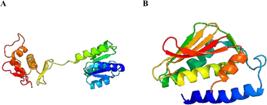
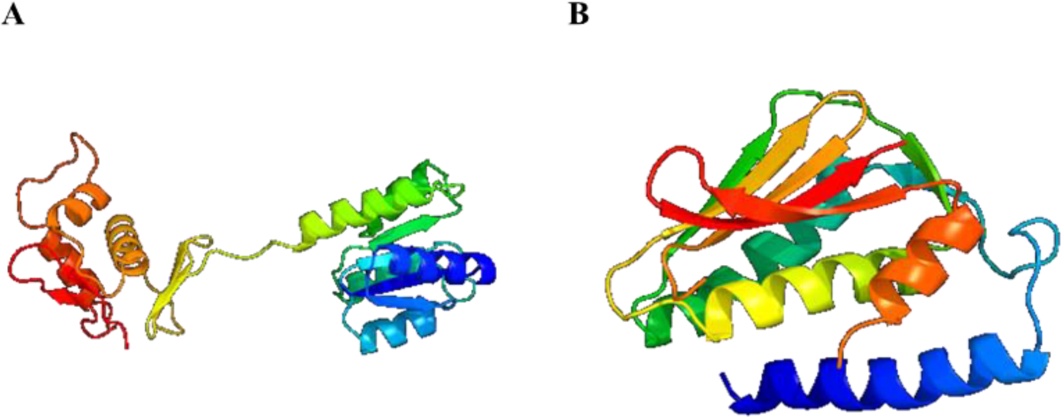
Predicted and validated of the 3D model of the QseB (A) and QseC (B) proteins structures generated using the Phyre2 predictor.
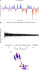
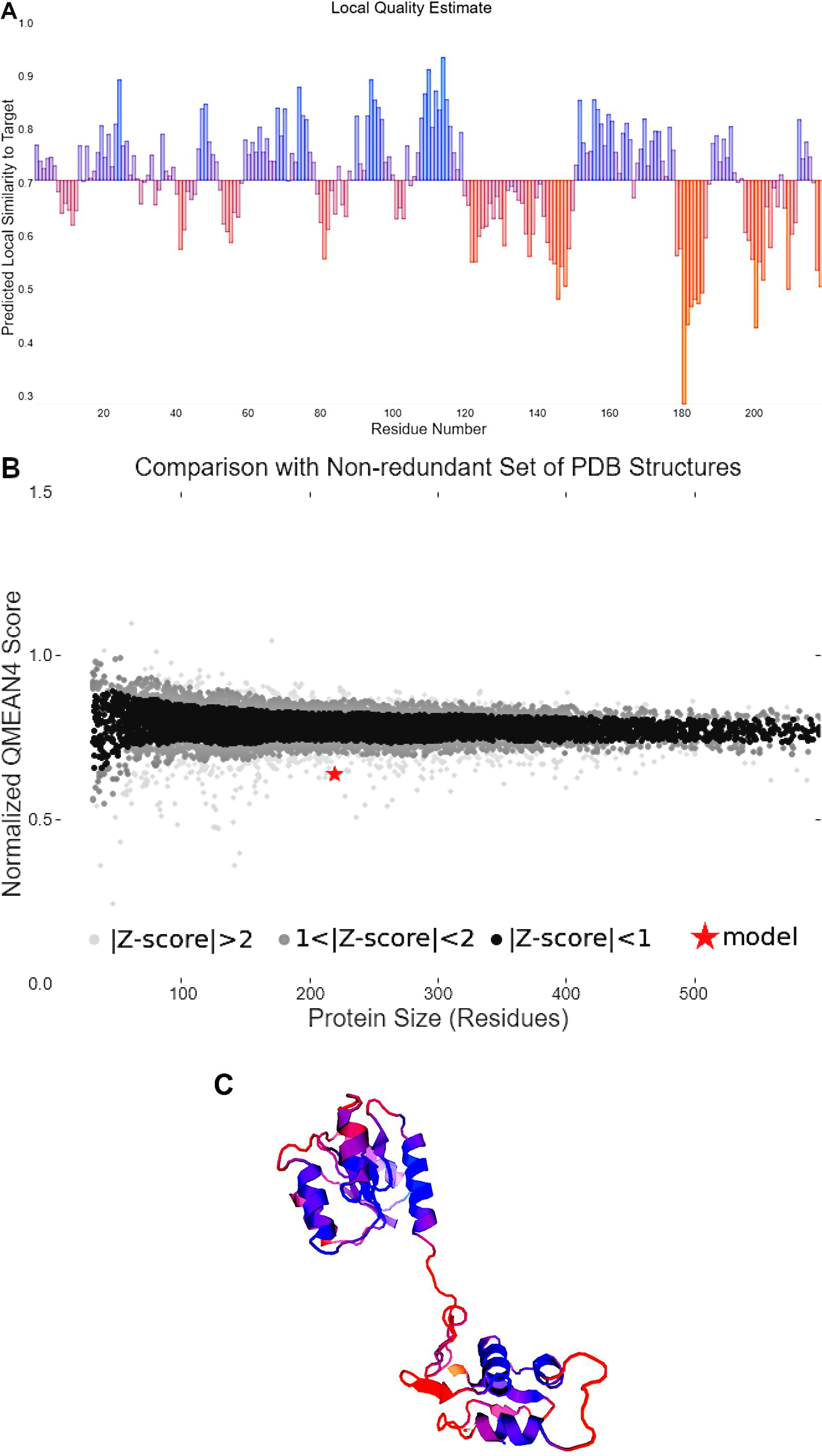
Quality parameters of the QseB protein model assessed by the QMEAN4 validator. (A) Local quality estimate; (B) Comparison wiht non-redundant set of PDB structures; (C) structure 3D.
Prediction of signal peptides, secretion pathway, subcellular localization and antigenic sites
The results from the SignalP-5.0 server indicated that QseB protein does not have a signal peptide in its structure, with scores for signal peptide, TAT signal peptide, lipoprotein signal peptide, and other (indicating the absence of any signal peptide by the program) being 0.0059, 0.0011, 0.001, and 0.992, respectively. Similarly, the QseC protein does not contain any signal peptides, with scores for signal peptide, TAT signal peptide, lipoprotein signal peptide, and other being 0.4183, 0.0035, 0.088, and 0.4902, respectively. The presence of signal peptide is indicated by the presence of a cleavage site, which was not indicated for any of the proteins.
The SecP scores for QseB and QseC proteins were 0.255702 and 0.096821, respectively, indicating that both proteins are not secreted into the extracellular environment. For bacterial sequences, SecP scores less than 0.5 indicate that the protein is not secreted into the extracellular environment. The CELLO and PSORTb servers were utilized to predict the cellular localization of each protein. The results indicate that QseB is a cytoplasmic protein (localization score of 9.97), whereas QseC is located in the cytoplasmic membrane (localization score of 10.00).
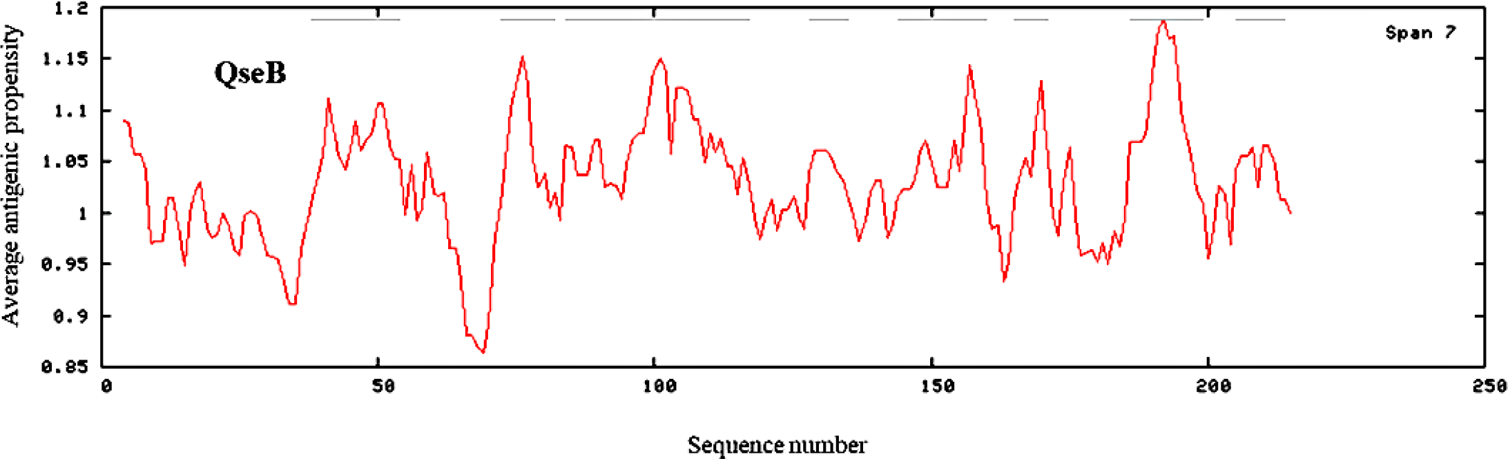
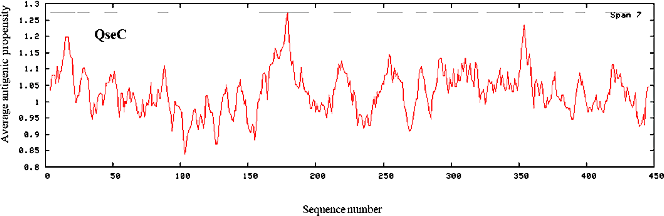
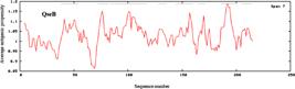
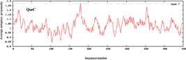
The results from the Antigenic Peptides program showed that QseB contains eight determinants in your sequence, with average antigenic propensity of 1.0293 (Figure 1). The QseC protein contains 14 antigenic determinants, with an average antigenic propensity of 1.0284 (Figure 2). These antigenic determinants present in sequence of QseB and QseC consist of fragments that start and end at various positions (Table 4).
Function prediction
To elucidate the functions of the proteins QseB and QseC, various databases including InterPro, Pfam, and SuperFamily were utilized. The results suggest that QseB belongs to the Signal transduction response regulators family and contains two motifs: Response_reg and Trans_reg_C. The Trans_reg_C domain, on the other hand, represents the C-terminal region that binds to the cell's DNA. The analysis indicates that QseC belongs to the two-component system sensor histidine kinase family and contains five motifs: HATPase_C, HisKA, 2CSK_N, HAMP, and Talin_IBS2B. Additionally, the results obtained from the ProFunc program, through global alignment with proteins deposited in the Protein Data Bank (PDB) database, showed that QseB protein binds to the cell's DNA, while QseC protein functions as a histidine kinase (Table 5).
Biological and biochemical function analysis of QseB and QseC proteins models by the ProFunc program.
Structural modeling
The models generated by I-TASSER and Phyre2 were assessed using the QMEAN and SAVER servers. Notably, models generated by Phyre2 exhibited superior Z-score values compared to those generated by QMEAN4, along with enhanced validation parameters from SAVES (Table 6). The quality of the QseB and QseC proteins models, validated via the SAVES server, is depicted through Ramachandran plots (Supplementary Figures 5 and 6). Moreover, ERRAT and Verify 3D scores were within acceptable ranges, further affirming the reliability of models generated using the Phyre2 predictor (Table 6).
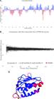
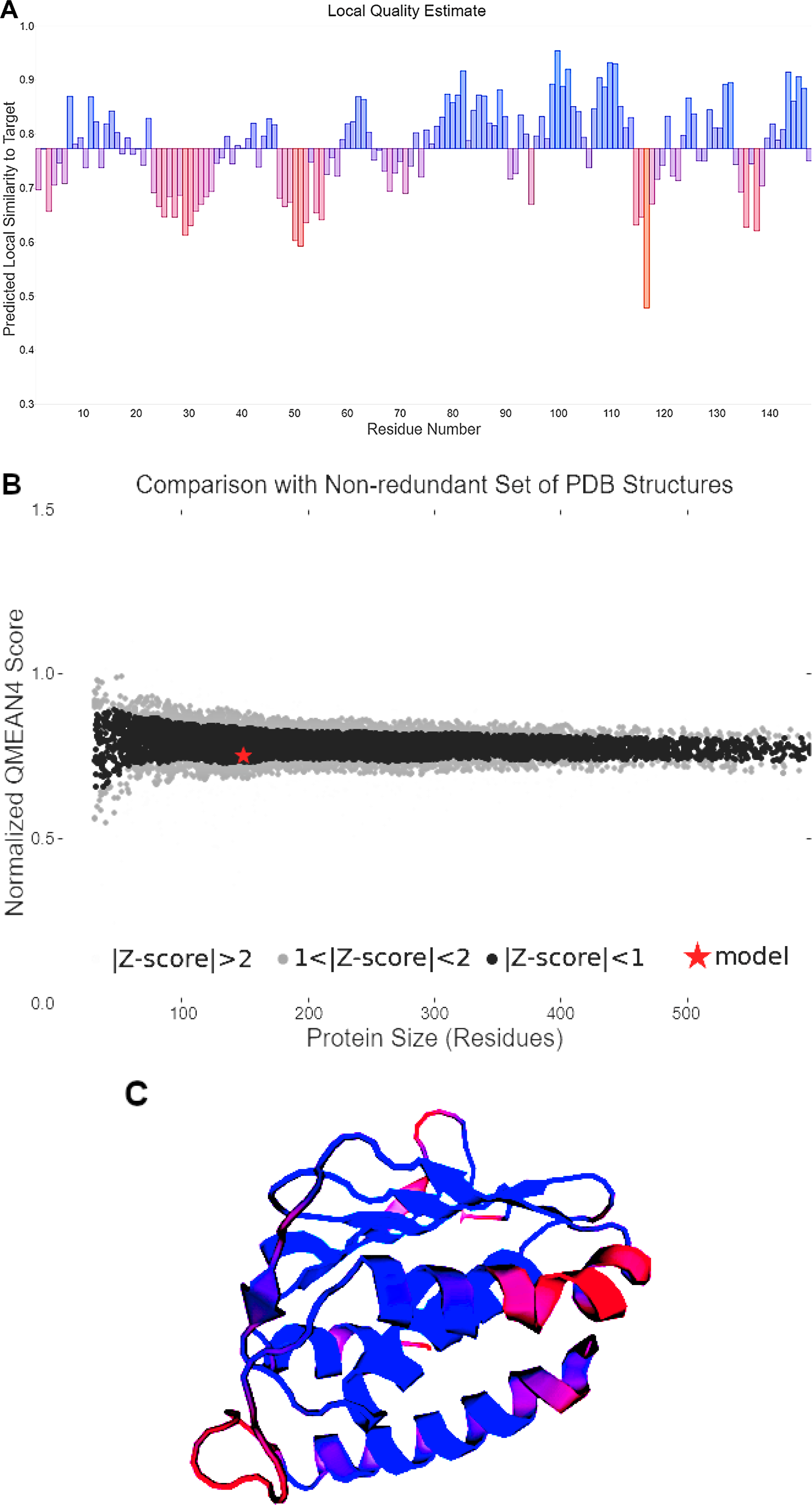
Quality parameters of the QseC model assessed by the QMEAN4 validator. (A) Local quality estimate; (B) Comparison wiht non-redundant set of PDB structures; (C) structure 3D.
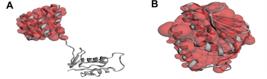
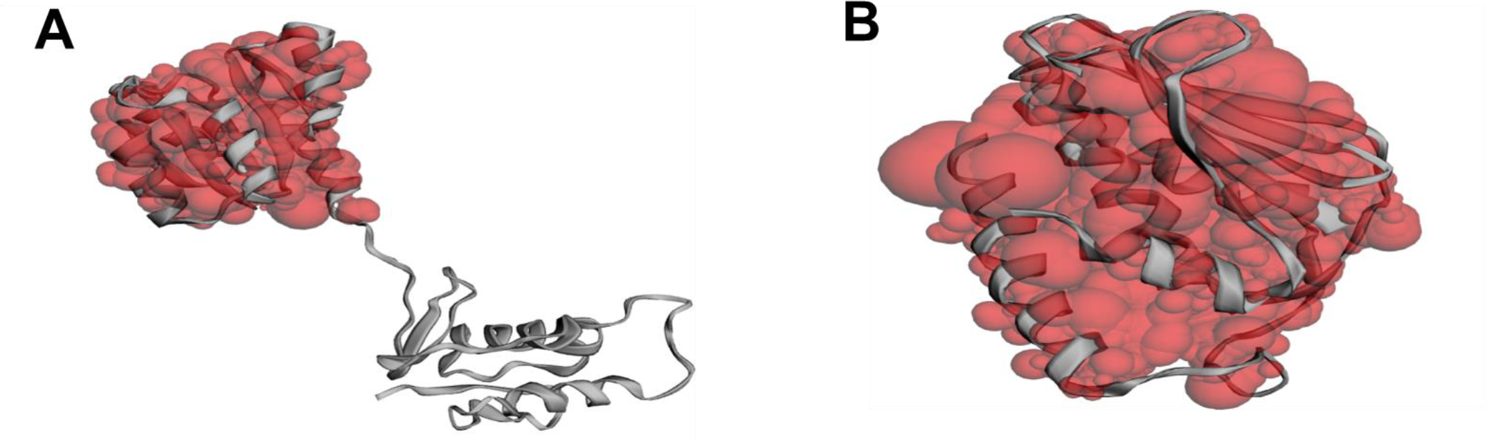
Binding site analysis. Presentation of the active site generated by the CASTp program for (A) QseB; (B) QseC proteins. The regions containing the active site amino acids residues are highlighted in red.
The QseB protein obtained a confidence level of 100% and full coverage based on the crystal structure of K. pneumoniae PMRA in complex with PMRA2 box DNA (PDB: 4S04). For the QseC protein, the Phyre2 predictor achieved a confidence level of 99.9% and covered 33% of the protein sequence structure corresponding to the cytoplasmic segment of histidine kinase QseC from E. coli K-12 (PDB: 3JZ3). The structural models generated are showed in Figure 3.
The analysis of the QseB model using the QMEAN4 program yielded detailed graphs of the local quality of each amino acid residue (Figure 4). The local quality is plotted against the residue positions in the model (Figure 4A). The quality varies on a scale from 0.0 to 1.0, represented by a color gradient from orange (0.0) to blue (1.0), where a score of 1.0 indicates high local quality of the residue position. For this model, regions of the sequence between amino acids residues 122-148 and 198-212 exhibited approximate confidence levels of 0.50 and 0.60, depicted on the graph as low peaks and shades ranging from light purple to orange. Between amino acids residues 181-186, the confidence was below 0.50, indicated by larger and orange bars. Importantly, these low-confidence regions may not only reflect a lack of a well-defined structure but could also play functional roles, such as facilitating the proper folding and dynamic flexibility of the overall protein.
In Figure 4B, the z-score of the analyzed model is plotted against the QMEAN value. It can be observed that the QseB model (marked by the red star) is positioned close to PDB models with z-scores >2, indicating it is a high-quality model. The Figure 4C shows the QseB protein model, where regions of lower confidence are shown in shades of orange, while regions of higher confidence are indicated in blue.
In Figure 5A, the local quality of residues in the QseC model is shown. This model has fewer residues with confidence scores below 0.50. The regions of lowest confidence are found between amino acids residues 23-34, 47-56, 115-120, and 134-139. Although these areas exhibit lower confidence, they may play important functional roles, such as providing the flexibility necessary for proper folding of adjacent regions. The z-score of the model (indicated by the red star) places it among PDB models with z-scores <1, and its QMEAN value of 0.56 classifies it as a model of good and reliable quality (Figure 5B). The QseC protein model shows few regions of lower confidence (shades of orange) and a large number of regions of higher confidence (shades of blue) (Figure 5C).
The predictive models for both proteins exhibited favorable QMEAN Z-scores and predictive local similarity.
Active site determination
The active site analysis of the modeled proteins was conducted using two functional analysis programs: ProFunc and CASTp. Both ProFunc and CASTp identified the binding sites of the protein models and specified the amino acid residues involved in their biochemical activity (Figure 6).
To analyze the QseB protein, the ProFunc program used the sequence from the crystal structure of K. pneumoniae PmrA in complex with PmrA box DNA (PDB: 4S04). This is the same protein used by the Phyre2 predictor for modeling the QseB protein in this study, yielding a similarity score of 392.81 and an E-value of 1.25E-07. For the QseC protein, the sequence from the structure of the cytoplasmic segment of histidine kinase QseC (PDB: 3JZ3) was used. Similarly, this sequence is the same one used by the Phyre2 predictor to model the QseC protein in this study. In this case, the obtained similarity score was 703.12, with an E-value of 0.00E+00. As an E-value < 1.00E-06 is required for high-quality analysis, these results validate the selection of the template sequences and support the reliability of the structural models generated for both proteins.
DISCUSSION
The clinical isolate of K. aerogenes Ea5A utilized in this study is known to exhibit resistance to a broad spectrum of antibiotics [21]. Indeed, infections caused by K. aerogenes in healthcare settings have become a pressing concern due to its ability to acquire resistance to various classes of antibiotics, posing a significant challenge for their management [35,36]. In Intensive Care Units (ICUs), K. aerogenes is one of the leading causes of nosocomial infections, resulting in a high mortality rate. Thus, it represents an emerging pathogen that demands attention from healthcare services [37,38].
Given the pathogen's relevance, the majority of studies focus on characterizing the resistance profile and identifying resistance genes in different isolates. Consequently, few studies aim to elucidate other mechanisms that may also contribute to the emergence of multidrug-resistant K. aerogenes strains. It is well known that QS systems play a key role in mediating microbial resistance mechanisms, including drug efflux pumps and biofilm formation [39,40]. Although little information is available on drug resistance and virulence controlled by QS mechanism in K. aerogenes, several research efforts should be made to elucidate the complex mechanisms contributing to the pathogenicity and antimicrobial resistance of this pathogen. This is important because the QS system become pharmacological targets for controlling infections caused by K. aerogenes [41].
The AI-3/epinephrine/norepinephrine signaling system is a type of QS mechanism that enables various enteric pathogens to detect AI-3 and catecholamines and induce the expression of virulence genes in the gut [42]. Thus, QS mediated by AI-3 and catecholamines functions as a general communication signal in bacteria, facilitating intergenus and interkingdom interactions [6].
In the present study, the chromosomal DNA of the K. aerogenesEa5A, collected from the rectum of a patient in the ICU, contains only one copy of the qseB and qseC genes. In silico analyses identified that the qseB and qseC genes encode a putative two-component system comprising a transcriptional regulatory protein (QseB) and a histidine kinase sensor (QseC). The identification of this system in various bacterial groups, such as Salmonella spp., different strains of E coli, Enterobacter cloacae, and Serratia marcescens, has been previously reported [13,41,43]. In enterohemorrhagic E. coli (EHEC), the AI-3/epinephrine/norepinephrine signaling system has been extensively explored. In this pathogen, the QseBC system regulates various functions, including motility, biofilm formation, and the activation of virulence genes [44]. In Enterobacter hormaechei, the QseBC system may be associated with colistin resistance [45]. Unfortunately, the information regarding the QseBC system is scarce in K. aerogenes. Therefore, further studies are needed to characterize the biological functions of qseB and qseC genes in K. aerogenes.
Using various bioinformatics tools, we characterized the different physicochemical properties of these proteins based on the predicted amino acids residues sequences. These proteins are hydrophilic, stable to temperature variation, not secreted by the cell, and lack a signal peptide. To our knowledge, there are no descriptions in the literature of secretion pathways or signal peptide detection for QseB and QseC proteins. The presence of a signal peptide is important for directing proteins to the membrane, activating translocation machinery for export, and inserting proteins into the membrane [46]. Our in silico prediction studies indicated that QseB is located in the cytoplasm, whereas QseC is associated with the cell membrane. SignalP 5.0 classifies signal peptides in prokaryotes into three types: Sec substrates cleaved by SPase I (Sec/SPI), Sec substrates cleaved by SPase II (Sec/SPII), and Tat substrates cleaved by SPase I (Tat/SPI). However, SignalP 5.0 cannot identify Tat substrates cleaved by SPase II or Sec substrates processed by SPase III [26], suggesting that QseC may possess an undetected signal peptide.
The results indicate that QseC protein has distinct domains enabling the recognition of autoinducers in the extracellular environment and the transmission of the signal to the cytoplasm. QseC is a membrane protein that interacts with AI-3, adrenaline, or norepinephrine through an extracytoplasmic sensory domain to undergo autophosphorylation [7,47]. Subsequently, QseC possesses phosphatase activity mediated by its ATPase domain, which can transfer its phosphate to the QseB response regulator [12,48]. In our study, we demonstrated that the QseB protein possesses two distinct domains: the Response_reg domain and the Trans_reg_C domain. In other microorganisms, the Response_reg domain of QseB typically receives signals from its cognate sensor kinase within bacterial two-component systems and is generally located at the N-terminus, preceding a DNA-binding effector domain. In contrast, the Trans_reg_C domain constitutes the C-terminal region responsible for binding to the cellular DNA [7,49].
On the other hand, the QseC protein contains five domains: HATPase_C, HisKA, 2CSK_N, HAMP, and Talin_IBS2B. In Salmonella and E. coli, the HATPase_C domain encompasses the conserved ATPase region of histidine kinases, while the HisKA domain plays a key role in dimerization and functions as a phospho-acceptor in these systems [47,49]. The 2CSK_N domain, located toward the N-terminus of bacterial two-component sensor kinases, is involved in the recognition of autoinducer-3 (AI-3), epinephrine, norepinephrine, and Fe(III). The HAMP domain, a cytoplasmic helical linker, is commonly found in various prokaryotic signaling proteins and mediates signal transduction. Finally, our findings also reveal the presence of the Talin_IBS2B domain, which is part of the talin adaptor protein complex that activates integrin-mediated cell adhesion and links integrins to the actin cytoskeleton. This specific domain is critical for integrin binding, interaction with acidic phospholipids, and localization to focal adhesions [50,51]. Taken together, these observations suggest that QseB and QseC in the clinical isolate of K aerogenes Ea5A may function as a canonical two-component regulatory system, modulating the expression of multiple genes involved in pathogenicity. Using the Antigenic Peptides program, we identified distinct antigenic determinants for QseB and QseC from the clinical isolate K. aerogenes Ea5A. These predicted antigenic sequences could potentially stimulate antibody responses, particularly for QseC, which includes an extracellular domain. Cell surface antigens are highly suitable targets for vaccine development due to their antigenicity and role in initial host-pathogen interactions [52,53]. Furthermore, they may be useful in the development of immunological methods for pathogen detection [54].
The modeling of the QseB and QseC proteins was carried out using the global alignment method, based on the prediction tool's search of sequences from the PDB database. This database exclusively contains structures validated by macromolecular crystallography, nuclear magnetic resonance spectroscopy, electron microscopy, and micro-electron diffraction, ensuring that the predictions are highly faithful to the native structures of the proteins. This PDB-based approach provides greater accuracy due to the reliance on rigorously validated techniques [55]. To confirm the quality of the models, the SAVES server analyses of the Ramachandran plots revealed a predominance of residues in favorable regions. For the QseB protein, only one residue was found in generously allowed regions, and eight residues were in additionally allowed regions. Similarly, for the QseC model, no residues were found in generously allowed regions, and eight residues were in additionally allowed regions. Importantly, no amino acid residues were detected in disallowed regions for either model. These results affirm the selection of the generated models and the high quality of the predictions obtained.
The qualitative energy analysis of the model generated by the QMEAN4 program corroborates the data obtained from the SAVES server for selecting the predicted model. The lower-quality regions of the model are predominantly those lacking defined secondary structure. Notably, despite having a lower QMEAN score compared to the QseC protein model, the QseB model exhibited optimal values in the SAVES model validator, further supporting the generated model. Protein modeling requires a meticulous analysis of obtained data and the presence of proteins previously modeled using more advanced techniques to ensure the reliability of the generated model. Moreover, the use of model quality validation tools, including Ramachandran plots, z-scores, and local and global quality assessments, enhances the reliability of the generated data [56,57]. These criteria were met in predicting the tertiary structure of the proteins in this study, as homologous sequences have been deposited in the PDB database, enabling more accurate modeling.
The binding site analyses of the models generated in this study confirm the information obtained from predictive functional analyses and binding site analyses, providing comprehensive insights into the amino acid residues involved in these functions. The data provided by the ProFunc program, which utilizes global alignment with crystallized proteins similar to those in this study, corroborate the findings of the CASTp program by identifying the same amino acid residues at the binding sites of the modeled proteins.
In the binding site analysis of the QseB protein, the region identified by CASTp spans amino acids 1-219. Secondary structure predictors indicate two domains within this region: the DNA-binding domain between amino acids 1-117, and the receiver domain (REC) between amino acids 124-218. The CASTp analysis of the QseC protein indicated that amino acid residues between 258-451 constitute a binding site for this protein, which, according to analyses by the Pfam and Superfamily programs, corresponds to the histidine kinase domain spanning amino acids 244-450 in Pfam and 292-449 in Superfamily. Interestingly, in EHEC, the functional integrity of QseC appears intrinsically linked to its periplasmic and transmembrane architecture, particularly for autophosphorylation and subsequent phosphotransfer to its cognate response regulator. Point mutations localized near the periplasmic face of the inner membrane profoundly disrupt QseC activity [58]. However, binding sites for the QseC sensor and membrane interaction of the QseC protein were not identified, possibly due to the absence of crystallized proteins in the PDB database containing similar proteins bound to these molecules. This limitation prevented the CASTp program from identifying these crucial binding sites of the QseC protein essential for its function in QS system. Determining the amino acid residues involved in the binding site of a protein can provide crucial insights into its interactions with molecules of interest and enhance understanding of the mechanisms underlying its activities [59].
CONCLUSIONS
Our analysis led to the identification of two genes associated with the QS system in the genome of the clinical isolate of K. aerogenes Ea5A. The identified genes, qseB and qseC, encode the QseBC two-component system responsible for detecting AI-3, epinephrine, and norepinephrine. The results showed that QseB is a cytoplasmic protein, while QseC is located in the cell membrane. Both proteins possess multiple antigenic sites, are not secreted, and lack signal peptides. In silico modeling methods were employed to predict the structures of QseB and QseC, proteins revealing theoretical similarities with homologous proteins deposited in databases. The utilization of various bioinformatics tools further elucidated important characteristics of these proteins, which are potentially valuable for future pathogen identification and control strategies. These findings highlight the need for additional structural investigations to achieve a comprehensive understanding of QseB and QseC proteins in K. aerogenes, as well as in vitro studies.
-
Funding:
This study was supported by the Conselho Nacional de Desenvolvimento Ciêntifico e Tecnológico (CNPq).
Acknowledgments:
The authors are gratefully to Universidade Estadual de Ciências da Saúde de Alagoas for its support and assistance with research in the state of Alagoas, Brazil.
Supplementary Material:
Data were provided as supplementary material files. https://1drv.ms/w/c/d71f86d4cf31527a/EaGrvkQlq6FDoXBT-lAizWEBdYwgN9UWLEN1mxEbGp20ZQ?e=hGLnxz
Data availability statement:
Research data are available in the body of the manuscript.
REFERENCES
- 1 Markus V, Paul AA, Teralı K, Özer N, Marks RS, Golberg K, et al. Conversations in the gut: the role of quorum sensing in normobiosis. Int J Mol Sc. 2023 Fev 1;24(4):3722-44.
- 2 Zeng X, Zou Y, Zheng J, Qiu S, Liu L, Wei C. Quorum sensing-mediated microbial interactions: mechanisms, applications, challenges and perspectives. Microbiol Res. 2023 Aug 1;273:127414.
- 3 Abisado RG, Benomar S, Klaus JR, Dandekar AA, Chandler JR. Bacterial quorum sensing and microbial community interactions. mBio. 2018 May 22;9(3):e02331-17.
- 4 Freitas LL, Carneiro DG, Oliveira GS, Vanetti MCD. N-acyl-homoserine lactone produced by Rahnella inusitata isolated from the gut of Galleria mellonella influences Salmonella phenotypes. Braz J of Microbiol. 2022 Jan 20;53(2):819-29.
- 5 Zhao X, Yu Z, Ding T. Quorum-sensing regulation of antimicrobial resistance in bacteria. Microorganisms. 2020 Mar 17;8(3):425.
- 6 Lv J, Zhu J, Wang T, Xie X, Wang T, Zhu Z, et al. The role of the two-component QseBC signaling system in biofilm formation and virulence of hypervirulent Klebsiella pneumoniae ATCC43816. Front Microbiol. 2022 Apr 5;6(13):817494.
- 7 Zhu Y, Dou Q, Du L, Wang Y. QseB/QseC: a two-component system globally regulating bacterial behaviors. Trends Microbiol. 2023 Jul 26;31(7):749-62.
- 8 Papenfort K, Bassler BL. Quorum sensing signal-response systems in Gram-negative bacteria. Nat Rev Microbiol. 2016 Aug 11;14(9):576-88.
- 9 Pereira CS, Thompson JA, Xavier KB. AI-2-mediated signalling in bacteria. FEMS Microbiol Rev. 2013 Jul 16;37(2):156-81.
- 10 Shi Q, Wen H, Xu Y, Zhao X, Zhang J, Li Y, et al. Virtual screening-based discovery of AI-2 quorum sensing inhibitors that interact with an allosteric hydrophobic site of LsrK and their functional evaluation. Front Chem. 2023 May 23;11:1185224.
- 11 Lallement C, Goldring WPD, Jelsbak L. Global transcriptomic response of the AI-3 isomers 3,5-DPO and 3,6-DPO in Salmonella Typhimurium. Arch Microbiol. 2023 Mar 16;205(4):117.
- 12 Moreira CG, Sperandio V. The epinephrine/norepinephrine/autoinducer-3 interkingdom signaling system in Escherichia coli o157:H7. Adv Exp Med Bio. 2016;874:247-61.
- 13 Njoroge J, Sperandio V. Enterohemorrhagic Escherichia coli virulence regulation by two bacterial adrenergic kinases, QseC and QseE. Infect Immun. 2012 Feb 20;80(2):688-703.
- 14 Milly TA, Tal-Gan Y. Targeting peptide-based quorum sensing systems for the treatment of gram-positive bacterial infections. Pep Sci. 2023 Mar;115(2):e24298.
- 15 Almeida FA, Freitas LL, Carneiro DG, Vanetti MCD. Chapter 55 - Bacterial cell-to-cell communication and its relevance to food safety. In: Knowles ME, Anelich LE, Boobis AR, Popping B. Present Knowledge in Food Safety. Academic Press. 2023;1:829-45.
- 16 Vargas ELG, Almeida FA, Freitas LL, Pinto UM, Vanetti MCD. Furanone and phytol influence metabolic phenotypes regulated by acyl-homoserine lactone in Salmonella Braz J Microbiol. 2022 Dec 1;53(4):2133-44.
- 17 Lima EMF, Almeida FA, Sircili MP, Bueris V, Pinto UM. N-acetylcysteine (NAC) attenuates quorum sensing regulated phenotypes in Pseudomonas aeruginosa PAO1. Heliyon. 2023 Fev 28;9(3):e14152.
- 18 Intra J, Carcione D, Sala RM, Siracusa C, Brambilla P, Leoni V. Antimicrobial resistance patterns of Enterobacter cloacae and Klebsiella aerogenes strains isolated from clinical specimens: a twenty-year surveillance study. Antibiotics. 2023 Apr 18;12(4):775.
- 19 Tindall BJ, Sutton G, Garrity GM. Enterobacter aerogenes hormaeche and Edwards 1960 (Approved lists 1980) and Klebsiella mobilis bascomb et al. 1971 (approved lists 1980) share the same nomenclatural type (ATCC 13048) on the approved lists and are homotypic synonyms, with consequences for the name Klebsiella mobilis Bascomb et al. 1971 (approved lists 1980). IJSEM. 2017 Fev 1;67(2):502-4.
- 20 Lustri BC, Sperandio V, Moreira CG. Bacterial chat: intestinal metabolites and signals in host-microbiota-pathogen interactions. Infect Immun. 2017 Nov 17;85(12):e00476-17.
- 21 Cabral AB, Maciel MAV, Barros JF, Antunes MM, Barbosa De Castro CMM, Souza Lopes AC. Clonal spread and accumulation of β-lactam resistance determinants in Enterobacter aerogenes and Enterobacter cloacae complex isolates from infection and colonization in patients at a public hospital in Recife, Pernambuco, Brazil. J Medl Microbiol. 2017 Jan 1;66(1):70-7.
- 22 Sievers F, Higgins DG. Clustal Omega for making accurate alignments of many protein sequences. Protein Sci. 2018 Set 7;27(1):135-47.
- 23 Gasteiger E, Hoogland C, Gattiker A, Duvaud S, Wilkins MR, Appel RD, et al. Protein identification and analysis tools on the ExPASy server. In: Walker, J.M. (eds) The Proteomics Protocols Handbook. Springer Protocols Handbooks. Humana Press. 2005:571-607.
- 24 Geourjon C, Deléage G. Sopma: significant improvements in protein secondary structure prediction by consensus prediction from multiple alignments. Bioinformatics. 1995 Dec 1;11(6):681-4.
- 25 Buchan DWA, Jones DT. The PSIPRED Protein Analysis Workbench: 20 years on. Nucleic Acids Res. 2019 Jul 2;47(W1):W402-7.
- 26 Almagro Armenteros JJ, Tsirigos KD, Sønderby CK, Petersen TN, Winther O, Brunak S, et al. SignalP 5.0 improves signal peptide predictions using deep neural networks. Nat Biotechnol. 2019 Feb 18;37(4):420-3.
- 27 Bendtsen JD, Kiemer L, Fausbøll A, Brunak S. Non-classical protein secretion in bacteria. BMC Microbiol. 2005 Oct 7;5(58):1-13.
- 28 Gardy JL, Laird MR, Chen F, Rey S, Walsh CJ, Ester M, et al. PSORTb v.2.0: Expanded prediction of bacterial protein subcellular localization and insights gained from comparative proteome analysis. Bioinformatics. 2005 Mar 1;21(5):617-23.
- 29 Yu C, Lin C, Hwang J. Predicting subcellular localization of proteins for Gram-negative bacteria by support vector machines based on n -peptide compositions. Protein Sci. 2004 May;13(5):1402-6.
- 30 Mistry J, Chuguransky S, Williams L, Qureshi M, Salazar GA, Sonnhammer ELL, et al. Pfam: the protein families database in 2021. Nucleic Acids Res. 2021 Jan 8;49(D1):D412-9.
- 31 Pandurangan AP, Stahlhacke J, Oates ME, Smithers B, Gough J. The Superfamily 2.0 database: a significant proteome update and a new webserver. Nucleic Acids Res. 2019 Jan 8;47(D1):D490-4.
- 32 Paysan-Lafosse T, Blum M, Chuguransky S, Grego T, Pinto BL, Salazar GA, et al. InterPro in 2022. Nucleic Acids Res. 2023 Jan 6;51(D1):D418-27.
- 33 Zhou X, Zheng W, Li Y, Pearce R, Zhang C, Bell EW, et al. I-TASSER-MTD: a deep-learning-based platform for multi-domain protein structure and function prediction. Nat Protoc. 2022 Aug 5;17(10):2326-53.
- 34 Kelley LA, Mezulis S, Yates CM, Wass MN, Sternberg MJE. The Phyre2 web portal for protein modeling, prediction and analysis. Nat Protoc. 2015 May 7;10(6):845-58.
- 35 Álvarez-Marín R, Lepe JA, Gasch-Blasi O, Rodríguez-Martínez JM, Calvo-Montes J, Lara-Contreras R, et al. Clinical characteristics and outcome of bacteraemia caused by Enterobacter cloacae and Klebsiella aerogenes: more similarities than differences. J Global Antimicrob Resist. 2021 Jun;25:351-8.
- 36 Kamio K, Espinoza JL. The Predominance of Klebsiella aerogenes among carbapenem-resistant Enterobacteriaceae infections in Japan. Pathogens. 2022 Jun 24;11(7):722.
- 37 Morgado S, Fonseca E, Vicente AC. Genomics of Klebsiella pneumoniae Species complex reveals the circulation of high-risk multidrug-resistant pandemic clones in human, animal, and environmental sources. Microorganisms. 2022 Nov 17;10(11):2281.
- 38 Takei K, Ogawa M, Sakata R, Kanamori H. Epidemiological characteristics of carbapenem-resistant Enterobacterales in Japan: a nationwide analysis of data from a clinical laboratory center (2016-2022). Pathogens. 2023 Oct 16;12(10):1246.
- 39 Sionov RV, Steinberg D. Targeting the holy triangle of quorum sensing, biofilm formation, and antibiotic resistance in pathogenic bacteria. Microorganisms. 2022 Jun 16;10(6):1239.
- 40 Wang M, Lian Y, Wang Y, Zhu L. The role and mechanism of quorum sensing on environmental antimicrobial resistance. Environ Pollut. 2023 Apr 1;322:121238.
- 41 Santajit S, Sookrung N, Indrawattana N. Quorum sensing in ESKAPE bugs: a target for combating antimicrobial resistance and bacterial virulence. Biology. 2022 Oct 6;11(10):1466.
- 42 Kendall MM, Sperandio V. Cell-to-Cell signaling in Escherichia coli and Salmonella EcoSal Plus. 2014 Nov 13;6(1):1-22.
- 43 Ji Y, Li W, Zhang Y, Chen L, Zhang Y, Zheng X, et al. QseB mediates biofilm formation and invasion in Salmonella enterica serovar Typhi. Microb Pathog. 2017 Mar;104:6-11.
- 44 Hughes DT, Clarke MB, Yamamoto K, Rasko DA, Sperandio V. The QseC adrenergic signaling cascade in enterohemorrhagic E. coli (EHEC). PLoS Pathog. 2009 Aug 21;5(8):e1000553.
- 45 Ding M, Shi J, Ud Din A, Liu Y, Zhang F, Yan X, et al. Co-infections of two carbapenemase-producing Enterobacter hormaechei clinical strains isolated from the same diabetes individual in China. J Medl Microbiol. 2021 Feb 1;70(3):001316.
- 46 Kaushik S, He H, Dalbey RE. Bacterial signal peptidesnavigating the journey of proteins. Front Physiol. 2022 Jul 26;13:933153.
- 47 Xie W, Dickson C, Kwiatkowski W, Choe S. Structure of the cytoplasmic segment of histidine kinase receptor QseC: a key player in bacterial virulence. Protein Pept Lett. 2012 Nov;17(11):1383-91.
- 48 Reading NC, Torres AG, Kendall MM, Hughes DT, Yamamoto K, Sperandio V. A novel two-component signaling system that activates transcription of an enterohemorrhagic Escherichia coli effector involved in remodeling of host actin. J Bacteriol. 2007 Mar 17;189(6):2468-76.
- 49 Weigel W, Demuth D. QseBC, a two-component bacterial adrenergic receptor and global regulator of virulence in Enterobacteriaceae and Pasteurellaceae. Mol Oral Microbiol. 2016 Oct;31(5):379-97.
- 50 Clarke MB, Hughes DT, Zhu C, Boedeker EC, Sperandio V. The QseC sensor kinase: a bacterial adrenergic receptor. PNAS. 2006 Jul 5; 106(27):10420-5.
- 51 Jagtap S. Two-Component Regulatory Systems in Microbial Pathogenesis. In: Verma, P. Industrial Microbiology and Biotechnology. Springer Nature. 2024 Jul 26:327-46.
- 52 Aguttu C, Okech BA, Mukisa A, Lubega GW. Screening and characterization of hypothetical proteins of Plasmodium falciparum as novel vaccine candidates in the fight against malaria using reverse vaccinology. J Genet Eng Biotechnol. 2021 Jul 16;19(1):103.
- 53 Maulana FK, Handijatno D. Computational evaluation of the B-cell epitope of 37-kDa outer membrane protein H Pasteurella multocida Type B from Nusa Tenggara Timur, IndonesiaTenggara Timur, Indonesia. IJVST. 2024 FeV;16(1):19-26.
- 54 Castillo-Henríquez L, Brenes-Acuña M, Castro-Rojas A, Cordero-Salmerón R, Lopretti-Correa M, Vega-Baudrit JR. Biosensors for the detection of bacterial and viral clinical pathogens. Sensors. 2020 Dec 4;20(23):6926.
- 55 Burley SK, Bhikadiya C, Bi C, Bittrich S, Chen L, Crichlow GV, et al. RCSB Protein Data Bank: celebrating 50 years of the PDB with new tools for understanding and visualizing biological macromolecules in 3D Protein Sci. 2022 Jan;31(1):187-208.
- 56 Binbay FA, Rathod DC, George AAP, Imhof D. Quality assessment of selected protein structures derived from homology modeling and alphafold. Pharmaceuticals. 2023 Nov 29;16(12):1662.
- 57 Sobolev OV, Afonine PV, Moriarty NW, Hekkelman ML, Joosten RP, Perrakis A, et al. A global ramachandran score identifies protein structures with unlikely stereochemistry. Structure. 2020 Nov 3;28(11):1249-1258.e2.
- 58 Parker CT, Russell R, Njoroge JW, Jimenez AG, Taussig R, Sperandio V. Genetic and mechanistic analyses of the periplasmic domain of the Enterohemorrhagic Escherichia coli QseC histidine sensor kinase. J Bacteriol. 2017 Mar 28;199(8):e00861-16.
- 59 Du H, Hu X, Duan H, Yu L, Qu F, Huang Q, et al. Principles of inter-amino-acid recognition revealed by binding energies between homogeneous oligopeptides. ACS Cent Sci. 2019 Jan 23;5(1):97-108.
-
Editor-in-Chief:
Paulo Vitor Farago
-
Associate Editor:
Jaiesa Zych Nadolny