Comparative Leaf Anatomy of Guettarda Species (Rubiaceae)
Abstract
This study presents a comparative anatomical analysis of three Guettarda species - Guettarda pohliana, Guettarda uruguensis, and Guettarda viburnoides - commonly known as the "veludinho" group due to their velvety leaves. As members of the Rubiaceae family, these species are of taxonomic and pharmacognostic interest. The study focuses on the leaf anatomy, identifying four distinct trichome types and describing their distribution and structure. In particular, differences in stomatal positioning, the presence of druses, and sheath extensions were noted. G. pohliana and G. uruguensis exhibited a biconvex midrib shape, while G. viburnoides presented a plano-convex shape. Phenolic compounds and crystals were identified, reinforcing their taxonomic importance within the Guettardeae tribe. This research provides the first anatomical data for G. uruguensis and G. pohliana. These results contribute to the anatomical characterization of the Guettarda genus, expanding the current knowledge of these species.
Keywords:
leaf anatomy; trichome; Guettarda; Rubiaceae; Brasilian flora.
HIGHLIGHTS
First anatomical data for G. uruguensis and G. pohliana.
Identifies four trichome types, emphasizing their distribution and morphology in each species.
Phenolic compounds and druse crystals were observed.
The results contribute to the structural knowledge of the genus Guettarda.
INTRODUCTION
The Rubiaceae family, comprising approximately 650 genera and 13.000 species, is the fourth-largest among angiosperms [1]. This cosmopolitan family is particularly abundant in tropical regions, especially within the Atlantic Forest [2,3]. Members of this family are characterized by opposite leaves and interpetiolar stipules [4,5], and many species hold significant medicinal and economic value. For instance, Coffea arabica L. is widely known for its caffeine content [6], while Uncaria tomentosa (Willd.) DC. (cat’s claw) contains quinic acid alkaloids and glycosides with immunomodulatory and anti-inflammatory properties [7]. The leaf anatomy of the Rubiaceae family is marked by consistent diagnostic features, including opposite phyllotaxy, interpetiolar stipules, dorsiventral mesophyll, and paracytic (rubiaceous) stomata restricted to the abaxial surface. Although anatomical data are still limited for the genus, species of Guettarda studied so far commonly exhibit uniseriate epidermis, calcium oxalate crystals in the form of druses, and various non-glandular trichomes. Anatomical studies have also reported the presence of bundle sheath extensions and idioblasts containing phenolic compounds and crystals [8,9,10].
Within the Rubiaceae family, the genus Guettarda L. belongs to the subfamily Cinchonoideae and the monophyletic tribe Guettardeae [12], comprising approximately 160 species [13]. Guettarda uruguensis Cham. & Schltdl., Guettarda viburnoides Cham. & Schltdl., are native to South America and exhibit distinct geographic distributions. G. uruguensis is primarily distributed in southern and southeastern Brazil, Uruguay, and northeastern Argentina, typically inhabiting dense ombrophilous forests of the Atlantic domain. Guettarda pohliana Müll. Arg. has a more limited range, occurring mainly in southeastern Brazil, particularly in remnants of the Atlantic Forest. In contrast, G. viburnoides is one of the most widespread species of the genus, found across multiple Brazilian biomes, including the Atlantic Forest, Cerrado, Caatinga, and reported in Bolivia and Paraguay. These species occur in various environments, ranging from mature forests to secondary vegetation and forest edges [14,15,16].
Species within the Guettarda genus are used in traditional medicine [17,18,19] and exhibit various biological activities [20,21]. Regarding the species analyzed in this study, research indicates that G. uruguensis [22], G. viburnoides [23,24], and G. pohliana [25] possess anti-inflammatory properties, while G. uruguensis [26] and G. viburnoides [23, 24] exhibit antioxidant activity. Antimicrobial effects have been reported for G. uruguensis [27] and G. viburnoides [28], whereas cytotoxic activity has been observed in G. uruguensis [26] and G. pohliana [29]. To evaluate the safety of these species, in vivo toxicological studies were conducted using mice treated with G. uruguensis [22] and G. viburnoides [11]. The results indicated no changes in toxicity markers, suggesting safety in the tested models and doses.
In this study, we performed a comparative leaf anatomical analysis of G. pohliana, G. uruguensis, and G. viburnoides, commonly known as "veludinho”. This popular name is likely associated with their epidermal characteristics, as their leaves exhibit dense pubescence, giving them a velvety texture. During the fruiting season, these species can be easily distinguished based on their fruit color, among other characteristics: G. pohliana produces red fruits, G. uruguensis bears purple fruits, and G. viburnoides has white fruits. The fruits of G. uruguensis are edible and can be consumed fresh or used in liqueur production. This species is classified as an unconventional food plant [30,31,32].
Although G. viburnoides has been previously studied morpho-anatomically [10,11], no such studies are available for G. uruguensis and G. pohliana. In G. viburnoides, leaf anatomy includes sinuous epidermal walls, druses and prismatic crystals, non-glandular trichomes with crystals, and secondary metabolites [11]. This study aims to identify and describe the anatomical characteristics of these species, providing the first leaf anatomical data for G. uruguensis and G. pohliana, contributing to a better understanding of the Guettarda genus.
MATERIAL AND METHODS
Plant material
Guettarda pohliana and G. viburnoides were collected in January 2013 in Campina do Monte Alegre-SP (23°32’09”S, 48°30’44”W), and G. uruguensis in February 2013 in Curitiba-PR, at Parque Municipal do Iguaçu (25°32’13”S, 49°13’33”W). G. pohliana and G. uruguensis are predominantly shrubs, while G. viburnoides may occur as a shrub or small tree [14,31]. The three species have opposite, pubescent leaves and tubular flowers, typical of the genus [4], and grow in ombrophilous forest and secondary vegetation of the Atlantic Forest biome [14]. Three adult individuals were sampled for each species, and three fully expanded leaves per individual were collected from the third to fifth node. Specimens were identified by a botanist from the Municipal Botanical Museum of Curitiba and deposited as exsiccates: G. pohliana (MBM297775), G. uruguensis (MBM386376), and G. viburnoides (MBM287987). Access to genetic material was authorized under registration 02001.00165/2013-47 by the Genetic Heritage Management Council (CGEN/MMA).
Anatomical studies
The samples were fixed in formalin-acetic acid-alcohol (FAA) solution [33] and stored in 70% ethanol until sectioning or processing for semi-permanent slides. Leaf sections were cut in the transverse direction. Semi-permanent slides were prepared from samples embedded in polyethylene glycol 1500 U.S.P. (PEG). For PEG inclusion, the samples were pre-infiltrated in a solution of PEG and 70% ethyl alcohol in a 1:1 ratio, in an oven at 56 °C for 12 hours, followed by infiltration with a pure PEG solution for 4 hours. Sections were cut with a thickness of 35 µm using a rotary microtome with a steel blade. The slides were stained with toluidine blue [34] or double-stained with basic fuchsin and Astra blue [35]. The sections were then mounted in glycerin [36], and sealed with colorless enamel. Histochemical tests with Sudan III [37] and ferric chloride [33] were employed to identify lipophilic and phenolic compounds. Photomicrographs were obtained using a microscope with an attached digital camera and image capture software for ultrastructural surface analysis using scanning electron microscopy [38]. The fixed samples underwent increasing ethanolic dehydration (80%, 90%, and 100%), with transfers occurring every ten minutes. These dehydrated samples were dried (Bal-Tec CPD-030). Subsequently, the samples were mounted on aluminum stubs using copper tape, gold-coated in the Balzers SCD-030 sputter coater, and observed and photographed using the JEOL JSM-6360LV scanning electron microscope.
RESULTS
Guetterda pohliana, G. uruguensis, and G. viburnoides exhibit morphological and anatomical characteristics consistent with the Rubiaceae family and the Guettarda genus. However, some characters may be considered relevant for identifying individual species.
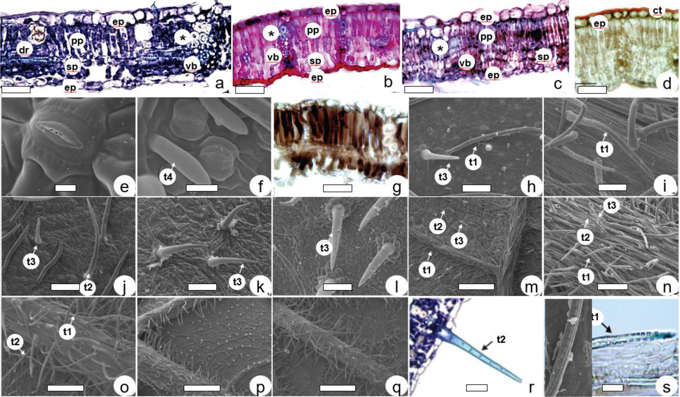
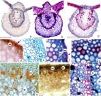
In cross-section, the lamina exhibits a single-layered epidermis in all three studied Guettarda species (Figures 1a, 1b, and 1c). The cuticle is thick on the adaxial surface and thin on the abaxial surface (Figure. 1d). Stomata are present on the abaxial surface (Figures 1e,1f, and 1g), classifying the laminae of all three species as hypostomatic. They are classified as paracytic or rubiaceous. Stomata are located at the same level as the epidermal cells in G. pohliana and G. uruguensis (Figures 1a,1b, 1e, and 1f). In G. viburnoides (Figures 1f, 1g), stomata project above the level of epidermal cells. The mesophyll exhibits a dorsiventral organization in all three species, with 1-2 layers of palisade parenchyma toward the adaxial surface and a reduced, poorly stratified spongy parenchyma on the abaxial side (Figures 1a-c). Phenolic compounds are detected in the mesophyll, especially in the palisade parenchyma region in all Guettarda species, as shown in G. viburnoides (Figure 1g). Along the mesophyll are small collateral vascular bundles surrounded by fibers with thick and non-lignified walls (Figures 1a-c). In G. pohliana, idioblasts containing druses are distributed in the palisade parenchyma (Figure 1a). All three species exhibited bundle sheath extensions.
Leaf anatomy of Guettarda species. G. pohliana (a, h, n, p, s), G. uruguensis (b, d, e, j, k, m), G. viburnoides (c, f, g, i, l, o, q, r). In cross-section (a-d, g, r, s). In frontal view (all others). Normal light microscopy (a-d, g, r, s). Scanning electron microscopy (e, f-q). Staining with toluidine blue (a, r), staining with safranin and astra blue (b, c), test with Sudan III (d), and with ferric chloride (g). Adaxial leaf surface (k, l), abaxial leaf surface (all others). Legends: ep: epidermis; pp: palisade parenchyma; sp: spongy parenchyma; dr: druse; *: sheath extension; vb: vascular bundle; (t1): type 1 trichome; (t2): type 2 trichome; (t3): type 3 trichome; (t4): type 4 trichome. Scale bars a, b, c, g, h, i, j, k, l, r, s = 50 µm; e = 5 µm; f = 10 µm; m, n = 100 µm; o = 200 µm; p, q = 500 µm.
The trichomes observed in Guettarda species exhibit four distinct morphological types. Type 1 trichomes are non-glandular (tector), apparently unicellular, long, and inclined on the surface, with thickened walls and internal crystals. They are present in all three species (Figures 1h, 1s). Type 2 trichomes are multicellular, erect, and long, with thinner walls that may present smooth or slightly ornamented surfaces; they also occur in all species (Figures 1j, 1r). Type 3 trichomes are smaller, non-glandular, erect, pointed, and thin-walled; they are present across all species analyzed (Figures 1k, 1l). Type 4 trichomes are apparently unicellular, inclined, with a rounded apex and thin walls, and were observed exclusively in G. viburnoides (Figure 1f). Trichomes occur on both adaxial and abaxial leaf surfaces, with a higher density on the abaxial surface (Figures 1m-1p); among the species analyzed, G. viburnoides exhibits the most dense indumentum (Figure 1q).
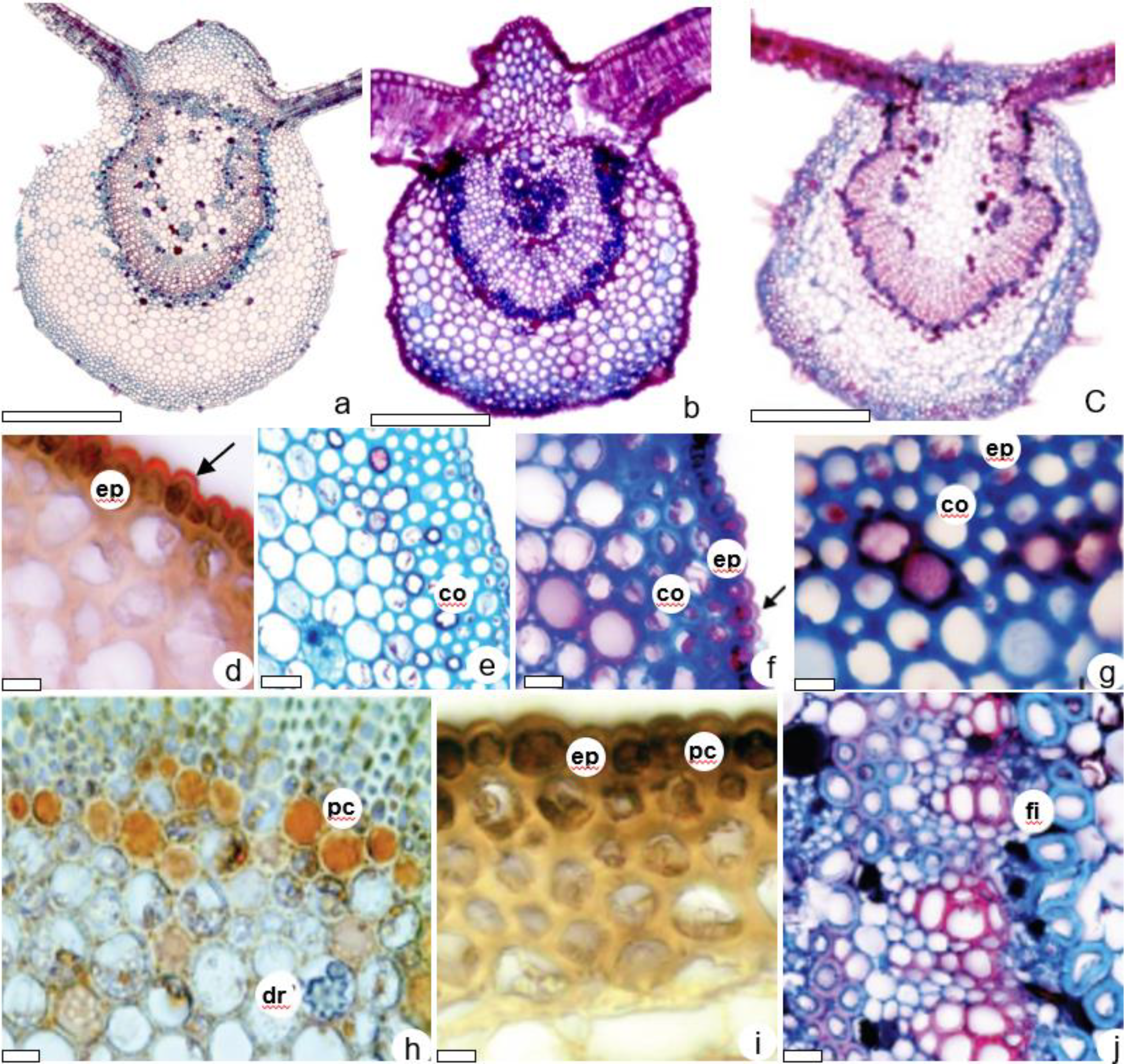
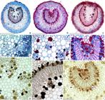
The midrib, in cross-section, shows different shapes. In G. pohliana (Figure 2a) and G. uruguensis (Figure 2b), the midrib is biconvex, with an adaxial surface prominent and tapered in G. uruguensis. G. viburnoides shows a plano-convex shape (Figure 2c). In G. pohliana (Figure 2d) and G. viburnoides (Figure 2f), the collenchyma is annular, whereas in G. uruguensis (Figure 2e) it is angular. The remaining fundamental tissue is parenchymatous with druses in all three species (Figure 2h). Phenolic compounds showed a positive reaction to the ferric chloride solution, observed in the midrib in the epidermis, cortex, and phloem (Figures 2h, 2i). The vascular system is represented by a horseshoe-shaped vascular bundle (Figures 2a, 2b, and 2c). Small vascular bundles occur in the medullary region, similar to G. pohliana (Figure 2a) and G. viburnoides (Figure 2c). In G. uruguensis, these bundles are condensed (Figure 2b). For G. pohliana, a vascular system surrounded by fibers was observed, with thick and non-lignified walls adjacent to the phloem (Figure 2j). All three species, G. pohliana, G. uruguensis, and G. viburnoides, exhibited druses.
Anatomy of Guettarda - leaf midrib in cross-section. G. pohliana (a, d, e, h, j), G. uruguensis (b, f, i). G. viburnoides (c, g). Scale bars a, b, c= 500 µm; e= 100 µm; d, f, g, h, i, j= 50 µm. [arrow - cuticle, ep - epidermis, co - collenchyma, dr - druse, fi - fibers, pc - phenolic compounds]
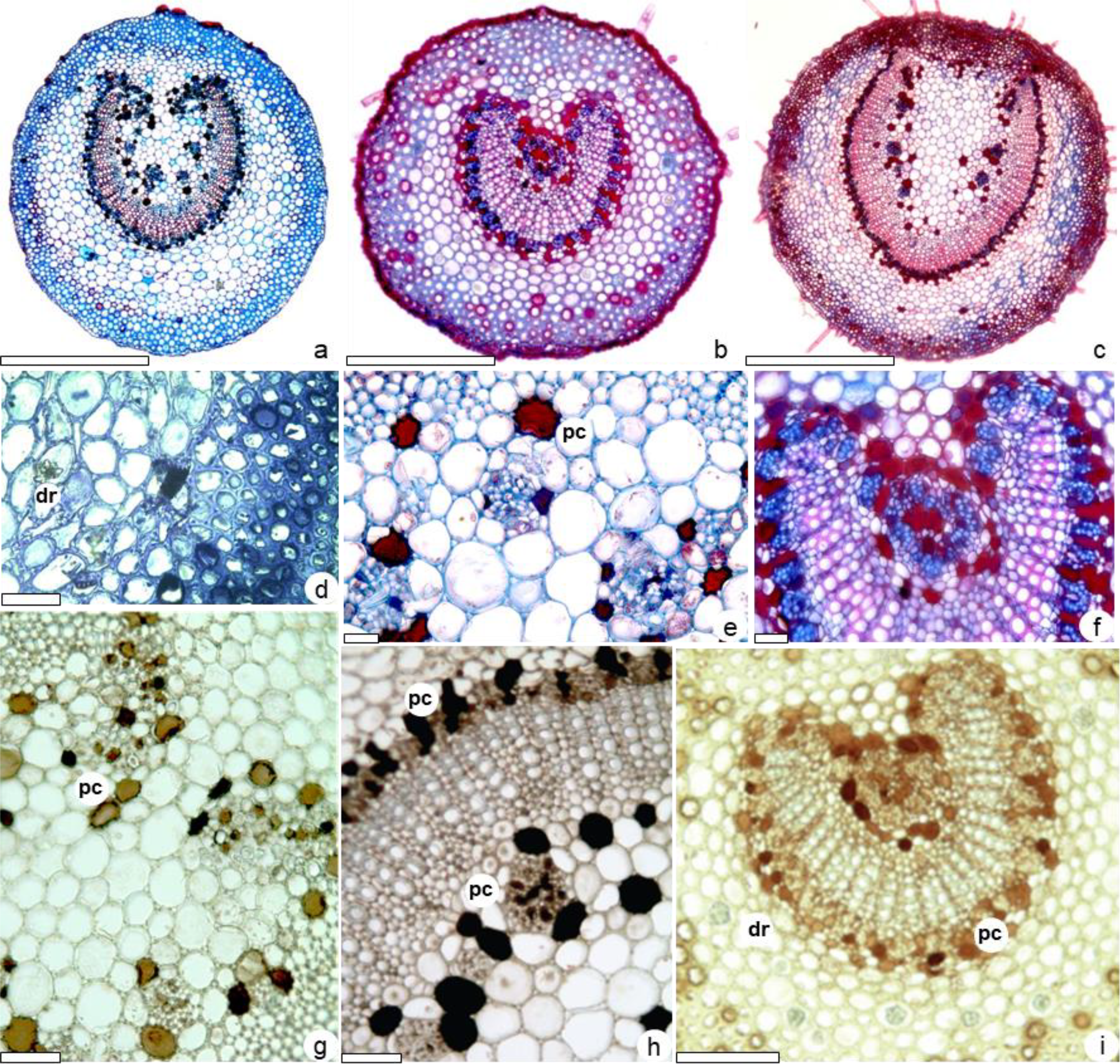
The petiole, in cross-section, has a rounded outline in all three species (Figures 3a, 3b, and 3c). The epidermis is single-layered, consisting of rounded cells in cross-section, with trichomes similar to those on the blade in all three species. Subepidermally, about five layers of angular collenchyma occur in G. uruguensis and G. viburnoides and a ring-like collenchyma in G. pohliana (Figure 3d,3f). The remaining fundamental tissue is parenchymatous, with druses in G. pohliana and G. uruguensis (Figure 3d). The vascular tissue consists of a horseshoe-shaped collateral vascular bundle, more open, with a broad pith in G. pohliana and G. viburnoides (Figure 3a,3c) and more closed in G. uruguensis (Figure 3b), with a poorly delimited pith. Small vascular bundles occur in the medullary region, similar to G. pohliana and G. viburnoides (Figure 3a, 3c, 3e); in G. uruguensis, they are condensed (Figure 3f). Phenolic compounds showed a positive reaction to the ferric chloride solution, observed in the petiole in the epidermis, cortex, and phloem (Figure 3e, 3g, 3h, 3i).
Anatomy of Guettarda - petiole in cross-section. G. pohliana (a, d, h, j), G. uruguensis (b, e, g, i). G. viburnoides (c, f). (e, g) Details of the medullary parenchyma showing phenolic compounds (pc). Scale bars a, b, c = 500 µm; d, e, f, g = 100 µm; h, i, j = 50 µm. dr - druse, pc - phenolic compound
DISCUSSION
The anatomical characteristics observed in G. pohliana, G. uruguensis, and G. viburnoides align with common traits reported for Rubiaceae species, particularly within the Guettarda genus. The presence of hypostomatic leaves with paracytic stomata, dorsiventral mesophyll, and both unicellular and multicellular covering trichomes reinforces their placement within the family [39,40,41].
One of the most notable findings in this study is the presence of bundle sheath extensions in all three species, supporting previous reports that suggested this characteristic as a potential taxonomic marker for Guettarda [10]. Our observations confirm that these structures, formed by fibers surrounding the vascular bundles, occur consistently across the studied species, strengthening their relevance for systematic classification.
Regarding epidermal structures, the differences in stomatal position provide distinguishing features among the species. While G. pohliana and G. uruguensis have stomata at the same level as the epidermal cells, G. viburnoides exhibits raised stomata [10]. This variation may be associated with adaptations to different microclimates, as suggested in other Rubiaceae species [36].
Trichome diversity is another key distinguishing factor among the species. The four trichome types identified in this study corroborate previous descriptions for G. viburnoides, with G. pohliana and G. uruguensis also exhibiting a similar range of structures. However, the exclusive presence of Type 4 trichomes in G. viburnoides, characterized by a rounded apex and thin walls, suggests a potential diagnostic trait for species identification. Trichomes containing prismatic crystals were previously described in Guettarda viburnoides [10,11], and their presence was confirmed in this study, especially in Type 1 trichomes. The detection of calcium oxalate crystals within certain trichomes reinforces their protective role, a feature commonly observed in Rubiaceae [13,12]. The presence of phenolic compounds and druses in the mesophyll and midrib of all three species aligns with previous phytochemical studies, which have reported the abundance of secondary metabolites in Guettarda [10,11]. The histochemical detection of phenolics in the epidermis, cortex, and phloem of the petiole further supports their role in defense mechanisms and suggests their importance in taxonomic differentiation. Cells containing phenolic compounds (positive for ferric chloride) and lipid content (positive for Sudan) were detected previously in Guettarda viburnoides [10,11]. This finding aligns with the established chemical profile of the Guettarda genus, which is known for its richness in secondary metabolites, including alkaloids, iridoids, triterpenes, phenolic acids, and flavonoids [42, 43, 44, 45]. In the Guettarda genus, phenolic compounds have been isolated. In G. pohliana, 5-caffeoylquinic acid, 4,5-dicaffeoylquinic acid [25,29], and shikimic acid were identified [20]. In G. uruguensis, quercetin was identified, along with long-chain esters of p-coumaric acid, including eicosanyl p-coumarate, triacontyl p-coumarate, and dotriacontyl p-coumarate [22]. G. viburnoides shows quercetin-3-O-β-D-galactopyranoside, quercetin-3-O-β-D-glucopyranoside, grandifloroside [23] and secoxiloganin [24]. The presence of these phytochemical groups highlights the importance of continued research within this genus.
In terms of vascular structure, the horseshoe-shaped vascular bundles found in the petiole of all species are consistent with descriptions for Rubiaceae [40]. The variation in vascular bundle organization between G. uruguensis (more condensed) and the other two species (more open, with a broader pith) may indicate ecological adaptations that warrant further investigation.
The anatomical traits described in this study, summarized in Table 1, provide valuable taxonomic markers for distinguishing Guettarda species. The combination of stomatal position, trichome types, vascular bundle arrangement, and bundle sheath extensions contributes to species differentiation. Furthermore, the detection of phenolic compounds and druses reinforces the importance of anatomical and histochemical data in taxonomic classification.
CONCLUSION
The anatomical characterization of G. pohliana and G. uruguensis, presented here for the first time, contributes to the structural knowledge of the genus Guettarda and provides descriptive data that may be useful for future investigations involving Brazilian Rubiaceae species. The findings indicate that G. pohliana, G. uruguensis, and G. viburnoides share common anatomical traits; however, distinct differences were observed. In summary, the three Guettarda species can be distinguished by specific anatomical traits. G. viburnoides exhibits raised stomata and exclusive Type 4 trichomes with the highest trichome density. G. uruguensis shows a tapered midrib, a narrow and poorly defined petiolar pith, and condensed vascular bundles. In contrast, G. pohliana presents a broad, well-delimited pith and a well-developed palisade parenchyma. These features contribute to the anatomical differentiation within the genus.
These results contribute to a deeper understanding of the taxonomic relationships and diversity within the Guettarda genus.
-
Funding:
This research was funded by Coordenação de Aperfeiçoamento de Pessoal de Nível Superior (CAPES), grant number 001.
Data Availability Statement:
Research data are available in the body of the manuscript.
Acknowledgments:
The authors thank the Municipal Botanical Museum of Curitiba for identifying the species and the Electron Microscopy Center at the Federal University of Paraná.
REFERENCES
- 1 Delprete PG, Jardim JG. Systematics, taxonomy and floristics of Brazilian Rubiaceae: an overview about the current status and future challenges. Rodriguésia. 2012;63:101-28.
- 2 Souza VC, Lorenzi H. [Systematic botany: illustrated guide for identifying the families of native and exotic phanerogams from Brazil, based on APG III]. 3a ed. Nova Odessa: Instituto Plantarum; 2012. 768 p.
- 3 Stehmann JR, Forzza RC, Salino A, Sobral M, Costa DP, Kamino LHY. [Plants of the Atlantic Forest]. Jardim Botânico do Rio de Janeiro; 2009. 515 p.
- 4 Delprete PG. Rubiaceae. In: Smith NP, editor. Flowering plant families of the American tropics. New York: Princeton University Press, New York Botanical Garden Press; 2004. p. 328-33.
- 5 Delprete PG. Rubiaceae - [Rubiaceae - Part 1: Introduction, Genera A-H]. In: Rizzo JA, coordinator. Flora dos estados de Goiás e Tocantins. Vol. 40. Goiânia: IRD/UFG, Universidade Federal de Goiás; 2010. p. 1-580.
- 6 Renda G, De Caterina R. Cafeine. In: Principles of Nutrigenetics and Nutrigenomics: Fundamentals of Individualized Nutrition; Elsevier: Amsterdam, The Netherlands, 2019; p. 335-40.
- 7 Heitzman ME, Neto CC, Winiarz E, Vaisberg AJ, Hammond GB. Ethnobotany, phytochemistry and pharmacology of Uncaria (Rubiaceae). Phytochemistry. 2005;66:5-29.
- 8 Solereder H. Systematic anatomy of the Dicotyledons. Oxford: Clarendon Press; 1908. 1182 p.
- 9 Metcalfe CR, Chalk L. Anatomy of the dicotyledons: leaves, stem, and wood in relation to taxonomy with notes on economic uses. Oxford: Clarendon; 1950.
- 10 Alexandrino CR, Moraes TMS, Cunha M. [Micromorphology and leaf anatomy of Rubiaceae species from Itatiaia National Park, RJ]. Floresta e Ambiente. 2011;18:275-88.
- 11 Oliveira PC Jr, Argandoña EJS, Faoro JAM, Silva RMMF, Konkiewitz EC, Ziff EB, Cabral MRP, Sarragiotto MH, Gauze GF, Rozada AMF, Monteiro LM, Manfron J, Formagio ASN. Guettarda viburnoides Cham. & Schltdl.: Neuroprotective Activities in Mice, Molecular Docking and Microchemical and Morphoanatomical Characteristics of Leaves and Stems. Curr Pharm Biotechnol. 2025;26:836-53.
- 12 Bremer B, Eriksson O. Time tree of Rubiaceae: phylogeny and dating the family, subfamily, and tribes. Int J Plant Sci. 2009;170:766-93.
- 13 Achille F, Motley TJ, Lowry II PP, Jérémie J. Polyphyly in Guettarda L. (Rubiaceae, Guettardeae) based on nrDNA ITS sequence data. Ann Missouri Bot Gard. 2006;93:103-21.
-
14 Jardim Botânico do Rio de Janeiro. Flora do Brasil 2020 em construção [internet]. Disponível em: http://floradobrasil.jbrj.gov.br/ Acesso em: 23 mar. 2025.
» http://floradobrasil.jbrj.gov.br/ -
15 Missouri Botanical Gardem. Tropicos.org. Disponível em: https://www.tropicos.org/ Acesso em: 23 mar. 2025.
» https://www.tropicos.org/ - 16 Stehlman JR, Forzza RC, Salino A, Sobral M,Costa DP, Kirizawa M. [Plants of the Atlantic Forest: identification guide]. Rio de Janeiro: Instituto de Pesquisas Jardim Botânico do Rio de Janeiro, 2009.
- 17 Agra MF, França PF, Barbosa-Filho JM. Synopsis of the plants known as medicinal and poisonous. Northeast Brazil. Rev Bras Farmacogn. 2007;17(1):114-40.
- 18 Agra MF, Silva KN, Basílio IJLD, França PF, Barbosa-Filho JM. Survey of medicinal plants used in the region Northeast of Brazil. Rev Bras Farmacogn. 2008;18(3):472-508.
- 19 Albuquerque AP, Monteiro JM, Ramos MA, Amorim ELC. Medicinal and magic plants from a public market in northeastern Brazil. J Ethnopharmacol. 2007;110(1):76-91.
- 20 Capasso A, Balderrama L, Sivila SC, De Tommasi N, Sorrentino L, Pizza C. Phytochemical and pharmacological studies of Guettarda acreana Planta Med. 1998;64(4):348-52.
- 21 Aquino R, De Simoe F, Pizza C, Cerri R, De Mello JF. Quinovic acid glycosides from Guettarda platypoda Phytochemistry. 1988;27(9):2927-30.
- 22 Duarte AFS, Soares DM, Dalarmi L, Krause MS, OCAMPOS FMM, Henneberg R, et al. Guettarda uruguensis (Rubiaceae): antioxidant and anti-inflammatory plant with quercetin and esters of p-coumaric acid. Nat Prod Res. 2024; (12):1-8.
- 23 Naressi MA, Manholer DD, Ames FQ, Bersani-Amado CA, Formagio ASN, Pereira ZV, et al. Chemical constituents, anti-inflammatory, and free-radical scavenging activities of Guettarda viburnoides Cham. & Schltdl. (Rubiaceae). Quim Nova. 2015;38(7):932-6.
- 24 Oliveira PC Jr, Argandoña EJS, Santos SM, Santos JM, Souza MF, Faoro JAM, Kassuuya CAL, Arena AC, Manoel BM, Silva RMM, Formagio ASN. Toxicological analysis and anti-inflammatory and antioxidant evaluations of extract, fractions and secoxyloganin obtained from Guettarda viburnoides Cham. & Schltdl. in mice. Journal of Ethnopharmacology. 2025;338(3):119090.
- 25 Testa G, Oliveira PRN, Silva CC, Schuquel ITA, Santin SMO. Constituintes químicos das folhas e avaliação anti-inflamatória de extratos das raízes e folhas de Guettarda pohliana Müll. Arg. (Rubiaceae). Quim Nova. 2012;35(3):527-9.
- 26 Duarte AFS, Szabo EM, Duarte IS, Merino FJZ, Oliveira VB, Miguel MD, Miguel OG. 2016. Evaluation of antioxidant capacity, phenolic content, antimicrobial and cytotoxic properties of Guettarda uruguensis flowers and fruits. Afr J Pharm Pharmacol. 10(47):1014-24
- 27 Duarte AFS, Hirota BCK, Oliveira VB, Campos R, Murakami FS, Miguel MD, Miguel OG. 2014 [Evaluation of antioxidant and antimicrobial activity of ethanolic extract and organic fractions from the stem bark of Guettarda uruguensis Cham. & Schltdl. (Rubiaceae)]. Rev Cienc Farm Basica Apl. 35(4):607-14.
- 28 Ramos, DF, Leitão GG, Costa FN, Abreu L, Villarreal JV, Leitão SG, Said y Fernández SL, Silva PEA. Investigation of the antimycobacterial activity of 36 plant extracts from the Brazilian Atlantic Forest. Rev Bras Cienc Farm, v. 44, n. 4, p. 669-74, 2008.
- 29 Oliveira PRN, Testa G, Sena SB, Costa WF, Sarragiotto MH, Santin MO. Chemical constituents of the roots of Guettarda pohliana Müll. Arg. (Rubiaceae). Quim Nova. 2008;31(4):755-8.
- 30 Kinupp VF. [Unconventional food plants from the metropolitan region of Porto Alegre, RS] [thesis]. Porto Alegre: Universidade Federal do Rio Grande do Sul; 2007. 336 p.
- 31 Lorenzi H. [Brazilian trees: Identification and cultivation manual of native tree species of Brazil]. Vol. 2. Nova Odessa: Ed. Plantarum; 1998. 352 p.
- 32 Lorenzi H. [Brazilian trees: Identification and cultivation manual of native tree species of Brazil]. Vol. 3. Nova Odessa: Ed. Plantarum; 2009. 384 p.
- 33 Johansen DA. Plant microtechnique. New York: McGraw-Hill; 1940.
- 34 O'Brien TP, Feder N, McCully ME. Polychromatic staining of plant cell walls by toluidine blue O. Protoplasma. 1964;59:368-73.
- 35 Roeser KR. Die Nadel der Schwarzkiefer-Massenprodukt und Kunstwerk der Natur. Mikrokosmos. 1972;61:33-6.
- 36 Kaiser E. Verfahren zur Herstellung einer tadellosen Glycerin-Gelatine. Bot Zentralb. 1880;25-6
- 37 Sass JE. Botanical microtechnique. 2nd ed. Ames: Iowa State College Press; 1951.
- 38 Souza W. [Basic techniques of electron microscopy applied to Biological Sciences]. Rio de Janeiro: Sociedade Brasileira de Microscopia Eletrônica; 1998.
- 39 Metcalfe CR, Chalk L. Anatomy of the dicotyledons: leaves, stem, and wood in relation to taxonomy with notes on economic uses. Oxford: Clarendon; 1950.
- 40 Bremekamp CEB. Remarks on the position, the delimitation, and the subdivision of the Rubiaceae. Acta Bot Neerl. 1966;15:1-33.
- 41 Solereder H. Systematic anatomy of the Dicotyledons. Oxford: Clarendon Press; 1908. 1182 p.
- 42 Kan-Fan C, Brillanceau MH, Husson HP. Plants of New Caledonia. 3 - Epi-antirhine, a new alkaloid from Guettarda heterosepala J Nat Prod. 1986;49(6):11302.
- 43 Brillanceau MH, Kan-Fan C, Kan SK, Husson HP. Guettardine, a possible biogenetic intermediate in the formation of Corynanthe-Cinchona alkaloids. Tetrahedron Lett. 1984;25(26):2767-70.
- 44 Ferrari F, Mesana I, Botta B, De Mello JF. Constituents of Guettarda platypoda J Nat Prod. 1986;49:1150-1.
- 45 Oliveira Junior PC, Argandoña EJS, Santos SM, Santos JM, Souza MF, Faoro JAM, Kassuuya CAL, Arena AC, Manoel BM, Silva RMM, Formagio ASN. Toxicological analysis and anti-inflammatory and antioxidant evaluations of extract, fractions and secoxyloganin obtained from Guettarda viburnoides Cham. & Schltdl. in mice. J Ethnopharmacol. 2025;338(3):119090.
-
Editor-in-Chief:
Paulo Vitor Farago
-
Associate Editor:
Jane Manfron