Molecular Mechanisms Underlying Sequence-Dependent Cytotoxicity of Cisplatin-Pemetrexed Therapy in A549 and BEAS-2B Cells
Abstract
Lung cancer continues to be a major contributor to cancer-related deaths globally. Antifolate agents and platinum-based medications form the basis of chemotherapy treatments. This study examined the distinct effects of pemetrexed disodium and cisplatin, both separately and sequentially, on cell cycle regulation in immortalized bronchial epithelial (BEAS-2B) and lung cancer (A549) cell lines. The MTT assay was used to measure cytotoxicity, and caspase-3, -7, and -9 activities were used to measure apoptotic activation. Quantitative real-time PCR (qRT-PCR) was used to analyze cell cycle gene expression profiles. Critically, while the sequential Cisplatin→Pemetrexed regimen maintained potent efficacy against A549 cells, it exhibited a distinct protective antagonistic interaction (CI>10) in non-malignant BEAS-2B cells, significantly mitigating cytotoxicity compared to cisplatin monotherapy. This treatment strategy significantly increased caspase-3, -7, and -9 activation in A549 cells. Gene expression analysis revealed downregulation of most cyclins, cyclin-dependent kinases, and DNA repair genes in A549 cells, whereas these genes were upregulated in BEAS-2B cells. These findings demonstrate preferential cytotoxicity toward lung cancer cells compared to immortalized bronchial epithelial cells, providing a molecular basis for optimizing combination chemotherapy strategies.
Keywords:
Antifolates; Platinum compounds; Cell cycle; Cytotoxicity; DNA repair.
HIGHLIGHTS
• Sequential cisplatin-pemetrexed shows higher cytotoxicity in lung cancer vs BEAS-2B.
• Combination therapy triggers strong apoptosis in A549, not BEAS-2B.
• Selective toxicity: DNA repair/cell cycle collapse in A549 vs upregulation in BEAS-2B .
INTRODUCTION
Lung cancer is a leading cause of cancer-related morbidity and mortality, representing a major global health concern. According to GLOBOCAN 2022 data [1], approximately 2.5 million new cases are diagnosed annually. Its poor prognosis is primarily due to aggressive disease progression, late-stage diagnosis, and limited effective treatments for advanced disease [2-5]. These statistics highlight the urgent need for improved screening, early detection, and novel therapies to increase survival rates [4, 6].
Currently available treatments for lung cancer are immunotherapy, targeted therapies, radiotherapy, chemotherapy, and surgery. The tumor location, disease stage, and patient's condition all affect treatment selection [7-9]. Chemotherapeutic agents, such as taxanes, nucleoside analogues, vinca alkaloids, topoisomerase inhibitors, and platinum derivatives, remain essential components of treatment regimens [10]. Conventional chemotherapy, however, has a number of drawbacks, such as high toxicity, poor effectiveness, and the emergence of drug resistance [11-15].
Drug resistance mechanisms often involve complex cellular defense pathways, such as enhanced DNA repair capacity, suppression of apoptosis, and dysregulation of cell cycle checkpoints [15, 16]. Since many chemotherapeutic agents exert their cytotoxicity by targeting dividing cells, the regulation of the cell cycle and its molecular components-including cyclins and cyclin-dependent kinases (CDKs)-is critical for therapeutic efficacy [17-19]. Therefore, identifying novel strategies to overcome these resistance mechanisms is essential for improving clinical outcomes [20, 21].
Cisplatin, a potent platinum-based chemotherapeutic agent, has been extensively utilized in oncology since its clinical introduction [22]. It exerts cytotoxic effects by forming DNA adducts and intrastrand crosslinks, which inhibit DNA replication and transcription, ultimately triggering apoptotic signaling pathways [23, 24]. However, its clinical utility is often hampered by severe adverse effects on normal tissues, necessitating combination strategies to enhance efficacy while minimizing toxicity [25, 26].
Pemetrexed disodium is a multi-targeted antifolate agent. It inhibits key enzymes in folate metabolism, thereby disrupting purine and pyrimidine biosynthesis. This disruption leads to S-phase arrest and then apoptosis [27-30]. Pemetrexed disodium has shown enhanced efficacy when combined with other agents, including those containing platinum derivatives [31, 32]. However, interactions between these drugs are complex. The specific sequence of administration can significantly alter the biological response. Although combination strategies are widely employed in clinical settings, treatment schedules are frequently determined empirically, and the precise molecular rationale for optimal drug sequencing remains limited. Optimizing the order of administration may widen the therapeutic window by boosting cytotoxicity in malignant cells while sparing non-malignant tissues.
Sequential chemotherapy regimens are designed to maximize dose intensity while minimizing overlapping toxicities and different toxic profiles [33]. The combination of cisplatin and pemetrexed disodium is rationalized by their distinct cytotoxic mechanisms and lack of cross-resistance [33, 34]. Clinically, this combination has demonstrated manageable safety and promising activity in refractory settings [34]. Mechanistically, sequential scheduling may exploit cell-cycle dynamics [33] and trigger autophagy through the regulation of AMPK/mTOR and PI3K/AKT/mTOR signaling pathways [35]. This approach seeks to potentially broaden the therapeutic window by leveraging protective interactions in non-malignant cells while maintaining high cytotoxicity in malignant models [33, 35].
This study evaluates the cytotoxic effects of cisplatin and pemetrexed disodium, given separately and sequentially, on A549 lung cancer and BEAS-2B bronchial epithelial cells. We used BEAS-2B as a surrogate for non-malignant bronchial epithelium to identify a treatment schedule with preferential toxicity to cancer cells. We examined how treatment schedules affect genes involved in cell cycle progression, apoptosis, and DNA repair. Our goal is to clarify the molecular basis of sequence-dependent selectivity to help optimize chemotherapy regimens with better tolerability.
MATERIAL AND METHODS
Chemicals
Cisplatin and pemetrexed disodium (Pemtrex) were obtained from Koçak Farma Pharmaceuticals (Türkiye). Cisplatin was dissolved in 0.9% saline, stored at 4 °C in the dark, freshly diluted before use to avoid degradation. Pemetrexed disodium was dissolved in sterile water, aliquoted and stored at -20 °C. Working concentrations were prepared from 1 mM stock solutions: cisplatin (5, 10, 20, 40, 80, and 160 μM) and pemetrexed disodium (0.125, 0.25, 0.5, 1, 2, 4, 8, and 16 μM).
Cell Culture
Human lung adenocarcinoma A549 (ATCC, CCL-185) cells and human non-malignant bronchial epithelial BEAS-2B (ATCC, CRL-3588) cells were generously provided by Prof. Dr. Engin Ulukaya from Istinye University. Both cell lines were grown as monolayer cultures in RPMI-1640 medium (Gibco, Cat. No. 11875093), enriched with 10% fetal bovine serum (FBS, Gibco, Cat. No. 10270106), and supplemented with penicillin (100 IU/mL) and streptomycin (100 μg/mL) (İ.E. Ulagay). Cells were grown at 37°C in a humidified incubator with 5% CO₂.
Cell Viability Assay
Cell viability was assessed via MTT assay. Cells were seeded at a density of 3 × 10⁴ cells per well in 96-well plates and allowed to adhere for 24 hours. Subsequently, cells were treated with cisplatin and pemetrexed, either individually, simultaneously (1:1) or in sequential combination (24-hour interval), at the specified concentrations. Untreated cells, cultured in complete medium without the addition of therapeutic agents, served as the control group. Following treatment periods of 24, 48, or 72 hours, 0.5 mg/mL MTT solution was added to each well and incubated for 4 hours at 37°C. The formazan crystals were dissolved in DMSO (Merck, Cat. No. D2650). Absorbance was measured at 570 nm using a microplate reader (BioTek ELx800) with a reference wavelength of 690 nm. Cell viability was expressed as a percentage of the untreated control, determined by calculating the ratio of the absorbance of treated samples to that of the control group where the viability of the control group was defined as 100% [36].
Caspase Activity Analysis
Caspase activation was detected using the CaspaTag™ Caspase-3,7 In Situ Assay Kit (Fluorescein, Cat. No. APT403, Millipore) and CaspaTag™ Caspase-9 In Situ Assay Kit (Fluorescein, Cat. No. APT409, Millipore) according to the manufacturer's protocols [37].
Gene Expression Analysis via qRT-PCR
Total RNA was extracted from A549 and BEAS-2B cell lines utilizing the PureLink RNA Mini Kit in accordance with the manufacturer’s protocol (Ambion, Cat. No. 12183-018A). cDNA synthesis was performed using 500 ng of total RNA with the High-Capacity RNA-to-cDNA Kit (Applied Biosystems, Cat. No. 4387406). Gene expression analysis was conducted on a StepOnePlus Real-Time PCR System (Applied Biosystems) by Power SYBR Green PCR Master Mix (Applied Biosystems, Cat. No. 4368706). GAPDH and β-actin served as reference genes for normalization. Relative gene expression levels were calculated using the 2-∆∆Ct method. The primer sequences for the genes analyzed are provided as supplementary data [38].
Statistical Analysis
All experiments were performed with a minimum of three independent biological replicates. For cell viability assays, each biological replicate comprised at least ten technical replicates per condition to ensure precision in dose-response modeling. For caspase activity and gene expression analyses, each biological replicate was performed in duplicate.
To evaluate cell viability, dose-response curves were generated in GraphPad Prism (v10.6) using a four-parameter logistic (4PL) non-linear regression. IC50 values and corresponding 95% CIs were calculated via the Python (v3.9) SciPy library, ensuring model reliability through R2 analysis. Comparative statistical analysis was performed using two-way ANOVA with Tukey’s post-hoc test for multiple comparisons. All results are presented as mean±SD of independent biological replicates. Furthermore, effect sizes for ANOVA components were reported as eta-squared (η2), categorized as small (0.01), medium (0.06), or large (≥0.14) according to Cohen [39].
Drug interactions between cisplatin and pemetrexed were evaluated using the Combination Index (CI) method described by Chou and Talalay [40]. The dose-effect parameters for each single agent were determined from the median-effect plot. Based on these parameters, CI values were calculated for simultaneous and sequential combinations using CalcuSyn software (Version 2.1). A CI value less than, equal to, or greater than 1 indicates synergism, additivity, and antagonism, respectively.
All statistical analyses for gene expression were performed using IBM SPSS Statistics v21.0. Data distribution and normality were evaluated using the Shapiro-Wilk test. For comparisons between independent groups, the independent samples t-test was applied, whereas comparisons of paired/matched measurements were analyzed using the paired samples t-test.
For all analyses, tests were two-tailed, and a p-value < 0.05 was considered statistically significant.
RESULTS
Dose-Response Characterization and IC50 Determination of Single Agents
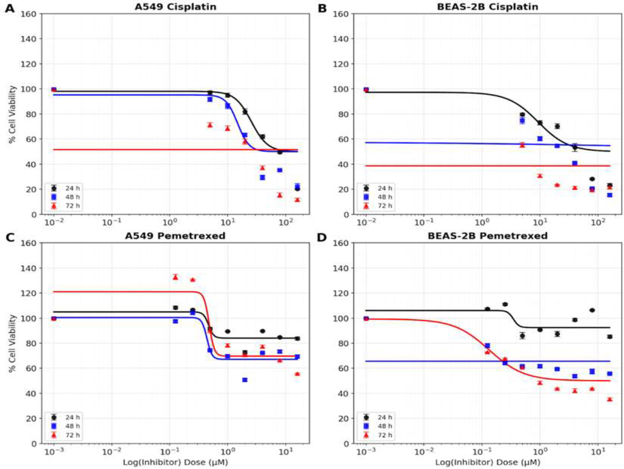
The cytotoxic potencies and time-dependent sensitivities of cisplatin (Cis) and pemetrexed disodium (Pem) were evaluated in A549 and BEAS-2B cells using the MTT assay across 24, 48, and 72-hour periods (Figure 1). To precisely quantify these effects, cell viability data were analyzed using a 4PL non-linear regression model. The determined IC50 values and 95% CIs are summarized in Figure 2 and Table 1.

Time-dependent viability of A549 and BEAS-2B cells following Cis and Pem administration for 24, 48, and 72 hours following treatment. (A) A549 - (B) BEAS-2B cells treated with Cis, (C) A549 - (D) BEAS-2B cells treated with Pem (*p<0.05, **p<0.01, ***p<0.001, ****p<0.0001).

Timeand dose-dependent cytotoxicity of Cis and Pem in A549 and BEAS-2B cell viability after 24, 48, and 72 h of treatment with Cis (0-160 µM; A, B) or Pem (0-16 µM; C, D). Curves represent 4PL non-linear regression fits.
Cisplatin showed distinct temporal patterns. A549 cells were resistant at 24 h, with a calculated IC50 of 77.70 µM. However, extending the exposure to 48 h, reduced the IC50 to approximately 19.96 µM; a modest increase was observed at 72 h (28.55 µM), suggesting reduced sensitivity. In contrast, non-malignant BEAS-2B cells exhibited higher intrinsic sensitivity, with an IC50 was 52.98 µM, slightly lower than that of A549 cells. Notably, prolonged exposure (72 h) resulted in a profound increase in cytotoxicity, with the IC50 dropping to 4.50 µM. These findings indicate that while both cell types are susceptible to Cis treatment, the non-malignant bronchial epithelium model demonstrates significant time-dependent sensitivity, particularly under chronic exposure conditions.
Pemetrexed exhibited a limited cytotoxicity within the tested concentration range in A549 cells, with maximal inhibition remaining insufficient to determine a precise IC50 (>16.0 µM) across all time points. The shallow dose-response slopes and non-monotonic fluctuations observed at 24h and 72h reflect the complex, often non-linear pharmacodynamics typical of antifolates in certain malignant models. In contrast, BEAS-2B cells displayed a distinct, time-dependent sensitivity shift. While showing tolerance at 24h and 48h (IC50>16.0 µM), these non-malignant cells became highly susceptible by 72h, reaching a potent IC50 of 0.21 µM. This late-onset cytotoxic transition, characterized by a steep response curve and narrow 95% confidence intervals, suggests that prolonged Pem exposure significantly impacts immortalized bronchial epithelial viability. Such a delayed but robust response in non-malignant cells may narrow the therapeutic window, highlighting a potential risk for cumulative toxicity in monotherapy regimens.
These findings demonstrate distinct sensitivity patterns between cancerous A549 cells and non-malignant BEAS-2B cells. The specific IC50 values obtained from the non-linear model supported the dose selection in subsequent combination studies, particularly emphasizing the notable intrinsic sensitivity of BEAS-2B cells to Cis.
Evaluation of Simultaneous and Sequential Combinations
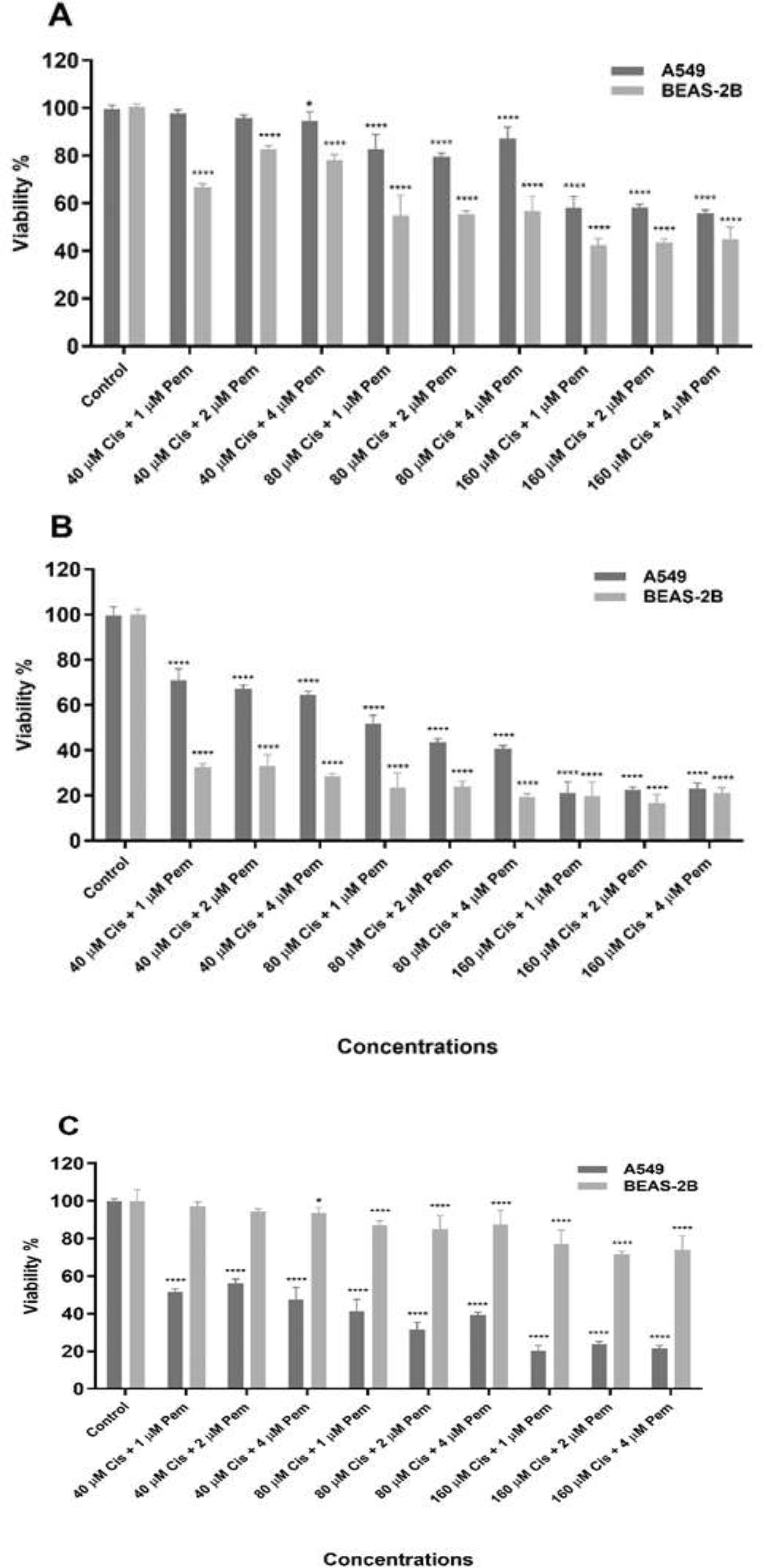
To determine whether the concurrent exposure to both drugs enhances cytotoxicity in a time-dependent manner, A549 and BEAS-2B cells were treated simultaneously with increasing concentrations of cisplatin and pemetrexed for 24, 48, and 72 h (Figure 3 A-C). Contrary to the anticipated therapeutic benefit, the simultaneous administration failed to potentiate the efficacy in A549 cells across all incubation periods. Instead, antagonistic interactions were consistently observed, particularly at 48 and 72 h, where cell viability remained significantly higher compared to cisplatin monotherapy. Conversely, in non-malignant BEAS-2B cells, the simultaneous regimen resulted in additive cytotoxicity in a time-dependent manner, leading to a further reduction in cell viability compared to single-agent treatments. This observation indicates that simultaneous exposure does not provide a favorable toxicity profile; rather, it exhibits a lack of differential sensitivity between the malignant and non-malignant cell lines across the tested time points. Consequently, these findings suggested that simultaneous administration might not be an optimal therapeutic strategy for limiting cytotoxicity in non-malignant bronchial epithelial cells.

Time-dependent cytotoxic effects of simultaneous Cis and Pem administration on A549 and BEAS-2B cells. Cells were co-treated with the indicated concentrations for (A) 24 h, (B) 48 h, and (C) 72 h. The simultaneous regimen exhibited antagonistic effects in A549 cells but additive cytotoxicity in BEAS-2B cells across the tested time points (*p<0.05, **p<0.01, ***p<0.001, ****p<0.0001).
Given these findings, we investigated sequential protocols with a 24-h interval. The sequence of administration significantly influenced the cytotoxic outcomes. The Pem→Cis sequence (Figure 4B) induced potent cytotoxicity in A549 cells (CI<1); however, it also caused substantial viability loss in BEAS-2B cells (>60% inhibition), indicating a similar sensitivity pattern in both cell lines for this specific schedule.

Comparative cytotoxicity of sequential drug treatments in A549 and BEAS-2B cells. (A) Cells were treated with Cis (24 h) followed by Pem (24 h) (Cis→Pem). This treatment resulted in significantly reduced cytotoxicity in BEAS-2B cells, while maintaining high inhibitory effects in A549 cells. (B) Cells were treated with Pem (24 h) followed by cisplatin (24 h) (Pem→Cis). This sequence resulted in high cytotoxicity in both cell lines (*p<0.05, **p<0.01, ***p<0.001, ****p<0.0001).
In contrast, the Cis→Pem sequence (Figure 4A) demonstrated a differential cytotoxic profile. While this sequence maintained effective inhibitory activity in A549 cells (71% inhibition at 160 µM cisplatin + 1 µM pemetrexed), it resulted in remarkably lower cytotoxicity in BEAS-2B cells compared to Cis monotherapy. Specifically, the inhibition rate in BEAS-2B cells dropped from 76% (Cis alone) to approximately 8% (combination). This observation suggests that the Cis→Pem sequence, particularly at the 160 µM Cis + 1 µM
Pem combination, exhibits a distinct toxicity profile where BEAS-2B cells show significantly higher tolerance compared to A549 cells. Therefore, this specific regimen was selected for further molecular mechanistic studies to investigate the underlying basis of this differential response.
To determine the nature of the interaction between Cis+Pem treatment, Combination Index (CI) were calculated based on the Chou-Talalay median-effect principle. In this analysis, CI<1, CI=1, and CI>1 indicate synergism, additive effect, and antagonism, respectively.
In A549 cells Pem→Cis sequence showed synergism at lower doses, while the Cis→Pem regimen (160 µM Cis + 1 µM Pem) displayed moderate antagonism (CI>1) yet maintained high cytotoxic efficacy (Table 2). Critically, the analysis in BEAS-2B cells revealed a distinct interaction pattern compared to cancer cells (Table 3). The Pem→Cis schedule showed additive effects, indicating a lack of differential sensitivity between the cell lines. In contrast, the Cis→Pem regimen (160 µM Cis + 1 µM Pem) exhibited strong antagonism (CI>10). This pronounced antagonistic interaction in the non-malignant model significantly reduced cytotoxicity compared to cisplatin monotherapy, suggesting a more favorable safety profile for this specific sequence.
To evaluate differential cytotoxicity between A549 and BEAS-2B cells, effect sizes were calculated for each two-way ANOVA. The Cis→Pem sequence exhibited the most pronounced differential response, with cell line identity as the dominant source of variation (η2=0.595, p<0.0001, 95% CI: 48.85-50.42%), indicating that malignant and non-malignant cells responded in a fundamentally distinct manner to this sequence.
Conversely, the Pem→Cis sequence showed negligible differential sensitivity; cell line identity accounted for only 1.0% of the total variation (η2=0.010, p<0.0001, 95% CI: 3.27-5.13%). In this regimen, dose was the primary driver of cytotoxicity (η2=0.775), suggesting a non-selective profile.
Simultaneous co-treatment revealed a time-dependent shift. At 24h and 48h, dose remained the primary determinant of viability (η2=0.721), despite a moderate cell line effect. By 72h, however, cell line identity became dominant (η2=0.616) driven by a reversal in sensitivity where BEAS-2B cells showed greater viability loss than A549 cells. These quantitative findings confirm that the Cis→Pem sequence uniquely maximizes selective cytotoxicity against malignant cells.
Caspase Activation Analysis
To evaluate apoptotic induction, caspase-3, -7, and -9 activities were measured following combination treatment (Figure 5). In A549 cells, all treatment groups showed increased caspase activation compared to controls. In contrast, BEAS-2B cells treated with 40-80 μM Cis followed by 1-4 μM Pem showed a decrease in all measured caspases relative to controls. Following 160 μM Cis treatment, caspase-3 and -7 activities increased in BEAS-2B cells, while caspase-9 activity decreased when combined with 1 or 4 μM Pem.

Caspase activity following Cis→Pem treatment. (A) Caspase-3, -7, and (B) Caspase-9 activities were measured. A549 cells show significantly higher activation than BEAS-2B cells under most conditions (*p<0.05).
The differential caspase activation patterns between A549 and BEAS-2B cells indicate selective induction of apoptosis in cancer cells, while non-malignant cells exhibit reduced apoptotic signaling under most treatment conditions except high-dose cisplatin.
Transcriptional Profiling of Cell Cycle and DNA Repair Genes
Building upon the dose-response curves, the 160 µM Cis concentration was utilized for molecular analyses. Interestingly, while this concentration appeared to impose a lethal threshold for the non-malignant BEAS-2B line, the sequential addition of 1 µM Pem seemed to confer a notable survival advantage, in contrast to the sustained toxicity in A549 cells. Consequently, the 160 µM Cis→1 µM Pem regimen was selected for molecular analyses to explore whether this differential response might be driven by the modulation of cell cycle progression. Mechanistically, the Cis+Pem regimen is suggested to impose convergent stresses: cisplatin-induced DNA adducts engaging DDR signaling and pemetrexed-mediated replication stress. A canonical response could involve the attenuation of G1/S drive, induction of CKIs, and remodeling of S-phase/G2 regulators. The present dataset suggests that both the baseline transcriptional landscape and the directionality of the Cis+Pem response may be strongly cell-line dependent, reflecting distinct stress-handling programs in malignant versus non-malignant cells.
Comparative Analysis of Baseline Gene Expression in A549 and BEAS-2B Cell Lines
The initial phase of the study involved characterizing the basal transcriptional landscapes of the A549 and BEAS-2B. The results suggest a significant dysregulation of cell cycle control mechanisms and DNA repair systems in A549 cells. Notably, the expression of CCND1, which typically facilitates the G1/S phase transition, was 27.91-fold higher in A549 cells than in BEAS-2B (p<0.0001; Figure 6A), followed by a marked upregulation of the cell cycle inhibitor CDKN1B (8.72-fold, p<0.0001; Figure 6B). This was accompanied by a marked upregulation of the Cyclin D-dependent kinases CDK4 (2.52-fold, p=0.002) and CDK6 (4.03-fold, p=0.020), collectively supporting a stronger baseline engagement of the G1 pathway in the malignant line (Figure 6E, 6F).

Comparative basal gene expression profiles of A549 and BEAS-2B cell lines. Relative mRNA expression levels of key regulators involved in cell cycle progression and DNA damage response (DDR) were determined by qPCR. (A) CCND1, (B) CDKN1B, (C) CDKN1A, (D) CDK2, (E) CDK4, (F) CDK6, (G) CCNB1, (H) CCNA1, (I) CCNA2, (J) CDK1, (K) CDKN1C, (L) XPA, (M) ATR, and (N) PARP1. Statistical significance is indicated as follows: *p < 0.05, **p < 0.01, ***p < 0.001, and ****p < 0.0001. 'ns' indicates non-significant differences (p > 0.05).
Furthermore, the elevated basal levels of XPA (10.34-fold, p=0.002) could indicate that these cancer cells maintain an enhanced adaptive capacity to counteract genomic instability, potentially contributing to their survival (Figure 6L). In contrast, PARP1 levels were found to be significantly lower in A549 cells (0.34-fold, p=0.004; Figure 6N). Interestingly, CDK2 expression was markedly reduced relative to BEAS-2B (0.24-fold, p<0.0001; Figure 6D), suggesting that baseline S-phase regulation differs between the two cell lines and that A549 may rely more prominently on the Cyclin D-CDK4/6 axis rather than on CDK2-centered control.
Several other targets exhibited borderline or non-significant baseline differences, including CDKN1A, CCNA1, CCNA2, CDKN1C, CDK1, and ATR (Figure 6C, 6H, 6I, 6K, 6J and 6M). These findings point toward a gene-specific rather than a uniform baseline divergence between the A549 and BEAS-2B cells.
Impact of Cis + Pem Combination on A549 Cancer Cells
Cis+Pem treatment in A549 cells, relative to untreated controls, exerted a profound inhibitory effect on cell cycle-related genes, inducing a coordinated pattern of attenuated progression across multiple checkpoints. Specifically, the G1/S-associated driver CCND1 (Figure 7A) was significantly decreased (0.68-fold, p=0.015), and its partner CDK6 was strongly suppressed (0.18-fold, p=0.029; Figure 7F), while CDK4 exhibited a non-significant downward trend (0.66-fold, p=0.066; Figure 7E).

Transcriptional response of A549 cells to Cis and Pem combination treatment compared to untreated (UT) controls. (A) CCND1, (B) CDKN1B, (C) CDKN1A, (D) CDK2, (E) CDK4, (F) CDK6, (G) CCNB1, (H) CCNA1, (I) CCNA2, (J) CDK1, (K) CDKN1C, (L) XPA, (M) ATR, and (N) PARP1. Statistical significance is indicated as follows: *p < 0.05, **p < 0.01, ***p < 0.001, and ****p < 0.0001. 'ns' indicates non-significant differences (p > 0.05).
Beyond the G1 phase, A549 cells exhibited pronounced suppression of S/G2 and G2/M modules, including CCNA1 (0.09-fold, p=0.001), CCNA2 (0.06-fold, p=0.002), CCNB1 (0.02-fold, p=0.003), and CDK1 (0.16-fold, p=0.033) (Figure 7H, 7I, 7G, 7J respectively). Collectively, these observations indicate a potent transcriptional brake on late-cycle execution, likely leading to G2/M phase arrest.
The marked induction of the DDR effector CDKN1A (3.63-fold, p<0.0001; Figure 7C) suggests that the observed arrest is likely mediated through a p21-dependent pathway in response to DNA damage and replication stress. Conversely, CDKN1B (0.15-fold, p<0.0001; Figure 7B) and CDKN1C (0.49-fold, p=0.035; Figure 7K) significantly declined, indicating that not all inhibitors are co-induced. Interestingly, CDK2 expression increased (2.63-fold, p=0.003; Figure 7D), potentially reflecting compensatory rewiring under therapy-induced stress.
A notable feature was the downshift in DDR/repair transcripts. Despite cisplatin’s known effects, A549 cells showed a profound and significant reduction in XPA (0.04-fold, p=0.004) and PARP1 (0.28-fold, p=0.011; Figure 7L, 7N). ATR levels also decreased, though the change did not reach statistical significance (0.40-fold, p=0.111; Figure 7M).
Taken together, these data suggest that the Cis+Pem regimen in cancer cells is characterized by broad cell-cycle suppression and a concurrent reduction in DNA repair transcripts. This pattern aligns with a decompensated response, potentially increasing therapeutic vulnerability at the analyzed time point.
Effects on BEAS-2B Normal Cells and Observations on Selective Toxicity
Unlike A549 cells, BEAS-2B cells exhibited a robust inductive transcriptional response to the Cis+Pem combination. Following treatment, CDKN1A expression was significantly induced by 17.63-fold (p=0.001; Figure 8C). This was accompanied by other important checkpoint signals, including CDKN1B (4.21-fold, p=0.024) and CDKN1C (10.85-fold, p<0.001) (Figure 8B and 8K). The simultaneous upregulation of these cyclin-dependent kinase inhibitors (CKIs) potentially indicates a compensatory mechanism for the inactivated p53/Rb axis in the immortalized BEAS-2B model, possibly through p53-independent stress-response pathways.

Transcriptional response of BEAS-2B cells to Cis and Pem combination treatment compared to untreated (UT) controls. (A) CCND1, (B) CDKN1B, (C) CDKN1A, (D) CDK2, (E) CDK4, (F) CDK6, (G) CCNB1, (H) CCNA1, (I) CCNA2, (J) CDK1, (K) CDKN1C, (L) XPA, (M) ATR, and (N) PARP1. Statistical significance is indicated as follows: *p < 0.05, **p < 0.01, ***p < 0.001, and ****p < 0.0001. 'ns' indicates non-significant differences (p > 0.05).
This checkpoint activation co-occurred with the upregulation of cell-cycle drivers, including G1-associated genes such as CCND1 (5.66-fold, p=0.015; Figure 8A), CDK2 (3.08-fold, p=0.021; Figure 8D), and CDK6 (4.13-fold, p=0.007; Figure 8F), while CDK4 showed a borderline upward trend (2.18-fold, p=0.092; Figure 8E). Furthermore, strong increases were noted in S/G2 and G2/M markers, specifically CCNA1 (21.86-fold, p=0.004), CCNA2 (10.27-fold, p=0.005), CCNB1 (10.59-fold, p=0.004), and CDK1 (7.81-fold, p=0.001) (Figure 8H, 8I, 8G and 8J). Rather than definitively indicating active proliferation, this broad transcriptional surge in both drivers and inhibitors likely reflects a regulated stress-adaptation program. In this context, transcriptional rebalancing and strategic checkpoint activation might serve as survival mechanisms for BEAS-2B cells under the pressure of platinum-induced stress.
In BEAS-2B cells, the NER component XPA increased 9.19-fold, indicating the activation of DNA adduct repair (Figure 8L). While PARP1 decreased (0.56-fold, p=0.031; Figure 8N), ATR (1.65-fold, p=0.081; Figure 8M) showed upward trends that did not reach statistical significance.
Collectively, Cis+Pem triggers a coordinated compensatory stress response in BEAS-2B cells. Unlike the suppression in A549 cells, this non-malignant context prioritizes checkpoint signaling and repair engagement, effectively mitigating treatment-induced damage.
Integrated Interpretation of Basal Differences and Differential Therapy Responses
As summarized in Table 4, the integration of baseline expression profiles with the responses to Cis+Pem treatment revealed distinct transcriptional trajectories for each cell line. At baseline, A549 cells demonstrated an enrichment of the Cyclin D pathway (CCND1, CDK4/6) and selected DDR/repair features (XPA) compared to BEAS-2B cells. However, following Cis+Pem exposure, A549 cells exhibited a widespread downregulation of cell-cycle executors (CCND1, CDK6, CCNA1/2, CCNB1, CDK1), accompanied by a simultaneous reduction in key DDR/repair transcripts, including PARP1 and XPA. In contrast, BEAS-2B cells responded to the same regimen with a predominant upregulation across hem cell-cycle modules and repair components, most notably XPA. This divergent response suggests that while malignant cells undergo a transcriptional collapse of survival and repair machinery, the non-malignant model initiates a protective stress-adaptation program. Furthermore, the robust induction of p21 observed in BEAS-2B cells, despite the absence of an intact p53/Rb axis due to immortalization, points toward the activation of alternative p53-independent pathways that may facilitate this in-vitro selectivity. These distinct molecular directionalities underscore the sequence-dependent efficacy of the combined therapy in targeting cancer cells while maintaining viability in the bronchial epithelial model.
Relative mRNA expression levels of cell cycle and DNA repair genes. Data are presented as fold change relative to the respective untreated (UT) controls, calculated using the 2-∆∆Ct method. The A549 vs BEAS-2B column indicates the differential expression ratio between the two cell lines under treatment conditions.
DISCUSSION
This study provides preliminary insights into the differential responses of lung cancer and non-malignant bronchial epithelial cells to sequential Cis-Pem therapy. The identification of distinct gene expression patterns between A549 and BEAS-2B cells suggests substantial progress in understanding how malignant cells might be selectively targeted while potentially maintaining non-malignant cell viability.
Our viability data appear to confirm previous studies demonstrating concentration-dependent cytotoxicity of cisplatin and pemetrexed in various cell lines [29, 41, 42]. The increased resistance of BEAS-2B cells to combination treatment, even at high concentrations, supports the therapeutic potential of this regimen.
Furthermore, the observed increase in caspase-3, -7, and -9 activation in A549 cells is consistent with established literature on drug-induced apoptosis [16, 26, 41, 42], whereas the differential activation patterns may indicate a more selective apoptotic induction in the malignant model.
An unexpected observation in the present study was the relatively high sensitivity of BEAS-2B cells to pemetrexed compared with A549 cells. Although BEAS-2B represents a non-tumorigenic bronchial epithelial model, its immortalization with SV40 large T antigen significantly alters cell-cycle regulation and proliferative behavior. Pemetrexed acts primarily as a multitargeted antifolate that inhibits thymidylate synthase and other folate-dependent enzymes involved in nucleotide biosynthesis. Cells with high proliferative activity and active DNA synthesis pathways are therefore particularly susceptible to antifolate agents. The increased responsiveness of BEAS-2B cells may reflect their immortalized phenotype, which can promote rapid proliferation and enhanced dependence on folate-mediated metabolic pathways. In contrast, cancer cells such as A549 frequently exhibit metabolic plasticity and adaptive mechanisms that can partially compensate for antifolate-induced metabolic stress. Consequently, the observed sensitivity difference likely reflects cell-line-specific metabolic and proliferative characteristics rather than a direct indicator of normal tissue toxicity. These considerations highlight the importance of interpreting BEAS-2B responses within the context of its immortalized epithelial phenotype.
Interestingly, the Cis→Pem treatment sequence demonstrated moderate antagonism in A549 cells according to the combination index analysis, despite producing substantial overall cytotoxicity. While synergistic interactions are often preferred in combination chemotherapy, antagonistic interactions do not necessarily eliminate therapeutic relevance. In the present study, the antagonistic interaction observed in A549 cells was considerably more pronounced in BEAS-2B cells, where combination index values indicated strong antagonism accompanied by a marked reduction in cytotoxic effects. This differential response suggests that the Cis→Pem sequence may generate a protective antagonistic interaction in non-tumorigenic epithelial cells while still maintaining effective cytotoxic activity against cancer cells. From a therapeutic perspective, such selective antagonism may contribute to an improved therapeutic window by reducing potential toxicity in normal epithelial tissues without completely compromising anticancer efficacy.
A coherent mechanistic framework emerges from our gene expression analysis, suggesting a transcriptional divergence following sequential Cis+Pem treatment. In A549 cells, the marked downregulation of essential cell cycle regulators, specifically CCNB1, CCNA2, and CDK1, indicates that the combination may effectively disrupt the molecular machinery required for the G2/M transition. Notably, this suppression coincided with a substantial decline in XPA expression. This state, which we characterize as transcriptional 'exhaustion,' could reflect a drug-induced impairment of DNA damage response (DDR) and nucleotide excision repair (NER) pathways, potentially leading to a state of synthetic lethality.
Conversely, the response observed in BEAS-2B cells aligns with a regulated stress-adaptation program. This cytoprotective mechanism is characterized by a robust induction of checkpoint-associated nodes, including p21, p27, and p57, alongside the upregulation of XPA. Such activation likely reflects an effort to induce transient cell cycle arrest to facilitate DNA repair. Furthermore, the unexpected elevation of CCNA1 and CCNB1 in these non-malignant cells might be interpreted as a compensatory survival strategy rather than a traditional proliferative signal.
The dual role of p21 in cellular homeostasis further elucidates these divergent patterns. Recent evidence suggests a context-dependent role for p21, where it may suppress apoptosis and promote the assembly of D-type cyclins with CDK4/6 to facilitate survival under stress [43]. In BEAS-2B cells, the co-induction of p21, CCND1, and CDK4/6 likely reflects such a regulated program, where p21 may act as a scaffold to maintain the integrity of Cyclin D-CDK complexes, thereby prioritizing repair. Conversely, in A549 cells, the substantial downregulation of CCND1 and CDK6, despite increased p21, suggests that the Cis+Pem regimen may bypass this potential pro-survival mechanism.
While our findings demonstrate a differential response between malignant and non-malignant models, it is important to acknowledge the limitations inherent to the use of the BEAS-2B cell line. BEAS-2B cells are immortalized via the SV40 large T-antigen, which is known to bind and functionally inactivate key tumor suppressor proteins, including p53 and Rb [44, 45]. Since Cis+Pem cytotoxicity often involves p53-mediated pathways, the observed effects may partially reflect the altered checkpoints of this immortalized model. Despite this inactivation, our results showed a robust induction of p21, which likely reflects the activation of p53-independent pathways. It is well-established that p21 can be induced by alternative factors, such as Sp1 or STAT3, which may bypass the need for functional p53 [43].
Collectively, these findings provide a plausible molecular basis for the observed selective cytotoxicity. The sequential regimen appears to exploit cancer-specific vulnerabilities while seemingly triggering self-preservation mechanisms in the normal epithelial model. Future investigations utilizing primary human bronchial epithelial cells (NHBE) are warranted to validate these findings in a more physiologically relevant context. Additionally, assessing these responses in in vivo models may reveal the optimal timing for drug administration.
CONCLUSION
In conclusion, the sequential administration of Cis+Pem appears to selectively eliminate A549 lung cancer cells by orchestrating a coordinated downregulation of cell cycle progression and DNA repair mechanisms. While this regimen leads to a notable suppression of G2/M regulators and a transcriptional collapse of XPA in the malignant context , non-malignant BEAS-2B cells seem to exhibit a distinct protective response. This cytoprotective program is characterized by the upregulation of cell cycle genes and the robust activation of DNA repair pathways, particularly the NER component XPA.
This divergence in molecular response suggests that the specific drug sequence exploits cancer-specific deficiencies, such as a compromised repair capacity, while simultaneously triggering self-preservation mechanisms in the non-transformed model. Our findings point toward a context-dependent role for p21, which likely acts as a survival scaffold in healthy cells to maintain the integrity of Cyclin D-CDK complexes. Furthermore, the induction of protective checkpoints in BEAS-2B cells, despite their p53-deficient status, highlights the potential engagement of p53-independent stress-adaptation pathways.
Based on these results, clinical practice could potentially benefit from sequential scheduling (Cis→Pem) to widen the therapeutic window and enhance treatment safety. This approach may exploit cancer-specific vulnerabilities while giving healthy bronchial tissues enough time to activate self-protection mechanisms. However, these sequence-dependent benefits should be validated in in vivo models and clinical trials to account for systemic pharmacokinetics and establish optimal dosing. Ultimately, this work provides a foundation for therapeutic strategies that target malignant cells while sparing healthy tissues.
-
Funding:
This study was funded by Scientific Research Projects Coordination Unit of Istanbul University. Project number: 23923 (FDK-2017-23923).
-
Institutional Review Board Statement: Not applicable.
-
Informed Consent Statement: Not applicable.
-
Use of Generative Artificial Intelligence: The authors declare that no generative artificial intelligence (AI) or AI-assisted technologies were used to generate or modify the scientific content of this manuscript, including the conception of the study, data collection, data analysis, interpretation of results, or creation of original text, figures, tables or graphical abstracts, apart from routine tools for spelling, grammar checking and reference management that do not create original scholarly content.
Supplementary Material
Acknowledgments:
The authors thank Prof. Dr. Engin Ulukaya and Dr. Nazlıhan Aztopal for kindly providing the cell lines.
Data Availability Statement:
Research data are only available upon request for corresponding author.
REFERENCES
-
1 Bray F, Laversanne M, Sung H, Ferlay J, Siegel RL, Soerjomataram I, et al. Global cancer statistics 2022: GLOBOCAN estimates of incidence and mortality worldwide for 36 cancers in 185 countries. CA Cancer J Clin. 2024;74(3):229-63. doi: 10.3322/caac.21834.
» https://doi.org/10.3322/caac.21834. -
2 Siegel RL, Miller KD, Jemal A. Cancer statistics, 2020. CA Cancer J Clin. 2020;70(1):7-30. doi: 10.3322/caac.21590.
» https://doi.org/10.3322/caac.21590. -
3 Sung H, Ferlay J, Siegel RL, Laversanne M, Soerjomataram I, Jemal A, et al. Global cancer statistics 2020: GLOBOCAN estimates of incidence and mortality worldwide for 36 cancers in 185 countries. CA Cancer J Clin. 2021;71(3):209-49. doi: 10.3322/caac.21660.
» https://doi.org/10.3322/caac.21660. -
4 Amicizia D, Piazza MF, Marchini F, Astengo M, Grammatico F, Battaglini A, et al. Systematic review of lung cancer screening: Advancements and strategies for implementation. Healthcare (Basel). 2023;11(14):2085. doi: 10.3390/healthcare11142085
» https://doi.org/10.3390/healthcare11142085 -
5 Rina A, Maffeo D, Minnai F, Esposito M, Palmieri M, Serio VB, et al. The genetic analysis and clinical therapy in lung cancer: Current advances and future directions. Cancers (Basel). 2024;16(16):2882. doi: 10.3390/cancers16162882
» https://doi.org/10.3390/cancers16162882 -
6 Li C, Wang H, Jiang Y, Fu W, Liu X, Zhong R, et al. Advances in lung cancer screening and early detection. Cancer Biol Med. 2022;19(5):591-608. doi: 10.20892/j.issn.2095-3941.2021.0690
» https://doi.org/10.20892/j.issn.2095-3941.2021.0690 - 7 Collins LG, Haines C, Perkel R, Enck RE. Lung cancer: Diagnosis and management. Am Fam Physician. 2007;75(1):56-63.
-
8 Liu SM, Zheng MM, Pan Y, Liu SY, Li Y, Wu YL. Emerging evidence and treatment paradigm of non-small cell lung cancer. J Hematol Oncol. 2023;16:40. doi: 10.1186/s13045-023-01436-2.
» https://doi.org/10.1186/s13045-023-01436-2. -
9 Alduais Y, Zhang H, Fan F, Chen J, Chen B. Non-small cell lung cancer (NSCLC): A review of risk factors, diagnosis, and treatment. Medicine (Baltimore). 2023;102(23):e32899. doi: 10.1097/MD.0000000000032899.
» https://doi.org/10.1097/MD.0000000000032899. -
10 Ke X, Shen L. Molecular targeted therapy of cancer: The progress and future prospect. Front Lab Med. 2017;1(2):69-75. doi: 10.1016/j.flm.2017.06.001
» https://doi.org/10.1016/j.flm.2017.06.001 -
11 He C, Poon C, Chan C, Yamada SD, Lin W. Nanoscale coordination polymers codeliver chemotherapeutics and siRNAs to eradicate tumors of cisplatin-resistant ovarian cancer. J Am Chem Soc. 2016;138(18):6010-9. doi: 10.1021/jacs.6b02486.
» https://doi.org/10.1021/jacs.6b02486. -
12 Cao ZT, Chen ZY, Sun CY, Li HJ, Wang HX, Cheng QQ, et al. Overcoming tumor resistance to cisplatin by cationic lipid-assisted prodrug nanoparticles. Biomaterials. 2016;94:9-19. doi: 10.1016/j.biomaterials.2016.04.001.
» https://doi.org/10.1016/j.biomaterials.2016.04.001. -
13 Zhang W, Shen J, Su H, Mu G, Sun JH, Tan CP, et al. Co-delivery of cisplatin prodrug and chlorin e6 by mesoporous silica nanoparticles for chemo-photodynamic combination therapy to combat drug resistance. ACS Appl Mater Interfaces. 2016;8(21):13332-40. doi: 10.1021/acsami.6b03881.
» https://doi.org/10.1021/acsami.6b03881. -
14 Chen DQ, Xie Y, Cao LQ, Fleishman JS, Chen Y, Wu T, et al. The role of ABCC10/MRP7 in anti-cancer drug resistance and beyond. Drug Resist Updat. 2024;73:101062. doi: 10.1016/j.drup.2024.101062.
» https://doi.org/10.1016/j.drup.2024.101062. -
15 Fan J, To KKW, Chen ZS, Fu L. ABC transporters affect tumor immune microenvironment to regulate cancer immunotherapy and multidrug resistance. Drug Resist Updat. 2023;66:100905. doi: 10.1016/j.drup.2022.100905.
» https://doi.org/10.1016/j.drup.2022.100905. -
16 Cong Y, Wang L, Wang Z, He S, Zhou D, Jing X, et al. Enhancing therapeutic efficacy of cisplatin by blocking DNA damage repair. ACS Med Chem Lett. 2016;7(10):924-8. doi: 10.1021/acsmedchemlett.6b00236
» https://doi.org/10.1021/acsmedchemlett.6b00236 -
17 Wenzel ES, Singh ATK. Cell-cycle checkpoints and aneuploidy on the path to cancer. In Vivo. 2018;32(1):1-5. doi: 10.21873/invivo.11197.
» https://doi.org/10.21873/invivo.11197. -
18 Feng J, Li S, Zhang B, Duan N, Zhou R, Yan S, et al. FGFC1 exhibits anti-cancer activity via inhibiting NF-κB signaling pathway in EGFR-mutant NSCLC cells. Mar Drugs. 2022;20(2):76. doi: 10.3390/md20010076.
» https://doi.org/10.3390/md20010076. -
19 Cheung AHK, Hui CHL, Wong KY, Liu X, Chen B, Kang W, et al. Out of the cycle: Impact of cell cycle aberrations on cancer metabolism and metastasis. Int J Cancer. 2023;152(8):1510-25. doi: 10.1002/ijc.34288.
» https://doi.org/10.1002/ijc.34288. -
20 Hahn WC, Bader JS, Braun TP, Califano A, Clemons PA, Druker BJ, et al. An expanded universe of cancer targets. Cell. 2021;184(5):1142-55. doi: 10.1016/j.cell.2021.02.020.
» https://doi.org/10.1016/j.cell.2021.02.020. -
21 Xiao Z, Osipyan A, Song S, Chen D, Schut RA, van Merkerk R, et al. Thieno[2,3-d]pyrimidine-2,4(1H,3H)-dione derivative inhibits d-dopachrome tautomerase activity and suppresses the proliferation of non-small cell lung cancer cells. J Med Chem. 2022;65(3):2059-77. doi: 10.1021/acs.jmedchem.1c01598.
» https://doi.org/10.1021/acs.jmedchem.1c01598. -
22 Kelland L. The resurgence of platinum-based cancer chemotherapy. Nat Rev Cancer. 2007;7(8):573-84. doi: 10.1038/nrc2167.
» https://doi.org/10.1038/nrc2167. -
23 Yen HC, Tang YC, Chen FY, Chen SW, Majima HJ. Enhancement of cisplatin-induced apoptosis and caspase 3 activation by depletion of mitochondrial DNA in a human osteosarcoma cell line. Ann N Y Acad Sci. 2005;1042:516-22. doi: 10.1196/annals.1338.047.
» https://doi.org/10.1196/annals.1338.047. -
24 Velma V, Dasari SR, Tchounwou PB. Low doses of cisplatin induce gene alterations, cell cycle arrest, and apoptosis in human promyelocytic leukemia cells. Biomark Insights. 2016;11:113-21. doi: 10.4137/BMI.S39445.
» https://doi.org/10.4137/BMI.S39445. -
25 Florea AM, Büsselberg D. Cisplatin as an anti-tumor drug: Cellular mechanisms of activity, drug resistance and induced side effects. Cancers (Basel). 2011;3(1):1351-71. doi: 10.3390/cancers3011351.
» https://doi.org/10.3390/cancers3011351. -
26 Tang C, Livingston MJ, Safirstein R, Dong Z. Cisplatin nephrotoxicity: New insights and therapeutic implications. Nat Rev Nephrol. 2023;19(1):53-72. doi: 10.1038/s41581-022-00631-7.
» https://doi.org/10.1038/s41581-022-00631-7. -
27 Calvert H. Pemetrexed (Alimta): A promising new agent for the treatment of breast cancer. Semin Oncol. 2003;30(2 Suppl 3):2-5. doi: 10.1053/sonc.2003.37267.
» https://doi.org/10.1053/sonc.2003.37267. -
28 Ramirez JM, Ocio EM, San Miguel JF, Pandiella A. Pemetrexed acts as an antimyeloma agent by provoking cell cycle blockade and apoptosis. Leukemia. 2007;21(4):797-804. doi: 10.1038/sj.leu.2404599.
» https://doi.org/10.1038/sj.leu.2404599. -
29 Buqué A, Muhialdin JS, Muñoz A, Calvo B, Carrera S, Aresti U, et al. Molecular mechanism implicated in pemetrexed-induced apoptosis in human melanoma cells. Mol Cancer. 2012;11:25. doi: 10.1186/1476-4598-11-25.
» https://doi.org/10.1186/1476-4598-11-25. -
30 Wu DM, Zhang P, Xu GC, Tong AP, Zhou C, Lang JY, et al. Pemetrexed induces G1 phase arrest and apoptosis through inhibiting Akt activation in human non-small lung cancer cell line A549. Asian Pac J Cancer Prev. 2015;16(4):1507-13. doi: 10.7314/apjcp.2015.16.4.1507.
» https://doi.org/10.7314/apjcp.2015.16.4.1507. -
31 Socinski MA, Smit EF, Lorigan P, Konduri K, Reck M, Szczesna A, et al. Phase III study of pemetrexed plus carboplatin compared with etoposide plus carboplatin in chemotherapy-naive patients with extensive-stage small-cell lung cancer. J Clin Oncol. 2009;27(28):4787-92. doi: 10.1200/JCO.2009.23.1548.
» https://doi.org/10.1200/JCO.2009.23.1548. -
32 Smit EF, Socinski MA, Mullaney BP, Myrand SP, Scagliotti GV, Lorigan P, et al. Biomarker analysis in a phase III study of pemetrexed-carboplatin versus etoposide-carboplatin in chemonaive patients with extensive-stage small-cell lung cancer. Ann Oncol. 2012;23(7):1723-9. doi: 10.1093/annonc/mdr563.
» https://doi.org/10.1093/annonc/mdr563. -
33 Kano Y, Akutsu M, Tsunoda S, Izumi T, Kobayashi H, Inoue K, et al. Schedule-dependent interactions between pemetrexed and cisplatin in human carcinoma cell lines in vitro. Oncol Res. 2006;16(2):85-95. doi: 10.3727/000000006783981215.
» https://doi.org/10.3727/000000006783981215. -
34 Kim JH, Kim SH, Jeon MK, Kim JE, Kim KH, Yun KH, et al. Pemetrexed plus cisplatin in patients with previously treated advanced sarcoma: a multicenter, single-arm, phase II trial. ESMO Open. 2021 Oct;6(5):100249. doi: 10.1016/j.esmoop.2021.100249.
» https://doi.org/10.1016/j.esmoop.2021.100249. -
35 Mohiuddin M, Kasahara K. Cisplatin and Pemetrexed Have Distinctive Growth-inhibitory Effects in Monotherapy and Combination Therapy on KRAS-dependent A549 Lung Cancer Cells. Cancer Genomics Proteomics. 2021 Jul-Aug;18(4):579-90. doi: 10.21873/cgp.20282.
» https://doi.org/10.21873/cgp.20282. -
36 Mosmann T. Rapid colorimetric assay for cellular growth and survival: application to proliferation and cytotoxicity assays. J Immunol Methods. 1983 Dec 16;65(1-2):55-63. doi: 10.1016/0022-1759(83)90303-4.
» https://doi.org/10.1016/0022-1759(83)90303-4. -
37 Qu Y, Wang W, Chen T, Yang Y, Zhang Y, Wang D. The neuroprotection of deproteinized calf blood extractives injection against Alzheimer's disease via regulation of Nrf-2 signaling. Aging. 2021;13(8):11150-69. doi: 10.18632/aging.202776.
» https://doi.org/10.18632/aging.202776. -
38 Livak KJ, Schmittgen TD. Analysis of relative gene expression data using real-time quantitative PCR and the 2(-Delta Delta C(T)) Method. Methods. 2001 Dec;25(4):402-8. doi: 10.1006/meth.2001.1262.
» https://doi.org/10.1006/meth.2001.1262. - 39 Cohen J. Statistical power analysis for the behavioral sciences. 2nd ed. Hillsdale, NJ: Lawrence Erlbaum Associates; 1988.
-
40 Chou TC. Drug combination studies and their synergy quantification using the Chou-Talalay method. Cancer Research. 2010;70(2):440-6. doi: 10.1158/0008-5472.CAN-09-1947.
» https://doi.org/10.1158/0008-5472.CAN-09-1947. -
41 Tong Y, Huang H, Pan H. Inhibition of MEK/ERK activation attenuates autophagy and potentiates pemetrexed-induced activity against HepG2 hepatocellular carcinoma cells. Biochem Biophys Res Commun. 2015;456(1):86-91. doi: 10.1016/j.bbrc.2014.11.038
» https://doi.org/10.1016/j.bbrc.2014.11.038 -
42 Wang Y, Ren H. Multi-omics sequencing revealed endostar combined with cisplatin treated non-small cell lung cancer via anti-angiogenesis. BMC Cancer. 2024;24:187. doi: 10.1186/s12885-023-11665-w
» https://doi.org/10.1186/s12885-023-11665-w -
43 Abbas T, Dutta A. p21 in cancer: intricate networks and multiple activities. Nat Rev Cancer. 2009;9(6):400-14. doi: 10.1038/nrc2657
» https://doi.org/10.1038/nrc2657 - 44 Reddel RR, Ke Y, Gerwin BI, McMenamin MG, Lechner JF, Su RT, et al. Transformation of human bronchial epithelial cells by infection with SV40 or adenovirus-12 SV40 hybrid virus, or transfection via strontium phosphate coprecipitation with a plasmid containing SV40 early region genes. Cancer Res. 1988;48(7):1904-9.
-
45 Ahuja D, Sáenz-Robles MT, Pipas JM. SV40 large T antigen targets multiple cellular pathways to elicit cellular transformation. Oncogene. 2005;24(52):7729-45. doi: 10.1038/sj.onc.1209046
» https://doi.org/10.1038/sj.onc.1209046
-
Editor-in-Chief:
Paulo Vitor Farago
-
Associate Editor:
Paulo Vitor Farago