Inhibitory Activity of Bauhinia forficata Link against Leishmania
Abstract
Bauhinia forficata is a Brazilian medicinal plant, named “cow's paw”. Aqueous leaf, seed and stem extracts were prepared and their protein content, and protease inhibitor (PI) activities were assessed. The highest protein concentration was observed in aqueous stem (BF-CA) and tris seed (BF-ST) extracts. Only stem and seed extracts inhibited trypsin, papain and pepsin. All extracts reduced the protease activity of Leishmania amazonensis extracellular fraction, and the greatest inhibition was observed for BF-CA. Thus, BF-CA was elected for the study of secondary metabolite composition by liquid chromatography-mass spectrometry analysis, toxicity against insect larvae and cytotoxicity to erythrocytes, macrophages and promastigote and amastigote from L. amazonensis, since good inhibitors of Leishmania proteases killed parasites in vitro, with future objectives to develop a sustainable formulations prototype to the treatment of cutaneous leishmaniasis. Quercetin and kaempferol glycosylated derivatives were the main metabolites of BF-CA, which showed very low cytotoxicity against erythrocytes, were nontoxic to Tenebrio molitor larvae and had low cytotoxicity against RAW macrophages (CC50 = 166.5 ± 8.61 µg of protein/mL). Regarding L. amazonensis, the half maximal inhibitory concentration (IC50) of BF-CA was 68.96 ± 9.91 µg of protein /mL for promastigotes and 34.61 ± 3.27 µg of protein /mL for amastigotes. Our results suggested that the combination of PIs and flavonoids was responsible for the significant leishmanicidal effects and low toxicity of B. forficata extract, associated with the reduced cost of this extract production, making an attractive and accessible adjuvant agent for leishmaniasis treatment.
Keywords:
Bauhinia forficata aqueous extracts; protease inhibitor activity; Leishmania proteases; flavonoids; leishmanicidal effect.
HIGHLIGHTS
The aqueous extract of B. forficata showed low cytotoxicity in an in vivo model of Tenebrio molitor.
BF-CA showed low cytotoxicity at lower concentrations in RAW cells.
The BF-CA showed cytotoxicity against the promastigote and amastigote forms of L. amazonensis.
In terms of selectivity index (SI), BF-CA was safer than pentamidine.
INTRODUCTION
The Bauhinia genus consists of about 300 species in tropical and subtropical forests of the world. They are trees and shrubs belonging to the Fabaceae family and are commonly known as "cow's paw" or "cow nail" due to the bifurcated and elongated leaves. They have been employed in recovery of degraded soils, as ornamental plants, production of paper, ropes and as food for humans and animals [1]. Bauhinia forficata is closely related to Bauhinia species, although it has characteristic thorns and white flowers. It is heliophytic plant endemic to the Atlantic Brazilian Forest [2]. It is an important Brazilian medicinal plant employed for different purposes, as hypoglycemic in diabetes mellitus, diuretic and as kidney tonic, against viral and fungal infections, cancers, inflammation, and respiratory and cardiovascular diseases [3-4]. The diversity of actions is related to the variety of chemical compounds, including secondary metabolites such as flavonoids, terpenoids, glycosides, lactones, steroids and quinones [5], moreover, proteins that are primary metabolites were also identified [6].
Plant protease inhibitors (PIs) are proteins that regulate the activity of endogenous proteases [7] and inhibit digestive proteases of mammals, insects and microorganisms, representing important defense mechanism [8]. They are mainly found in Fabaceae, Brassicaceae, Poaceae seeds and Solanaceae tubers and are classified as serine, cysteine, aspartic or metalloproteases PIs. Serine PIs are predominant, and trypsin inhibitors are the most abundant [9]. They are potential therapeutic agents for viral and parasite infections, cancers, inflammation, respiratory, cardiovascular and neurodegenerative diseases [10], because they inhibit the activity of viral, parasite [11] and tumor cells proteases [12] inducing cell death, attenuating gastric ulcer [13] and emphysema [14]. Trypsin inhibitors have been reported in seeds of Bauhinia species [6,15], however, PI activity has never been studied in B. forficata.
Leishmania protozoa infect the mononuclear phagocytic system cells of skin, mucosae and viscera of mammals, including humans, dogs, cats, rodents, monkeys, horses, being responsible for a broad spectrum of chronic diseases called leishmaniasis. This disease ranges from self-healing cutaneous lesions to mucocutaneous ulcers and long-lasting diffuse cutaneous involvement in immune response-deficient hosts and causing a lethal visceral form called kala-azar [16]. It is an important neglected disease, endemic in more than 98 countries in the tropical and subtropical regions of America, Asia, Africa and Europe and its incidence is increasing because it is associated with poverty and environmental changes, mainly deforestation [17]. Drug therapy is the only way to treat human cases and pentavalent antimony compounds (e.g., Glucantime) have been the pillar of antileishmanial therapy since the 1940s [18]. Although they are effective, they produce serious side effects, the treatment is costly; parasites persist in the scars of cured patients and drug resistance has been observed [19]. Second-line drugs (e.g., pentamidine), have been used where the antimonial treatment unresponsiveness. However, they are more toxic and expensive than antimonial and have a lower therapeutic index [20]. Thus, new molecules for leishmaniasis treatment should be investigated, such as PIs, because the inhibition of Leishmania proteases by natural PIs killed Leishmania with low cytotoxic to host cells [21-24]. Based on these considerations, the aims of the present manuscript were to investigate the PI activity of aqueous B. forficata extracts using reference and L. amazonensis proteases, to identify the major secondary metabolites in the most active extract, to study its toxicity in Tenebrio molitor larvae and investigate the cytotoxicity in erythrocytes, macrophages, promastigotes and amastigotes forms of L. amazonensis.
MATERIAL AND METHODS
Plant material
The cultivation of B. forficata (wfo-0000173906) complied with the standards of organic agriculture according to the Ministry of Agriculture, Livestock and Supply (MAPA) and the National Health Surveillance Agency (ANVISA) for the cultivation of medicinal plants [25]. The plant organs were collected in the morning on sunny days in different seasons of the year from the Agroecological Phytomedication Platform (PAF) on the campus of the Oswaldo Cruz Foundation (FIOCRUZ) in the state of Rio de Janeiro, Brazil (S: 22° 56´ 24.10‘‘/ W: 43° 24´ 09.22’’). A plant specimen was deposited in the Botanical Garden of Rio de Janeiro under registration number RB-511.138- JB-RJ.
Preparation of B. forficata extracts and protein content determination
All extracts were obtained from fresh leaves, seeds and stems of B. forficata and were prepared as previously described [26]. Briefly, leaves were powdered using N2, and proteins were extracted using water and buffers for 2 h at 25 °C, with gentle stirring and centrifugation at 10.000 x g for 30 min at 4 °C (Eppendorf centrifuge 5810R). The supernatants were collected and lyophilized. The extracts obtained from leaves were an aqueous extract (BF-A) using distilled water and a phosphate extract (BF-P) using sodium phosphate 50 mM pH 6.5. Fresh seeds and stems were homogenized in distilled water and buffers using a blender, and the supernatants obtained after centrifugation were lyophilized to produce different extracts. Aqueous extracts were obtained from seeds (BF-SA) using distilled water, using sodium phosphate 50 mM pH 6.5 (BF-SP) and Tris HCl 50 mM pH 7.5 (BF-ST). The stem extract BF-CA was prepared using distilled water. All lyophilized extracts were stored at -20°C. The protein content of extracts was determined by the Bradford method (λ = 595 nm) to minimize the interference of plant alkaloids and polyphenols [27], using bovine serum albumin (BSA) as protein standard.
Parasites and growth conditions and preparation of Leishmania fraction III
Promastigotes (1 x 10⁹ cells) of Leishmania amazonensis (IFLA/BR/67/PH8) was cultivated in the Laboratory de Immunomodulation e Protozoology (Institute Oswaldo Cruz- FIOCRUZ) in sterile culture flasks at 27 ºC in Schneider´s insect medium (Sigma-Aldrich) supplemented with 10% (v/v) heat-inactivated fetal bovine serum (FBS) and 50 µg/ml of gentamicin and maintained in Biochemical Oxygen Demand (BOD) chamber. All experiments were performed with parasite cultures in the stationary phase of growth (on day 5 of cultivation). Cell growth was estimated by counting the parasites in a Neubauer chamber and the cell viability was assessed with 0.4% (v/v) Trypan blue dye exclusion. The protozoa solution was centrifuged (200 x g for 15 min at 4 °C) (Eppendorf centrifuge 5810R) and the promastigotes were resuspended in RPMI medium (Sigma-Aldrich) supplemented with 10% FBS.
Cultures (1 x 10⁹ cells) were harvested by centrifugation (3.000 x g for 15 min at 4 °C) and the parasites were washed three times in cold phosphate buffer saline (PBS) (3.000 x g for 15 min at 4 °C) and stored for further analysis. Solid (NH4)2SO4 was added to the culture supernatants to 45% saturation. After gently stirring at 4 °C overnight, the suspension was centrifuged (12.000 x g for 60 min at 4 °C). The pellet was collected and resuspended in Tris-HCl 10 mM pH 7.5, at one-tenth of the original volume and dialyzed overnight against 200 volumes with same buffer at 4 °C. After removal of the insoluble material by centrifugation (10.000 x g for 60 min at 4 °C), fraction III, which is rich in secreted proteases, was obtained as described previously [28]. Fraction III was stored at -20 °C.
Inhibitory effect of B. forficata extracts on protease activity
Inhibitory assays were performed by pre-incubating the references proteases trypsin, papain and pepsin (1 mg/mL) with 10 μg of protein from B. forficata extracts for 30 min at 25 °C. The reaction was started with the addition of the casein substrate (0.5 mg/mL) in the buffer for 30 min with slight agitation. The reactions were stopped with trichloroacetic acid and absorbance was measured at 280 nm. Trypsin, papain and pepsin are representative proteases of the serine, cysteine and aspartic classes, respectively. The 50 mM buffers used in the inhibition assays were Tris-HCl pH 8.2, sodium phosphate pH 5.5, and sodium acetate pH 4.5, for trypsin, papain and pepsin, respectively.
The inhibitory activity of B. forficata extracts was assayed with Leishmania proteases from fraction III obtained from culture supernatant of L. amazonensis which are known to have serine proteases with activity against L-TAME [28]. Ten μg of protein from fraction III was incubated for 30 min with 10 μg of protein from B. forficata extracts at 25 °C. Next, 125 μM L-TAME was added and the reaction proceeded for 15 min. Absorbance was measured at 247 nm. Controls were carried out, for both experiments, in parallel using the same enzyme solutions free of extracts. Inhibition was expressed as a percentage of the control activity (100%). The buffer used was Tris-HCl 50 mM pH 7.5. The data represents the average and standard error of the three independent experiments performed in quadruplicates.
Analysis of secondary metabolites of B. forficata stem extract
Thin layer chromatography (TLC)
About 2.5 mg of BF-CA were diluted with 0.5 mL of methanol or ethanol, submitted to ultrasound (UstraSonic Cleaner USC-1800A) during 5 min, and applied (10 μL) in the spots on silica gel 60 F254 TLC plates (Merck). The separation occurred by the migration of substances dragged by specific solvents from the base plate to the top (front). Then, the plates were dry, and the developers were nebulized. Both mobile phases and developers were specific for each class of secondary metabolites according to Wagner methodology [29].
Liquid chromatography-mass spectrometry (LC-MS) analysis of secondary metabolites of B. forficata stem extract
BF-CA (10 mg) was diluted in 500 μL of methanol/water (8:2) and filtered, and 100 μL of deionized water was added. The sample was analyzed using a Shimadzu Prominence liquid chromatography system with two Shimadzu LC-20AD automatic injector (SIL-20A HT) pumps. A C18 Phenomenex Gemini (250 x 4.6 mm - 5 μm) column was used in the analyses. The mobile phase was acidified by ultrapure water (0.1% HCOOH) and HPLC grade methanol, also acidified (0.1% HCOOH), at a flow rate of 1.0 mL/min, with the methanol gradient: 0 min, 5% B; 1-60 min, 5-100% B; 60-70 min, 100% B. The injection volume was 10 μL. The LC was coupled to a mass spectrometer (Amazon X, Bruker, Massachusetts, USA) equipped with electrospray ionization (ESI) and an ion-trap (IT) type analyzer in negative mode, under the following conditions: 5 kV capillary voltage, capillary temperature 325 °C, entrainment gas (N2) flow 12 L/min, nitrogen nebulizer pressure at 10 psi. The acquisition range was m/z 100-1500, with two or more events.
Assays of B. forficata stem extract toxicity and cytotoxicity
In vivo toxicity assay using Tenebrio molitor
The acute toxicity of BF-CA was determined using the in vivo model of the larval insect Tenebrio molitor. The larvae, acquired and cultivated at the Immunophysiology Laboratory of Maranhão Federal University (UFMA), weighing on average 100 mg, were randomly distributed into four test groups on Petri dishes at concentrations of 1000, 500, 250 and 100 mg of extract, and a control group containing 15 larvae each received PBS. Using the insulin syringe, 5 µL of BF-CA solution and control PBS were inoculated into the larvae caudal region. The larvae were left at 25 °C and the survival rate was observed for 7 days at 24 h intervals. Differences were considered significant when p < 0.05 using the Kaplan-Meier Test [30].
Hemolysis assays with sheep erythrocytes
This study was performed in accordance with the guidelines of the Guide for the Care and Use of Laboratory Animals of the Brazilian National Council of Animal Experimentation (COBEA) and had the approval of the Animal Ethics Committee of the Federal University of Maranhão (license number 23115.005441/2017-62). Hemolytic activity was determined by the method of Almaaytah and coauthors [31]. About 10 mL of sheep blood was collected into a tube containing ethylenediaminetetraacetic acid (EDTA) and centrifuged at 2,000 rpm for 10 min at 20 °C. The plasma was removed, and the erythrocytes were washed three times with PBS. A 100 µL aliquot of erythrocyte suspension was mixed with 200 µL of extract in a 96-well plate at different BF-CA concentrations (14, 7, 3.5, 0.87, 0.44, 0.22, and 0.11 mg of protein/mL) and incubated for 1 h at 37 °C. Positive and negative controls received 100 µL of 1% Triton X-100 and PBS, respectively. After incubation, the samples were centrifuged at 2,000 rpm for 10 min at 20 °C and supernatants (100 or 150 µL) were removed, transferred to a microplate, and the absorbance was measured at 450 nm. Hemolytic activity was expressed in relation to the action of Triton X-100 and calculated by the following formula: Hemolysis % = [(Abs450nm sample treated with the extract - Abs450nm sample treated with PBS) / (Abs450nm sample treated with Triton X-100-Abs450nm treated with PBS)] x 100.
In vitro cytotoxicity assay using RAW 264.7 cells macrophages
Macrophages of the RAW 264.7 murine lineage is monocyte/macrophage-like cells originating from an Abelson leukemia virus transformed cell line derived from BALB/c mice, that were donated by the Laboratory of Applied Immunology - Rio de Janeiro. These cells were maintained in sterile culture bottles with RPMI medium containing penicillin (100 µg/mL), streptomycin (100 U/mL), amphotericin B (0.25 µg/mL) and supplemented with 5% FBS in the presence of 5% CO2 at 37 ºC (the cells were in the 14th passage). About 5 x 10⁵ /mL of cells were grown in RPMI medium supplemented with 1% FBS. It was used to reduce the effect of growth factors and nutrients, allowing the effect of the extract to be more accurate. A lower concentration reduces the basal stimulation of the cells, making them less susceptible to interference and more sensitive to the compound [32].
Cells were added to flat-bottomed 96-well plates in a volume of 100 µL per well and conditioned in 5% CO2 for 1 h to adhere to the plate bottom. After this period, the macrophages were incubated with different BF-CA concentrations (1000 - 7.81 mg/mL corresponding to 100 - 0.78 µg/mL of protein) to calculate the concentration that reduced cell viability by 50% (CC50). The selection of the BF-CA concentration range was based on similar studies described in the literature, which demonstrated the efficacy and safety of the bioactive compounds across broad ranges [33,34]. Wells with untreated cells (only with Schneider medium) and cells treated with pentamidine (100 - 1.56 µg/mL) were used as controls. The choice of pentamidine was based on its well-documented cytotoxic activity, thereby enabling a more robust evaluation of the extract's relative safety and efficacy in comparison to a widely used reference compound. After 48 h incubation, 3-(4,5-dimethyithiazol2-yl)-2,5-diphenyltetrazolium bromide (MTT) (Sigma - 5 mg/mL) was added to the culture, followed by a new incubation (3 h at 37 °C). Cell viability was evaluated based on MTT metabolism, which was proportional to absorbance values at 540 nm. After 3 h incubation, 100 µL of dimethyl sulfoxide (DMSO) per well was added, and the reactions were read using an Elisa 540 nm reader, and the cytotoxicity percentage was determined according to the calculation: percent cytotoxicity (%) = (1 - Abs540nm of the test/mean of the negative control) x 100 [33,34]. Absorbance values were tabulated in an Excel spreadsheet, and the arithmetic mean of the negative control (untreated cells) was calculated to correct the data relative to the negative control, for determining the percentage of cytotoxicity. The spreadsheet was imported into GraphPad Prism software version 8.0.2 for the determination of the dose-response curve and calculation of the cytotoxic concentration for 50% of the cells (CC50) through nonlinear regression.
In vitro cytotoxicity assay using L. amazonensis promastigotes
Leishmania amazonensis (IFLA/BR/67/PH8) promastigote (5 x 10⁵ /mL) were cultured in the Laboratory of Immunophysiology (UFMA) in sterile culture flasks containing Schneider’s medium, gentamicin (50 µg/mL), and supplemented with 10% Fetal Bovine Serum (GIBCO), kept in BOD chamber at 27 °C. They were obtained in the stationary phase from 5-day cultures. The solution containing the protozoa was centrifuged (200 x g for 15 min), and the promastigotes were resuspended in RPMI medium supplemented with 10% fetal bovine serum.
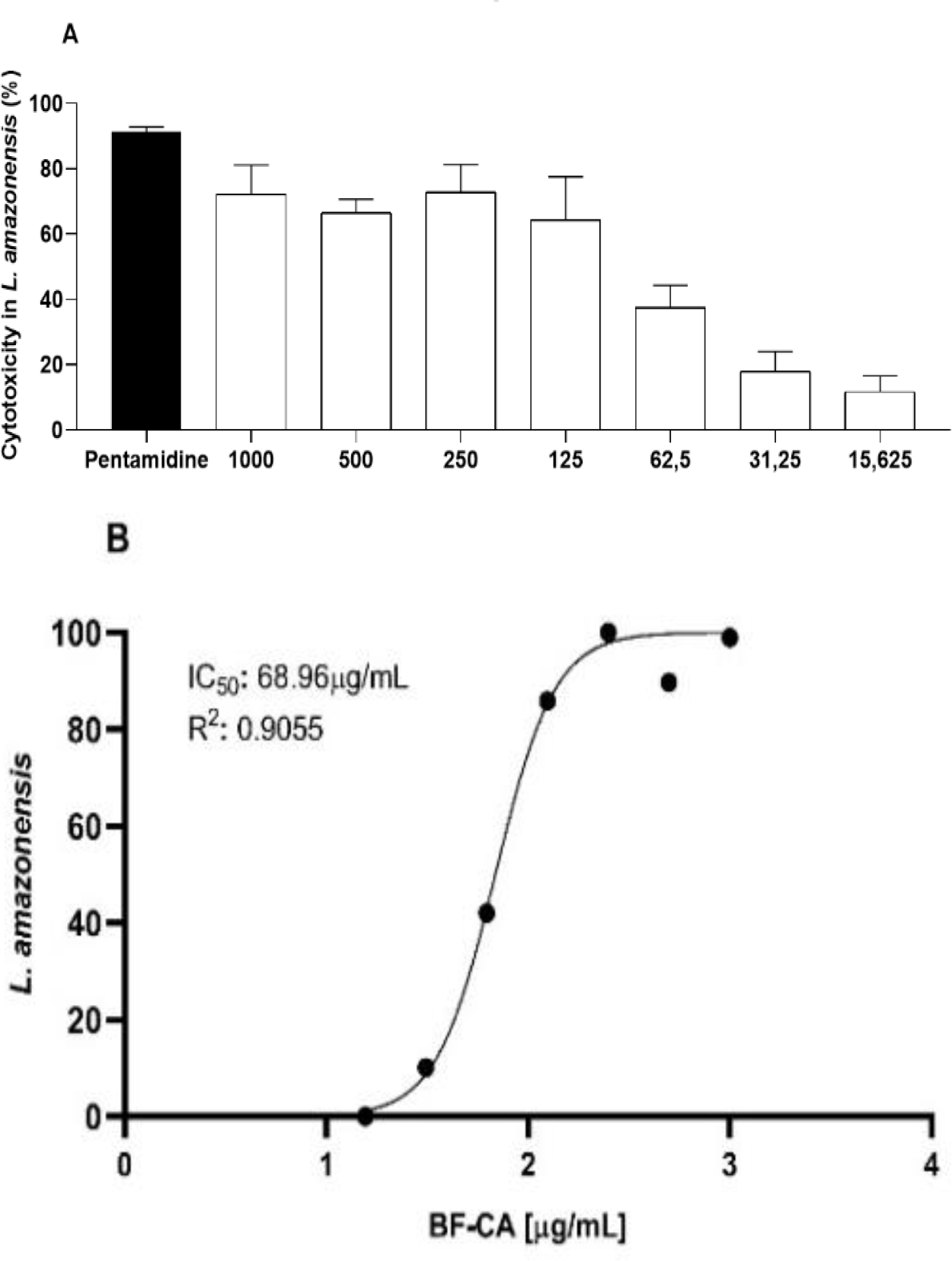
Promastigotes were added to 96-well plates (5 x 10⁵ /mL per well - 10 µL) with different BF-CA concentrations (1000; 500; 250; 125; 62.5; 31.25; 15.625 µg/mL), that were used to perform serial dilutions for dose screening to determine the inhibitory concentration for 50% of the parasites (IC50) [31,34]. Pentamidine (100; 50; 25; 12.5; 6.25; 3.125; 1.625; 0.78125 µg/mL) was used as positive control and the negative control was prepared only with Schneider medium. After 48 h of incubation, 10 µL of MTT (5 mg/mL) were added and incubated again for 3 h for the formazan crystals formation. Finally, 100 µL of DMSO (1:1) were added and its absorbance measured with a spectrophotometer at 540 nm after 24 h (the next day). The concentration of the compounds that reduced the response by 50% compared to the control group (IC50) was obtained by nonlinear regression using GraphPad Prism software version 8.0.2.
In vitro cytotoxicity assay using L. amazonensis amastigotes
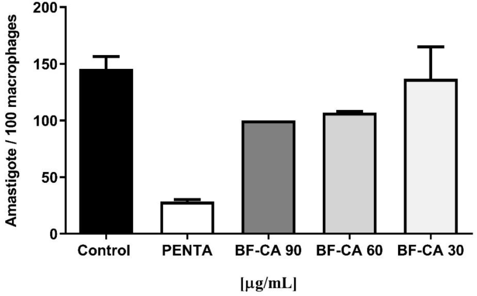
RAW cells (1 x 10⁵ in 500 µL of RPMI) were seeded on sterile glass coverslips in 24-well plates with medium containing penicillin, streptomycin, amphotericin B and supplemented with 5% FBS and incubated for 1 h at 37 °C in 5% CO₂ to allow macrophages adhesion. After incubation, the supernatant was removed and 1 x 10⁶ L. amazonensis promastigotes (ten parasites per cell) were added to each well. Plates were incubated again for 24 h at 33 °C in 5% CO₂ to allow the parasite phagocytosis by macrophages. Next, the wells were washed with PBS, and the infected cells with amastigotes were treated with BF-CA (30, 60 and 90 µg of protein/mL) and pentamidine at 5 µg/mL during 48 h. The coverslips were stained with Giemsa and placed on a slide to evaluate the number of intracellular parasites, considering the count of 100 macrophages, using a light microscope, bright field, using 100 x magnification [35].The concentration of compounds that reduced the response by 50% in relation to the control group (IC50) was obtained by non-linear regression using GraphPad Prism software version 8.0.2.
Statistical analysis
The data were analyzed using one-way ANOVA followed by Tukey-Kramer test (for three or more groups) or Student's t-test (for two groups). Values of P < 0.05 were considered significant. IC50 and CC50 values were estimated from the mean ± standard deviation values of two independent experiments, performed in triplicate. All data were analyzed using GraphPad Prism version 8.0.2.
RESULTS
Preparation of B. forficata extracts, protein content determination and characterization of PI activity of B. forficata extracts
Leaves, seeds and stems of B. forficata are the most frequent parts used in folk medicine and therefore aqueous extracts of these organs were investigated in the present study. As shown in Table 1, B. forficata extracts have different yields and protein contents, the highest yield and protein concentrations were observed for Tris seed (BF-ST) and stem (BF-CA) extracts.
Protein content, extract yield and percentage of reference and Leishmania amazonensis extracellular fraction proteases activities inhibition by B. forficata extracts.
Seed extracts showed excellent inhibition against trypsin (Table 1). On the other hand, leaf extracts did not affect the protease activity. These results indicate the presence of serine-type PIs in stem and seeds of B. forficata. The papain activity was eliminated by seed and stem extracts, whereas leaf extracts did not affect it, indicating that stem and seeds of B. forficata are important sources of cysteine-type PIs. Aspartic protease inhibition has been evaluated using pepsin. BF-ST and BF-CA inhibited about 55 to 60% of pepsin activity, whereas leaf extracts seem to lack pepsin, papain or trypsin PIs. The lowest activity of PI was precisely observed for pepsin. The L. amazonensis fraction III was obtained from the parasite-free culture supernatant and contains proteins and enzymes secreted by Leishmania involved in parasite-host interaction [28]. All B. forficata extracts inhibited the activity of fraction III proteases, at different levels. The greatest inhibitory effect was achieved by BF-CA, which completely abolished the protease activity (Table 1). Taken together, these results indicated that the activity of the major types of PIs was not found in B. forficata leaves, but was observed in stem and seed extracts, mainly serine and cysteine PIs. This is the first time that PI activities were identified in B. forficata. Therefore, BF-CA was elected to investigate the chemical composition, the toxicity and cytotoxic effects on Leishmania because it had the most expressive inhibition of fraction III protease activity, because good inhibitors of Leishmania proteases killed parasites in vitro [36]. Besides, BF-CA has high protein content compared to other extracts.
Analysis of secondary metabolites of B. forficata stem extract
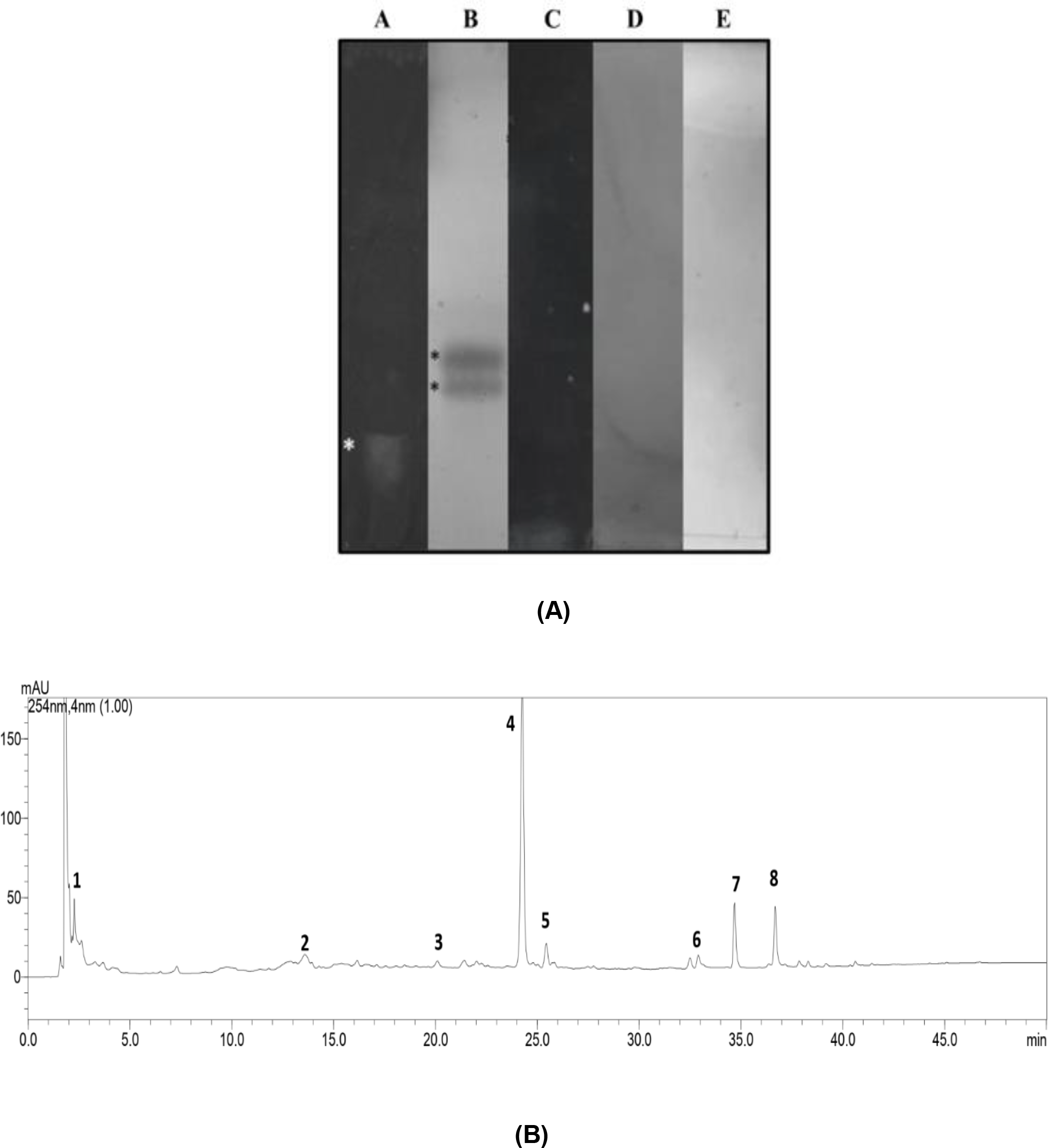
TLC is a preliminary analysis of secondary metabolites of plants. BF-CA had flavonoids (A) and terpenes (B), but not coumarins (C), alkaloids (D) and saponins (E). The presence of flavonoids and terpenes in high polar extract is explained because both class of metabolites can be glycosylated. Water was employed mainly to extract soluble proteins, that are very polar molecules. The comparison of the present results has been difficult with literature, because we used water in the preparation of B. forficata stem extract. Organic solvents are routinely employed to extract secondary metabolites, which are molecules with lower polarity than proteins, which are objects of this study. TLC analysis showed the presence of flavonoids (1 band) and terpenes (2 bands) and the absence of alkaloids, coumarins and saponins in BF-CA (Figure 1A).
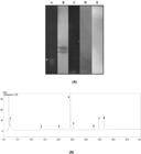
Analysis of secondary metabolites of BF-CA. A. Classification of extract by TLC according to the classes of flavonoids (A), terpenes (B), coumarins (C), alkaloids (D) and saponins (E); B. Chromatographic profile of BF-CA on HPLC-PDA-MS.
Flavonoids are especially abundant in legumes [37], and thus, BF-CA was submitted to HPLC-PDA and the chromatogram suggested the presence of compounds with characteristics related to flavonoids and phenolic acids (Figure 1B).
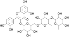
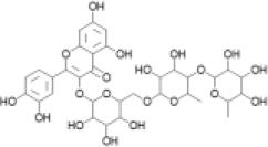
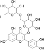
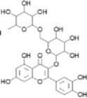
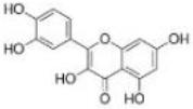
The most abundant secondary metabolite was quercetin 3-rhamnosyl(1->4)-rhamnosyl-(1->6)-glycoside, a glycosylated flavonoid with recognized antioxidant activity. The combined presence of these metabolites with antioxidants and anti-inflammatory activities may contribute to the observed biological activity of the extract (Table 2).
Compounds identified in B. forficata stem aqueous extract and the main fragments obtained by HPLC-ESI-IT/MS.
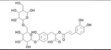
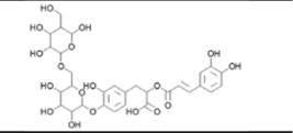
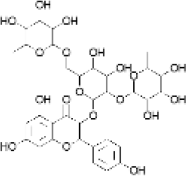
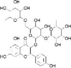
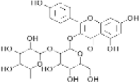
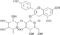
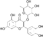
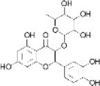
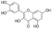
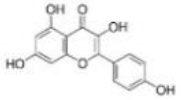
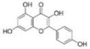
Phenolic acid (diCQA) showed a precursor ion at m/z 683 [M-H]-(C330H35O18), which permitted to identify the peak 1 as rosmarinic acid-diglucoside. Peak 2 was identified as 3 Kaempferol 3-O-(2,6-di-O-rhamnopiranosil)-galactopyranoside which showed a precursor ion at m/z 739 [M-H]-(C33H40O19) in the negative ionization mode. This precursor ion was fragmented in product ions at m/z 593, produced by the loss of a rhamnosyl unit, and at m/z 447, produced by the loss of a second rhamnosyl unit. Peak 3 was identified as quercetin 3-rhamnosyl(1->4)-rhamnosyl-(1->6)-glucoside. This peak had a precursor ion at m/z 755 [M-H]-(C33H40O20) and its MS/MS spectrum showed product ions at m/z 609 and m/z 301, which are due to the loss of a rhamnosyl residue, glucose and rhamnose together, respectively. Peak 4 had a precursor ion at m/z 593 [M-H]-(C27H30O15) and its MS/MS spectrum exhibited a product ion at m/z 447 attributed to the elimination of terminal rhamnosyl. Also in the negative ionization mode, this m/z 447 precursor produced m/z 285 formed after the elimination of a glucose residue. Therefore, this peak was identified as Kaempferol-3-O-glucorhamnoside. Compound 5 had a precursor ion at m/z 609 [M-H]-(C27H30O16) in the negative ionization mode, and its MS/MS spectrum showed product ions characteristic for rutin at m/z 463 due to loss of a rhamnosyl unit, and at m/z 301, formed after loss of hexose residue (162 u) or the direct loss of a rutinoside residue (rhamnosyl-(α1→6)-glucose) unit. Therefore, these data and comparison with the literature revealed that compound 5 is quercetin-3-O-rutinoside (rutin). Peak 6, it exhibited a mass spectrum with a precursor ion at m/z 447 [M-H]-(C21H20O11). This precursor ion showed an MS/MS spectrum with a product ion at m/z 301, which corresponds to the loss of a rhamnosyl unit. This peak showed MS/MS spectrum characteristics of quercetin 3-rhamnoside, and the aglycone quercetin (peak 7) was identified due to the characteristic fragment m/z 151. Finally, peak 8 was identified as kaempferol due to precursor ions at m/z 285 [M-H]-(C15H10O6). The MS/MS spectrum in the negative ionization mode showed ions at m/z 267 and at m/z 151, which are characteristic of fragmentation patterns for this substance, and comparison with the literature confirmed its identity. The BF-CA compounds were extracted with water, so they all had polar characteristics, including their secondary metabolites that have carbohydrates in their structure. This can explain why apolar substances such as alkaloids were not found in this B. forficata stem extract.
Toxicity and cytotoxicity assays of B. forficata extracts
In vivo toxicity assay using Tenebrio molitor
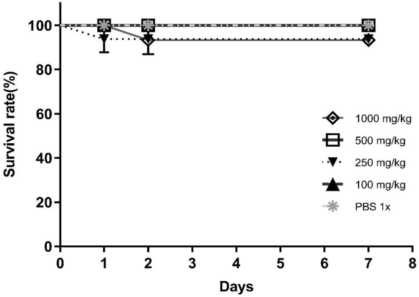
T. molitor survival was evaluated by inoculation of 5 µL BF-CA between the 4th and 5th metamere, from tail to head, in the ventral portion. Larvae were observed for seven days, with the removal of dead individuals. In vivo assay indicated that average survival was 93.3% in the highest dose group (1000 mg/kg), while the survival rate was 100% at the doses of 500 and 100 mg/kg compared to control, suggesting a very low toxicity of BF-CA (Figure 2).
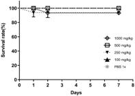
Survival rate of T. molitor larvae until 8 days after application of different concentrations of BF-CA (mg/kg) at day 0.
Hemolysis assays with sheep erythrocytes
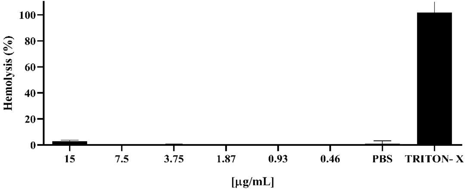
BF-CA did not induce erythrocytes hemolysis after incubation for 1 h at 37 °C, and therefore was not cytotoxic at any tested concentrations (1.0, 0.5, 0.25, 0.125, 0.063, and 0.032 μg of protein/mL) when compared to the negative control (human erythrocytes incubated with PBS) and positive 1% Triton X-100 control (erythrocytes incubated with triton) (Figure 3).
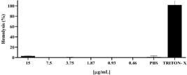
Hemolytic analysis in sheep erythrocytes with different concentrations of BF- CA (µg of protein/mL) and using 1% Triton X-100 as a positive hemolysis control.
In vitro cytotoxicity assay using RAW cell macrophages
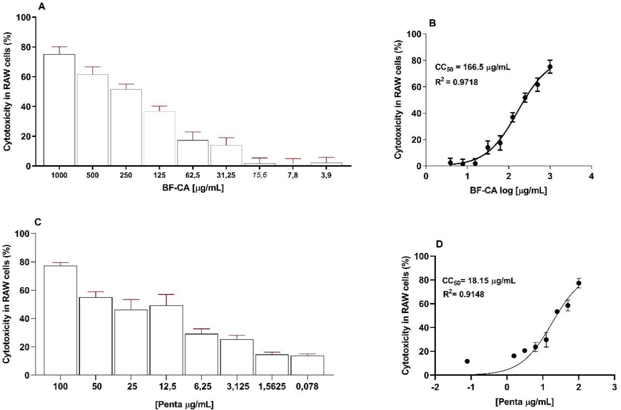
The effect of BF-CA on macrophages was dose-dependent, and the extract was expressively cytotoxic at the highest concentrations (1000, 500, and 250 µg/mL) (Figure 4). However, at the lowest extract concentrations, cytotoxicity was very low (Figure 4 A). Pentamidine, the positive control employed in the treatment of leishmaniasis [38], was more toxic than BF-CA (Figure 4 B). The cytotoxic concentration that reduced 50% of macrophage viability (CC50) was analyzed and BF-CA showed a value of about 166.50 ± 8.61 µg protein/mL (R2 = 0.9718) compared (Figure 4 C) to about 18.15 ± 9.34 µg/mL (R2 = 0.9148) for pentamidine (Figure 4 D). Thus, pentamidine was about 9.17 times more cytotoxic to the RAW cells than the extract.
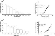
Cytotoxic effects on macrophages RAW 264.7 with different concentrations of BF-CA (A) and its nonlinear regression curve of CC50 (B) and pentamidine (C) and its nonlinear regression of CC50 curve (D). All data represent the mean ± standard deviation of two independent experiments performed in triplicates, analyzed using GraphPad Prism version 8.0.2. Values of p < 0.05 were considered significant.
In vitro cytotoxicity assay using L. amazonensis promastigotes and amastigotes
The determination of BF-CA cytotoxicity on L. amazonensis amastigotes (Figure 6) considered the IC50 value (~60 µg of protein /mL) previously obtained for promastigotes (Figure 5B). So, 30 µg of protein /mL below 60 µg of protein /mL and 30 µg of protein /mL above, i.e. 30, 60 and 90 protein /mL were assayed.
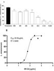
Cytotoxic effects on L. amazonensis promastigotes with different concentrations of BF-CA (µg of protein/mL) (A) and nonlinear regression curve of IC50 (B). Values of p < 0.05 were considered significant.
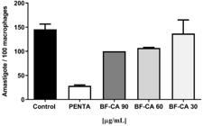
Cytotoxic effect on L. amazonensis amastigotes cells with different concentrations of BF-CA (µg of protein/mL). It means that p < 0.05, when compared with the control group.
BF-CA was cytotoxic to L. amazonensis amastigotes, and the effect was dependent on extract protein concentration (Figure 6). It is necessary at least four data points to obtain a reliable regression analysis, however, we estimated using five concentrations, and these three data points were the best, and IC50 of BF-CA was about 34.61 ± 3.27 µg of protein/mL (R2 = 0.9020) and 0.9 ± 0.16 µg/mL (R2 = 0.9395) for pentamidine. BF-CA had anti-amastigote activity, but the efficacy of the standard drug pentamidine is higher than the extract, because pentamidine IC50 is lower than BF-CA about 38.45 times (Table 3).
Cytotoxicity, antileishmanial activity, and selectivity index for BF-CA in comparison with pentamidine.
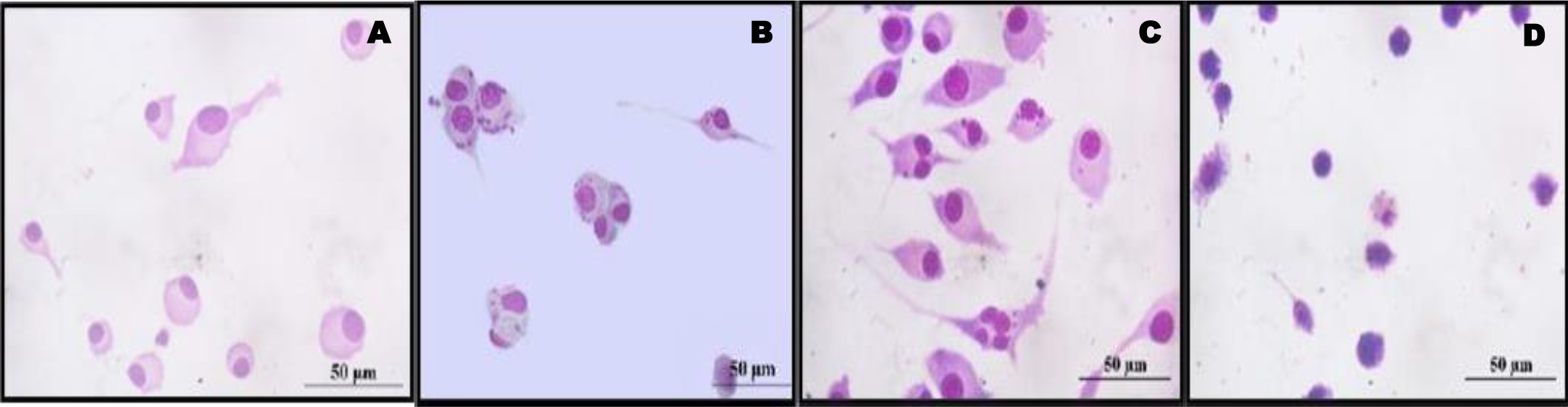
The Figure 7 results corroborated with the findings of cytotoxicity of BF-CA on macrophages and Leishmania. The extract BF-CA reduced the number of intracellular amastigotes in the host cell without affecting macrophage morphology (Figure 7C). On the other hand, although pentamidine killed intracellular amastigotes, it greatly affected the structural integrity of macrophages (Figure 7D). Thus, BF-CA was more selective to L. amazonensis than pentamidine.
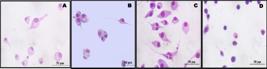
Anti-Leishmania activity of BF-CA (60 µg/mL) and Pentamidine (5 µg/mL) on macrophages infected by amastigotes L. amazonensis observed by light microscopy. Macrophages control (A), L. amazonensis-infected macrophages control (B), L. amazonensis-infected macrophages treated with BF-CA (C) or with Pentamidine (D), respectively.
DISCUSSION
In the present study we assayed B. forficata aqueous extracts, from the most employed organs in folk medicine, which contain substances of great polarity such as proteins and polypeptides, as natural PIs. All substances and extracts reported in other B. forficata studies were prepared using organic solvents or high temperatures, conditions that denature the structure of PIs, thus, the PI activity observed in the present manuscript was due to the action of these polypeptides.
PI activity was study using reference and Leishmania fraction III proteases. Reference proteases are valuable tools for the identification of PI activity, because the major types of PIs are serine, cysteine and aspartic that inhibit trypsin, papain and pepsin respectively [23]. Serine proteases are the most studied enzymes, and their inhibitors are the largest PI family in nature [41]. Pancreatic trypsin (EC 3.4.21.4) is the representative serine protease and trypsin-like enzymes are found in all organisms, performing various biological functions [9]. In this study, B. forficata seed and stem extracts presented serine-type PIs. In plants, PIs are almost exclusively studied in seeds, since they accumulate these PIs in these organs to protect proteins and the genetic content against attack by predators [42]. Furthermore, trypsin-like PIs have already been described in seeds of other Bauhinia species [6,15]. Papain (EC 3.4.22.2) isolated from papaya (Carica papaya L.) is the most studied cysteine protease [43], and its activity was eliminated by B. forficata seed and stem extracts, indicating that these organs are important sources of cysteine-type PIs. Furthermore, a trypsin PI isolated from B. bauhinioides inhibited the activity of cruzipain, the main cysteine protease of Trypanosoma cruzi [15]. Bovine pepsin (EC 3.4.4.1) was employed to evaluate the aspartic proteases PIs [44] in B. forficata and only seed, and stem extracts presented pepsin-type PIs. The lowest activity of PI was precisely observed for pepsin since these types of PIs are the rarest in nature [42]. PI activity has been extensively studied in Bauhinia genus, however, there are no reports about this activity in B. forficata. The L. amazonensis fraction III contains proteins and enzymes released by Leishmania that are involved in host-parasite interaction, such as LSPIII, the 115 kDa serine protease [28]. BF-CA completely abolished the protease activity of Leishmania fraction III. These results indicated that the activity of the major types of PIs was not found in B. forficata leaves, but it was observed in stem and seed extracts, mainly serine and cysteine PIs. The novelty of the present work is the first time that PI activities were identified in B. forficata. Besides, many PIs have been isolated and characterized in seeds of other species of the Bauhinia genus such as B. bauhinioides, B. petandra, B. ungulata, B. variegata, B. mollis, B. purpurea, B. pulchella and B. rufa [13-15,45], but they have never been identified in other parts of these species. Therefore, BF-CA was elected to investigate the main secondary metabolites, the toxicity on T. molitor larvae and cytotoxicity on sheep erythrocytes, murine macrophages and L. amazonensis since good inhibitors of Leishmania proteases can kill promastigotes and amastigotes in vitro [36].
Flavonoids are especially abundant in legumes, and they are the legume chemical markers. They are polar substances when glycosylated and have a great diversity of biological and pharmacological activities, including antioxidant, anticancer, anti-inflammatory, antimicrobial activities, and neuroprotective and cardio-protective effects [37,46-48]. Rosmarinic acid-diglycoside, derivates of quercetin and kaempferol glycosylated and their aglycones were identified. Rosmarinic acid is widely distributed among plants, especially those of the Lamiaceae family, however, the diglycoside form is rare in nature and it was first reported in B. forficata [47]. On the other hand, kaempferol and quercetin are found in many plants, especially in the Fabaceae family, but glycosylated forms are also rarely observed. The aqueous stem extract of B. forficata contains a high concentration of glycosylated flavonoids, which could explain the low toxicity on T. molitor larvae and low cytotoxicity on sheep erythrocytes and murine macrophages, and the anti-Leishmania activity synergically with PIs. Besides, quercetin and its derivatives (7,8-dihydroxyflavone) and (3-O-rutinoside-7-O-rhamnoside) demonstrated anti-Leishmania activity against L. amazonensis promastigotes and low cytotoxicity for host cells [49,50].
T. molitor larvae is a toxicity model for preliminary screening and acute toxicity assessment evaluation of new bioactive compounds, reducing the number of mammals in preclinical tests, and the costs of drug research. Besides, it responds similarly to vertebrates when exposed to toxic compounds [51]. BF-CA was not toxic for T. molitor; thus, this extract can be employed for future essays in infection models.
According to the International Standard Organization (ISO, 2009), in vitro cytotoxicity tests are the first stage to investigate the safety of material before in vivo tests [52]. Erythrocytes are frequently used because they lack organelles and have only one membrane. So, they are suitable for studies about interaction between compounds and cell membranes [53]. BF-CA was not toxic to sheep erythrocytes. On the other hand, methanol/water seed extracts from Nigella sativa and Lepidium sativum [54] were much more toxic than BF-CA when tested under similar experimental conditions. Most medicinal plant extracts are prepared using organic solvents and their composition is very different from the B. forficata extracts obtained using water or buffers, which contain polar substances. Thus, it is difficult to compare our results with literature.
Macrophages are the Leishmania host cells, and in vitro cytotoxicity assays using these cells are crucial to estimate the selectivity index (SI) and study the BF-CA safety [50]. CC50 value indicated that BF-CA had low cytotoxicity on macrophages, and it was less toxic than pentamidine used as control. However, pentamidine is a second-line leishmanicide drug with high cytotoxicity [38]. Similar CC50 results were obtained using a crude extract of Diospyros gracilescens Gürke trunk, which was the most active extract in bio-guided isolation of anti-leishmanial natural products [50]. Glycosylated flavonoids were abundant in BF-CA, and the lower cytotoxicity can be explained by these metabolites in extract. Besides, water employed for BF-CA preparation, extracted lesser toxic polar substances. The use of polar solvents reduced the generation of chemical waste, that is according to the principles of green chemistry and environmental sustainability [53].
BF-CA was cytotoxic to L. amazonensis promastigotes and amastigotes. It reduced the number of amastigotes in macrophage, in other words, BF-CA substances permeated the cytoplasmic and megasome membranes of macrophage and killed amastigotes, without affected its morphology and viability. Although pentamidine was more toxic for L. amazonensis than BF-CA, the extract was safer, because the drug induced important structural changes in macrophages (Figure 7). Due to SI value, BF-CA was considered promising anti-leishmanial compounds with moderate activity. Studies about the extract formulations may enhance its selectivity. However, BF-CA is a candidate for leishmaniasis treatment, as it was a very good inhibitor of Leishmania proteases, was not toxic or cytotoxic for normal cells, was selective against Leishmania and it was water soluble. On the other hand, most plant extracts have metabolites with low water solubility.
There is a correlation that good inhibitors of L. amazonensis serine proteases have anti-Leishmania activity by inducing expressive morphological changes where proteases are localized [36]. BF-CA was also a good inhibitor of L. amazonensis protease activity, and it showed leishmanicide effects. Thus, the present results support that L. amazonensis serine proteases are promising targets for the development of specific inhibitors for leishmaniasis chemotherapy. However, other types of Leishmania proteases, such as metalloprotease GP63 [54], cysteine proteases [55] and aspartic proteases [56] are important to parasite survival and infection [55,57]. Based on these findings, we believe that the presence of serine, cysteine and aspartic protease inhibitors and flavonoids such as glycosylated flavonoids, rosmarinic acid, kaempferol, and quercetin in BF-CA, may be involved in anti-Leishmania activity. Aglycones of quercetin, kaempferol and rosmarinic acid had expressive anti-Leishmania effect [46,47]. However, the anti-Leishmania activity of glycosylated flavonoids has never been studied.
The novelty of this work relies on the aqueous extraction of polar substances, such as polypeptides with PIs activity and glycosylated flavonoids from B. forficata. It is the first report about the PI activities of B. forficata, flavonoids composition, non-toxic effects on organisms and normal cells and selective anti-Leishmania activity. As a future prospect, the development of a low-cost topical formulation to treat skin lesions with using BF-CA could represent an adjuvant treatment of leishmaniasis by reducing the dose of toxic and costly available drugs.
-
Funding:
This study was financed in part by the Coordenação de Aperfeiçoamento de Pessoal de Nível Superior - Brasil (CAPES) - Finance Code 001.
Acknowledgments:
The authors are grateful to Dr. Valerio Morelli from Farmanguinhos - FIOCRUZ for supplying B. forficata. Clara Bárbara Carneiro Echebaster and Bruna Cristina dos Santos for the initial studies with B. forficata extracts. Joana Neres Ferreira Assenço for the analysis of cytotoxicity assays in L. amazonensis. Josivan Regis Farias and Arthur André Castro da Costa for the in vivo toxicity test with larvae of T. molitor. Luis Douglas Miranda Silva for maintenance RAW cells and L. amazonensis. Rosane Nassar Meireles Guerra Liberio for the infrastructure, reagents, materials and assay protocols. Aldilene da Silva Lima for assistance in chemical characterization of BF-CA. Érika Maria Gomes Ferreira Teixeira and Thiago Maldonado Brazil for the identification of secondary metabolites by TLC assays.
Data Availability Statement:
Research data are available in the body of the manuscript.
REFERENCES
-
1 Hayashi AH, Appezzato-da-Glória B. Resprouting from roots in four brazilian tree species. Rev Biol Trop. 2009; 57(3): 789-800. https://doi.org/10.15517/rbt.v57i3.5493
» https://doi.org/10.15517/rbt.v57i3.5493 -
2 Vaz AMSF. Bauhinia. In: Lista de Espécies da Flora do Brasil. Jardim Botânico do Rio de Janeiro. Available from: https://floradobrasil2015.jbrj.gov.br/FB22811, accessed in April 2024.
» https://floradobrasil2015.jbrj.gov.br/FB22811 -
3 Cechinel-Zanchett CC, de Andrade SF, Cechinel-Filho V. Ethnopharmacological, phytochemical, pharmacological and toxicological aspects of Bauhinia forficata: a mini-review covering the last five years. Nat Prod Comm. 2018; 13(7): 1934578X1801300732. http://dx.doi.org/10.1177/1934578X1801300732
» http://dx.doi.org/10.1177/1934578X1801300732 -
4 Tonelli CA, de Oliveira SQ, Silva Vieira AAD, Biavatti MW, Ritter C, Reginatto FH, et al. Clinical efficacy of capsules containing standardized extract of Bauhinia forficata Link (pata-de-vaca) as adjuvant treatment in type 2 diabetes patients: a randomized, double blind clinical trial. J Ethnopharmacol. 2022; 282:114616. https://doi.org/10.1016/j.jep.2021.114616
» https://doi.org/10.1016/j.jep.2021.114616 -
5 Silva-López RE, Santos BC. Bauhinia forficata Link (Fabaceae) Rev Fitos. 2015; 9: 217-32. https://doi.org/10.5935/2446-4775.20150018
» https://doi.org/10.5935/2446-4775.20150018 -
6 Silva-Lucca RA, Andrade SS, Ferreira RS, Sampaio MU, Oliva ML. Unfolding studies of the cysteine protease baupain, a papain-like enzyme from leaves of Bauhinia forficata: effect of pH, guanidine hydrochloride and temperature. Molecules. 2013; 19(1): 233-46. https://doi.org/10.3390/molecules19010233
» https://doi.org/10.3390/molecules19010233 - 7 Silva-López RE. Protease inhibitors originated from plants: useful approach for development of new drugs. Rev Fitos. 2009; 4(1): 108-19.
-
8 Cotabarren J, Lufrano D, Parisi MG, Obregón WD. Biotechnological, biomedical, and agronomical applications of plant protease inhibitors with high stability: a systematic review. Plant Sci. 2020; 292: 110398. https://doi.org/10.1016/j.plantsci.2019.110398
» https://doi.org/10.1016/j.plantsci.2019.110398 -
9 Van der Hoorn RAL, Klemenčič M. Plant proteases: from molecular mechanisms to functions in development and immunity. J Exp Bot. 2021; 72(9): 3337-9. https://doi.org/10.1093/jxb/erab129
» https://doi.org/10.1093/jxb/erab129 -
10 Cid-Gallegos MS, Corzo-Ríos LJ, Jiménez-Martínez C, Sánchez-Chino XM. Protease inhibitors from plants as therapeutic agentsa review. Plant Foods Hum Nutr. 2022; 77(1): 20-9. https://doi.org/10.1007/s11130-022-00949-4
» https://doi.org/10.1007/s11130-022-00949-4 -
11 Chen TY, Zhou M, Lin MQ, Liang ST, Yan Y, Wang SM, et al. Research progress on the SERPINE1 protein and chronic inflammatory diseases of the upper respiratory tract: a literature review. Int Arch Allergy Immunol. 2021; 182(11): 1097-102. https://doi.org/10.1159/000516195
» https://doi.org/10.1159/000516195 -
12 Yoo Im S, Ramalho Bonturi C, Miti Nakahata A, Ryuichi Nakaie C, Pott A, Pott VJ, et al. Differences in the inhibitory specificity distinguish the efficacy of plant protease inhibitors on mouse fibrosarcoma. Plants (Basel). 2021; 10(3):602. https://doi.org/10.3390/plants10030602
» https://doi.org/10.3390/plants10030602 -
13 Valois MV, de Oliveira C, Lapa AJ, Souccar C, Oliva MLV. Bauhinia protease inhibitors attenuate gastric ulcer by blocking neutrophil enzymes. Planta Med. 2021; 87(1-02): 169-76. https://doi.org/10.1055/a-1202-4799
» https://doi.org/10.1055/a-1202-4799 -
14 Martins-Olivera BT, Almeida-Reis R, Theodoro-Júnior OA, Oliva LV, Neto Dos Santos Nunes N, Olivo CR, et al. The plant-derived Bauhinia bauhinioides kallikrein proteinase inhibitor (rBbKI) attenuates elastase-induced emphysema in mice. Mediators Inflamm. 2016; 2016: 5346574. https://doi.org/10.1155/2016/5346574
» https://doi.org/10.1155/2016/5346574 -
15 Oliva ML, Mendes CR, Juliano MA, Chagas JR, Rosa JC, Greene LJ, et al. Characterization of a tissue kallikrein inhibitor isolated from Bauhinia bauhinioides seeds: inhibition of the hydrolysis of kininogen related substrates. Immunopharmacol. 1999; 45(1-3):163-9. https://doi.org/10.1016/s0162-3109(99)00075-2
» https://doi.org/10.1016/s0162-3109(99)00075-2 -
16 Kaye PM, Cruz I, Picado A, Van Bocxlaer K, Croft SL. Leishmaniasis immunopathology-impact on design and use of vaccines, diagnostics and drugs. Semin Immunopathol. 2020; 42(3):247-64. https://doi.org/10.1007/s00281-020-00788-y
» https://doi.org/10.1007/s00281-020-00788-y -
17 World Health Organization (WHO). Leishmaniasis. www.who.int Available from: https://www.who.int/health-topics/leishmaniasis#tab=tab Accessed in April 2024.
» www.who.int» https://www.who.int/health-topics/leishmaniasis#tab=tab -
18 Pinheiro AC, de Souza MVN. Current leishmaniasis drug discovery. RSC Med Chem. 2022; 13(9):1029-43. https://doi.org/10.1039/D1MD00362C
» https://doi.org/10.1039/D1MD00362C -
19 Garza-Tovar TF, Sacriste-Hernández MI, Juárez-Durán ER, Arenas R. An overview of the treatment of cutaneous leishmaniasis. Fac Rev. 2020; 9: 28. https://doi.org/10.12703/r/9-28
» https://doi.org/10.12703/r/9-28 -
20 Palić S, Beijnen JH, Dorlo TPC. An update on the clinical pharmacology of miltefosine in the treatment of leishmaniasis. Int J Antimicrob Agents. 2022; 59(1): 106459. https://doi.org/10.1016/j.ijantimicag.2021.106459
» https://doi.org/10.1016/j.ijantimicag.2021.106459 -
21 Silva-López RE. Immunocytochemistry of proteases in the study of Leishmania physiology and host-parasite interaction. Applications of Immunocytochemistry. InTech; 2012. https://dx.doi.org/10.5772/32954
» https://dx.doi.org/10.5772/32954 -
22 De Almeida Nogueira NP, Morgado-Díaz JA, Menna-Barreto RF, Paes MC, da Silva-López RE. Effects of a marine serine protease inhibitor on viability and morphology of Trypanosoma cruzi, the agent of chagas disease. Acta Trop. 2013; 128(1):27-35. https://doi.org/10.1016/j.actatropica.2013.05.013
» https://doi.org/10.1016/j.actatropica.2013.05.013 -
23 Pacheco JS, Teixeira E, Paschoal RG, Torres-Santos EC, Simone SG, Silva-López RE. Antileishmanial effects of Crotalaria spectabilis Roth aqueous extracts on Leishmania amazonensis An Acad Bras Cienc. 2023; 95(suppl 1): e20220613. https://doi.org/10.1590/0001-3765202320220613
» https://doi.org/10.1590/0001-3765202320220613 - 24 Silva-López RE. Proteases de Leishmania: novos alvos para o desenvolvimento racional de fármacos. Quím Nova. 2010; 33: 1541-1548.
-
25 Agência Nacional de Vigilância Sanitária, MEDICAMENTOS - GUIA nº 74, versão 1, 4 de outubro de 2024. Available from: https://anexosportal.datalegis.net/arquivos/1874686.pdf, accessed in April 2024.
» https://anexosportal.datalegis.net/arquivos/1874686.pdf -
26 Gonçalves RN, Gozzini Barbosa SD, da Silva-López RE. Proteases from Canavalia ensiformis: active and thermostable enzymes with potential of application in biotechnology. Biotechnol Res Int. 2016; 2016: 3427098. https://doi.org/10.1155/2016/3427098
» https://doi.org/10.1155/2016/3427098 -
27 Bradford MM. A rapid and sensitive method for the quantitation of microgram quantities of protein utilizing the principle of protein-dye binding. Anal Biochem. 1976; 72: 248-54. https://doi.org/10.1016/0003-2697(76)90527-3
» https://doi.org/10.1016/0003-2697(76)90527-3 -
28 Silva-López RE, Coelho MG, De Simone SG. Characterization of an extracellular serine protease of Leishmania (Leishmania) amazonensis Parasitol. 2005; 131(Pt 1): 85-96. https://doi.org/10.1017/s0031182004006675
» https://doi.org/10.1017/s0031182004006675 -
29 Wagner H, Bladt S. Plant drug analysis: a thin layer chromatography atlas: Springer Science & Business Media; 1996. http://dx.doi.org/10.1007/978-3-642-00574-9
» http://dx.doi.org/10.1007/978-3-642-00574-9 -
30 Kaplan EL, Meier P. Nonparametric estimation from incomplete observations. J Am Statistic Assoc. 1958; 53(282): 457-81. https://doi.org/10.2307/2281868
» https://doi.org/10.2307/2281868 -
31 Almaaytah A, Tarazi S, Alsheyab F, Al-Balas Q, Mukattash T. Antimicrobial and antibiofilm activity of mauriporin, a multifunctional scorpion venom peptide. Int J Pep Res Therapeutic. 2014; 20(4): 397-408. https://doi.org/10.1007/s10989-014-9405-0
» https://doi.org/10.1007/s10989-014-9405-0 -
32 van der Valk J. Fetal bovine serum-a cell culture dilemma. Science. 2022; 375(6577): 143-144. https://doi.org/10.1126/science.abm1317
» https://doi.org/10.1126/science.abm1317 -
33 da Costa CS, Marques EM, do Nascimento JR, Lima VAS, Santos-Oliveira R, Figueredo AS, et al Design of liquid formulation based on F127-loaded natural dimeric flavonoids as a new perspective treatment for leishmaniasis. Pharmaceutics. 2024; 16: 252. https://doi.org/10.3390/pharmaceutics16020252
» https://doi.org/10.3390/pharmaceutics16020252 -
34 Lopes ACCB, do Nascimento JR, Camara MBP, Lima AdS, Lopes GLN, do Nascimento MO, et al Chemical characterization, leishmanicidal activity and in vitro cytotoxicity of the essential oil extracted from Pectis brevipedunculata (Gardner) Sch.Bip. and its incorporation into microemulsion systems. Pharmaceutics. 2024; 16: 87. https://doi.org/10.3390/pharmaceutics16010087
» https://doi.org/10.3390/pharmaceutics16010087 - 35 Espíndola MR. Comparação de métodos utilizados para ensaios de citotoxicidade in vitro na avaliação de compostos com atividade antiplasmodial: Universidade Federal de São João Del Rei, 2016.
-
36 Silva-López RE, Morgado-Díaz JA, Chávez MA, Giovanni-De-Simone S. Effects of serine protease inhibitors on viability and morphology of Leishmania (Leishmania) amazonensis promastigotes. Parasitol Res. 2007; 101(6): 1627-35. https://doi.org/10.1007/s00436-007-0706-5
» https://doi.org/10.1007/s00436-007-0706-5 -
37 Yao LH, Jiang YM, Shi J, Tomas-Barberan FA, Datta N, Singanusong R, et al Flavonoids in food and their health benefits. Plant Food Hum Nutr. 2022; 77(3): 369-90. https://doi.org/10.1007/s11130-004-0049-7
» https://doi.org/10.1007/s11130-004-0049-7 -
38 Anjum A, Shabbir K, Din FU, Shafique S, Zaidi SS, Almari AH, et al. Co-delivery of amphotericin B and pentamidine loaded niosomal gel for the treatment of Cutaneous leishmaniasis Drug Deliv. 2023; 30(1): 2173335. https://doi.org/10.1080/10717544.2023.2173335
» https://doi.org/10.1080/10717544.2023.2173335 -
39 Najm M, Hadighi R, Heidari-Kharaji M, Alipour M, Hajizadeh M, Rafiei-Sefiddashti R, et al Anti-leishmanial activity of Artemisia persica, A. spicigera, and A. fragrance against Leishmania major Iran J Parasitol. 2021; 16(3): 464-473. https://doi.org/10.18502/ijpa.v16i3.7100
» https://doi.org/10.18502/ijpa.v16i3.7100 -
40 Staffen IV, Banhuk FW, Tomiotto-Pellissier F, da Silva Bortoleti BT, Pavanelli WR, Ayala TS, Menolli RA. Chalcone-rich extracts from Lonchocarpus cultratus roots present in vitro leishmanicidal and immunomodulatory activity. J Pharm Pharmacol. 2022; 74(1): 77-87. https://doi.org/10.1093/jpp/rgab155
» https://doi.org/10.1093/jpp/rgab155 -
41 Rawlings ND, Bateman A. How to use the MEROPS database and website to help understand peptidase specificity. Protein Sci. 2021; 30(1): 83-92. https://doi.org/10.1002/pro.3948
» https://doi.org/10.1002/pro.3948 -
42 Parisi MG, Ozón B, Vera González SM, García-Pardo J, Obregón WD. Plant protease inhibitors as emerging antimicrobial peptide agents: a comprehensive review. Pharmaceutics. 2024; 16(5):582. https://doi.org/10.3390/pharmaceutics16050582
» https://doi.org/10.3390/pharmaceutics16050582 -
43 Amri E, Mamboya F. Papain, a plant enzyme of biological importance: a review. Am J Biochem Biotechnol. 2012; 8(2): 99-104. https://doi.org/10.3844/ajbbsp.2012.99.104
» https://doi.org/10.3844/ajbbsp.2012.99.104 -
44 Headey SJ, MacAskill UK, Wright MA, Claridge JK, Edwards PJB, Farley PC, et al Solution structure of the squash aspartic acid proteinase inhibitor (SQAPI) and mutational analysis of pepsin inhibition. J Biol Chem. 2010; 285(35): 27019-25. https://doi.org/10.1074/jbc.M110.137018
» https://doi.org/10.1074/jbc.M110.137018 -
45 Roma RR, Dias LP, Santos ALE, Silva RRS, Santos MHC, Rocha BAM et al Purification, characterization and evaluation of the anticoagulant effect of an uncompetitive trypsin inhibitor obtained from Bauhinia pulchella (Benth) seeds. Curr Protein Pept Sci. 2024; 25(2): 172-82. https://doi.org/10.2174/1389203724666230908114115
» https://doi.org/10.2174/1389203724666230908114115 -
46 Qattan MY, Khan MI, Alharbi SH, Verma AK, Al-Saeed FA, Abduallah AM, Al Areefy AA. Therapeutic importance of kaempferol in the treatment of cancer through the modulation of cell signalling pathways. Molecules. 2022; 27(24):8864. https://doi.org/10.3390/molecules27248864
» https://doi.org/10.3390/molecules27248864 -
47 Lu YH, Hong Y, Zhang TY, Chen YX, Wei ZJ, Gao CY. Rosmarinic acid exerts anti-inflammatory effect and relieves oxidative stress via Nrf2 activation in carbon tetrachloride-induced liver damage. Food Nutr Res. 2022; 66:8359. http://dx.doi.org/10.29219/fnr.v66.8359
» http://dx.doi.org/10.29219/fnr.v66.8359 -
48 Miceli N, Buongiorno LP, Celi MG, Cacciola F, Dugo P, Donato P, et al Role of the flavonoid-rich fraction in the antioxidant and cytotoxic activities of Bauhinia forficata Link. (Fabaceae) leaves extract. Nat Prod Res. 2016; 30(11): 1229-39. https://doi.org/10.1080/14786419.2015.1050671
» https://doi.org/10.1080/14786419.2015.1050671 -
49 Tasdemir D, Kaiser M, Brun R, Yardley V, Schmidt TJ, Tosun F, et al Antitrypanosomal and antileishmanial activities of flavonoids and their analogues: in vitro, in vivo, structure-activity relationship, and quantitative structure-activity relationship studies. Antimicrob Agents Chemother, 2006; 50(4): 1352-64. https://doi.org/10.1128/AAC.50.4.1352-1364.2006
» https://doi.org/10.1128/AAC.50.4.1352-1364.2006 -
50 Njanpa CAN, Wouamba SCN, Yamthe LRT, Dize D, Tchatat BMT, Tsouh PVF, et al Bio-guided isolation of anti-leishmanial natural products from Diospyros gracilescens L. (Ebenaceae). BMC Complement Med Ther. 2021; 21(1): 106. https://doi.org/10.1186/s12906-021-03279-1
» https://doi.org/10.1186/s12906-021-03279-1 -
51 Brai A, Poggialini F, Vagaggini C, Pasqualini C, Simoni S, Francardi V, et al. Tenebrio molitor as a simple and cheap preclinical pharmacokinetic and toxicity model. Int J Mol Sci. 2023; 24(3): 2296. https://doi.org/10.3390/ijms24032296
» https://doi.org/10.3390/ijms24032296 -
52 Markowicz-Piasecka M, Huttunen KM, Mikiciuk-Olasik E, Sikora J. Biocompatible sulfenamide and sulfonamide derivatives of metformin can exert beneficial effects on plasma haemostasis. Chem Biol Interact, 2018; 280: 15-27. https://doi.org/10.1016/j.cbi.2017.12.005
» https://doi.org/10.1016/j.cbi.2017.12.005 -
53 Sousa AP, Ferreira MDL, Fernandes DA, Cordeiro LV, Souza MFV, Pessoa HLF et al. In silico, in vitro and ex-vivo toxicological profiling of 5,7,4’-trihydroxyflavone-8-C-β-glucopyranoside - vitexin. Rev Ciênc Farm Básica Apl. 2021; 42: e709. https://doi.org/10.4322/2179-443X.0709
» https://doi.org/10.4322/2179-443X.0709 -
54 Ouattar H, Zouirech O, Kara M, Assouguem A, Almutairi SM, Al-Hemaid FM, et al. In vitro study of the phytochemical composition and antioxidant, immunostimulant, and hemolytic activities of Nigella sativa (Ranunculaceae) and Lepidium sativum seeds. Molecules, 2022; 27(18): 5946. https://doi.org/10.3390/molecules27185946
» https://doi.org/10.3390/molecules27185946 -
55 Pleissner D, Kümmerer K. Green chemistry and its contribution to industrial biotechnology. Adv Biochem Eng Biotechnol. 2020; 173: 281-298. https://doi.org/10.1007/10_2018_73
» https://doi.org/10.1007/10_2018_73 -
56 Chan A, Ayala JM, Alvarez F, Piccirillo C, Dong G, Langlais S et al The role of Leishmania GP63 in the modulation of innate inflammatory response to Leishmania major infection. PLoS One. 2021; 16(12): e0262158. https://doi.org/10.1371/journal.pone.0262158
» https://doi.org/10.1371/journal.pone.0262158 -
57 Rawat A, Roy M, Jyoti A, Kaushik S, Verma K, Srivastava VK. Cysteine proteases: battling pathogenic parasitic protozoans with omnipresent enzymes. Microbiol Res. 2021; 249:126784. https://doi.org/10.1016/j.micres.2021.126784
» https://doi.org/10.1016/j.micres.2021.126784 -
58 Yuan X, Sun J, Kadowaki T. Aspartyl protease in the secretome of honey bee trypanosomatid parasite contributes to infection of bees. Parasit Vectors. 2024;17(1):60. https://doi.org/10.1186/s13071-024-06126-7
» https://doi.org/10.1186/s13071-024-06126-7
-
Editor-in-Chief:
Paulo Vitor Farago
-
Associate Editor:
Jane Manfron