Anti-Biofilm and Human Nail Permeation Properties of Two Green Propolis Extracts
Abstract
Onychomycosis is the most common nail infection, it has been attributed to fungal biofilms, and its treatment success is uncertain yet, but some natural products seem promising for topical treatment. The use of natural residues can also contribute to environmental sustainability and serve as valuable sources of bioactive compounds. This study aimed to evaluate the effectiveness of alcoholic propolis extract (PE) and a by-product extract (WPE) on monospecies mature fungal biofilms, and their permeation capacity in the human nail. The extracts were applied on seven-day biofilms produced by Fusarium oxysporum and Trichophyton rubrum, two important and antifungal resistant agents of onychomycosis. Photoacoustic spectroscopy evaluated the permeation of extracts across the nail fungal biofilms. Both extracts were able to significantly reduce the formed biofilms: WPE inhibited biofilms from both fungi at 137.50 μg/mL and PE was effective at 856.26 μg/mL (F. oxysporum) and 428.13 μg/mL (T. rubrum). Furthermore, both extracts showed a nail complete permeation, and WPE was the most efficient. Thus, our results confirm that these extracts would be safe and effective options for topical treatment for onychomycosis.
Keywords:
Trichophyton rubrum; Fusarium oxysporum; biofilms; biological products; dermatomycoses.
HIGHLIGHTS
Two different propolis extracts significantly reduced the biofilms formed by filamentous fungi.
A by-product extract is as efficient as the original alcoholic propolis extract.
Both extracts completely permeated the nail, with WPE being the most efficient.
Both extracts would be safe and effective for topical treatment of onychomycosis.
INTRODUCTION
Onychomycosis (OM) is a fungal infection, the most common nail disease worldwide, mainly caused by dermatophytes being the most prevalent Trichophyton spp., non-dermatophyte fungi (NDMs) like Fusarium spp. also have been increasingly isolated [1,2]. It is clear biofilms play a relevant role in the pathogenesis of OM [3]. Recently, differences between fungal biofilms formed on the nail have been associated with the symptoms of OM [4]. These authors attributed the paronychia and pain observed in OM due to Fusarium spp. to the strong ability of this fungus to form a biofilm with intense extracellular matrix (ECM) production, on the other hand, the chronic aspect of OM caused by Trichophyton spp. was associated with lower production of biofilm on the nail. Furthermore, fungal organization in biofilms surrounded by a protective ECM contributes to treatment resistance, supposedly it justifies the high rates of recurrence and chronicity of the OM [5-7].
Systemic therapeutic options for onychomycosis have some limitations, such as side effects and drug interactions [1,8], so topical treatment with natural products is promising to aid in treatment. Some studies have already demonstrated the effectiveness of propolis extracts in treating biofilms formed by fungi capable of infecting human nails [9-11]. In addition, a few studies on the by-product, originating from the propolis extraction process, have shown to be promising to be used in several areas. The use of raw materials has a positive impact from an environmental and economic point of view. The growing ecological awareness has led to an increased use of natural residues, especially in various industries, where these byproducts not only contribute to environmental sustainability but also serve as valuable sources of bioactive compounds. As the extract of by-product of propolis has already shown excellent antifungal activity [12,13], it could be an important option in the treatment of onychomycosis. Recently, two natural extracts containing propolis showed excellent antibiofilm activity against mature biofilm preformed on the nail by the yeast Candida albicans [14]. However, the behavior of these compounds on biofilms produced by filamentous fungi is not yet known.
The aim of this study was to perform a comparative analysis between the alcoholic extract of propolis 30% (w/w) and the extract 50% (w/w) from propolis by-product, obtained through the extraction process of the former, on mature biofilms experimentally produced by two of the main onychomycosis agents. Both extracts were also evaluated regarding the permeation capacity through nail fragments infected by the same fungi, simulating a topical treatment of onychomycosis.
MATERIAL AND METHODS
Propolis extracts
The green propolis used in this study was collected from an apiary of Apis mellifera L. bees, located within a eucalyptus reserve, in the northwest of the state of Paraná (23º24’2” S, 52º1’50” W), Brazil. This research was registered in Brazil with SISGEN N° AC7A2F5. alcoholic extract of propolis (PE; 30%, w/w) was prepared by turbo-extraction using 96% ethanol (v/v) as solvent. A propolis residue (by-product) was obtained during the PE preparation (extraction and filtration processes). This by-product was also submitted to extraction, as previously described [13]. Briefly, the propolis residue was submitted to turbo-extraction using the by-product:ethanol ratio of 50:50 (w/w). The final dispersion was also filtered through filter paper, resulting in the extract of propolis by-product (WPE). The physicochemical characteristics of both extracts were previously determined [13].
Fungal strain, biofilm formation and antifungal activity
Two filamentous fungal strains, obtained from onychomycosis Trichophyton rubrum (CMRP2912) and Fusarium oxysporum (CMRP2925) were subcultured in Sabouraud Dextrose Agar (SDA; DifcoTM, Detroit, United States) overnight at 25 °C, before the experiments. Biofilms were prepared as previously [13]. Suspension containing 1 × 10⁷ colony-forming units per milliliter (CFU/mL) were prepared in Roswell Park Memorial Institute 1640 medium (RPMI Medium 1640; Gibco, Grand Island, NY, United States), with L-glutamine (without sodium bicarbonate) and 0.165 M 3-(N-morpholino) propanesulfonic acid (pH 7.2) as the buffer, supplemented with 2% glucose. Aliquots of this suspension were placed into 96-well flat-bottomed microtitration plates (Orange Scientific, Braine-l’Alleud, Belgium) and incubated at 35 °C in a shaker at 110 rev min-1 for 7 days, with daily renewal of culture medium.
After that, the fungal biofilms were treated with 200 μL containing twice the minimum inhibitory concentration (MIC) in total phenol content (TPC) of each extract determined against planktonic cells from each fungus [13]. Thus, PE was tested at 856.26 μg/mL of TPC for F. oxysporum and 428.13 μg/mL of TPC for T. rubrum, and WPE tested at 137.50 μg/mL of TPC for both fungi, and the plates were re-incubated at 35 °C for 24 hours. Untreated controls (200 μL of RPMI 1640 medium and preformed biofilm) were also included. The number of cultivable cells was expressed as log of CFU/mL [13]. Treated and untreated biofilms were also analyzed by scanning electron microscopy (SEM), as previously described [9]. Prior to observation, plates were mounted on gold-plated aluminum stubs and observed with an FEI QuantaTM 250 scanning electron microscope (Leo, MA, USA). All tests were performed in triplicate, and on independent days. Significant differences among means were identified using the ANOVA test followed by the Bonferroni multiple-comparison test. The data were analyzed using Prism 8 software (GraphPad, San Diego, CA, USA). Values of p < 0.05 were considered statistically significant.
Biofilm formation by filamentous fungi and nail permeance analysis of PE and WPE
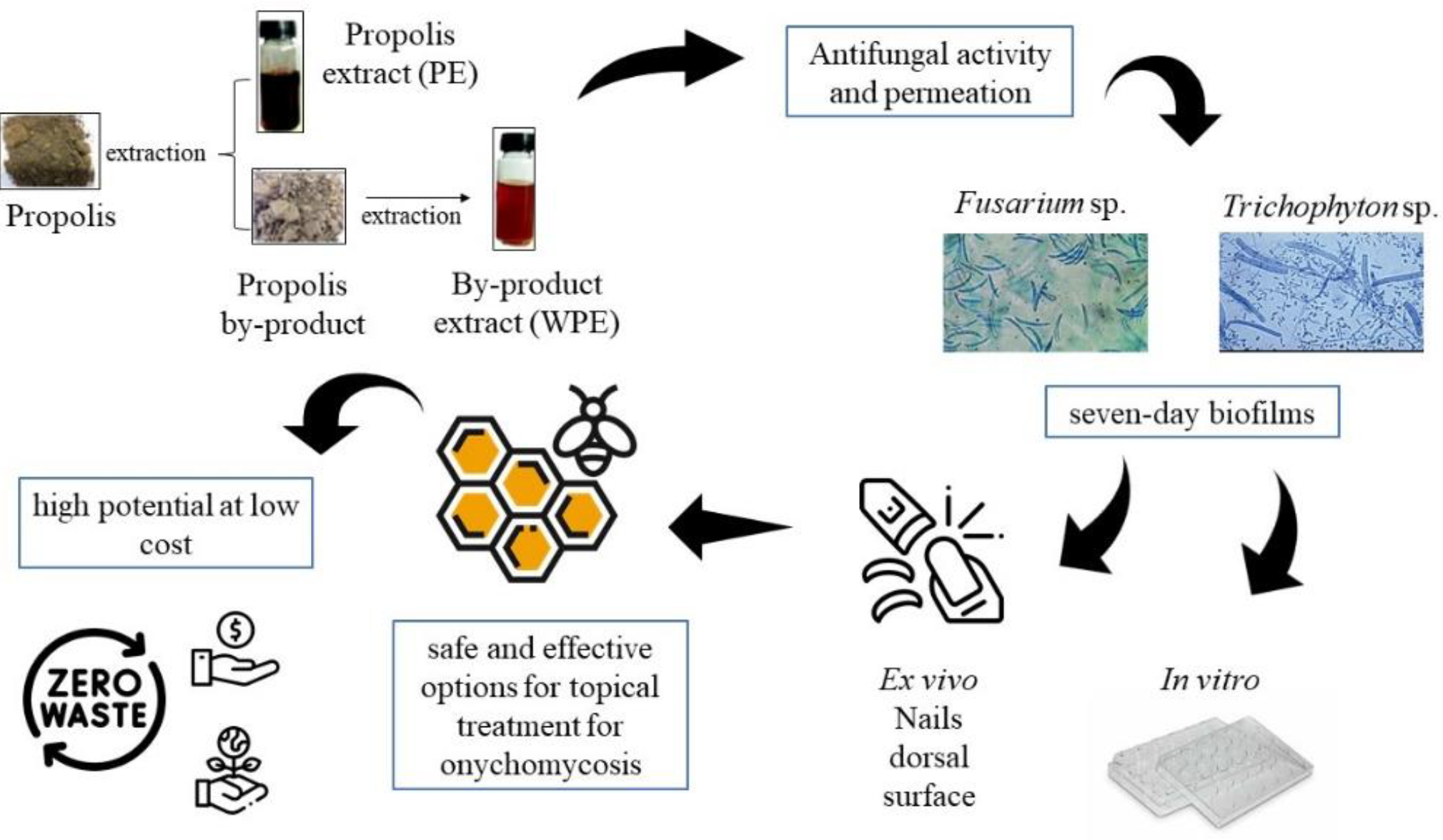
Biofilms of these fungi were also formed for seven days on the dorsal surface of nail fragments [18]. These were used for an evaluation on the permeation of PE and WPE extracts by photoacoustic spectroscopy (PAS), as previously described [10]. First, the spectra of extracts and of the infected nails (with biofilm) were analyzed separately, for control. Nail spectra were obtained 24 h after application of 2 µL of the extracts on its dorsal surface. To assess the permeation of PE and WPE in the nail, the samples were illuminated first on the dorsal side and then on the ventral side to detect the optical absorption band of the extracts. To be indicative of permeation along the thickness of the nail, the extract bands should be detected on the ventral side, opposite the application. The present study was carried out as shown in Figure 1.
RESULTS
In vitro antibiofilm activity of PE and WPE extracts
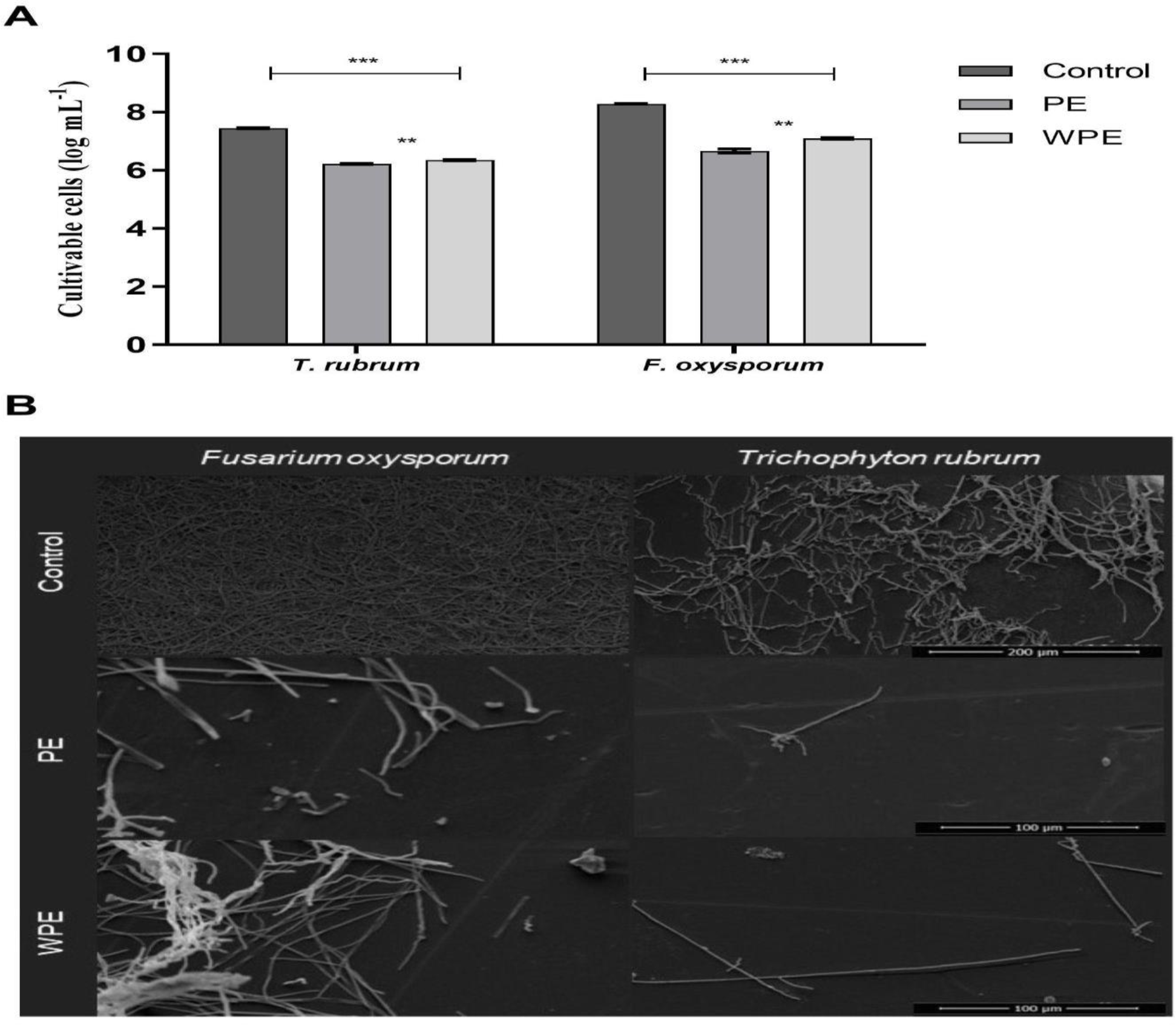
Both extracts were able to reduce at least one log of the cultivable cells from biofilms formed by the two fungi (p < 0.001) about the untreated control (Figure 2A). There was also a significant difference between the two extracts on biofilms of F. oxysporum (p < 0.001) and T. rubrum (p < 0.01). The effectiveness of PE and WPE on mature biofilms was evidenced by SEM images (Figure 2B), the drastic reduction in the amount of CFU accompanied by significant disorganization of the biofilms structure is visible.
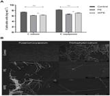
A - Antibiofilm effect of PE and WPE extracts at twice the MIC on seven-day preformed biofilms, in flat-bottomed polystyrene plates. ** Statistical difference between the PE and WPE extracts. *** Statistical difference between controls and treated biofilms. B - Morphological illustration, by SEM of biofilms of T. rubrum and F. oxysporum treated with PE and WPE extracts (magnification of 1000; scale bar = 100μM) and untreated (magnification of 500; scale bar = 200μM)
Evaluation of permeation of PE and WPE extracts in infected nails, using Photoacoustic Spectroscopy (PAS)
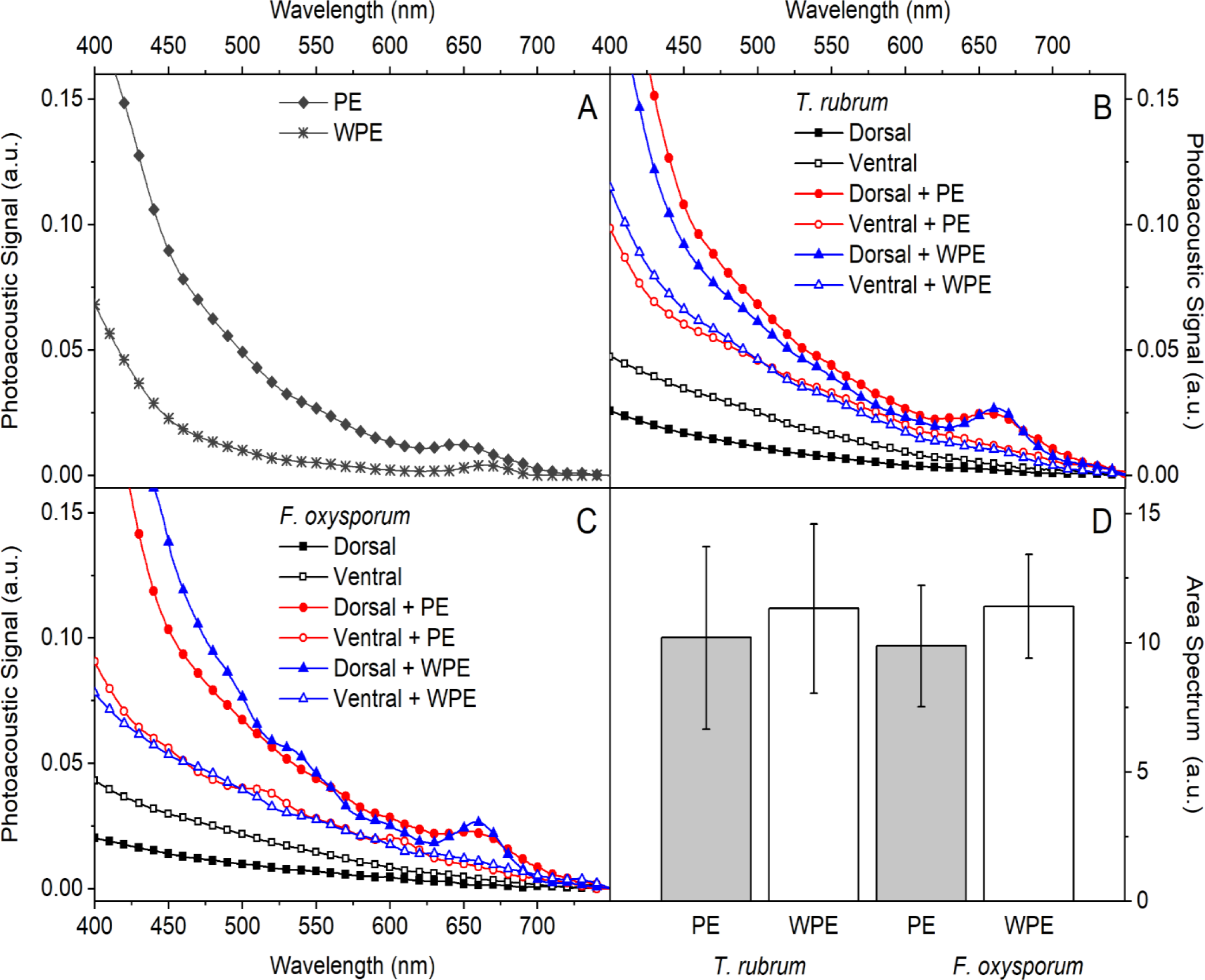
The PE and WPE extracts were evaluated individually in the region of 400 to 750 nm by PAS, before being applied to the nail. The optical absorption spectra obtained are shown in Figure 3A, in which both showed broadband absorption below 700 nm.
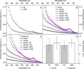
Optical absorption spectra of PE and WPE extracts, applied once on the dorsal surface and evaluated, after 24 h, by the dorsal (filled markers) and ventral (empty markers) surfaces of human nails. Red circles indicate nails previously infected and treated with PE; Blue triangles indicate nails previously infected and treated with WPE; Black squares indicate infected and untreated nails. (A) Extracts alone. (B) Nail infected by T. rubrum. (C) Nail infected by F. oxysporum. (D) Area under the curve obtained through the integral of the ventral spectra, after 24 h of a single application of the PE and WPE extracts.
Human nail fragments with biofilm experimentally produced on their dorsal surface, by one of the two fungi at a time, were treated with PE or WPE, also from the dorsal surface, provided the respective optical absorption spectra: T. rubrum (Figure 3B), F. oxysporum (Figure 3C). Reading from the dorsal surface was performed just to confirm the presence of extracts at the place of application. The absorption spectra obtained are similar to those of pure extracts (filled markers, Figure 3B and C). The spectrum obtained from the ventral surface of the nail after application of the extracts showed an increase in absorption in the region below 700 nm, when compared to the spectra of the infected nail alone (empty markers, Figure 3B and C). This increase confirms a complete permeation since PE and WPE were applied on the dorsal surface and they were detected on the ventral surface. To quantify the permeation, the area under the ventral spectra curve was integrated (Figure 3D). It was clear that WPE permeated the nails better than PE.
DISCUSSION
This study shows a comparative study between the action of the alcoholic extract of propolis (PE) and the extract of its by-product (WPE), on mature monospecies biofilms produced by Fusarium oxysporum and Trichophyton rubrum, two filamentous fungi, important agents of onychomycosis. The use of propolis ethanolic extracts for the treatment of these type of infections constitutes an important strategy, considering the safety and effectiveness of propolis as antifungal compound. Moreover, the use of propolis by-product constitutes an exclusive and an environmentally friendly purpose. WPE may yet be an important source of active compounds and show equivalent or better antifungal activity against these types of biofilms than PE. The results reinforced the action of both extracts PE and WPE on the nail biofilm produced by another agent, the yeast C. albicans [14]. Our group had already proven the effectiveness of PE and WPE in inhibiting the initial phase of biofilm formation of these filamentous fungi [13]. However, evaluating the impact of these extracts on preformed biofilms (mature) is extremely important, since onychomycosis is a consequence of this condition, and it is related to old biofilms [4,6,7]. The fungal organization in biofilms is highly complex and the cells are surrounded by a matrix that provides protection and resistance to antifungal treatment [15].
In the current study, PE and WPE at twice the MIC, previously determined [13] showed an important action of disrupting preformed biofilms by the evaluated fungi. These results were surprising since fungal cells from the biofilms are usually much more resistant than their planktonic counterparts. A 1000-fold decrease in susceptibility has already been documented in mature bacterial and fungal biofilms, requiring high doses of antimicrobials for an effective treatment [5]. Multiple components of the biofilm contribute to this acquired resistance, among them the difficulty of penetration of the agents due to the presence of ECM, as well as the presence of efflux pumps, persistent cells, and reduction of ergosterol [5,6]. Recently, the volatile organic compound 2-ethyl-1-hexanol (2EH) was described for the first time as a component of quorum sensing in the ex vivo biofilm of F. oxysporum on human nails, which was capable of modulating the biofilm formed, suggesting that this molecule plays an important role in the biofilm's resistance to antifungals [16].
The results obtained with the PE used in this study corroborate the effectiveness of other propolis extracts evaluated on biofilms formed by the same species [9-11]. This is the first time that WPE was evaluated on mature biofilms formed by filamentous fungi and there was a significant reduction against the two evaluated fungi (Fig. 2). This reinforces our idea that WPE, an extract obtained from a by-product during the PE extraction process, could also be used in the treatment of onychomycosis. We emphasize that onychomycosis is a chronic infection attributed to fungi organized in the form of mature biofilms and that currently none of the antifungals available on the market are capable of disorganizing previously formed biofilms. The eradication of mature biofilms is probably associated with the amount of polyphenols since the release of these compounds from WPE is faster than from PE [17]. Additionally, the fact that PE has a higher resin content [12], probably results in a more prolonged release and action of polyphenols in biofilms, justifying the better effect of PE. Interestingly, SEM showed the disorganization of mature biofilms by both extracts, which is important both to prove the effectiveness of the treatment and to prevent reinfection of the nail by other agents that could take advantage of the structure of the remaining old biofilm.
As important as the antibiofilm action of the extracts is the permeation capacity of PE and WPE across nails infected by fungi, an important feature for the topical treatment of onychomycosis [3]. Our results showed that both extracts were able to permeate nails infected by a dermatophyte and an NDM. Probably this property has been favored by the ability of filamentous fungi to easily invade the nail since their hyphae facilitate the penetration across the nail’s layers [7,11,18]. Furthermore, the porosity of the infected nail has been reported as a factor that facilitates the permeation of drugs in the human nail tissue, due to the increase in its thickness [3,19,20]. This concept was formed on classical onychomycosis, caused by dermatophyte fungi, corroborating our findings. Therefore, it is possible to hypothesize that the porosity caused in the nail during an infectious process, by filamentous fungi facilitates the permeation of extracts in the nail. Furthermore, the polarity and chemical compositions of propolis enable good retention and permeation [21,22]. It was also evident that WPE permeated the nails better, probably because it has a lower content of dry residue (less resin) [12,13], which would facilitate its diffusion through the nail. Further studies must be accomplished in order to prove this hypothesis.
These results have shown the potential application of both PE and WPE for the treatment of onychomycosis. Despite the different physicochemical properties and permeation trough the nail of these two types of propolis extracts, it is necessary to improve the final dosage form for their delivery on the nail. The development of formulations displaying improved rheological performance, bioadhesiveness and that can control the propolis extract delivery is necessary. Thus, the propolis extract can be administered on the nail in a suitable way, improve the stay at the site of application and control the PE or WPE delivery.
CONCLUSION
It was demonstrated that both PE and WPE could disrupt mature biofilms of important onychomycosis agent fungi, in addition to permeating infected nails. This reinforces the idea that these extracts would be safe and effective options for topical treatment for onychomycosis. Highlighting WPE, a compound produced in large quantities which would be discarded, in our studies it presented excellent antibiofilm activity and high permeation in the nails showing high potential at low cost. Further studies are necessary to develop final pharmaceutical dosage forms for improved administration and control delivery of PE and WPE on nail.
-
Funding:
This work received partial financial support from Coordenação de Aperfeiçoamento de Pessoal de Nível Superior - Brasil (CAPES) [Finance Code 001, through a scholarship awarded to ILEB]; Conselho Nacional de Desenvolvimento Científico e Tecnológico - Brasil [Process number 3128262023-0 given to TIES] and INCT-CERBC, 2022, Process number 406645/2022-1.
-
Institutional Review Board Statement:
Not applicable.
-
Informed Consent Statement:
Not applicable.
Acknowledgments:
The authors thank the Research Support Center Complex (COMCAP) at the UEM, maintained by FINEP and Fundação Araucária for supporting our research laboratory.
Data Availability Statement:
Research data are available in the body of the manuscript.
REFERENCES
-
1 Gupta AK, Stec N, Summerbell RC, Shear NH, Piguet V, Tosti A, et al. [Onychomycosis: a review]. J Eur Acad Dermatol Venereol. 2020; 34:1972-90. doi:10.1111/jdv.16394
» https://doi.org/10.1111/jdv.16394 -
2 Maskan Bermudez N, Rodríguez-Tamez G, Perez S, Tosti A. [Onychomycosis: Old and New]. J Fungi (Basel). 2023; 9: 559. doi:10.3390/jof9050559
» https://doi.org/10.3390/jof9050559 -
3 Costa P de S, Mendes V, Veiga FF, Negri M, Svidzinski TIE. [Relevant insights into onychomycosis’ pathogenesis related to the effectiveness topical treatment]. Microb Pathog. 2022; 169:105640. doi:10.1016/j.micpath.2022.105640
» https://doi.org/10.1016/j.micpath.2022.105640 -
4 Veiga FF, de Castro-Hoshino LV, Rezende PST, Baesso ML, Svidzinski TIE. [Insights on the etiopathogenesis of onychomycosis by dermatophyte, yeast and non-dermatophyte mould in ex vivo model]. Exp Dermatol. 2022; 31(11): 1810-14. doi:10.1111/exd.14643
» https://doi.org/10.1111/exd.14643 -
5 Gupta AK, Daigle D, Carviel JL. [The role of biofilms in onychomycosis]. J Am Acad Dermatol. 2016; 74: 1241-46. doi:10.1016/j.jaad.2016.01.008
» https://doi.org/10.1016/j.jaad.2016.01.008 -
6 Gupta AK, Foley KA. [Evidence for biofilms in onychomycosis]. G Ital Dermatol Venereol. 2019; 154: 50-5. doi:10.23736/S0392-0488.18.06001-7
» https://doi.org/10.23736/S0392-0488.18.06001-7 -
7 Veiga FF, de Castro-Hoshino LV, Sato F, Baesso ML, Silva S, Negri M, et al. [Characterization of a biofilm formed by Fusarium oxysporum on the human nails]. Int J Dermatol. 2022; 61: 191-8. doi:10.1111/ijd.15747
» https://doi.org/10.1111/ijd.15747 -
8 Gupta AK, Elewski B, Joseph WS, Lipner SR, Daniel CR, Tosti A, et al. [Treatment of onychomycosis in an era of antifungal resistance: Role for antifungal stewardship and topical antifungal agents]. Mycoses. 2024; 67: e13683. doi:10.1111/myc.13683
» https://doi.org/10.1111/myc.13683 -
9 Galletti J, Tobaldini-Valerio FK, Silva S, Kioshima ÉS, Trierveiler-Pereira L, Bruschi M, et al. [Antibiofilm activity of propolis extract on Fusarium species from onychomycosis]. Future Microbiol. 2017; 12:1311-21. doi:10.2217/fmb-2017-0052
» https://doi.org/10.2217/fmb-2017-0052 -
10 Veiga FF, Gadelha MC, da Silva MRT, Costa MI, Kischkel B, de Castro-Hoshino LV, et al. [Propolis Extract for Onychomycosis Topical Treatment: From Bench to Clinic]. Front Microbiol. 2018; 9: 779. doi:10.3389/fmicb.2018.00779
» https://doi.org/10.3389/fmicb.2018.00779 -
11 Vasconcellos-Pontello V, Veiga FF, Gadelha MC, Ribeiro M, Negri M, Estivalet Svidzinski TI. [The Success of Topical Treatment of Onychomycosis Seems to Be Influenced by Fungal Features]. Evid Based Complement Alternat Med. 2018; 5553634. doi:10.1155/2021/5553634
» https://doi.org/10.1155/2021/5553634 -
12 De Francisco L, Pinto D, Rosseto H, Toledo L, Santos R, Tobaldini-Valério F, et al. [Evaluation of radical scavenging activity, intestinal cell viability and antifungal activity of Brazilian propolis by-product]. Food Res Int. 2018; 105: 537-47. doi:10.1016/j.foodres.2017.11.046
» https://doi.org/10.1016/j.foodres.2017.11.046 -
13 Barros ILE, Veiga FF, Jarros IC, Corrêa JL, Dos Santos RS, Bruschi ML, et al. [Promising effect of propolis and a by-product on planktonic cells and biofilm formation by the main agents of human fungal infections]. An Acad Bras Cienc. 2022; 94: e20210189. doi:10.1590/0001-3765202220210189
» https://doi.org/10.1590/0001-3765202220210189 -
14 Barros ILE, Veiga FF, de Castro-Hoshino LV, Souza M, Malacrida AM, Diniz BV, et al. [Performance of Two Extracts Derived from Propolis on Mature Biofilm Produced by Candida albicans] Antibiotics (Basel). 2022; 12. doi:10.3390/antibiotics12010072
» https://doi.org/10.3390/antibiotics12010072 -
15 Pereira F de O. [A review of recent research on antifungal agents against dermatophyte biofilms]. Med Mycol. 2021; 59: 313-26. doi:10.1093/mmy/myaa114
» https://doi.org/10.1093/mmy/myaa114 -
16 Veiga FF, Marcomini EK, Salvador A, Chiavelli LUR, Barros ILE, de Castro LV, et al. [Detection of 2-ethyl-1-hexanol and its modulating effect in biofilm of Fusarium oxysporum] Mol Microbiol. 2023. doi:10.1111/mmi.15194
» https://doi.org/10.1111/mmi.15194 -
17 Rosseto HC, Toledo L de AS de, Francisco LMB de, Esposito E, Lim Y, Valacchi G, et al. [Nanostructured lipid systems modified with waste material of propolis for wound healing: Design, in vitro and in vivo evaluation]. Colloids Surf B Biointerfaces. 2017;158: 441-52. doi:10.1016/j.colsurfb.2017.07.029
» https://doi.org/10.1016/j.colsurfb.2017.07.029 -
18 Veiga FF, de Castro-Hoshino LV, Sato F, Bombassaro A, Vicente VA, Mendes V, et al. [Fusarium oxysporum is an onychomycosis etiopathogenic agent]. Future Microbiol. 2018; 13:16, 1745-56. doi:10.2217/fmb-2018-0245
» https://doi.org/10.2217/fmb-2018-0245 -
19 Baraldi A, Jones SA, Guesné S, Traynor MJ, McAuley WJ, Brown MB, et al. [Human nail plate modifications induced by onychomycosis: implications for topical therapy]. Pharm Res. 2015; 32: 1626-33. doi:10.1007/s11095-014-1562-5
» https://doi.org/10.1007/s11095-014-1562-5 -
20 McAuley WJ, Jones SA, Traynor MJ, Guesné S, Murdan S, Brown MB. [An investigation of how fungal infection influences drug penetration through onychomycosis patient’s nail plates]. Eur J Pharm Biopharm. 2016; 102:178-84. doi:10.1016/j.ejpb.2016.03.008
» https://doi.org/10.1016/j.ejpb.2016.03.008 -
21 Toledo L de AS de, Bavato MI, Rosseto HC, Cortesi R, Bruschi ML. [Pharmaceutical films made from the waste material from the preparation of propolis extracts: development and characterization]. Braz J Pharm Sci. 2015; 51:847-59. doi:10.1590/s1984-82502015000400011
» https://doi.org/10.1590/s1984-82502015000400011 -
22 De Toledo L de AS, Rosseto HC, Ravani L, Cortesi R, Bruschi ML. [Waste material of propolis as a film forming agent intended to modify the metronidazole release: preparation and characterization]. Curr Drug Deliv. 2016; 13:1152-64. doi:10.2174/1567201813666151113121827
» https://doi.org/10.2174/1567201813666151113121827
-
Editor-in-Chief:
Paulo Vitor Farago
-
Associate Editor:
Paulo Vitor Farago