Preliminary Evaluation of HSA and DNA Interactions with Indole-Thiosemicarbazone Compounds and Molecular Docking Studies
Abstract
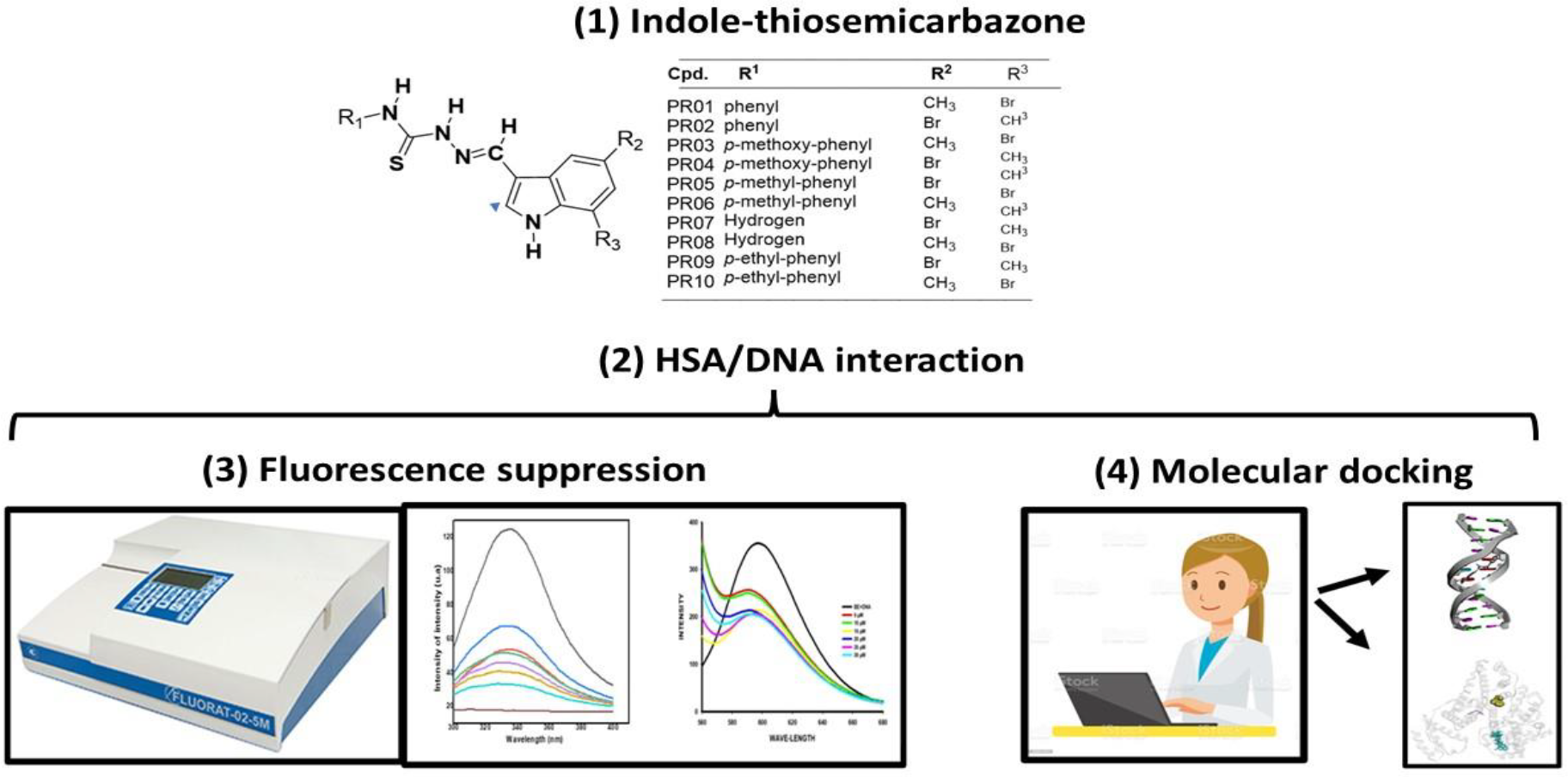
Indole-thiosemicarbazones have different biological activities. The present study evaluated the preliminary interaction of these compounds with biomolecules, specifically human serum albumin (HSA) and DNA, using fluorescence techniques. The suppression results (Ksv) for HSA ranged from 3.5 x 10⁴ to 4.6 x 10⁵ L/mol, while for DNA, they ranged from 1.4 x 10⁴ to 5.9 x 10⁴ L/mol. The suppression was classified as weak to strong for HSA and moderate for DNA. The bimolecular suppression constant (Kq) showed values between 6.0 x 10¹² and 8.2 x 10¹³ L/mol/s for HSA and from 1.4 x 10¹² to 5.9 x 10¹² L/mol/s for DNA, suggesting a static suppression mechanism. Compound PR09 stood out, presenting a binding constant (Ka) greater than 105 L/mol for HSA, indicating a strong interaction. Additionally, PR05, PR06, PR07, and PR09 demonstrated strong interactions with DNA. The values of the number of binding sites (n) indicated that PR01, PR02, and PR09 bind to multiple sites on HSA, while PR04, PR05, PR06, and PR09 interact with more than one site on DNA. All interactions were spontaneous, with ∆G negative. The distance between the compounds and tryptophan on HSA was less than 8 nm, suggesting high energy transfer efficiency. In molecular docking studies, PR05 showed the highest affinity for DNA (-11.15 kcal/mol), while PR09 had the highest affinity for HSA (-10.00 kcal/mol). PR07 exhibited the lowest binding energies for DNA (-8.21 kcal/mol) and for HSA (-7.38 kcal/mol). This study demonstrates that the evaluated compounds have potential as new drug candidates.
Keywords:
thiosemicarbazone; interaction; macromolecules.
HIGHLIGHTS
Indole-thiosemicarbazone compounds are potential drug candidates.
For albumin, the compounds were able to promote weak to strong suppression.
For DNA, the compounds were able to promote moderate suppression.
The in silico studies proved the interaction of the compounds with the macromolecules.
INTRODUCTION
Thiosemicarbazones are an important class of compounds widely studied in medicinal chemistry due to their diverse biological applications, including anti-inflammatory, antibacterial, antifungal, antiviral, and antiparasitic activities [1]. The indole nucleus is frequently found in nature and constitutes several molecules present in plants, bacteria, and animals, such as alkaloids and hormones, including serotonin and melatonin. It is also a component of the amino acid tryptophan [2]. This structure is commonly utilized in the development of new molecules, known for promoting various biological activities, including antitumor, antibacterial, antiviral, antiparasitic, and anti-inflammatory effects [3]. In this context, indole-thiosemicarbazone compounds, which arise from the combination of an indole nucleus with a thiosemicarbazone, have garnered interest in research due to their diverse biological properties and potential applications in medicine [4].
Several studies have highlighted the versatility of indole-thiosemicarbazone compounds in exhibiting diverse biological activities. Silva and coauthors [5] evaluated these compounds as potential leishmanicidal agents and found that their toxicity in macrophage cells ranged from 53.23 to 357.97 μM. In terms of leishmanicidal activity, the effective concentrations varied between 12.31 and over 481.52 μM against Leishmania amazonensis and between 4.36 and 23.35 μM against Leishmania infantum. Ultrastructural analysis revealed that these compounds caused significant damage to the parasites, including shrinkage of the cell body, shortening and loss of the flagellum, severe mitochondrial swelling, and cytoplasmic vacuolization, ultimately leading to cellular inviability. Under the evaluated experimental conditions, these compounds demonstrated promising potential as leishmanicidal agents.
Jacob and coauthors [6] investigated the anti-inflammatory properties of indole-thiosemicarbazone compounds, specifically LT76, LT81, and LT87. These compounds were found to inhibit the in vitro production of TNF-α and NO, while also stimulating IL-4. Additionally, they demonstrated the ability to inhibit COX-2 in vitro assays. In animal models, LT76 (64.8% inhibition after 6 h), LT81 (89% after 6 h), and LT87 (100% after 4 h) significantly reduced edema in mice inoculated with carrageenan, outperforming the efficacy of indomethacin. Immunohistochemical analysis further confirmed that the groups treated with these compounds exhibited reduced COX-2 expression, with results comparable to or superior to those achieved with indomethacin.
Jacob and coauthors [7] evaluated indole-thiosemicarbazone compounds as potential antitumor agents, investigating their interactions with key molecular targets, including human serum albumin (HSA), DNA, and human topoisomerase IIα (topo). They also assessed the cytotoxic activity of these compounds in normal cells and various tumor cell lines, such as DU-145, Jurkat, MCF-7, T-47D, and J774A.1. The results indicated that the compounds could interact with HSA without compromising the protein's integrity. Furthermore, DNA and topoisomerase emerged as primary molecular targets for inhibition. In vitro assays demonstrated that the compounds effectively inhibited the growth of the DU-145, Jurkat, MCF-7, T-47D, and J774A.1 tumor cell lines.
Santos and coauthors [8] reported that indole-thiosemicarbazone compounds exhibited good oral bioavailability in in silico studies. In in vitro assays, these compounds demonstrated antioxidant activity and low toxicity in normal mammalian cells, while effectively inhibiting the growth of tumor cell lines, including T-47D, MCF-7, Jurkat, and DU-145. These findings suggest that indole-thiosemicarbazone compounds hold significant potential as antitumor agents.
Given the potential of these structures, studies with various biomolecules have been conducted to identify possible interaction mechanisms, focusing on evaluating their transport in the bloodstream and/or potential toxic effects [7]. The binding of drugs to plasma proteins plays a crucial role in influencing their pharmacological properties. Such binding can increase the drug's half-life by preventing its rapid elimination from circulation, while also enhancing its stability and reducing toxicity, ultimately contributing to improved therapeutic outcomes. This interaction occurs reversibly, and the drug’s therapeutic effects are attributed only to the unbound fraction circulating in the body [9].
If the binding between a drug and plasma proteins is weak, the drug will be rapidly metabolized and excreted, leading to a short-lived therapeutic effect. Conversely, stronger binding can prolong the drug’s retention time in the body, potentially increasing toxicity and side effects, while also enhancing therapeutic benefits [7, 9]. Given the significant influence of plasma proteins on therapeutic outcomes, it is essential to investigate the ability of drug candidates to bind to proteins like human serum albumin (HSA). Albumin is the most abundant plasma protein, comprising about 55-60% of all plasma proteins. It plays crucial roles in maintaining osmotic pressure, exhibiting antioxidant properties, participating in platelet aggregation and cell signaling, and facilitating the transport and distribution of drugs, hormones, and fatty acids. By binding to specific sites, albumin directly influences drug bioavailability and, consequently, their mechanisms of action [9]. Structural damage to this protein can impair these essential functions and cause harm to the body.
Another extensively studied biomolecule is DNA, which plays a key role in fundamental biological processes, such as protein synthesis and cell replication. The binding of small molecules to DNA can alter its structure, function, and stability, disrupting these processes and potentially leading to cell death. This mechanism serves as a potential foundation for therapies aimed at treating and preventing diseases [7,9]. As a result, there is a growing demand for studies focused on discovering new drugs that act at the DNA level, offering greater selectivity and reduced cytotoxicity.
The objective of this study was to evaluate the interaction of indole-thiosemicarbazone compounds with key biomolecules (HSA and DNA), with the interaction mechanism assessed through molecular docking simulations.
MATERIAL AND METHODS
Reagents
The reagents used for the synthesis and analysis of thiosemicarbazones were: hydrazine solution (CAS:302-01-2), methylene chloride (CAS:75-09-2), 7-Bromo-5-methylindole-3-carboxaldehyde ( CAS: 16077-60-4), 5-Bromo-7-methylindole-3-carboxaldehyde (CAS: 16076-86-1), phenyl isothiocyanate (CAS: 103-72-0), 4-Methoxyphenyl isothiocyanate (CAS: 2284) -20-0), 4-Methylphenyl isothiocyanate (CAS: 622-59-3), ethyl alcohol (CAS: 64-17-5), glacial acetic acid (CAS:1186-52-3), Ethidium bromide (CAS:1239-45-8) 4′,6-Diamidine-2′phenylindole dihydrochloride (DAPI) (CAS:28718-90-3), Tris[hydroxymethyl]-aminomethane (Tris HCl - Sigma/Merck), Human Serum Albumin (HSA - Sigma/Merck), DNA - Deoxyribonucleic acid sodium salt from salmon Tests (CAS: 438545-06-3). All reagents provided by Sigma/Merck. The solvents were ethyl alcohol, dichloromethane, dimethyl sulfoxide (DMSO), in addition to glacial acetic acid, provided by Dinâmica.
Indole-thiosemicarbazone compounds
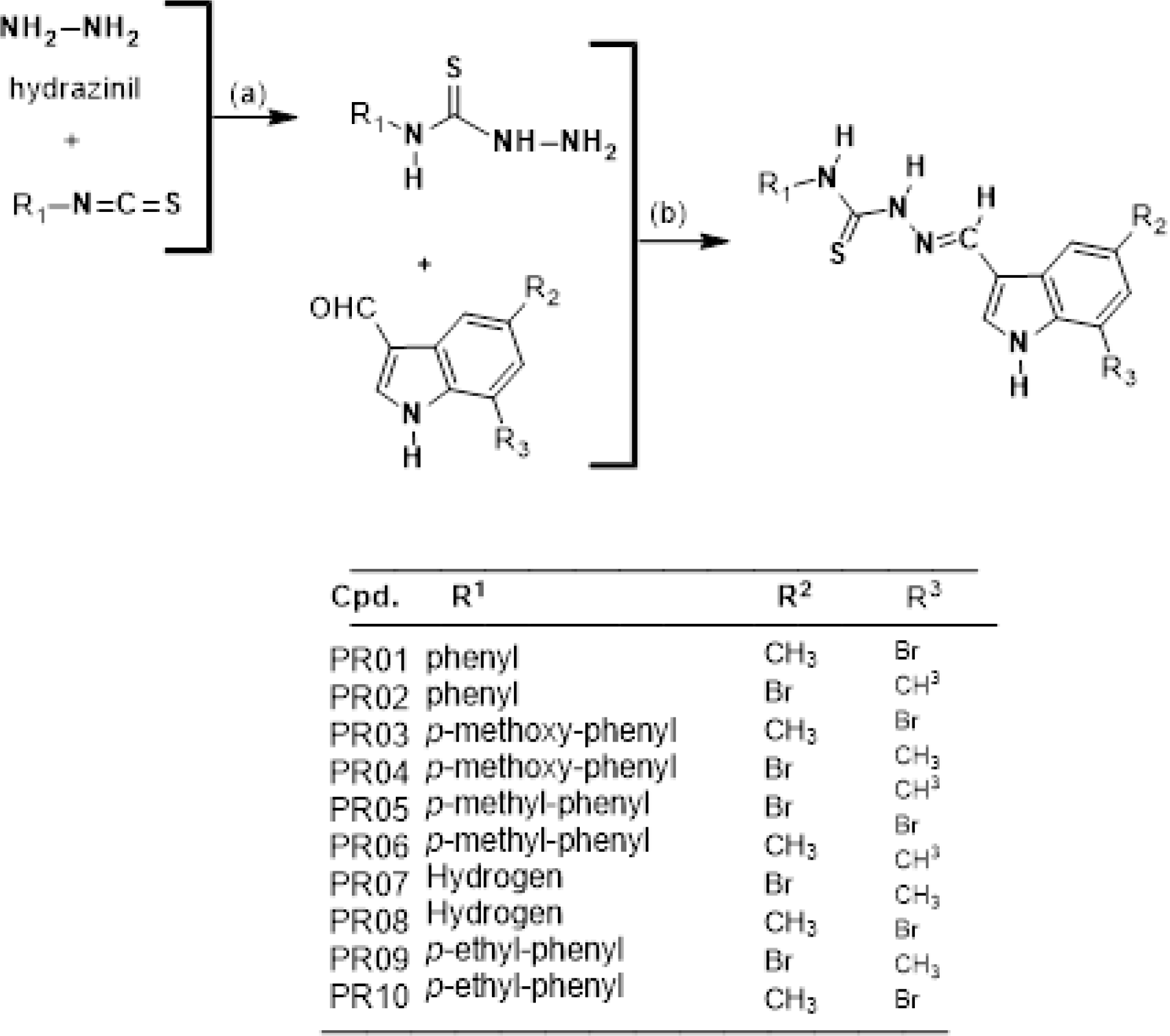
The synthesis was carried out at the Chemistry and Therapeutic Innovation Laboratory of the Federal University of Pernambuco (UFPE), Recife, Pernambuco, Brazil and published by Silva and coauthors [5]. Obtaining the indole-thiosemicarbazone compounds (PR1 - PR10) was carried out in two steps (Figure 1). Initially (a), the thiosemicarbazides were obtained from the linker hydrazinyl (hydrazine) with the unsubstituted and substituted isothiocyanates. Then (b), the thiosemicarbazones react with the substituted 3-indole-carboxaldehydes in the presence of acetic acid as a catalyst, originating the substituted Thiosecarbazones (PR1 - PR10).
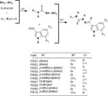
Reagents and Conditions: (a) hydrazine, substituted isothiocyanate, chloroform, temperature 30 ± 0.5 ºC; (b) thiosemicarbazide, substituted 3-indole-carboxaldehyde, absolute ethanol, acetic acid as catalyst, temperature: 75 ± 0.5 ºC.
Interaction assays by fluorescence spectroscopy
Interaction study of thiosemicarbazone/albumin compounds
The study of the interaction between the compounds and human serum albumin (HSA) was performed through fluorescence spectroscopy, following methodology adapted from Jacob and coauthors [7], Santos and coauthors [9] and Xiao and coauthors [10]. The compounds were diluted in DMSO and diluted in concentrations ranging from 5 to 80 µM and a 10 µM HSA solution (100 nM Tris HCl buffer, pH 7.5), in a final volume of 1mL. The systems were submitted to fluorescence analysis in a Jasco FP-6300 fluorometer, using a quartz cuvette with a 1cm optical path. The parameters used were Emission intensity scan between 300 and 400 nm, excitation length of 285 nm, emission and excitation bands of 2.5 nm, with medium response and high sensitivity, at a scanning speed of 200 nm/min.
Thiosemicarbazones/DNA compound interaction study
The fluorescent emission spectroscopy assays were performed according to the methodology proposed by Jacob and coauthors [7] and Nagaraj and coauthors [11] with few modifications. A solution of salmon sperm DNA (DNA) was prepared in Tris HCl buffer (10 mmol/L, pH = 7.4 ± 0.10) with 0.1 mol/L NaCl for ionic strength adjustment. Nucleic acid purity was determined by spectrophotometer (Perkin Elmer, Lambda 650) at wavelengths at 260 and 280 nm. ABS260/ABS280 ratio values ranging between 1.8 and 1.9 indicate that the DNA is free of protein contamination. In addition, to calculate the DNA concentration, ABS260 was used, with a molar extinction coefficient of 6600 L/mol. The compounds were previously solubilized in 1% DMSO and diluted in Tris HCl buffer (10 mmol/L, pH = 7.4 ± 0.10) in concentrations ranging from 0 to 30 µM. Interaction assays were performed using fixed concentrations of ethidium bromide (10 μM) and DNA (100 μM) with a reaction volume of 1.0 mL and incubated for 10 min at 25 ºC. Measurements were then performed in a rectangular quartz cuvette with a path length of 1 cm, in the JASCO FP-6300 spectrofluorimeter (Tokyo, Japan). The spectra were from the reaction system containing different concentrations of compounds (0 to 30 µM) were obtained using the following parameters: excitation wavelength of 526 nm and emission scan between 550 - 700 nm.
Obtaining interaction parameters
The interaction parameters of the compounds between albumin and DNA were obtained through the linearized Stern-Volmer equations (Equations 1 and 2) and Hill (Equation 3), in addition, the Gibbs free energy was calculated (Equation 4).
Where: F0 and F are the relative fluorescence intensities in the absence and presence of compound, respectively, [C] is the compound concentration (µM), Ksv is the Stern-Volmer constant (L/mol), Kq is the constant of bimolecular rate of suppression (L/mol/s), n is the number of binding sites and τ0 is the average lifetime of the fluorophore in the excited state, which has an approximate value of ~ 5.6 × 10- 9 s (HSA) and 10- 8 s (DNA).
In addition to the interaction parameters were the Forster-type resonance energy transfer (FRET) parameters. These experiments were carried out according to the methodology proposed by Siddiqi and coauthors [12] with few modifications. Compounds were dissolved in 1% DMSO at a concentration of 40 µM. Then they were analyzed in a spectrophotometer at wavelengths ranging from 280 and 650 nm. Albumin fluorescence spectra were obtained in a Jasco spectrofluorimeter, model FP-6300, and in a Lambda model spectrophotometer, Perkin Elmer (USA) also at wavelengths ranging from 280 and 650 nm. FRET was determined from the superimposition of the fluorescence absorption and emission spectra. Using equations 5, 6 and 7, it was possible to determine the overlapping area between the absorption and fluorescence spectra (J), the Forster distance (R0), the energy transfer efficiency (E) and the distance between the donor and the acceptor (r).
Where: k2 is a factor describing the relative orientation in space of the donor and acceptor transition dipoles and is numerically equal to 2/3, n is the refractive index and is equal to 1.33, ϕD is the quantum yield of donor fluorescence which is numerically equal to 0.13. ID(λ) is the normalized emission intensity at a given wavelength. ɛA(λ) is the excitation coefficient of the receiver at a given wavelength.
Docking molecular
Docking molecular analyzes were performed using AutoDock4.2.6 in combination with the Lamarckian genetic algorithm [13].
Preparation of the ligand structure
The 3D structures were built with Avogadro 1.2.0 software) and fully optimized with the PM6 semi-empirical method implemented in MOPAC2016 [14,15]. Optimized binders were saved as pdb files. Using AutoDockTools-1.5.6, the nonpolar hydrogens were mixed with the corresponding carbons, then the partial charges of the atoms were calculated using the Gasteiger procedure implemented in the AutoDockTools package. Linkers rotary links were defined, structures were saved as pdbqt and used for docking studies.
Target structure preparation
The crystallographic structures of the DNA used for intercalation (d(CGATCG) 2 hexamer complexed with ellipticin, PDB ID: 1Z3F) and minor groove ligation (d(CGCGAATTCGCG) 2 dodecamer, PDB ID: 1BNA) were obtained from the Protein Data Bank. Using Dassault Systèmes BIOVIA Discovery Studio Visualizer (v16.1.0.15350) [16], water molecules (and intercalated ellipticin present in 1Z3F) were removed and polar hydrogens were added to the macromolecules. Then, using AutoDockTools, the Gasteiger loads were added and the structures were saved as pdbqt for the docking studies.
The crystallographic structure of HSA was obtained from the Protein Data Bank (PDB ID:1AO6). Using Dassault Systèmes BIOVIA Discovery Studio Visualizer (v16.1.0.15350), water molecules were removed and polar hydrogens were added to the macromolecule. Then, using AutoDockTools, Kollman loads were added and the structure was saved as pdbqt for the docking studies.
Docking procedure
The 3D grid was created by the Autogrid algorithm to generate the grid parameters file. Grid spacing was 0.0375 nm in each dimension. For 1BNA, the DNA structure was placed inside a box composed of 80 × 90 × 120 grid points. For 1Z3F, the grid box was centered on the cocrystallized intercalating agent and contained 40 × 50 × 40 grid points. For HSA, the grid box was centered at coordinates x = 29,535, y= 31,826, and z=23,500 (the center of the protein) and contained 126 × 126 × 126 grid points.
The Lamarckian genetic algorithm in AutoDock4.2.6 was applied to search for the best conformation and orientation of the ligands. Global optimization was started with a population of 150 randomly placed individuals with a maximum of 2500000 energy evaluations and a maximum of 27000 generations. During each docking experiment, 100 runs were performed, generating 100 conformations. The resulting docking poses were analyzed using AutoDockTools and Discovery Studio Visualizer.
RESULTS AND DISCUSSION
Evaluation of the interaction of indole-thiosemicarbazone compounds with HSA and DNA
A preliminary study of the interaction of the compounds with albumin and DNA is necessary to evaluate a possible mechanism [17]. Albumin is the most abundant protein in plasma, produced in the liver; it can interact with different exogenous and endogenous compounds present in the blood, transporting them throughout the body [18]. Therefore, compounds are sought that interact with albumin without altering its macromolecular conformation [17,18]. In addition to albumin, a target macromolecule for the study of the mechanism of compounds is DNA. This is because damage to this macromolecule compromises the functioning of the organism [7,19].
Figures S1, S2, S3 and S4 of the supplementary material show the fluorescence spectra of the interaction of the compounds with HSA and DNA respectively. In this study, it was observed that the fluorescence emission of HSA and DNA was suppressed by indole-thiosemicarbazone compounds (with the addition of different concentrations of compounds). Regarding HSA, the addition of compounds promoted a gradual decrease in the maximum fluorescence intensity of HSA (Figures S1 and S2), which indicates that the position of interaction between compounds and HSA is located close to tryptophan residues [20]. With regard to DNA, the decrease in suppression (Figures S3 and S4) is an indication that an interaction has occurred.
Table 1 shows the interaction results of the compounds with albumin and with DNA. The parameters were obtained by linearized Stern-Volmer equations (Equations 1 and 2) and Hill (Equation 3). In addition, the Gibbs free energy was calculated (Equations 4). Figures S5 and S6 of the supplementary material show the linearization of the interaction data of the compounds with HSA and DNA respectively.
Linearized fluorescence results for the Stern-Volmer and Hill equations to obtain the association constants and number of binding sites and free energy values respectively.
The results of the suppression of the interaction of the compounds with albumin and with DNA were adjusted using the linearized Stern-Volmer equation (Table 1). It was observed that Ksv values for albumin ranged from 3.5 x10⁴ to 4.6 x10⁵ L/mol. Regarding DNA, Ksv values ranging from 1.4 x10⁴ to 5.9 x10⁴ L/mol were obtained, respectively. Through these results, it is possible to classify the suppression intensity according to an arbitrary scale proposed by Santos and coauthors [9]. Thus, compounds that show Ksv results ranging from 1x10³ to 1x10⁴ L/mol promote weak suppression, moderate suppression between 1x10⁴ and 1x10⁵ L/mol, and strong suppression ranging from 1x10⁵ to 1x10⁶ L/mol. For albumin, the compounds were able to promote weak to strong suppression. For DNA, the deletion was classified as moderate.
The bimolecular suppression constant (Kq) reflects the dynamic suppression efficiency, and can be used as a parameter to indicate the suppression mechanism. The value of Kq is calculated from the ratio between the SternVolmer constant (Ksv), and the lifetime of the fluorophore in the excited state (τ0), which for HSA is approximately ~ 5.6 × 10⁻⁹ s. The maximum value of the biomolecular suppression constant for a dynamic suppression event in water is limited to the value of 1010 L/mol/s [20].
Therefore, fluorescence quenching events, where the bimolecular quenching constant exceeds the maximum value for dynamic quenching, are considered static quenching events. That is, the fluorescence intensity of the fluorophore is suppressed by formation of a non-fluorescent complex between species in the ground state [20, 21]. The Kq values obtained by the interaction of the compounds with albumin and DNA were greater than 1010 L/mol/s, indicating a possible static suppression.
The literature presents different suppression results of thiosemicarbazone compounds interacting with DNA and albumin. Jacob and coauthors [7] evaluating indole-thiosemicarbazone compounds obtained Ksv values ranging from 1.12 × 10⁴ to 7.72 × 10⁴ L/mol for albumin indicating that the suppression of the compounds is moderate. As for DNA, they obtained values ranging from 3.90 × 10³ to 1.78 × 10⁶ L/mol using DAPI as a marker for these assays, the compounds were able to promote suppression ranging from weak to strong. Santos and coauthors [9] evaluating the interaction of different thiosemicarbazones with albumin obtained Ksv values that ranged from 3.5 × 10³ to 4.6 × 10⁶ L/mol, showing that the compounds were able to promote suppression ranging from weak to strong. In addition, they found that thiosemicarbazones and thiazoles compounds showed values ranging from 1.1 × 10³ to 9.0 × 10⁴ L/mol, indicating that suppression ranged from moderate to weak with DNA using ethidium bromide as a marker. In both studies the k values were greater than 1010 L/mol/s.
Since the mechanism is static, the binding constant (Ka) and the number of binding sites (n) can be obtained using the modified double logarithmic regression curve (Equation 3). Through this equation it is possible to obtain a relationship between the intensity of fluorescence suppression and the concentration of suppressors (compounds).
The results (Table 1) showed that most of the compounds presented binding constant values (Ka) < 105 L/mol when interacting with albumin (from moderate to weak). These values indicate that the exogenous species (compounds) binds weaklier to the protein and, consequently, increases its pharmacological action, since there is a greater number of exogenous species free to cross the cell membrane and interact with biological receptors [22, 23]. However, only compound PR09 showed strong interaction. Compounds that showed values from n to 1.0 interact with only one binding site, higher values indicate that the compounds can interact with more than one site.
This same analysis was performed for the interaction with DNA. Interactions were classified as moderate to weak with Ka values < 105 L/mol and strong values with Ka values > 105 L/mol. Only compounds PR05, PR06 and PR09 were able to provide strong interaction. However, it is worth mentioning that in the body the compounds need to cross different barriers to the active site, so even if these compounds present a strong interaction, their concentration is reduced until they interact with the DNA. Regarding the binding site, the compounds that showed values from n to 1.0 interact with only one binding site, higher values indicate that the compounds can interact with more than one site.
Finally, the Gibbs free energy was determined, all interactions between the compounds and the different macromolecules (HSA/DNA) were spontaneous, the interactions that presented more negative ∆G values showed greater spontaneity when compared to the ∆G values less negative [24]. These results show that the interaction of the compounds with the different macromolecules is related to the chemical structure of the compounds.
Different studies can be found in literature; however, it is worth noting that the varying results are directly related to the chemical structure of the compounds. In other words, different structures lead to different interaction outcomes. Findik and coauthors [25], evaluating the binding interaction of the Cu(II) complex derived from thiosemicarbazone with DNA/BSA, obtained the following Ka values for DNA: 2.x107 L/mol and Ksv of 2.x106 L/mol. For albumin, the Ka was 7.18 x10⁶ L/mol and Ksv was 3 x10⁶ L/mol, respectively. Karthikeyan and coauthors [26], studying the interaction of different thiosemicarbazone derivatives with HSA, reported Kq values ranging from 0.860 x10¹¹ L/mol/s to 1.316 x10¹¹ L/mol/s, Ka values ranging from 6.77 x10⁵ L/mol to 7.87 x10⁵ L/mol, the number of binding sites (n) ranging from 1.05 to 1.06, and ∆G values ranging from -21.757 kJ/mol to -25.395 kJ/mol at a temperature of 298 K.
In addition to the suppression parameters, energy transfer was evaluated. This phenomenon has many applications in energy conversion processes. For example, the photodynamic action used in the treatment of cancer is a consequence of energy transfer [27]. The distance between the tryptophan residue (donor) and the drug (acceptor) can be calculated according to Förster's theory of non-radiative energy transfer. The energy transfer efficiency (E) is related to the distances R0 and r between donor and acceptor and J which represents the overlapping integral of the fluorescence emission spectrum of the donor (HSA) with the absorption spectrum of the acceptor (compound) (the overlay graphics are shown in Figures S7 and S8 of the supplementary material).
Table 2 presents Förster's non-radiative energy transfer parameters. These parameters were calculated only for albumin since the DNA linked to ethidium bromide interacting with the compounds was not able to promote an overlapping area.
Results of the Förster resonance energy transfer (FRET) parameters for the interaction of indole-thiosemicarbazone compounds with HSA.
Through the results presented in Table 2, it is possible to observe that the bonding distance (r) of the compounds is < 8 nm and 0.5 R0 < r < 1.5 R0 indicating that the transfer of energy from HSA to the compounds occurs with high probability [27]. Similar results were obtained by Yu and coauthors [28], Karthikeyan and coauthors [26], Tarai and coauthors [29] and Silva and coauthors [30], for different thiosemicarbazones.
Molecular docking
Molecular docking is a computational approach used in drug discovery to predict binding interactions between small molecules and target proteins. This study evaluated the interaction of compounds in DNA through binding in the minor cleft of DNA. This study refers to the interaction of molecules, such as drugs or ligands, with the narrow portion of the DNA double helix (PDB ID: 1BNA). This cleft is a region preferred by certain compounds that bind specifically, usually without significantly distorting the DNA structure, which can influence biological processes such as replication and transcription [31]. Also in DNA, the mechanism of DNA intercalation (PDB ID: 1Z3F) was evaluated, which occurs when flat molecules insert themselves between the nitrogenous bases of DNA, distorting the double helix. This type of interaction can alter the structural stability of DNA and interfere with processes such as replication and transcription, being explored in the research of antitumor compounds and antibiotics [32]. Finally, the interaction of the compounds with HSA (PDB ID: 1AO6), a protein responsible for drug transport in the body, was evaluated in order to verify how these compounds bind to this macromolecule [30]. The results of this in silico study were evaluated based on the free binding energy (kcal/mol) between the compounds and the target macromolecule and are presented in Table 3.
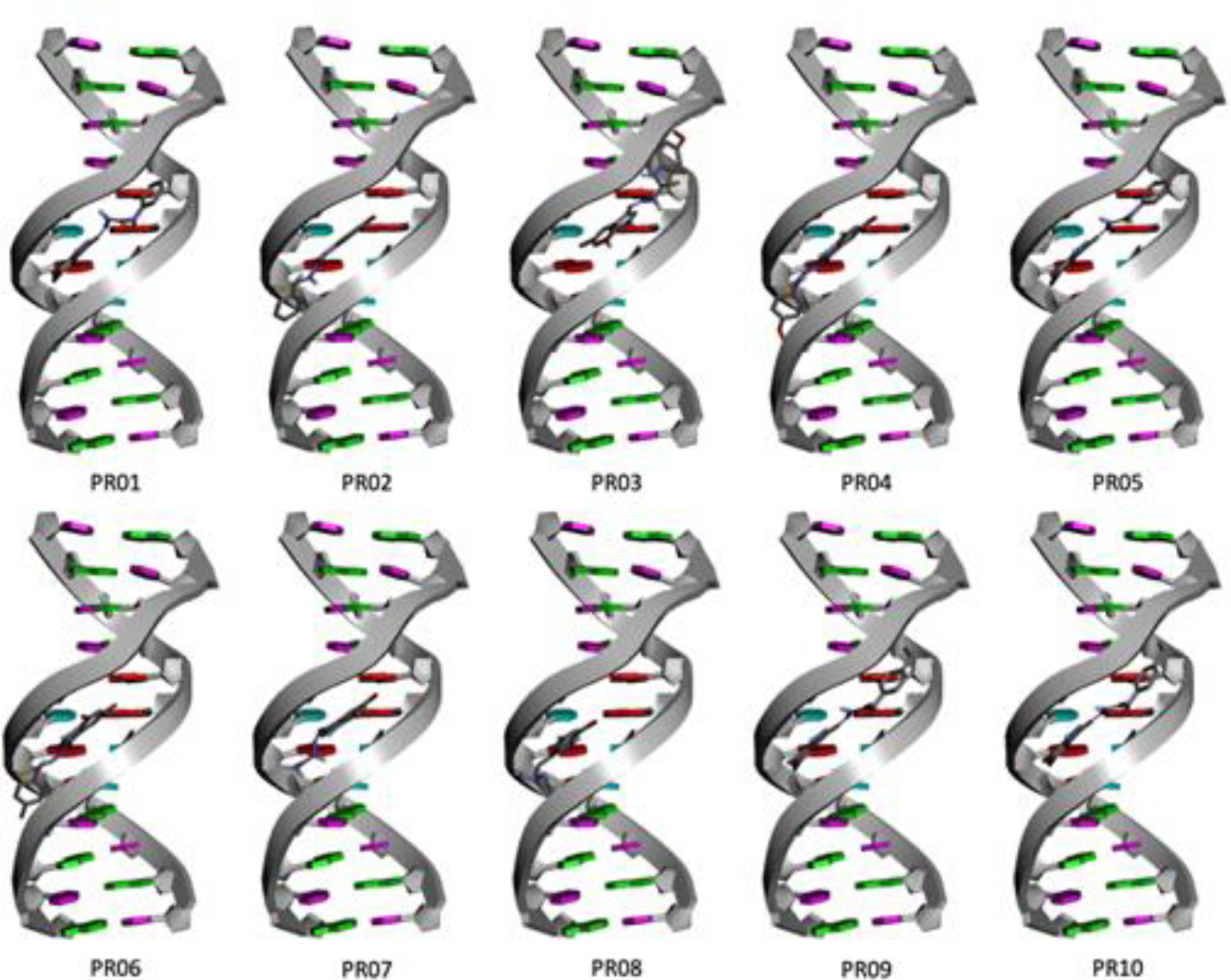
The results presented in Table 3 showed that all binding free energy values were negative for the three targets evaluated, which indicates that all interactions analyzed have a high probability of occurring. For the DNA Minor Groove Binding target (1BNA), PR05 demonstrated the highest binding energy of -11.15 kcal/mol, suggesting a strong interaction with the DNA minor groove. On the other hand, PR07 showed the lowest binding energy of -8.21 kcal/mol. The anchoring poses are represented in Figure 2.
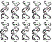
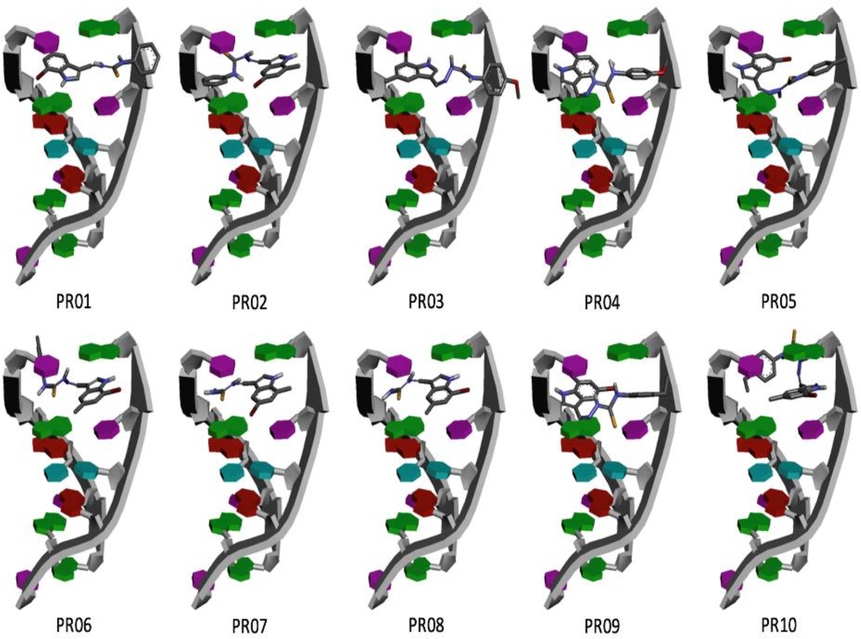
Regarding target DNA intercalation (1Z3F) (Figure 3), PR05 showed the strongest interaction with a binding energy of -8.88 kcal/mol, while PR03 and PR04 exhibited binding energies of -7.99 kcal/mol, respectively. In the docking simulations, the results revealed that the indole rings of the compounds intercalated with the DNA base pairs. This intercalation phenomenon involves the insertion of compounds between adjacent base pairs, resulting in a stacked arrangement of aromatic rings. The stacking interaction between the aromatic rings of compounds and DNA bases is known to contribute to the stabilization of the compound-DNA complex and may have implications for modulating gene expression or inhibiting DNA-related processes [32].
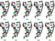
Poses of DNA (PDB ID: 1Z3F) anchoring of tested compounds in the DNA intercalation binding mode.
The literature presents different thiosemicarbazones capable of interacting with the minor groove and intercalating into DNA, with results close to those obtained in this work. In the study of the interaction with the minor groove of DNA, Krishna and coauthors [33] obtained energies ranging from -9.53 to -8.82 kcal/mol for N-substituted hydrazinecarbothioamide compounds. Takroni and coauthors [34], when evaluating the interaction with new copper (II) complexes derived from 1,3,4-thiadiazole-thiosemicarbazone, found energies ranging from -5.759 to -4.132 kcal/mol. Ghassemzadeh and coauthors [35] studied nickel (II) complexes containing acenaphthenequinone, based on bis-Schiff base thiosemicarbazone, and obtained binding free energies ranging from -8.89 to -8.50 kcal/mol. Regarding the intercalation mechanism, Ribeiro and coauthors [36] evaluated 4-quinoline-thiosemicarbazone compounds and found energies ranging from -9.78 to -7.06 kcal/mol. Basheer and coauthors [37] studied octahedral nickel (II) and iron (III) complexes based on thiosemicarbazone and obtained energies between -7.6 and -7.5 kcal/mol. Jacob and coauthors [7], when evaluating indole-thiosemicarbazone compounds, obtained energies ranging from -10.16 to -9.41 kcal/mol for the minor groove, and for DNA intercalation, they obtained energies ranging from -9.54 to -8.84 kcal/mol, respectively. These results demonstrate that thiosemicarbazone compounds are capable of interacting with DNA in different regions.
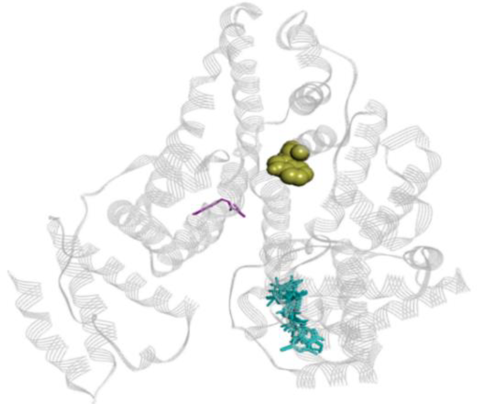
Finally, the energy results for human serum albumin (HSA) (1AO6), presented in Table 3, showed that compound PR09 exhibited the highest binding energy of -10.00 kcal/mol, indicating a strong affinity for HSA. In contrast, PR07 exhibited the lowest binding energy of -7.38 kcal/mol. However, it is worth noting that none of the compounds were able to alter the conformation of HSA. Figure 4 presents the representation of the interaction between albumin and compound PR09.
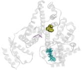
Docking poses of compounds tested in human serum albumin (HSA) (PDB ID: 1AO6). Residue TRP214 is in yellow, PR09 in violet, other compounds are in cyan.
Different studies have shown that thiosemicarbazone compounds can be easily transported by albumins without causing damage to the structure of the macromolecule. Ribeiro and coauthors [36] evaluated 4-quinoline-thiosemicarbazone compounds and found energies ranging from -11.51 to -7.58 kcal/mol. Jacob and coauthors [7], when evaluating indole-thiosemicarbazone compounds, obtained energies ranging from -11.09 to -8.30 kcal/mol. Lighvan and coauthors [38] obtained energies ranging from -6.66 kcal/mol to -6.19 kcal/mol for the tetranuclear cyclopalladated complexes using thiosemicarbazone-derived ligand.
CONCLUSION
The compounds studied in the interaction of indole-thiosemicarbazone with human albumin (HSA) and DNA showed promising results, with compounds PR05 and PR09 standing out in particular. Compound PR09 exhibited the highest association constant (Ka) value, indicating a strong interaction with HSA, along with negative Gibbs free energy (∆G) values, suggesting that the interaction occurs spontaneously. PR05, although also showing good Ka and ∆G values, did not stand out as much as PR09 in its interaction with albumin. Regarding the interaction with DNA, PR05 was the most notable, presenting one of the best binding energies in both DNA intercalation and binding to the minor groove, indicating a strong interaction. PR09 also demonstrated good performance in terms of DNA binding energy, reflecting its significant interaction capacity. Thus, compounds PR05 and PR09 are considered the most promising in the study. While PR05 is distinguished by its strong interaction with DNA, PR09 is notable for its interaction with albumin. These results indicate the potential of these compounds for applications in pharmacological and therapeutic research, reinforcing the relevance of indole-thiosemicarbazones in the development of new therapeutic agents.
-
Funding:
This work was supported by the Coordination for the Improvement of Higher Education Personnel (CAPES), the Pernambuco State Research Support Foundation - FACEPE (Process APQ-0498-4.03/19), the FACEPE researcher fixation grant (Process BFP-0038-04.03/21; Process BFP-0087-4.03/23), and the National Council for Scientific and Technological Development - CNPq (Process 306865/2020-3).
Acknowledgments:
The authors thank the Center for Technological Platforms at the Aggeu Magalhães Research Center (FIOCRUZ Pernambuco).
REFERENCES
-
1 Gupta S, Singh N, Khan T, Joshi S. Thiosemicarbazone derivatives of transition metals as multi-target drugs: A review. Results Chem. 2022;100459. https://doi.org/10.1016/j.rechem.2022.100459
» https://doi.org/10.1016/j.rechem.2022.100459 -
2 Acharya PT, Bhavsar ZA, Jethava DJ, Patel DB, Patel HD. A review on development of bio-active thiosemicarbazide derivatives: Recent advances. J Mol Struct 2021;1226:129268. https://doi.org/10.1016/j.molstruc.2020.129268
» https://doi.org/10.1016/j.molstruc.2020.129268 -
3 Kumar S. A brief review of the biological potential of indole derivatives. Future J Pharm Sci. 2020;6(1):1-19. https://doi.org/10.1186/s43094-020-00141-y
» https://doi.org/10.1186/s43094-020-00141-y -
4 Pasha AR, Khan A, Ullah S, Halim SA, Alharthy RD, Anwar MU, et al. Indole-based thiosemicarbazones for neurodegenerative diseases as prolyl oligopeptidase inhibitors. J Mol Struct. 2024;1312:138666. https://doi.org/10.1016/j.molstruc.2024.138666
» https://doi.org/10.1016/j.molstruc.2024.138666 -
5 Silva PR, Oliveira JF, Silva AL, Queiroz QF, Feitosa APS, Duarte DMFA, et al. Novel indol-3-yl-thiosemicarbazone derivatives: Obtaining, evaluation of in vitro leishmanicidal activity and ultrastructural studies. Chem. Biol. Interact. 2020;315:108899. https://doi.org/10.1016/j.cbi.2019.108899
» https://doi.org/10.1016/j.cbi.2019.108899 -
6 Jacob ÍT, Gomes FO, Miranda MD, Almeida SM, Cruz-Filho IJ, Peixoto CA, et al. Anti-inflammatory activity of novel thiosemicarbazone compounds indole-based as COX inhibitors. Pharmacol Rep. 2021;73:907-925. https://doi.org/10.1007/s43440-021-00221-7
» https://doi.org/10.1007/s43440-021-00221-7 -
7 Jacob IT, Cruz Filho IJ, Alves JEF, Souza FM, Azevedo RDS, Marques DSC, et al. Interaction study with DNA/HSA, anti-topoisomerase IIα, cytotoxicity and in vitro antiproliferative evaluations and molecular docking of indole-thiosemicarbazone compounds. Int J Biol Macromol 2023;234:123606. https://doi.org/10.1016/j.ijbiomac.2023.123606
» https://doi.org/10.1016/j.ijbiomac.2023.123606 -
8 Santos KLD, Marques DSC, Jacob IT, Silva PRD, Machado DC, Souza TRC, et al. In silico prediction of ADMET parameters and in vitro evaluation of antioxidant and cytotoxic activities promoted by indole-thiosemicarbazone compounds. An Acad Bras Ciênc 2024;96(3). https://doi.org/10.1590/0001-3765202420230811
» https://doi.org/10.1590/0001-3765202420230811 -
9 Santos FNS, Bezerra Junior NS, Oliveira JF, Duarte DMFA, Soares JCS, Marques DSC, et al. Synthesis, characterization, antioxidant and antiparasitic activities new naphthyl-thiazole derivatives. Int. J. Parasitol. 2023;248:108498. https://doi.org/10.1016/j.exppara.2023.108498
» https://doi.org/10.1016/j.exppara.2023.108498 -
10 Xiao W, Li Y, Xiong Y, Chen Z, Li H. Fluorescence turn-on detection of human serum albumin based on the assembly of gold nanoclusters and bromocresol green. Anal. Bioanal. 2023:1-12. https://doi.org/10.1007/s00216-023-04717-4
» https://doi.org/10.1007/s00216-023-04717-4 -
11 Nagaraj R, Murugesa S, Jeyaraj DR, Arumugam S, Shunmugasundaram G, Radhakrishnan NA. Spectroscopic studies on DNA interaction and anticancer activities of pharmacologically active pyrimidine derivative mixed ligand Co (II) and Ni (II) complexes. J. Mol. Struct. 2022;1252:132079. https://doi.org/10.1016/j.molstruc.2021.132079
» https://doi.org/10.1016/j.molstruc.2021.132079 -
12 Siddiqi MK, Alam P, Chaturvedi SK, Nusrat S, Ajmal MR, Abdelhameed AS, et al. Probing the interaction of cephalosporin antibiotic-ceftazidime with human serum albumin: A biophysical investigation. Int J Biol Macromol. 2017;105:292. https://doi.org/10.1016/j.ijbiomac.2017.07.036
» https://doi.org/10.1016/j.ijbiomac.2017.07.036 -
13 Morris GM, Huey R, Lindstrom W, Sanner MF, Belew RK, Goodsell DS, et al. AutoDock4 and AutoDockTools4: Automated docking with selective receptor flexibility. J Comput Chem. 2009;30:2785. https://doi.org/10.1002/jcc.21256
» https://doi.org/10.1002/jcc.21256 -
14 Stewart JJP. Optimization of parameters for semiempirical methods V: Modification of NDDO approximations and application to 70 elements. J Mol Model 2007;13:1173. https://doi.org/10.1007/s00894-007-0233-4
» https://doi.org/10.1007/s00894-007-0233-4 - 15 Stewart JJ. Mopac2016. Stewart Computational Chemistry: Colorado Springs, CO, USA, 2016;650.
- 16 Dassault Systémes BIOVIA, Discovery Studio Visualizer, version 16.1.0.15350, Dassault Systemes. San Diego, 2016.
-
17 Tian FF, Jiang FL, Han XL, Xiang C, Ge YS, Li JH, et al. Synthesis of a novel hydrazone derivative and biophysical studies of its interactions with bovine serum albumin by spectroscopic, electrochemical, and molecular docking methods. J. Phys. Chem. B. 2010;114:14842. https://doi.org/10.1021/jp105766n
» https://doi.org/10.1021/jp105766n -
18 Larsen MT, Kuhlmann M, Hvam ML, Howard KA. Albumin-based drug delivery: harnessing nature to cure disease. Mol. Cell. Ther. 2006;4:1. https://doi.org/10.1186/s40591-016-0048-8
» https://doi.org/10.1186/s40591-016-0048-8 -
19 Palchaudhuri R, Hergenrother PJ. DNA as a target for anticancer compounds: methods to determine the mode of binding and the mechanism of action. Curr. Opin. Biotechnol. 2007;18:497. https://doi.org/10.1016/j.copbio.2007.09.006
» https://doi.org/10.1016/j.copbio.2007.09.006 -
20 Shu Y, Xue W, Xu X, Jia Z, Yao X, Liu S, et al. Interaction of erucic acid with bovine serum albumin using a multi-spectroscopic method and molecular docking technique. Food Chem. 2015;173:31. https://doi.org/10.1016/j.foodchem.2014.09.164
» https://doi.org/10.1016/j.foodchem.2014.09.164 -
21 Li JC, Li N, Wu QH, Wang Z, Ma JJ, Wang C, et al. Study on the interaction between clozapine and bovine serum albumin. J Mol Struct 2007;833:184. https://doi.org/10.1016/j.molstruc.2006.09.019
» https://doi.org/10.1016/j.molstruc.2006.09.019 -
22 Neto GLB, Baptista EAM, Becca GHS, Nakatani HS, Souza VRD. [Competitive interactions of ruthenium complexes containing dimethyl sulfoxide and N-heterocyclic ligands with human serum albumin]. Quím Nova 2020;43:261. https://dx.doi.org/10.21577/0100-4042.20170488
» https://dx.doi.org/10.21577/0100-4042.20170488 -
23 Zhang RJ, Kou SB, Hu L, Li L, Shi JH, Jiang SL. Exploring binding interaction of baricitinib with bovine serum albumin (BSA): multi-spectroscopic approaches combined with theoretical calculation. J Mol Liq. 2022;354:118831. https://doi.org/10.1016/j.molliq.2022.118831
» https://doi.org/10.1016/j.molliq.2022.118831 -
24 Galleano M, Verstraeten SV, Oteiza PI, Fraga CG. Antioxidant actions of flavonoids: thermodynamic and kinetic analysis. Arch Biochem Biophys 2010;501:23. https://doi.org/10.1016/j.abb.2010.04.005
» https://doi.org/10.1016/j.abb.2010.04.005 -
25 Findik M, Uçar A, Akgemci E. Spectroscopic analyses on the binding interaction of thiosemicarbazone-derivated Cu (II) complex with DNA/BSA. CBUJOS 2021;17(4):387-95. https://doi.org/10.18466/cbayarfbe.902377
» https://doi.org/10.18466/cbayarfbe.902377 -
26 Karthikeyan S, Bharanidharan G, Kesherwani M, Mani KA, Srinivasan N, Velmurugan D, et al. Insights into the binding of thiosemicarbazone derivatives with human serum albumin: spectroscopy and molecular modelling studies. J Biomol Struct Dyn. 2016;34(6):1264-81. https://doi.org/10.1080/07391102.2015.1075905
» https://doi.org/10.1080/07391102.2015.1075905 -
27 Matei I, Hillebrand M. Interaction of kaempferol with human serum albumin: a fluorescence and circular dichroism study. J Pharm Biomed Anal 2010;51:768. https://doi.org/10.1016/j.jpba.2009.09.037
» https://doi.org/10.1016/j.jpba.2009.09.037 -
28 Yu W, Shi L, Hui G, Cui F. Synthesis of biologically active thiosemicarbazone and characterization of the interaction with human serum albumin. J Lumin. 2013;134:491-7. https://doi.org/10.1016/j.jlumin.2012.07.040
» https://doi.org/10.1016/j.jlumin.2012.07.040 -
29 Tarai SK, Tarai A, Mandal S, Nath B, Som I, Bhaduri R, et al. Cytotoxic behavior and DNA/BSA binding activity of thiosemicarbazone based Ni (II) complex: bio-physical, molecular docking and DFT study. J Mol Liq. 2023;383:121921. https://doi.org/10.1016/j.molliq.2023.121921
» https://doi.org/10.1016/j.molliq.2023.121921 -
30 Silva MJC, Marques DSC, Bomfim Nascimento PH, Mendes RFV, Buonafina-Paz MDS, Machado DC, et al. Synthesis of trimethoxy-benzylidene-hydrazine-carboxamide compounds: antioxidant, antimicrobial and antiparasitic agent, evaluation of the interaction with BSA and ADMET parameters. Chem Pap 2024;78(3):1897-913. https://doi.org/10.1007/s11696-023-03214-3
» https://doi.org/10.1007/s11696-023-03214-3 -
31 Farahat AA, Kumar A, Wenzler T, Brun R, Paul A, Guo P, et al. Investigation of the effect of structure modification of furamidine on the DNA minor groove binding and antiprotozoal activity. Eur J Med Chem. 2023;252:115287. https://doi.org/10.1016/j.ejmech.2023.115287
» https://doi.org/10.1016/j.ejmech.2023.115287 -
32 Gupta S, Paul K. DNA damage and intercalation by elinafide modified bis-naphthalimides for their anticancer activity. J Mol Liq. 2023;382:121980. https://doi.org/10.1016/j.molliq.2023.121980
» https://doi.org/10.1016/j.molliq.2023.121980 -
33 Krishna PM, Reddy NG, Harish BG, Patil YP, Nethaji M. Synthesis, structural studies, molecular docking and DNA binding studies of 4N-substituted hydrazinecarbothioamides. J Mol Struct. 2019;1175:97-104. https://doi.org/10.1016/j.molstruc.2018.07.053
» https://doi.org/10.1016/j.molstruc.2018.07.053 -
34 Takroni KM, Farghaly TA, Harras MF, El-Ghamry HA. Synthesis, structure elucidation, DNA binding and molecular docking studies of novel copper (II) complexes of two 1,3,4-thiadiazolethiosemicarbazone derivatives. Appl Organomet Chem. 2020;34(10). https://doi.org/10.1002/aoc.5860
» https://doi.org/10.1002/aoc.5860 -
35 Ghassemzadeh M, Moghadam ME, Saeidifar M, Jahangiri S, Yarahmadi A, Aghapoor K, et al. Novel Nickel (II) complex bearing acenaphthenequinone based thiosemicarbazone bis-Schiff Base: Synthesis, characterization, linkage isomers, antitumor activity, DFT, and DNA docking simulation. J Mol Struct. 2024;1313:138716. https://doi.org/10.1016/j.molstruc.2024.138716
» https://doi.org/10.1016/j.molstruc.2024.138716 -
36 Ribeiro AG, de Almeida SMV, de Oliveira JF, de Lima Souza TRC, Dos Santos KL, de Barros Albuquerque AP, et al. Novel 4-quinoline-thiosemicarbazone derivatives: Synthesis, antiproliferative activity, in vitro and in silico biomacromolecule interaction studies and topoisomerase inhibition. Eur J Med Chem. 2019;182:111592. https://doi.org/10.1016/j.ejmech.2019.111592
» https://doi.org/10.1016/j.ejmech.2019.111592 -
37 Basheer SM, Rasin P, Kumar SLA, Kumar MS, Sreekanth A. Investigation on DNA/Protein interaction of thiosemicarbazone based octahedral nickel (II) and iron (III) complexes. J Mol Struct. 2022;1260:132913. https://doi.org/10.1016/j.molstruc.2022.132913
» https://doi.org/10.1016/j.molstruc.2022.132913 -
38 Lighvan ZM, Ramezanpour A, Pirani S, Akbari R, Arman M, Mahdavi M, et al. Structural and spectroscopic studies of the interaction of 2, 3-dimethyl-4-phenylthiazol-5-carbaldehyde thiosemicarbazone with DNA and bovine serum albumin. J Biomol Struct Dyn. 2023;41(6):3034-43. https://doi.org/10.1080/07391102.2023.2178620
» https://doi.org/10.1080/07391102.2023.2178620
-
Editor-in-Chief:
Paulo Vitor Farago
-
Associate Editor:
Jaiesa Zych Nadolny