Varronia curassavica Jarq. Essential Oils Inhibit the Formation of the Biofilm of Xanthomonas campestris pv. campestris in vitro.
Abstract
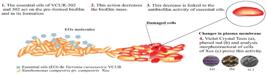
The aim of this study was to investigate the anti-biofilm activity of two essential oils from Varronia curassavica Jaq. Two genotypes (VCUR-202 and VCUR-302), previously identified as exhibiting high antimicrobial activity against Xanthomonas campestris, were selected for this study. Use of Crystal Violet (CV) and Phenol Red (PR) staining of biofilm biomass demonstrated that both Essential Oils (EOs) had strong effect on both biofilm formation and preformed biofilm. The EOs effects on biofilm was confirmed with scanning electron microscopy where the changes in biofilm structure were noticed. Furthermore, results obtained show that sub-inhibitory concentrations of EOs were able to inhibit biofilm formation. Our findings show that EOs from V. curassavica exhibit strong anti-biofilm activity and might be used as a potential agent to controlX. campestris biofilm. Significance and Impact of the Study: This study provides useful information for the development of natural treatments for black rot caused by X.campestris. Studies on the cellular mechanisms involved in antimicrobial and antibiofilm activities are ongoing. These steps are essential for future in vivo tests using these essential oils to control black rot caused by Xcc.
Keywords:
Medicinal plants; Phytobacteria; Disease; Anti-biofilm; Black rot.
HIGHLIGHTS
Essential oils from two genotypes of V. curassavica exhibit strong anti-biofilm activity
EOs was effective in sub-inhibitory concentrations
EOs had effect at the cellular ultrastructure and in the biofilm structure.
Potential agent to controlX. campestris biofilm.
INTRODUCTION
Xanthomonas campestris are phytopathogens responsible for black rot, a disease of Brassicas that is characterized by yellow V-shaped lesions starting at the leaf margins and progressing to the center through the vascular tissue, usually resulting in leaf necrosis [8].
X. campestris can occur in planktonic form (alone), filaments, or biofilms. Biofilm is formed mainly by extracellular polysaccharide (EPS) mainly a gum called xanthan [49], which is formed by pentose units such as mannose-(β-1,4), glucuronic acid-(β-1,2), manose- (β-1,3), and celobiosis [9]. Xanthan is responsible for forming aggregates of X. campestris [27]. These aggregates are present in the matrix of the biofilm, and its function is to facilitate bacteria fixation within the plant's vascular system and adhesion on the surfaces of leaves, roots and seeds. Thus, biofilm plays a key role in the disease cycle and infection of the host plant [16-39].
The infection is characterized by events that include the interaction between host-pathogen, movement of the bacteria towards the host plant and mainly by adhesion to the plant surface and subsequent penetration and multiplication inside the host plant [42]. This adhesion and survival inside the plant is only possible due to the formation of biofilm on the leaf surface. Therefore, the formation of biofilm allows the bacteria to infect the host plant, consequently causing black rot in cruciferous plants [6-46]. This disease develops at any stage of the development of the infected plant, with severity levels reaching the entire aerial part of the plant, compromising the crop's productivity due to the reduction of photosynthetic leaf area and loss of the commercial value of the plant agricultural product [40]. In view of this, it is essential to establish control strategies as a way of reducing the impacts of the disease, since black rot represents a risk to the cruciferous production chain, both for small and large farmers [51].
However, farmers do not have an efficient and specific physical, chemical, or biological method to control Xcc that can be used as a phytosanitary control of black rot, either in its primary or secondary stage of the disease. Thus, as a strategy, the effectiveness of the use of essential oils (EOs) in the control of black rot disease, caused by Xcc, has been investigated.
EOs are compounds used by plants as a defense mechanism, for growth and development, plant-plant communication, plant-microorganism, plant-insects, or physiological response and environmental stresses [30-23]. They are characterized as liquid, oily compounds that can be volatile, semi-volatile, or not volatile, at room temperature. Chemically, Carbons and Hydrogens form EOs, with alkenes being the main class of molecules belonging to EOs [34]. These hydrocarbons bring together simple or complex molecules, with varied chemical composition, presenting carbonic, homocyclic, heterocyclic, or acyclic structures in addition to the presence of oxygenated groups, which further expands the structural diversity of EOs [35-28-38]. This wide chemical diversity of bio-active compounds makes EOs a valuable source of raw material, as they reveal a wide spectrum of antimicrobial activity, both in vitro and in vivo [11-21-31-45-52]. However, there is still a few studies on the action of EOs on phytopathogens. Therefore, the search for new innovative antibacterial agents remains a major challenge, being essential to control the damage from black rot disease, caused by Xanthomonas spp. In view of this, in the absence of an efficient natural control and absence of studies on the action of EOs on Xanthomonas spp. and how to reduce the damage caused by black rot disease, the Varronia curassavica EO has been studied.
Varronia curassavica (whaling grass), also known as Cordia verbenacea or Cordia curassavica (Jacq.) [5], is an important medicinal plant, occurring in several Brazilian biomes. Its EOs has proven biological activity against a wide diversity of phytopathogenic microorganisms [11-22-50] its antimicrobial activity and synergistic effect against X. campestris has only recently been established [12]. In addition, V. curassavica EOs are safe, non-toxic to human health and the environment [2;37]. However, its effect on the action of X. campestris at the biofilm level is still unknown, adding to this, there are few studies that explore the action of anti-biofilm activity on phytopathogens, especially against the Xanthomonas species.
Thus, the objective of this study was to evaluate the anti-biofilm action of the EO of two genotypes of Varronia curassavica Jacq. (VCUR-202, -302) selected for having high antimicrobial activity and proven synergistic effect [12] on the target phytopathogen Xanthomonas campestris pv. campestris.
MATERIAL AND METHODS
Plant material, Essential oil extraction and characterization
The two genotypes were botanically identified by Dr. Ana Paula Prata of the Federal University of Alagoas (Maceió, Brazil). The voucher of the species studied was deposited in the Herbarium of the Federal University of Sergipe, Department of Biology (voucher number ASE 9404). Essential oil extraction from leaves and characterization were done and published in da Silva and co-authors [12].
The access to plant material was registered under the ID number A8CCB3B in the National System of Management of Genetic Heritage and Traditional Knowledge (SisGen) according to Article 4 of Decree No. 8.772/2016 of the Ministry of Environment in Brazil.
Microorganisms and growth conditions
Xanhomonas campestris pv campestris 629IBSBF (Xcc-629IBSBF) was obtained from Biologic Institute (São Paulo, Brazil). For all experiments Xcc was cultivated in Yeast Malt (YM) culture medium (3 g/L yeast extract, 3 g/L malt extract, 5 g/L peptone, 10 g/L sucrose, pH 6.0) at 30° ± 2 0C, under orbital agitation (150 rpm). In all experiments a standardized inoculum (106 CFU/mL) was used. For this a colony of the pure culture, grown in a Petri dish containing solid YM medium, was inoculated in 15 mL of sterile YM medium, overnight under orbital agitation (150 rpm / 30 ± 2 0C). Then, 500 µL of this culture was re-inoculated in 50 mL of sterile YM. After 12 hrs, the optical density (OD600nm) was measured, in order to obtain OD corresponding to 0.5 ± 0.042.
Growth kinetics
The growth kinetics of Xcc in the presence of EOs (VCUR-202 and -302) at concentrations equivalent to 2x, 1x, 1/2x, 1/4x, and 1/8x, MIC (Minimum Inhibitory Concentration) was evaluated during 24 hrs. MIC values of 500 µg/mL, for both EOs, were previously defined [12]. The cultures (100 µL of bacteria and 100 µL of EOs) in 96 microplates were kept in incubation with orbital shaking at 30 ± 2 0C and bacterial growth was monitored every hour by measuring the optical density (OD600nm) using a spectrophotometer. As control, 100 µL of Xcc-629IBSBF in 100 µL of liquid YM medium was used and as a sterile control, 100 µL of liquid YM medium were used in 100 µL of EOs and 100 µL of EOs in 100 µL DMSO (1%). The rate of inhibition (%) of the growth of Xcc-629IBSBF represented the difference in the mean absorbance of each treatment in relation to the control growth. The experiments were carried out in biological triplicates, with five technical repetitions.
Anti-biofilm activity
Biofilm formation inhibition assay
The ability of V. curassavica EOs to inhibit the formation of the Xcc-629IBSBF biofilm in vitro was evaluated according to Hertiani T and co-authors [22] with some modifications. Initially, 500 µL aliquots of the culture (106 CFU/mL) were distributed in 24-well plates and incubated for 48 hours with different concentrations (2x, 1x, 1/2x, 1/4x, and 1/8x MIC) of the EOs, VCUR-202 and -302. As a control, wells containing cell suspension were included without the addition of EOs.
After 48 hrs, the plates were gently washed with sterile water (500 µL/well) to remove non-adherent cells. The biofilm adhered to the wells was fixed by adding 1,000 µL methanol for 15 min (40 0C), and then the methanol was removed and air dried at room temperature. Subsequently, 1,000 µL of 0.1% Crystal Violet (CV) was added for 15 min and then the plates were washed and air dried again. Then, to solubilize the CV in the biofilm, 1,000 µL of ethanol (96%) was added to each well, allowing to act for 5 min under orbital agitation (100 rpm / 25 0C).
Finally, 200 µL of the suspension from each well was transferred to a 96-well microplate. The absorbance intensity of the CV was measured through the microplate reader using a wavelength of 570 nm. Each experiment was repeated independently on three different days.
Biokinetic assay (BKA) for inhibition of preformed biofilm
The Biokinetic assay was performed as described by Berlutti and co-authors [4] and was used to indirectly monitor the effect of the EOs, VCUR-202 and -302, on preformed biofilm. For this, the YM culture medium with Phenol Red (YM-PR medium) was used: 3 g/L yeast extract, 3 g/L malt extract; 5 g/L peptone; sucrose 10 g/L; 25 mg/L, Phenol Red, with pH adjustment 7.2 ± 0.1, with a light red color after sterilization.
Initially 500 µL of cell suspension (106 CFU/mL) were inoculated in 50 mL of YM medium containing sterile microspheres 5 cm in diameter Then, the cultures were in incubated, for biofilm formation, without shaking at 30 ± 2 0C for 48 hrs. After incubation, the microspheres, with the presence of the biofilm, were transferred to 24-well microplates. Each well with a sphere received 500 µL of the essential oil (2x, 1x, 1/2x, 1/4x, and 1/8 x MIC) dissolved in culture medium YM + liquid PR. As a negative control, microspheres were used immersed in 500 µL of the YM culture without the addition of EOs and as a sterile control, 500 µl of the YM medium in 500 µL of YM-PR, 500 µL of the liquid YM-PR medium in 500 µL of the EOs, and 1.000 µL of the YM-PR medium, 1.000 µL of the YM medium and 1.000 µL of the PR medium. After 6 hrs, 200 µL of each well were transferred to a 96-well plate and the absorbance was measured through the microplate reader at a wavelength of 595 nm. All experiments were carried out in biological triplicates, with five technical repetitions.
Scanning electron microscopy (SEM)
SEM was used to determine morphological changes in cells and biofilm of Xcc-629IBSBF treated with EOs. For this, 106 CFU/mL of cells in suspension were incubated with different concentrations of EO (control, 2x, 1x, 1/2x, 1/4x, and 1/8x MIC), in liquid YM for 6 hrs, and centrifuged at 5.000 g/5 min/30° ± 2 0C. After centrifugation, the cells were washed with PBS (0.1 M / pH = 7.4) three times and fixed with 1: 1: 2 (v / v) 4% glutaraldehyde, 2% paraformaldehyde, and 0.1 M PBS (pH = 7.4) for 1 h at room temperature.
After fixation, the cells were washed twice in 0.1 M PBS (pH = 7.4) and dehydrated in graduated series using glacial ethanol in the percent concentrations of 50, 70, 80, 90, 95, and 100% at 4 0C for 10 min. Then, a 1:1 mixture of Ethanol and HMDS (Hexamethyldisilazane) for 10 min replaced ethanol. The dehydrated cells were finally dispersed in 100% HMDS and a 10 µL aliquot was added to a sterile cover slip and dried in the hood for 12 hrs. As a positive control, bacterial suspensions, in the same concentration, without the presence of EO, were used as living cells (106 CFU/mL). Samples with the presence of the biofilm were fixed, as previously described.
After drying, the cells were sputter-coated with gold (approximately 30 nm thickness) through a Denton Vacuum (Desk V) sputtering system for 60 seconds. Finally, the cells were observed using SEM (JEOL 5700) with magnification of up to 5 kx. For the analysis of the bacterial surface, the free software image J Lab were used. The image analysis algorithms using binary threshold did not result in data that correspond to the increasing trend of cell density on the surface of gold-coated samples (SEM), which were measured in parallel by an established method. This digital analysis test was able to discern subtle differences in the accumulation of surface accumulated over the time of exposure to EO.
Statistical analysis
The data were analyzed using the Graphism Prism software of the Statistical Analysis System (version 7; San Diego, CA, USA, 2012). The analysis of variance (ANOVA) was performed using a standard ANOVA procedure. P <0.05 was considered statistically significant.
RESULTS
Kinetic analysis
MIC and MBC (Minimum Bactericidal Concentration) from EO-VCUR-202 and EO-VCUR-302 for Xcc-629IBSBF have been already determined in our early work [12]. The kinetic growth analysis under varying proportions of the minimum inhibitory concentration (MIC) conducted in this study was essential for understanding the antimicrobial activity at sub-inhibitory concentrations. Additionally, it allowed for the determination of the optimal incubation time required to assess the ultrastructural effects of essential oils on bacterial cells and biofilms using electron microscopy. The treatment of Xcc with EO-VCUR-302 and EO-VCUR-202 reduced the cell growth even in sub-inhibitory concentrations equivalent to 1/8x MIC after 1-24 hrs incubation (Table 1).
Effect of Antimicrobial Activity of VCUR-202 and -302 EOs on cell growth rate (%) of Xcc over 24 hours of incubation.
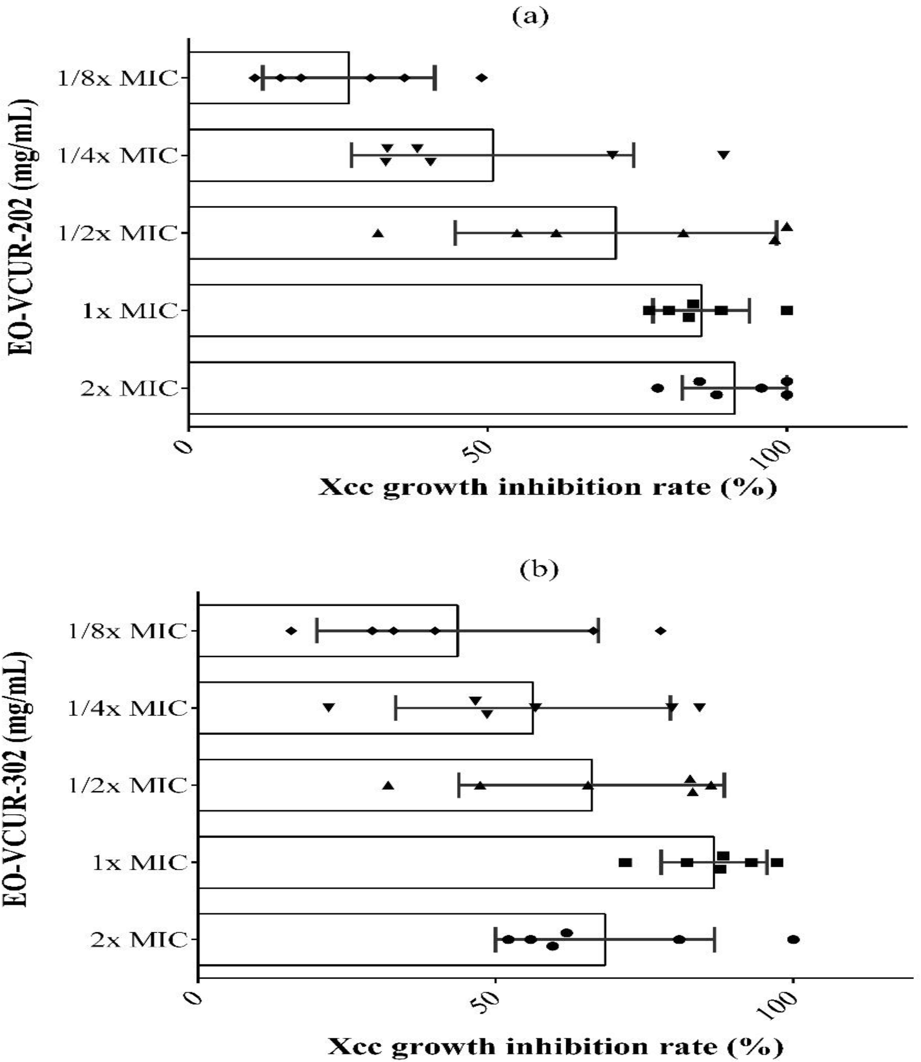
At the end of 6 hrs, the growth inhibition (% to not treated cells) was statistically different (p <0.05) for the concentrations of 1x, 1/2x, 1/4x, and 1/8x MIC of EO-VCUR-202 and 2x, 1x, 1 / 2x, and 1/8x MIC for the EO-VCUR-302. Thus, the 6 hrs incubation was used to demonstrate the effect of different concentrations of EOs on biofilm formation using Phenol Red and electron microscopy (Figure 1a,b).

Antimicrobial activity after 6 hrs of exposure to the EOs of VCUR-202 (a) and -302 (b) on X. campestris ( 2x;1x;•2x ;▲4 x ■d 1/8 ♦ MIC). Test performed in liquid medium. All data represent means ± standard error of the mean of 3 independent experiments (* p <0.05; ** p <0.01; *** p <0.001).
Anti-biofilm activity
Inhibition of Biofilm formation
For analysis of biofilm formation, it was used staining with the Crystal Violet. It can be observed a strong inhibition at 2x and 1x MIC for both EOs (VCUR-202 and 302), showing (0.063% ± 0.013; 0,384% ± 0.042; 0.425% ± 0.058, 0.681% ± 0.122) respectively.Even sub-inhibitory concentrations (1/2x for EO-VCUR-202 and 1/2x, 1/4x, and 1/8x MIC for EO-VCUR-302) resulted in lower production or inhibition of biofilm in presence of EOs (Figure 2a, b).

Inhibition of biofilm formation assay using Crystal Violet (CV). Absorbances obtained after 48 hrs of adhesion and biofilm formation in XCC not treated and treated with EO-VCUR-202 (a) and -302 (b) in the different concentrations related to MIC.
Inhibition of preformed biofilm
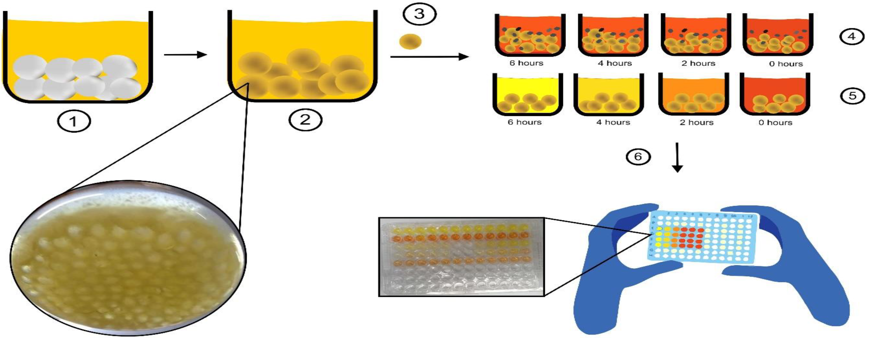
The colorimetric method used quantifies the microbial metabolism indicated by the color change (red to yellow) of the Phenol Red (PR) indicator present in the Yeast Malt (YM) culture medium. The color change of the PR is the result of microbial activity, and this change is correlated to the concentration of bacteria. Thus, a decrease in the number of viable bacteria present in microspheres (preformed biofilm) could be observed for both EOs and related with absorbance. For VCUR-202 the absorbance (OD595nm) varied from 0.789 ± 0.96 (not treated cells) to 0.002 ± 0.027 (2x MIC), to 0.001 ± 0.011 (1xMIC), and 0.221 ± 0.023 (1/2x MIC) (Figure. 3a). For EO-VCUR-302, changes in biofilm metabolism were observed for 2x MIC concentration (Figure 3b). These data are reinforced by the scientific illustration, which summarizes the methodological adaptation, used in this article. This is the first article to apply Phenol Red for this purpose (Figure 4).
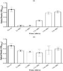
Inhibition of preformed biofilm by viability test based on colorimetric analysis using phenol red pH indicator. Absorbances obtained after 48 hrs of biofilm formation and 6 hrs treatment with EO-VCUR-202 (a) and -302 (b) in the different concentrations related to MIC.
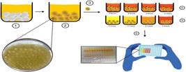
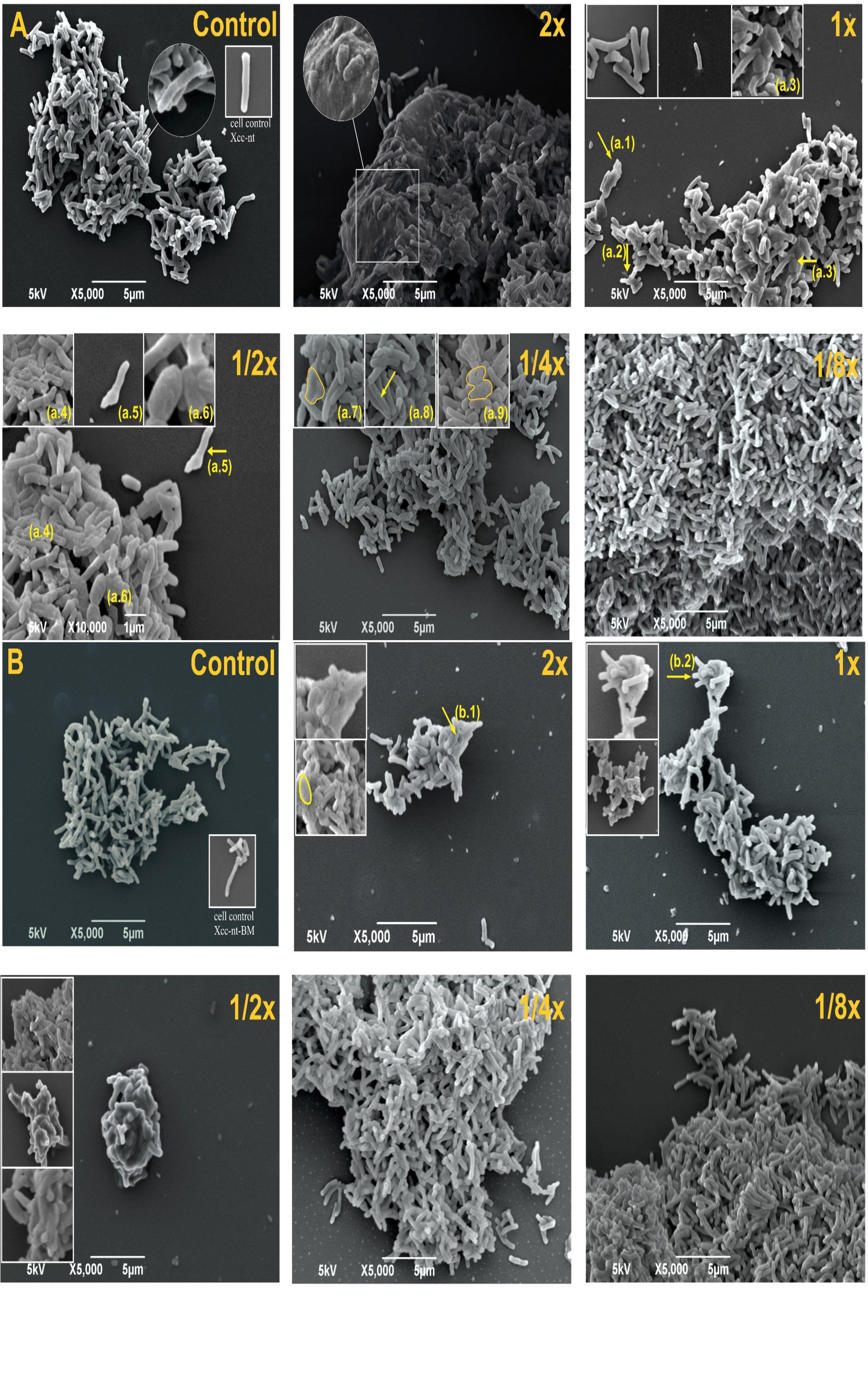
(1) Growth of microspheres in culture medium containing YM + Xcc for 48 hrs for biofilm formation. (2) After 48 hrs, microspheres with the presence of Xcc biofilm adhering to its surface were transferred (3) to 24-well microplates containing culture medium supplemented with phenol red and VCUR essential oil (4) and medium of culture supplemented with phenol red without the addition of VCUR essential oil (5). After 6 hrs, a change in the color of the culture medium from Red to yellow is observed as the culture medium acidifies, in the wells without the presence of the essential oil (5). The decrease in the rate of growth of previously adhered biofilm was measured using the microplate reader at a wavelength of 595 nm. to presenting an ovoid appearance as marked by yellow arrows and inserts (Fig. 5A. a.1, a.2, a.3, a.4, a.5, a.6, a.7, a.8 and a.9). Cellular leakage (Figure 5A. a.2), cell debris among intact cells (Figure 5A. a.4) were also observed. The treatment with EO-VCUR-302 at a concentration of 2x, 1x, and 1/2x MIC, cells formed large lumps with colloidal and surface topography (Fig. 5B. b.1 and b.1).
Scanning electron microscopy (SEM)
SEM was used to observe morphological changes in Xanthomonas campestris pv campestris cells and biofilm after exposure to EOs. When comparing the images of the untreated cells (control, Fig 5A and B) with those exposed to EOs, in the concentrations of 2x, 1x, 1/2x, 1/4x EO-VCUR-202 and 2x, 1x, 1/2x EO-VCUR-302, changes were observed, both at the ultrastructural cellular level and in the biofilm structure (Fig 5A-B). The images show that there was no cellular alteration evidenced by intact surface and with bacillary appearance after treatment with EO-VCUR-202 in the concentration of 1/8x MIC (Fig 5A) and 1/4x and 1/8x MIC for VCUR-302 (Fig 5B). However, a large number of cells were damaged or had their membrane ruptured when treated with EO-VCUR-202 and -302, in concentrations equivalent to 2x and 1x MIC (Figure 5A-B). These cells showed a surface roughness, with the formation of some pores on the entire surface, in addition
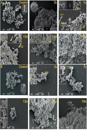
Effects of EOs-VCUR-202 (5a) and 302 (5b) on X. campestris pv campestris cell ultrastructure and biofilm performed by scanning electron microscopy. A - cells treated with concentration equivalent to 2x MIC; B- 1x MIC; C-1/2 x MIC; D- 1/4x; E-1/8 x MIC and F- cells untreated. Images obtained after 6 hrs treatment (or not) with EOs. Yellow arrows and inserts (c.1, c.2, c.3 e d.1, d.2. d.3) show ultrastructural modifications.
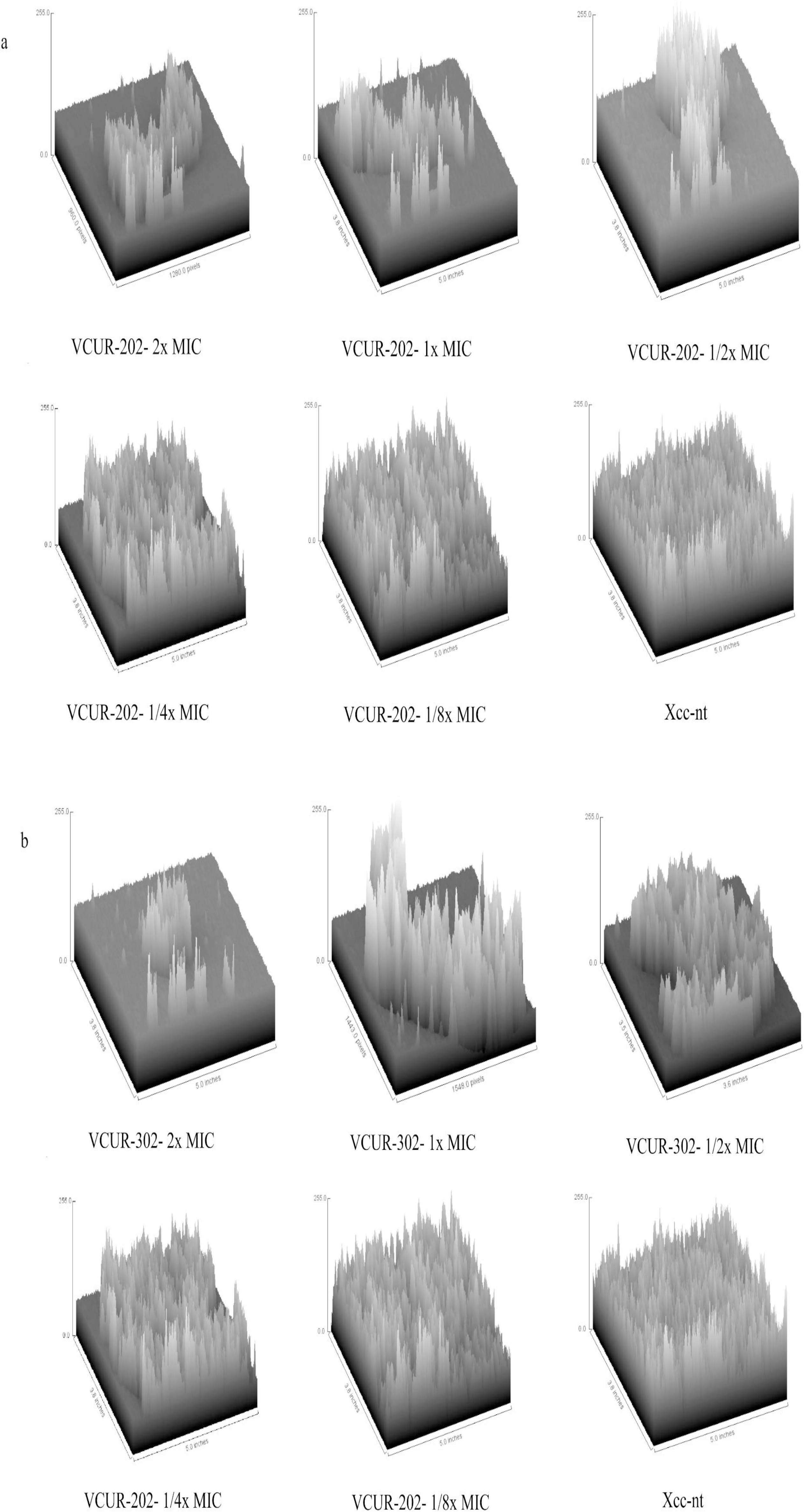
Analysis of the topographic layer of the Xcc cells shows changes in the biofilm in cells treated with EOs when compared to the untreated control (Figure 6a, b). This result visually confirms what was indicated in the violet and phenol red crystal tests Based on the observations of the topographic analysis of the Xcc biofilm, both assays showed distinct effects when treated with OE-VCUR-202 and -302 at concentrations of 2x, 1x and 1/2x MIC, compared to the control of non-Xcc cells. treated with the OE (Figure 6a,b). Xcc cells treated with OEs at sub-inhibitory concentrations 1/4x, 1/8x MIC of the OE of VCUR-202 and -302 exhibited a biofilm architecture similar to that produced by Xcc not treated with OE, which presented a topography with more bacterial aggregates. These distinct biofilm patterns prove the effect of oil on the aggregate of cells present in the biofilm (Figure 6a,b).
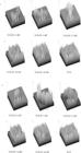
Representation of the 3D image processing of Xanthomonas campestris pv. campestris biofilm: The SEM image is filtered and linearized to obtain a binary representation of the biofilm. These binary data are transformed into 3D, the cells being represented by cubes (pseudo-cells) with defined size approximately 0.2-0.6 µm thick by 0.8-2.9 µm length to quantify the distribution of the spatial property of the structure. In these images, each biofilm cube is colored in gray scales, according to the density of the local biomass. Figures show the representation of the 3D image processing of the Xcc biofilm treated with different concentrations ssential oil of VCUR-202 (6a) and VUCR-302 (6b) in relation to the control
DISCUSSION
Black rot in crucifers is one of the most destructive diseases in vegetable-producing areas. However, the strategies used to control this disease, including sanitation, resistant varieties, antibiotics, cupric compounds, and fungicides cannot completely eliminate the disease [53]. In addition to the lack of efficient strategies for combating it, there are few natural products based on microorganisms or plants used to control black rot caused by Xanthomonas campestris [11-13]. In the world market, for example, in the United States Environmental Protection Agency (active ingredients of the biopesticide), there are 33 products registered with specific agencies for combating phytopathogens in general. A low number when compared to traditional forms of control (chemical fungicide / bactericide)
Therefore, the use of V. curassavica EOs may be a viable and safe alternative. Different studies have shown that EOs of V. curassavica did not present significant levels of acute toxicity in laboratory animals [2-3-14-18-37-43-54], do not present any allergic or irritating reaction in humans, and its main constituents are non-toxic to mammals, birds, and fish [26]. In addition to these advantages, their constituents are non-persistent in the environment after 24 hrs on soil or water surfaces, being degraded in the environment or volatilized when in plant foliage, minimizing residual contact [24].
Regarding the antimicrobial activity against Xcc, we had already demonstrated that the V. curassavica EOs (VCUR-202 and -302) present inhibition of Xcc alone and a strong inhibition in synergism [12] In the current study, we demonstrated that the V. curassavica EOs had a significant effect on Xcc bacterial growth inhibition kinetics (even in sub-inhibitory concentrations (Table 1). These results confirm the bactericidal effect, reducing bacterial growth. This action was observed even when the bacteria were inoculated in solutions with EOs in their exponential phase (106 CFU / mL), which demonstrates the high bactericidal power of these EOs. Other studies have revealed the effect of inhibiting growth kinetics in bacteria and phytopathogenic bacteria by essential oils [7-20-33-47].
In addition to the antimicrobial effect of EOs, their anti-biofilm effect is currently being studied on different bacterial species. Kang and co-authors [25] investigated the anti-biofilm activity of thyme essential oil on planktonic growth, and biofilm formation of Bacillus cereus and showed that thyme EOs inhibited the formation of biofilm. Shafiei and co-authors [48], demonstrated that the Acacia mangium leaf extract exhibited an inhibitory effect on the biofilm formation of Xanthomonas oryzae pv. oryzae with inhibition rate of 81.25% at a concentration of 12.5 mg/mL.
Our data demonstrate that VCUR-202 and -302 EOs are effective in inhibiting Xcc biofilm formation with an inhibition rate of 100%, at a concentration of 0.5 mg/mL. The observations corroborate the data by Shafiei and co-authors [48], who observed that methanol leaf extract from Acacia mangium has an anti-biofilm effect against Xanthomonas oryzae pv. oryzae. Biofilm is considered the most common growth state for many microbials, and its formation is important as a virulence factor for a wide range of microorganisms.
Consequently, aiming at the inhibition and formation of the Xcc biofilm, EOs are a strong candidate in the potential strategic control of black rot disease, caused by Xcc. Therefore, in order to better understand the inhibitory action of the EOs VCUR-202 and -302, tests were performed to evaluate the effectiveness of the EOs in inhibiting the biofilm formation by Xcc.
The absence of blue color (after staining with Crystal Violet) indicated the non-adhesion of the biofilm on the walls of the 24-well microplates, clearly showing the absence of Xcc biofilms treated with EOs VCUR-202 and -302 compared to the controls (Figures 2a, b). In experiments using Phenol Red, the results obtained by changing the color of the culture medium clearly showed that the EOs VCUR-202 and -302 dramatically reduced the biofilm that previously adhered to the microspheres (preformed biofilm) (Figure 4). The decrease in the growth rate of the biofilm that previously adhered to the microsphere (BPAM) treated with EOs VCUR-202 and -302 is due to the non-acidification of the culture medium, since the quantification of the colorimetric indicator (Red Phenol) is established when there is acidification of the culture medium as the bacteria colony develops in the culture medium, and this change in pH is quantified by the change from red to yellow color as the medium acidifies. The results obtained confirm the hypotheses of the experiment with Crystal Violet and show a reduction in biofilm metabolism in response to EOs. In the experiment, the already mature biofilm present in the spheres was exposed to EOs, which shows the effects of oils on the complex shape of the film, commonly associated with resistance to the active ingredients used as antimicrobials. Our work is the first to report this analysis technique with phytopathogens.
Moreover, digital images acquired by scanning electron microscopy show morphological changes in Xcc-629IBSBF cells, after exposure to EOs. Our findings are in agreement with results previously reported by 55. Diao W-R and co-authors [55], who demonstrated that the EO of the Foeniculum vulgare Mill seed causes changes in bacillary bacteria, including changes in the surface. Mirzaei-Najafgholi and co-authors [56] suggested that Citrus EO induces irregular changes in Xanthomonas citri subsp. citri resulting in severely collapsed and crushed bacterial morphology. Our results are in line with these reports, with VCUR-202 and -302 also altering the bacterial morphology of Xcc, resulting in wrinkled and collapsed cells.
The images acquired from the surface of the biofilm treated and not treated with the EOs were subjected to digital image analysis using an image analysis algorithm for quantification of biofilm, using the ImageJ software. It allowed us to observe that there was a reduction in cell density in the totality of the surface of biofilm when cells treated and not treated with EOs were registered. In this way, it can reinforce the effect of EOs on the inhibition of biofilm.
The two EOs evaluated regarding anti-biofilm activity have different main compounds. VCUR-202 EO have Germacrene-D-4-ol (32.16%) and Epizonarene (16.67%) as main compounds, and VCUR-302, ar-Curcumene (20.37%), β-Sesquiphellandrene (18.53%), and E-Caryophyllene (16.47%) (Da Silva et.al., 2020). These differences may explain the better performance of EO-VCUR-202 on biofilm inhibition. Our results indicated that EO-VCUR-202 is a strong candidate for antibiofilm action. Comparing with other studies, compounds such as E-Caryophyllene and Germacrene-D, found in Varronia curassavica, have been linked to antibiofilm activities. E-Caryophyllene demonstrated effectiveness against biofilms of Staphylococcus aureus and Pseudomonas aeruginosa [56], while Germacrene-D inhibited biofilms of Candida albicans and Escherichia coli [57]. Additionally, a study on the essential oil of Varronia dardani also reported antibiofilm activity, highlighting the potential of species from this genus for biofilm control [58]. In addition, all biofilm adhesion tests showed the high performance of EO-VCUR-202 even in sub-inhibitory concentrations. This fact is important, since biofilm-forming phytopathogenic bacteria, such as X. campestris, are more resistant to the antimicrobial action of fungicides / bactericides. Thus, in addition to the EO-VCUR-202, the EOs VCUR-302 being efficient in reducing the growth of planktonic populations they are also effective in treatment against the preformed biofilm. In this study, it was shown that the VCUR-202 and 302 EOs, in addition to inhibiting the planktonic form of X. campestris pv campestris, also inhibit biofilm formation. Studies on the cellular mechanisms involved in antimicrobial and antibiofilm activities are ongoing. These steps are essential for future in vivo tests using these essential oils to control black rot caused by Xcc.
CONCLUSION
In this study, it was shown that the VCUR-202 and 302 EOs, in addition to inhibiting the planktonic form of X. campestris pv campestris, also inhibit biofilm formation. Studies on the cellular mechanisms involved in antimicrobial and antibiofilm activities are ongoing. These steps are essential for future in vivo tests using these essential oils to control black rot caused by Xcc.
-
Funding: This study was funded, in part, by Brazil's Conselho Nacional de Desenvolvimento Científico e Tecnológico (CNPq); Fundação de Apoio à Pesquisa e a Inovação Tecnológica do Estado de Sergipe (Fapitec/SE); Coordenação de Aperfeiçoamento de Pessoal de Nível Superior (CAPES; Finance Code 001); and Financiadora de Estudos e Projetos (FINEP).).
Acknowledgments:
None
REFERENCES
-
1 Andrade FP, Venzon M, das Dôres RGR, Franzin ML, Martins EF, de Araújo GJ, et al. [Toxicity of Varronia curassavica Jacq. Essential Oil to Two Arthropod Pests and Their Natural Enemy]. Neotrop Entomol. 2021 Oct; 50(5):835-845. doi: 10.1007/s13744-021-00906-x. Epub 2021 Aug 16. PMID: 34398399.
» https://doi.org/10.1007/s13744-021-00906-x. -
2 Hoeltgebaum MP, Lauterjung MB, Montagna T, Candido-Ribeiro R, Vieira W, Bernardi AP, et al. [Genetic and demographic aspects of Varronia curassavica Jacq. in a heterogeneous coastal ecosystem]. An Acad Bras Cienc. 2020 Sep 7;92(suppl 2):e20180532. doi: 10.1590/0001-3765202020180532. PMID: 32901674
» https://doi.org/10.1590/0001-3765202020180532. - 3 Bayeux MC, Fernandes AT, Foglio MA, Carvalho JE. [Evaluation of the antiedematogenic activity of artemetin isolated from Cordia curassavica DC]. Braz J Med Biol Res [Internet]. 2002;35:1229-32.
- 4 Berlutti F, Rosso F, Bosso P, Giansanti F, Ajello M, De Rosa A, et al. [Quantitative evaluation of bacteria adherent to polyelectrolyte HEMA-based hydrogels]. J Biomed Mater Res [Internet]. 2003;67A:18-25.
- 5 Brandão DS, Mendes ADR, Santos RR, Rocha SMG, Leite GLD, Martins R. [Floral biology and reproductive system da erva-baleeira (Varronia curassavica Jacq.)]. Rev Bras Plantas Med [Internet]. 2015;17:562-9.
- 6 Buchbauer G, Hemetsberger S. [Handbook of Essential Oils]. [Internet]. Baser KHC, Buchbauer G, editors. Handb. Essent. Oils Sci. Technol. Appl. Second Ed. CRC Press; 2015.
- 7 Buldain D, Buchamer A V, Marchetti ML, Aliverti F, Bandoni A, Mestorino N. [Combination of Cloxacillin and Essential Oil of Melaleuca armillaris as an Alternative Against Staphylococcus aureus]. Front Vet Sci [Internet]. 2018;5:177.
- 8 Cerutti A, Jauneau A, Auriac M-C, Lauber E, Martinez Y, Chiarenza S, et al. [Immunity at Cauliflower Hydathodes Controls Systemic Infection by Xanthomonas campestris pv campestris]. Plant Physiol [Internet]. 2017;174:700-16.
- 9 Crossman L, Dow JM. [Biofilm formation and dispersal in Xanthomonas campestris]. Microbes Infect [Internet]. 2004;6:623-9.
- 10 Da Silva ACR, Ferro JA, Reinach FC, Farah CS, Furlan LR, Quaggio RB, et al. [Comparison of the genomes of two Xanthomonas pathogens with differing host specificities]. Nat [Internet]. 2002;417:459-63.
- 11 Da Silva RS, de Oliveira MMG, de Melo JO, Blank AF, Corrêa CB, Scher R, et al. [Antimicrobial activity of Lippia gracilis essential oils on the plant pathogen Xanthomonas campestris pv. campestris and their effect on membrane integrity]. Pestic Biochem Physiol [Internet]. 2019;160.
- 12 Da Silva RS, de Oliveira MMG, Silva KP, da Silva Vasconcelos Rodrigues I, dos Santos Pinto V, Blank AF, et al. [Synergistic effect of Cordia curassavica Jacq. essential oils association against the phytopathogen Xanthomonas campestris pv. campestris]. Environ Sci Pollut Res [Internet]. 2020;27.
- 13 Da Silva RS, Moutinho BL, dos Santos DR, Vasconcelo-Rodrigues ISS, Talamini V, Fernandes MF, et al. [Using antagonistic soil bacteria and their cell-free filtrates to control the black rot pathogen Xanthomonas campestris pv. campestris]. J Phytopathol [Internet]. 2018;166:494-501.
- 14 De Carvalho PMM, Rodrigues RFO, Sawaya ACH, Marques MOM, Shimizu MTT. [Chemical composition and antimicrobial activity of the essential oil of Cordia verbenacea D.C]. J Ethnopharmacol. 2004;95:297-301.
- 15 Ehlert PAD, Blank AF, Arrigoni-Blank MF, Paula JWA, Campos DA, Alviano CS. [Hydrodistillation time in the extraction of essential oil from seven species of medicinal plants]. Rev Bras Plantas Med. 2006;8:79-80.
- 16 Dow JM, Crossman L, Findlay K, He Y-Q, Feng J-X, Tang J-L. [Biofilm dispersal in Xanthomonas campestris is controlled by cell-cell signaling and is required for full virulence to plants]. Proc Natl Acad Sci.2003; 100(19): 995-1000.
- 17 Gilbert B, Favoreto R. [Cordia verbenacea DC. Boraginaceae]. Rev Fitos. 2012;7:17-25.
- 18 Gonçalves NZ, Lino Júnior RS, Rodrigues CR, Rodrigues AR, Cunha LC. [Acute oral toxicity of Celtis iguanaea (Jacq.) Sargent leaf extract (Ulmaceae) in rats and mice]. Rev Bras Plantas Med. 2015;17:1118-24.
- 19 Gupta M, Vikram A, Bharat N. [Black rot-A devastating disease of crucifers: A review]. Agric Rev. 2013;24(4)269.
- 20 Habbadi K, Meyer T, Vial L, Gaillard V, Benkirane R, Benbouazza A, et al. [Essential oils of Origanum compactum and Thymus vulgaris exert a protective effect against the phytopathogen Allorhizobium vitis]. Environ Sci Pollut Res [Internet]. 2018;25:29943-52.
- 21 Hernandez T, Canales M, Teran B, Avila O, Duran A, Garcia AM, et al. [Antimicrobial activity of the essential oil and extracts of Cordia curassavica (Boraginaceae)]. J Ethnopharmacol [Internet]. 2007;111:137-41.
- 22 Hertiani T, Pratiwi S. [Effect of Indonesian medicinal plants essential oils on Streptococcus mutans biofilm]. Maj Farm Indones. 2011;22(3):174-81.
- 23 Hoyos JMÁ, Alves E, Rozwalka LC, de Souza EA, Zeviani WM. [Antifungal activity and ultrastructural alterations in Pseudocercospora griseola treated with essential oils]. Ciênc. Agrotec. [Internet]. 2012;36:270-84.
- 24 Irchhaiya R, Kumar A, Yadav A, Gupta N, Kumar S, Gupta N, et al. [Metabolites in Plants and Its Classification]. World J Pharm Pharm Sci. 2015;4(1):287-305.
- 25 Isman MB, Machial CM. [Chapter 2 Pesticides based on plant essential oils: from traditional practice to commercialization]. Adv Phytomedicine [Internet]. 2006;3:29-44.
- 26 Kang J, Liu L, Wu X, Sun Y, Liu Z. [Effect of thyme essential oil against Bacillus cereus planktonic growth and biofilm formation]. Appl Microbiol Biotechnol.2018;102(23):10209-18.
- 27 Koul O, Suresh W, Dhaliwal G. [Essential oils as green pesticides: Potential and constraints]. Biopestic Int. 2008;
- 28 Lu X-H, An S-Q, Tang D-J, McCarthy Y, Tang J-L, Dow JM, et al. [RsmA Regulates Biofilm Formation in Xanthomonas campestris through a Regulatory Network Involving Cyclic di-GMP and the Clp Transcription Factor]. van Schaik W, editor. PLoS One [Internet]. 2012;7:1-11.
- 29 Matias EFF, Alves EF, Santos BS, Sobral de Souza CE, Alencar Ferreira JV De, Santos de Lavor AKL, et al. [Biological Activities and Chemical Characterization of Cordia verbenacea DC. as Tool to Validate the Ethnobiological Usage]. Evidence-Based Complement Altern Med [Internet]. 2013;2013:1-7.
- 30 Mazid M, Khan TA, Mohammad F. [Role of secondary metabolites in defense mechanisms of plants]. Biol. Med. 2011.
- 31 Meccia G, Rojas LB, Velasco J, Díaz T, Usubillaga A, Arzola JC, et al. [Chemical composition and antibacterial activity of the essential oil of Cordia verbenacea from the Venezuelan Andes]. Nat Prod Commun [Internet]. 2009;4:1119-22.
- 32 Michielin EMZ, Salvador AA, Riehl CAS, Smânia A, Smânia EFA, Ferreira SRS. [Chemical composition and antibacterial activity of Cordia verbenacea extracts obtained by different methods]. Bioresour Technol [Internet]. 2009;100:6615-23.
- 33 Mirzaei-Najafgholi H, Tarighi S, Golmohammadi M, Taheri P. [The Effect of Citrus Essential Oils and Their Constituents on Growth of Xanthomonas citri subsp. citri]. Molecules [Internet]. 2017;22:591.
- 34 Moreira MR, Alvarez MV, Ponce AG. [Essential oils]. Postharvest Manag Approaches Maint Qual Fresh Prod. 2016.
- 35 Murari AL, De Carvalho FH, Heinzmann BM, Michelot TM, Hörner R, Mallmann CA. [Composition and antibacterial activity of the essential oils of Senecio crassiflorus var. crassiflorus] Quim Nova. 2008;31(5):1081-4.
- 36 Nizio DA de C, Brito F de A, Sampaio TS, Melo J de O, Silva FLS da, Gagliardi PR, et al. [Chemical diversity of native populations of Varronia curassavica Jacq. and antifungal activity against Lasiodoplodia theobromae]. Ind Crops Prod [Internet].2015;76:437-48.
- 37 Osho A, Otuechere CA, Adeosun CB, Oluwagbemi T, Atolani O. [Phytochemical, sub-acute toxicity, and antibacterial evaluation of Cordia sebestena leaf extracts]. J Basic Clin Physiol Pharmacol [Internet]. 2016;27.
- 38 Pandey AK, Kumar P, Singh P, Tripathi NN, Bajpai VK. [Essential Oils: Sources of Antimicrobials and Food Preservatives]. Front Microbiol [Internet].2017;7:1-14.
- 39 Padmavathi AR, Bakkiyaraj D, Pandian SK. [Biochemical and Molecular Mechanisms in Biofilm Formation of Plant-Associated Bacteria]. Biofilms Plant Soil Heal [Internet]. Wiley; 2017;11:195-214.
- 40 Pereira L, Vendrame DC, Moita AW. [Branch-type broccoli in the Federal District Agronomic performance and susceptibility to black rot of branch-type broccoli cultivars in the Federal District. Embrapa Hortaliças]. 2018;8:7-22.
- 41 Queiroz TB, Mendes ADR, Silva JCR, Fonseca FSA, Martins ER. [Content and chemical composition of baleeira essential oil (Varronia curassavica Jaqc.) depending on collection times] Rev Bras Plantas Med [Internet]. 2016;18:356-62.
- 42 Reis V, Olivares F. [Routes of Penetration and Infection of Plants by Bacteria]. Embrapa Agrobiologia. 2006;34:43.
- 43 Ribeiro FV, Barrella GE, Casarin RCV, Cirano FR, Foglio MA, Pimentel SP. [Effect of crude extract and essential oil of Cordia verbenacea in experimental periodontitis in rats]. Brazilian J Oral Sci. 2012;11:42-46.
- 44 Rodrigues FFG, Rodrigues FFG, Almeida SCX, Campos AR, Oliveira LGS, Saraiva ME, et al. [Chemical composition, antibacterial and antifungal activities of essential oil from Cordia verbenacea DC leaves]. Pharmacognosy Res [Internet]. 2012;4:161.
- 45 Rodriguez-Garcia I, Cruz-Valenzuela MR, Silva-Espinoza BA, Gonzalez-Aguilar GA, Moctezuma E, Gutierrez-Pacheco MM, et al. [Oregano ( Lippia graveolens ) essential oil added within pectin edible coatings prevents fungal decay and increases the antioxidant capacity of treated tomatoes]. J Sci Food Agric [Internet]. 2016;96:3772-8.
- 46 Dos Santos LA, Bandeira DDA, da Silva JP, da Silveira EB, Gomes AMA, Mariano R de L. [Characterization of isolates of Xanthomonas campestris pv campestris from organic production systems and reaction of brassicas to black rot]. Hortic Bras [Internet]. 2008;26:486-91.
- 47 Selestino Neta MC, Vittorazzi C, Guimarães AC, Martins JDL, Fronza M, Endringer DC, et al. [Effects of β-caryophyllene and Murraya paniculata essential oil in the murine hepatoma cells and in the bacteria and fungi 24-h time-kill curve studies]. Pharm Biol [Internet]. 2017;55:190-7.
- 48 Shafiei SNS, Ahmad K, Ikhsan NFM, Ismail SI, Sijam K. [Suppression of Xanthomonas oryzae pv. oryzae biofilm formation by Acacia mangium methanol leaf extract]. Braz J Biol. 2020;81(1):11-7.
- 49 Sharma A, Gautam S, Wadhawan S. [Xanthomonas]. Encycl Food Microbiol [Internet]. Elsevier; 2014. p. 811-7.
- 50 Da Silva AC, de Souza PE, Machado J da C, da Silva BM, Pinto JEBP. [Effectiveness of essential oils in the treatment of Colletotrichum truncatum-infected soybean seeds]. Trop Plant Pathol [Internet]. 2012;37:305-13.
- 51 Vicente JG, Holub EB. [Xanthomonas campestris pv. campestris (cause of black rot of crucifers) in the genomic era is still a worldwide threat to brassica crops]. Mol Plant Pathol [Internet]. 2013;14:2-18.
- 52 Vishwakarma P, Pandey AK, Mishra P, Singh P, Tripathi NN. [Enhancement of Shelf Life of Button Mushroom, Agaricus bisporus (Higher Basidiomycetes) by Fumigant Application of Lippia alba Essential Oil]. Int J Med Mushrooms [Internet]. 2015;17:87-92.
- 53 Liu Z, Wang H, Wang J, Lv J, Xie B, Luo S, et al. [Physical, chemical, and biological control of black rot of Brassicaceae vegetables: A review]. Front Microbiol [Internet]. 2022;13:1-13.
- 54 De Sousa DP, Damasceno ROS, Amorati R, Elshabrawy HA, de Castro RD, Bezerra DP, et al. [Essential Oils: Chemistry and Pharmacological Activities]. Biomolecules [Internet]. 2023;13:1144.
- 55 Diao W-R, Hu Q-P, Feng S-S, Li W-Q, Xu J-G. [Chemical Composition and Antibacterial Activity of the Essential Oil from Green Huajiao (Zanthoxylum schinifolium) against Selected Foodborne Pathogens]. J Agric Food Chem [Internet]. 2013;61:6044-9.
- 56 Chen P, Horton LB, Mikulski RL, Deng L, Sundriyal S, Palzkill T, et al. [2-Substituted 4,5-dihydrothiazole-4-carboxylic acids are novel inhibitors of metallo-β-lactamases]. Bioorg Med Chem Lett [Internet]. 2012;22:6229-32.
- 57 Almeida, RBA, Akisue, G, Cardoso, LML, Junqueira, JC, Jorge, AOC. [Antimicrobial activity of the essential oil of Cymbopogon citratus (DC) Stapf. on Staphylococcus spp., Streptococcus mutans and Candida spp]. Rev. Bras. Pl. Med. 2013.
- 58 Veloso CAG, Souza PHS, Nóbrega FP, Medeiros ACD, Fechine IM, Melo JIM, et al. Chemical composition of Varronia dardani (Taroda) J.S. Mill essential oil and its antibiofilm activity. Braz J Dev [Internet]. 2020;6:12887-98.
-
Editor-in-Chief: Paulo Vitor Farago
-
Associate Editor: Fábio André dos Santos