
Este estudo avaliou o preparo do canal radicular em molares decíduos com sistema manual e mecanizado [ProTaper Next e Self-Adjusting File (SAF)], por meio de parâmetros morfológicos bi (2D) e tridimensionais (3D) em microtomografia computadorizada (micro-CT). Canais radiculares de 24 molares decíduos foram preparados com limas manuais (M), ProTaper Next (PTN) e SAF (n=8/grupo). Os dentes foram escaneados antes e após o preparo do canal radicular e as imagens de micro-CT pré e pós-operatória foram reconstruídas. As mudanças nos parâmetros morfológicos 2D (área, perímetro, circularidade, menor e maior diâmetro) e 3D [volume, área de superfície, índice de estrutura do modelo (SMI)], bem como o transporte do canal radicular e a presença de perfurações laterais foram avaliadas (Kruskal-Wallis and ANOVA; a=0.05). SAF apresentou menores mudanças no diâmetro menor, volume e área de superfície quando comparado com M e PTN (p<0.05). PTN mostrou canais mais circulares após o preparo. A análise 3D revelou maior transporte para o grupo M. PTN e SAF mostraram maior centralização do preparo do canal radicular, especialmente em áreas de curvatura. SAF e M apresentaram, respectivamente, o menor (0,05±0,02 e 0,07±0,04) e o maior (0,14±0,11 e 0,29±0,17) transporte apical. Foi possível observar menor porcentagem de perfurações laterais para os grupos SAF (4,2%) e PTN (7,7%) comparados ao grupo M (47,8%) (p<0.05). Conclui-se que o preparo mecanizado dos canais radiculares em molares decíduos, apresentou melhor capacidade de modelagem do que as limas manuais, promovendo preparos mais centralizados e menor ocorrência de perfurações laterais e transporte do canal.
Article • Braz. Dent. J. 28
(4)
• Jul-Aug 2017 • https://doi.org/10.1590/0103-6440201601552 linkcopiar
 Mechanical Preparation Showed Superior Shaping Ability than Manual Technique in Primary Molars - A Micro-Computed Tomography Study
Autoria
person Lídia Regina da Costa Hidalgo
schoolDepartment of Paediatric Dentistry, Dental School of Ribeirão Preto, USP - Universidade de São Paulo, Ribeirão Preto, SP, BrazilUniversidade de São PauloBrazilRibeirão Preto, SP, BrazilDepartment of Paediatric Dentistry, Dental School of Ribeirão Preto, USP - Universidade de São Paulo, Ribeirão Preto, SP, Brazil
person Léa Assed Bezerra da Silva
schoolDepartment of Paediatric Dentistry, Dental School of Ribeirão Preto, USP - Universidade de São Paulo, Ribeirão Preto, SP, BrazilUniversidade de São PauloBrazilRibeirão Preto, SP, BrazilDepartment of Paediatric Dentistry, Dental School of Ribeirão Preto, USP - Universidade de São Paulo, Ribeirão Preto, SP, Brazil
person Graziela Bianchi Leoni
person Jardel Francisco Mazzi-Chaves
schoolDepartment of Restorative Dentistry, School of Dentistry of Ribeirão Preto, USP - Universidade de São Paulo, Ribeirão Preto, SP, BrazilUniversidade de São PauloBrazilRibeirão Preto, SP, BrazilDepartment of Restorative Dentistry, School of Dentistry of Ribeirão Preto, USP - Universidade de São Paulo, Ribeirão Preto, SP, Brazil
person Emily Eduardo da Silva Carvalho
person Alberto Consolaro
schoolDepartment of Paediatric Dentistry, Dental School of Ribeirão Preto, USP - Universidade de São Paulo, Ribeirão Preto, SP, BrazilUniversidade de São PauloBrazilRibeirão Preto, SP, BrazilDepartment of Paediatric Dentistry, Dental School of Ribeirão Preto, USP - Universidade de São Paulo, Ribeirão Preto, SP, Brazil
person Manoel Damião Sousa-Neto
schoolDepartment of Restorative Dentistry, School of Dentistry of Ribeirão Preto, USP - Universidade de São Paulo, Ribeirão Preto, SP, BrazilUniversidade de São PauloBrazilRibeirão Preto, SP, BrazilDepartment of Restorative Dentistry, School of Dentistry of Ribeirão Preto, USP - Universidade de São Paulo, Ribeirão Preto, SP, Brazil
Correspondence: Manoel Damião Sousa-Neto, Rua Célia de Oliveira Meirelles 350, 14024-070 Ribeirão Preto, SP, Brasil. Tel: +55-16-3603-6783. e-mail: sousanet@forp.usp.br
SCIMAGO INSTITUTIONS RANKINGS
Department of Paediatric Dentistry, Dental School of Ribeirão Preto, USP - Universidade de São Paulo, Ribeirão Preto, SP, BrazilUniversidade de São PauloBrazilRibeirão Preto, SP, BrazilDepartment of Paediatric Dentistry, Dental School of Ribeirão Preto, USP - Universidade de São Paulo, Ribeirão Preto, SP, Brazil
Department of Endodontics, School of Dentistry, University of Ribeirão Preto, Ribeirão Preto, SP, BrazilUniversity of Ribeirão PretoBrazilRibeirão Preto, SP, BrazilDepartment of Endodontics, School of Dentistry, University of Ribeirão Preto, Ribeirão Preto, SP, Brazil
Department of Restorative Dentistry, School of Dentistry of Ribeirão Preto, USP - Universidade de São Paulo, Ribeirão Preto, SP, BrazilUniversidade de São PauloBrazilRibeirão Preto, SP, BrazilDepartment of Restorative Dentistry, School of Dentistry of Ribeirão Preto, USP - Universidade de São Paulo, Ribeirão Preto, SP, Brazil
Figuras | Tabelas
 Thumbnail
 Thumbnail
 Thumbnail
 Thumbnail
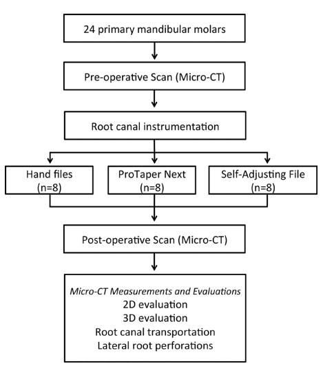
imageFigure 1 Diagram representation of study desig open_in_new
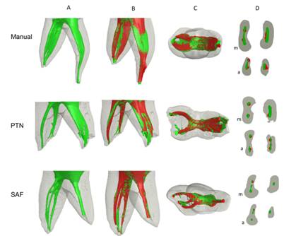
imageFigure 2 Representative 2D and 3D reconstructions of the internal and external anatomy of the primary mandibular first molars before and after root canal preparation with hand, ProTaper Next and SAF files. (A) Preoperative 3D models of the internal root canal anatomy. (B) Buccal view of superimposed 3D models before (green) and after (red) root canal preparation. (C) Apical view of superimposed 3D models before (green) and after (red) root canal preparation. (D) Representative cross-sections of the superimposed root canals before (green) and after (red) preparation at the middle (m) and apical (a) thirds. open_in_new
imageFigure 3 Distribution (%) of lateral perforations after root canal preparation with hand, ProTaper Next and SAF files. Different letters mean statistically significant difference (p<0.05). open_in_new

imageFigure 4 Apical view of 3D models and representative cross-sections of groups HF, PTN and SAF showing irregularities and thinner dentin walls due to physiological root resorption before canal preparation, and the location and extent of perforations observed after canal preparation (black arrows). open_in_new

table_chartTable 1
Morphometric data (media ± standard deviation) of the mesial and distal roots canals of primary mandibular molars before and after instrumentation with hand, ProTaper Next and SAF files at 1 mm from the apical foramen.
| Mesial root | Distal root | |||||
|---|---|---|---|---|---|---|
| Experimental Groups | Experimental Groups | |||||
| Manual | PTN | SAF | Manual | PTN | SAF | |
| Length (mm) | 7.79±1.25 | 7.80±1.39 | 7.89±0.63 | 7.08±2.74 | 6.14±1.67 | 7.78±2.02 |
| Area (mm2) (initial) | 0.26±0.40 | 0.15±0.14 | 0.23±0.32 | 0.38±0.33 | 0.37±0.34 | 0.27±0.29 |
| After preparation | 0.34±0.42 | 0.22±0.13 | 0.25±0.32 | 0.51±0.34 | 0.52±0.39 | 0.30±0.30 |
| Δ | 0.07±0.05A | 0.07±0.04A | 0.02±0.01B | 0.13±0.10A | 0.14±0.09A | 0.03±0.03B |
| Perimeter (mm) (initial) | 2.08±1.71 | 1.55±0.81 | 2.29±1.97 | 3.21±2.41 | 3.38±2.74 | 2.48±1.68 |
| After preparation | 2.42±1.74 | 1.89±0.63 | 2.37±1.97 | 3.70±2.28 | 3.77±2.68 | 2.63±1.56 |
| Δ | 0.35±0.24A | 0.34±0.32A | 0.07±0.04B | 0.35±0.22A | 0.29±0.13AB | 0.10±0.08B |
| Roundness (initial) | 0.46±0.16 | 0.48±0.17 | 0.42±0.24 | 0.36±0.17 | 0.30±0.19 | 0.32±0.17 |
| After preparation | 0.56±0.18 | 0.62±0.22 | 0.48±0.26 | 0.42±0.18 | 0.46±0.28 | 0.38±0.20 |
| Δ | 0.10±0.09AB | 0.16±0.13A | 0.05±0.05B | 0.03±0.03B | 0.19±0.23A | 0.03±0.04B |
| Major diameter (mm) (initial) | 0.82±0.71 | 0.62±0.34 | 0.94±0.86 | 1.28±1.04 | 1.43±1.18 | 1.04±0.73 |
| After preparation | 0.92±0.70 | 0.70±0.30 | 0.98±0.87 | 1.41±0.98 | 1.54±1.22 | 1.10±0.71 |
| Δ | 0.07±0.06A | 0.06±0.06A | 0.04±0.02A | 0.06±0.07A | 0.02±0.04A | 0.04±0.02A |
| Minor diameter (mm) (initial) | 0.37±0.21 | 0.30±0.13 | 0.31±0.18 | 0.39±0.21 | 0.31±0.17 | 0.31±0.17 |
| After preparation | 0.48±0.21 | 0.46±0.10 | 036±0.17 | 0.58±0.17 | 0.52±0.07 | 0.39±0.16 |
| Δ | 0.11±0.07A | 0.16±0.07A | 0.05±0.04B | 0.19±0.12A | 0.21±0.13A | 0.07±0.07B |
| Volume (mm3) (initial) | 6.35±3.81 | 5.88±3.61 | 6.81±4.10 | 7.90±4.09 | 7.79±5.57 | 7.56±5.22 |
| After preparation | 7.97±3.95 | 7.57±3.72 | 7.30±4.07 | 9.57±4.46 | 9.41±6.19 | 7.98±5.57 |
| Δ | 1.62±0.90A | 1.69±0.52A | 0.50±0.29B | 1.67±1.10A | 1.62±0.78A | 0.42±0.55B |
| Surface Area (mm2) (initial) | 52.83±16.85 | 46.99±20.34 | 55.93±21.15 | 52.80±19.01 | 50.85±26.18 | 49.67±24.84 |
| After preparation | 57.01±16.22 | 50.74±19.70 | 57.38±20.51 | 56.41±19.27 | 52.80±26.82 | 50.70±24.69 |
| Δ | 4.19±2.92A | 3.75±2.59A | 1.45±1.59B | 2.31±1.45A | 2.23±1.61A | 0.39±0.41B |
| SMI (initial) | 1.82±0.44 | 2.12±0.45 | 1.70±0.50 | 1.76±0.53 | 1.60±0.35 | 1.97±0.25 |
| After preparation | 2.12±0.35 | 2.25±0.43 | 1.85±0.57 | 1.90±0.55 | 1.90±0.37 | 2.07±0.26 |
| Δ | 0.25±0.16A | 0.16±0.07AB | 0.05±0.03B | 0.16±0.08A | 0.18±0.18A | 0.10±0.05A |
-
Δ, mean increase (± standard deviation) of the analysed parameter. Different superscript letters in the same line indicate statistical significant difference between groups (Tukey test, P < .05). Within groups, values with bold letters in the same column were not statistically different (paired t test, P > .05).
table_chartTable 2
Transportation (in mm) of the mesial and distal roots canals of primary mandibular molars after instrumentation with hand, ProTaper Next and SAF files.
| Manual | PTN | >SAF | ||||
|---|---|---|---|---|---|---|
| Mean±SD | Range | Mean±SD | Range | Mean±SD | Range | |
| Mesial root | 0.14±0.11B | 0.04-0.33 | 0.09±0,04AB | 0,04-0,15 | 0,05±0.02A | 0.02-0.09 |
| Distal root | 0.29±0.17B | 0.04±0.55 | 0.20±0.24AB | 0.03-0,65 | 0.07±0.04A | 0.03-0.15 |
-
Different superscript letters in the same row indicate statistically significant difference among groups (Tukey’s test, p<0.05).
Como citar
location_on
Fundação Odontológica de Ribeirão Preto
Av. do Café, S/N, 14040-904 Ribeirão Preto SP Brasil, Tel.: (55 16) 3602-3982, Fax: (55 16) 3633-0999 -
Ribeirão Preto -
SP -
Brazil
E-mail: bdj@forp.usp.br
E-mail: bdj@forp.usp.br
rss_feed
Acompanhe os números deste periódico no seu leitor de RSS
Versão para download de PDF
Artigos relacionados
Lista de links para artigos relacionados. Os links abrem em nova aba.
Versões e tradução automática
Escolha a versão original do texto ou utilize um serviço de tradução automática.
Versão original do texto
Como citar
Escolha um formato para exportar ou selecione um estilo de citação. O conteúdo abaixo pode ser atualizado após a seleção.