 Radiomic characterization of sinus lift grafts using texture analysis technique in cone-beam computed tomography images
ABSTRACT
Objective This study aims to apply texture analysis (TA) to cone-beam computed tomography (CBCT) images as a predictive tool for characterizing Geistlich Bio-Oss® bone grafts used in maxillary sinus lift (SL) surgeries.
Material and Methods Eleven CBCT scans of patients who underwent bilateral sinus lift (SL) procedures with Bio-Oss® grafts were selected from the Radiology database of ICT-UNESP. The scans had been acquired using an I-CAT Next Generation scanner (voxel size: 0.25 mm; field of view: 16.0 × 8.0 cm) and exported in DICOM format to OnDemand 3D software. Five parasagittal slices from each case were saved in bitmap format. Texture analysis was performed using MaZda 3.20 software. A 4.0 × 4.0 mm region of interest (ROI) was positioned at the center of the graft, and texture features were extracted using the gray-level co-occurrence matrix (GLCM). All analyses were performed by a single calibrated evaluator. Data normality was assessed using the Shapiro–Wilk test. Since the variables did not present a normal distribution, data were transformed into ranks. Spearman’s correlation coefficient was used to evaluate the association between directions and distances of texture measurements. A mixed linear model was applied for each texture parameter, considering maxillary sinus (right/left), section (S0–S4), and their interaction as fixed effects, with patient as a random effect. Intra-examiner reproducibility was assessed using the intraclass correlation coefficient (ICC). Statistical significance was set at 5%.
Results Most texture parameters showed a homogeneous distribution between the analyzed sides and slices, except for the SumAverg parameter, which indicated higher density on the right side.
Conclusion The TA proved to be an effective tool for the quantitative characterization of grafts, revealing symmetrical and homogeneous bone integration of the biomaterial, with potential clinical application in surgical planning and monitoring.
KEYWORDS:
Bone transplantation; Diagnosis, computer-assisted; Maxillary sinus; Radiomics; Spiral cone-beam computed tomography
Objetivo Este estudo teve como objetivo aplicar a técnica de Análise de Textura (AT) em imagens de tomografia computadorizada de feixe cônico (TCFC) como ferramenta preditora para caracterização dos enxertos ósseos Geistlich Bio-Oss®, utilizados em cirurgias de levantamento de seio maxilar (sinus lift – SL).
Material e Métodos Foram selecionados 11 exames de TCFC, com SL bilateral e uso de Bio-Oss®, do banco de dados da Radiologia do ICT-UNESP. As imagens foram obtidas em tomógrafo I-CAT Next Generation (voxel 0,25 mm, FOV 16,0 × 8,0 cm) e exportadas em formato DICOM para o software OnDemand 3D, onde foram salvos cinco cortes parasagitais por caso em formato bitmap. As imagens foram analisadas no software MaZda 3.20, com uma região de interesse (ROI) de 4,0 × 4,0 mm posicionada sobre o centro do enxerto, utilizando a matriz de coocorrência de níveis de cinza (MOC). A AT foi realizada por um único avaliador calibrado. A normalidade dos dados foi avaliada por meio do teste de Shapiro–Wilk. Como as variáveis não apresentaram distribuição normal, os dados foram transformados em ordens. O coeficiente de correlação de Spearman foi utilizado para avaliar a associação entre as direções e as distâncias das medidas de textura. Um modelo linear misto foi aplicado para cada parâmetro de textura, considerando o seio maxilar (direito/esquerdo), a seção (S0–S4) e sua interação como efeitos fixos, com o paciente como efeito aleatório. A reprodutibilidade intraexaminador foi avaliada por meio do coeficiente de correlação intraclasse (ICC). A significância estatística foi fixada em 5%.
Resultados A maioria dos parâmetros de textura apresentou distribuição homogênea entre os lados e cortes analisados, com exceção do parâmetro SumAverg, que indicou maior densidade no lado direito.
Conclusão A AT demonstrou ser uma ferramenta eficaz para a caracterização quantitativa dos enxertos, revelando integração óssea simétrica e homogênea do biomaterial, com potencial aplicação clínica no monitoramento e planejamento cirúrgico.
PALAVRAS-CHAVE:
Enxerto ósseo; Diagnóstico por computador; Seio maxilar; Radiômica; Tomografia computadorizada de feixe cônico
INTRODUCTION
Osseointegrated dental implants are widely used in the rehabilitation of edentulous patients, requiring the presence of supporting bone for their success. Osseointegration, a concept established by Brånemark, refers to the direct connection between the implant and the bone, without interposed fibrous tissue, which ensures stability and improves masticatory function, aesthetics, and patient comfort [1]. Implant rehabilitation, however, can be hampered by maxillary sinus pneumatization and alveolar bone loss, especially in the posterior regions of the maxilla [2].
Maxillary sinus lifting is considered a predictable and effective technique for bone augmentation in atrophic regions of the posterior maxilla and has been used since 1980 [3,4]. The success rate of the procedure depends on the quality of the graft and subsequent bone integration [5,6].
In addition, clinical studies and biomechanical reviews reinforce that both the quality and volume of bone tissue are determinants for the predictability of dental implants. Guided bone regeneration, associated with different biomaterials and surgical techniques, has been effective in restoring conditions favorable to osseointegration, especially in regions with marked resorption [7]. Complementarily, biomechanical aspects, such as load distribution at the implant-abutment interface, directly influence the stability and longevity of prosthetic rehabilitation, being essential factors in rehabilitation planning [8]. Several graft materials are used, including autogenous, xenogeneic, allogeneic, and alloplastic bone [9].
Cone-beam computed tomography (CBCT) has established itself as a reliable tool in the planning and monitoring of dental surgical procedures. In addition to providing high-resolution images with low distortion, CBCT has been used in quantitative analyses, such as texture analysis (TA), which allows the quantification of radiographic patterns using mathematical algorithms applied to the gray-scale co-occurrence matrix [10].
Radiomic analysis and TA have been successfully employed in different clinical contexts, revealing subtle changes in bone and soft tissues that are often imperceptible to the naked eye [10-12]. Recent studies have demonstrated their applicability in the diagnosis of periodontal lesions and in the assessment of dental implant stability [11,13]. However, there are no studies using TA in CBCT images to evaluate the characteristics of bone grafts in maxillary sinus lift procedures, evidencing a gap in the literature.
Considering the potential of texture analysis to identify structural changes in tissues, this study is based on the hypothesis that TA parameters applied to CBCT can characterize the quality of grafts used in maxillary sinus lift procedures. The main objective is to evaluate, through texture analysis, CBCT images of bone grafts in sinus lift procedures, contributing objective data for the follow-up and prognosis of this surgical intervention.
MATERIAL AND METHODS
This study was approved by the Human Research Ethics Committee of ICT Unesp, São José dos Campos campus, São Paulo. Number – CAAE: 7446725.1.0000.0077. All images analyzed in this study belong to the image bank of the Radiology department of ICT Unesp, São José dos Campos campus, São Paulo.
The CBCT images were previously obtained using an I-CAT Next Generation CT scanner (Imaging Science International, Hattfield, PA, USA), using a protocol consisting of a field of view (FOV) of 16.0 x 8.0 cm, covering the region of interest of the maxilla, using a voxel of 0.25 mm, obtained at 120 kV with an exposure time of 26.9 seconds and 37.07 mAs. The exams were acquired in completely edentulous upper patients as a means of planning surgery for bilateral sinus lifts (SL). Initially, CBCT was acquired to quantify the degree of alveolar bone resorption in the posterior regions of the maxilla and the actual need for grafts and SL.
A voxel size of 0.25 mm was used in the present study, consistent with clinically feasible CBCT protocols. The aim was not to assess bone microarchitecture directly, but to determine whether CBCT-derived texture features could identify relevant image-pattern differences within standardized regions. Previous studies have shown that CBCT texture analysis can successfully evaluate dental implant stability [13], medication-related osteonecrosis of the jaw [14], and periapical bone healing [15]. Although voxel size may influence bone structure analysis [16], all scans were acquired using standardized parameters. Therefore, the results should be interpreted as texture-based image biomarkers rather than direct measurements of bone microstructure.
In this study, CBCT scans performed after SL procedures, carried out after 6 months, were evaluated to analyze the degrees of integration with the grafts. All grafts used belonged to the same type of exogenous material - Geistlich Bio-Oss®.
Sample calculation
Eleven CBCT scans (11 patients) were evaluated, involving 22 maxillary sinuses with bilateral Geistlich Bio-Oss® exogenous bone grafts. This sample was based on a previous study by Queiroz et al. (2023) [14] and was a convenience sample.
Inclusion, non-inclusion and exclusion criteria
CT scans performed between 5 and 6 months after bilateral sinus lift surgery using Geistlich Bio-Oss® exogenous bone grafts in completely edentulous patients. All scans belonged to the ICT Unesp Radiology Department archive and were obtained using the protocol described above. All examinations recorded the entire grafting region, as well as the entire graft structure, with no motion artifacts or pathological changes in the maxillary sinuses. The images belonged to individuals of both sexes and ages ranging from 32 to 71 years. The sample did not include examinations that did not show bilateral bone grafts in completely edentulous patients; the presence of motion artifacts that impaired image quality; images obtained using CT scan technical protocols other than those described; and examinations that indicated pathological changes in the maxillary sinuses. Smokers and patients with systemic changes that could alter the bone integration process were not included in the study. These data were accessed through the individuals' medical records. In addition to those for whom it was not possible to confirm the graft material (Geistlich Bio-Oss®) used.
Image selection
The DICOM images from the CBCT were exported to the OnDemand 3D software (Cybermed, Seoul, South Korea) and analyzed in the Dental module. In the Dental window, the standard 2X enhancement filter was applied, and the brightness and contrast (window) values were set to 4083 for window width and 1077 for window center. These values were standardized for all exams. Adjustments were made to the sagittal and occlusal planes (with the hard palate parallel to the horizontal plane) using the “axis slice” tool. Using the “arc and curve” tool, a panoramic curve was drawn in the center of the upper arch, over the central axial slice, thus generating the panoramic slice and the respective parasagittal (or transverse) slice.
Five parasagittal slice were selected individually from each maxillary sinus (S0 to S4), covering the regions corresponding to the grafts, one more distal (S0), one in the middle region of the graft (S2), one more mesial (S4), and two intermediate, between the ends and the middle region of the graft, S1 and S3, respectively (Figure 1).

Panoramic CBCT section with the respective parasagittal sections of each maxillary sinus (S0 to S4) to be analyzed by TA.
These five parasagittal sections were exported in bitmap format (.bmp) were individually analyzed using MaZda 3.20 software (Institute of Electronics, Technical University of Lodz, Poland) to calculate the texture of the grafts.
Texture analysis
The selected sections were exported to MaZda 3.20 software (Institute of Electronics, Technical University of Lodz, Poland). In this software, the previously determined ROI (4.0x4.0mm) positioned in the center of the region corresponding to the graft image was delimited using the “Draw Polygon” tool.
TA was performed using a second-order statistical approach, through co-occurrence matrix (MOC) analysis, analyzing the same parameters used in the study by [13] (Figure 2). After determining the ROI, the TA of this region was calculated using the co-occurrence matrix (MOC) in four different directions and three distances, relative to 11 texture parameters - AngScMom, Contrast, Correlat, SumOfSqs, InvDfMom, SumAverg, SumVarnc, SumEntrp, Entropy, DifVarnc, and DifEntrp.

MaZda 3.20 software, illustrating ROI delimitation on the center of the graft in the parasagittal section, as well as determination of texture parameters.
After calculating the texture parameters, the values for the second-order statistical approach were obtained through co-occurrence matrix analysis. In this study, the same 11 parameters used in the study by Costa et al. (2023) [10]; Costa et al. (2021) [13]; Gonçalves et al. (2020) [17]; and Queiroz et al. (2023) [14] were analyzed.
After performing the TA process in the MaZda 3.20 software, the results were displayed in an orderly manner and tabulated in software for statistical analysis. This process was performed individually, in each ROI of each exam previously selected in the region.
Evaluator calibration
All analyses were performed by a single evaluator trained in the use of the system and then calibrated for texture analysis. This calibration was performed using a set of representative pilot images not included in the final sample. This process involved repeating the segmentation of regions of interest (ROI) at two different times, with a minimum interval of seven days, allowing the calculation of the intraclass correlation coefficient (ICC) to assess intra-examiner reproducibility. The ICC value obtained was greater than 0.90, indicating excellent agreement and ensuring the reliability of the data collected for the study.
Statistical analysis
As the response variables did not show a normal distribution (Shapiro-Wilk test), they were transformed into ranks for the application of the mixed model. Spearman's correlation coefficient was calculated between the four directions (horizontal, vertical, 45° and 135°) and three distances for each parameter. A high correlation was observed between the different directions, so to simplify the analysis and avoid multicollinearity, we chose to use the average of the directions for each parameter. Subsequently, descriptive statistics (mean, standard deviation, median, minimum, and maximum) were calculated for each of the 11 texture parameters. These statistics were presented by section (S0 to S4) and by maxillary sinus (right and left).
For the inferential analysis, a mixed linear model was used for each parameter, with the fixed effects of sinus (right or left) and section (S0 to S4) and the interaction between sinus and section, in addition to a random effect for the patient, aiming to adjust the dependence of the repeated measures. This approach was chosen because it allows for the joint evaluation of the effects of side and section, in addition to the interaction between these two factors. Statistical significance was set at 5% (p < 0.05) for all analyses.
RESULTS
This study included 11 patients, 10 women and 1 man. The participants' ages ranged from 32 to 71 years, with a mean of 52.0 years and a standard deviation of 11.5 years. Eleven texture parameters were analyzed: AngScMom, Contrast, Correlat, SumOfSqs, InvDfMom, SumAverg, SumVarnc, SumEntrp, Entropy, DifVarnc, and DifEntrp. Each parameter is described in Table I, which presents descriptive statistics stratified by maxillary sinus (right and left). In addition, the p-values derived from the mixed linear model are presented, which evaluate the main effects of sinus and section, as well as the interaction between both. For data analysis, a mixed linear model was adopted, as it is suitable for data structures in which multiple measurements are obtained from the same individual—in this case, different sections and sinuses per patient. This type of model allows for the simultaneous evaluation of the main effects of side (right vs. left sinus) and section (S0 to S4), considering intra-individual dependence through a random effect for the patient. By using a single analysis to investigate both factors, the model contributes to reducing the risk of type I error, avoiding the need for multiple isolated comparisons. In addition, an interaction term between sinus and slice was included in the model, allowing us to verify whether the pattern of variation in the slices differs between sides.
Table I summarizes the pooled descriptive statistics and the results of the mixed linear model. No significant effects of slice position or sinus × slice interaction were observed for any texture parameter. A significant difference between the right and left maxillary sinuses was found only for SumAverg (p < 0.001), with higher values in the right sinus.
Pooled texture parameters of maxillary sinus graft and mixed-model analysis. Values are presented as mean ± standard deviation. P-values correspond to the effects of sinus side, slice position (S0–S4), and sinus × slice interaction.
Analyzing Table 1 , in general, the statistical analysis did not identify significant differences between the maxillary sinuses, between the sections, or in the interaction between these factors for most of the parameters studied. The only exception was observed for the SumAverg parameter, which showed a statistically significant difference between the sinuses (p < 0.001): the right side had higher values than the left side. The other parameters showed no significant differences for the sinus effect, nor for the effects of section or the sinus × section interaction.
Figure 3 shows the variation in the mean of each parameter across the sections, allowing graphical visualization of the differences between the sinuses. Each graph shows the sections (from S0 to S4) on the horizontal axis and the mean of the analyzed parameter on the vertical axis; the lines represent the two maxillary sinuses, allowing visual comparisons between sides and along the length of the graft.

Variation in the average of each texture parameter of the grafts along the cuts (S0 to S4) in the right and left maxillary sinuses.
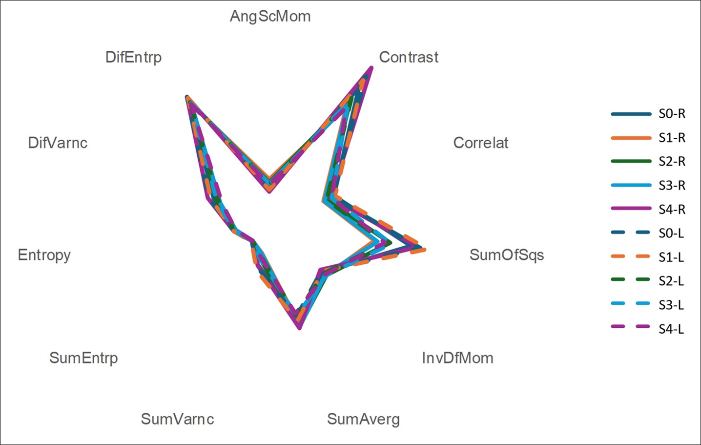
Figure 4 shows the radar chart with the averages of the eleven texture parameters evaluated, organized by section (S0 to S4) and maxillary sinus (right and left). To facilitate the simultaneous visualization of variables with different scales, some parameters were rescaled: AngScMom was multiplied by 100; Correlat, InvDfMom, and DifEntrp were multiplied by 10; while SumAverg and SumVarnc were divided by 10. The other parameters were kept in their original scale. The solid lines represent the mean values of the right sinus, and the dotted lines represent the values of the left sinus. In general, similar patterns are observed between the sides and sections.

Radar chart showing the average texture parameters per maxillary sinus and section (some parameters were rescaled to fit the chart).
DISCUSSION
The evaluation of bone graft behavior used in the sinus lift technique in maxillary sinuses is important for planning dental implant placement. This evaluation has usually been performed using CBCT scans, with varying post-surgical times, using subjective methodologies related to visual inspections of graft tissue aspects in CBCT images. The image evaluation methodology using the texture analysis (TA) technique allows statistical data to be extracted through different parameters that characterize tissue behavior, providing objective information about them, favoring the treatment plan and its consequent success.
In this study, 11 texture parameters extracted from CBCT images were analyzed, performed between 5 and 6 months after the sinus lift procedure in bilateral maxillary sinuses, related to a specific type of exogenous grafting material - Geistlich Bio-Oss®, in order to characterize the behavior of this material, relating it to the side of its installation (right or left maxillary sinus), as well as its mesiodistal extension.
The results showed homogeneous distributions of the material in both right and left maxillary sinuses, as well as in the 5 sections (S0 to S4) analyzed on each side. Most of the 11 texture parameters evaluated showed stable behavior and no statistically significant differences. This homogeneity indicates an organized and uniform pattern of bone integration, consistent with adequate distribution and stability of the biomaterial used in the grafting technique.
To understand the results and how they enable the characterization of the graft material analyzed, it is necessary to analyze each texture parameter individually, correlating its values with its meaning.
The findings related to the AngScMom parameter, which measures image uniformity, indicated high values in the images of the grafts in both maxillary sinuses—right and left—which translates into few variations in gray tones in the images and, therefore, homogeneous texture, indicating good integration of the biomaterial [10]. Contrast is the parameter responsible for quantifying the intensity of local variation, with high values indicating greater contrast between neighboring pixels. In this study, the mean values of the graft materials indicated a moderately contrasting and similar pattern between the sides, consistent with good graft distribution [14].
The Correlat parameter measures the linearity between gray tones of neighboring pixels, with high values indicating good tissue organization [18]. In this study, it was observed that it was slightly higher for the graft in the maxillary sinuses on the left side, but without statistical significance. The median values indicate that the grafts presented a moderate pattern organization. Regarding SumOfSqs, which reflects heterogeneity, the values found in our results indicate uniform distribution of the graft, which is positive for its osseointegration [13]. The only parameter that showed a statistically significant difference between the sides (p<0.001), being higher on the right side, was SumAverg, which is related to density. Thus, a higher density or mineralization was observed in the grafts on the right side compared to the contralateral side. Parameters such as InvDfMom, SumVarnc, Entropy, SumEntrp, DifEntrp, and DifVarnc did not show statistically significant differences between sides and sections. These are related to the degree of heterogeneity, irregularity, microstructural complexity, and tissue disorder [14,15,19]. The values observed here in these parameters suggest that the graft materials analyzed are characterized by low local irregularity, being well distributed and stable, organized, with complex but organized structures, indicating balanced remodeling of the grafts, signaling positive for osseointegration.
These findings corroborate the growing evidence that texture analysis using CBCT images is capable of detecting subtle bone microstructures, often not observed by conventional analysis, as demonstrated by [10]. In this study, the authors were able to differentiate odontogenic from non-odontogenic sinusitis based on textural characteristics, reinforcing the sensitivity of the technique even in anatomically complex regions such as the maxillary sinus. Such evidence indicates that the parameters used in the present study are sufficiently sensitive to detect changes, but no marked structural differences were found between the sides, suggesting a stable regeneration pattern.
Another study, applied to bone pathology in CBCT images [14], showed that texture analysis made it possible to identify abnormal patterns associated with drug-induced osteonecrosis, characterized by elevations in heterogeneity parameters (such as Contrast and Entropy). The absence of these patterns in our study reinforces the hypothesis that the grafts evaluated are in good internal condition, with no textural signs compatible with necrosis, inflammation, or integration failures.
SumAverg was the only texture parameter to show a statistically significant difference, with higher values in the right-side grafts (p < 0.001) compared to the left-side grafts.
This parameter reflects the average gray tone intensity and is influenced by tissue density and local mineralization. Costa et al. (2021) [13], in a study that correlated texture parameters with implant stability, observed that high SumAverg values were associated with the presence of denser and more stable bone tissue. Therefore, our findings may indicate local asymmetry in the density or bone maturation of sinus lift grafts, although clinically this variation may not have a significant impact if other parameters remain homogeneous, as observed in our results. Similarly, Nussi et al. (2023) [18] demonstrated that changes in texture parameters may be related to factors such as age, gender, and functional load, even in the absence of pathologies. This study focused on the bone marrow of mandibular condyles in CBCT images and highlights the influence of biological and anatomical variables on bone composition, serving as a reference for the interpretation of asymmetric findings related to SumAverg in this study.
Previous studies have reinforced the performance of the texture analysis technique as a clinical biomarker. Among these, Lopes et al. (2025) [15] investigated the effect of different intracanal medications on periapical bone healing, demonstrating that texture analysis allowed the quantification of the evolution of the repair process, with a progressive reduction in the entropy parameter during the course of treatment. This reduction represents an increase in tissue organization, indicating that homogeneous patterns, such as those found in the present study, can be interpreted as a sign of bone maturation.
The contribution of texture analysis to the study of bone behavior was also highlighted by Gonçalves et al. (2020), who used this technique to detect early furcation lesions in CBCT images, observing superior performance compared to pure subjective visual inspection. This reinforces the fact that texture parameters, even if subtle, can signal pathological or regenerative processes with high accuracy, thus making them valuable tools for longitudinal monitoring of grafts.
From a methodological point of view, Barioni et al. (2024) [19] highlights that texture analysis has transformed the paradigms of dento-maxillofacial imaging by enabling a quantitative and reproducible approach, reducing the subjectivity associated with image interpretation. This study fits into this context, adopting objective parameters for evaluating graft structure in sinus lift surgeries, contributing to the development of standardized protocols in implant dentistry and reconstructive surgery.
The statistical approach adopted in this study—with mixed linear models and data transformation into ranks based on the absence of data normality—is appropriate for dependent and repeated data, such as those collected in multiple sections and different sides of the same patient. This approach was also used by Ogawa et al. (2024) [20] when comparing condylar changes in CT and MRI, enabling the control of individual variations and leading to more robust results.
It is important to highlight several limitations inherent to this study, such as the small sample size, which may have limited the ability to detect subtle differences between the groups; the impossibility of correlating the findings with histological data, given that histological analysis remains the gold standard for confirming biological graft incorporation and osseointegration, however, for obvious ethical and practical reasons, histological validation is not feasible in the routine clinical follow-up of maxillary sinus lift procedures; the lack of longitudinal follow-up, considering that CBCT images were analyzed at a single postoperative time point between 5 and 6 months, which could limit the understanding of the structural evolution of bone tissue throughout the healing process; and, additionally, the potential influence of minor variations in CBCT image acquisition protocols.These observations can be considered for future studies, characterizing this as a pilot study.
Another limitation of this study is that patient motion during CBCT acquisition could not be objectively measured. Although all scans were reviewed and examinations presenting visible motion artifacts were excluded, subtle micro-movements may still have occurred without producing detectable image degradation. As this was a retrospective study, this limitation is unavoidable and should be considered when interpreting the texture analysis results.
From a clinical point of view, the findings support the applicability of texture analysis as a quantitative assessment tool for graft quality and newly formed bone, with potential use in rehabilitation protocols, implant planning, bone regeneration monitoring, and biomaterial evaluation.
CONCLUSION
Texture analysis of CBCT images demonstrated homogeneous trabecular texture patterns in Geistlich Bio-Oss® grafted regions following maxillary sinus augmentation. These findings suggest that texture analysis may provide objective quantitative information regarding the tomographic characteristics of grafted areas, complementing conventional CBCT visual assessment. However, the biological significance of these imaging-derived texture features requires confirmation through studies including histological correlation and clinical outcomes.
Acknowledgements
The authors acknowledge that no individuals or organizations, other than the listed authors, made substantive contributions to this study.
Data availability
The research data are available from the corresponding author upon reasonable request.
How to cite:
Funding
Regulatory Statement
REFERENCES
-
1 Bhalla N, Dym H. Update on maxillary sinus augmentation. Dent Clin North Am. 2021;65(1):197-210. https://doi.org/10.1016/j.cden.2020.09.013 PMid:33213709.
» https://doi.org/10.1016/j.cden.2020.09.013 -
2 Aghaloo TL, Misch C, Lin GH, Iacono VJ, Wang HL. Bone augmentation of the edentulous maxilla for implant placement: a systematic review. Int J Oral Maxillofac Implants. 2016;31(Suppl):s19-30. https://doi.org/10.11607/jomi.16suppl.g1 PMid:27228250.
» https://doi.org/10.11607/jomi.16suppl.g1 - 3 Boyne PJ, James RA. Grafting of the maxillary sinus floor with autogenous marrow and bone. J Oral Surg. 1980;38(8):613-6. PMid:6993637.
-
4 Tatum H Jr. Maxillary and sinus implant reconstructions. Dent Clin North Am. 1986;30(2):207-29. https://doi.org/10.1016/S0011-8532(22)02107-3 PMid:3516738.
» https://doi.org/10.1016/S0011-8532(22)02107-3 - 5 Del Fabbro M, Testori T, Francetti L, Weinstein R. Systematic review of survival rates for implants placed in the grafted maxillary sinus. Int J Periodontics Restorative Dent. 2004;24(6):565-77. PMid:15626319.
-
6 Al-Moraissi EA, Alkhutari AS, Abotaleb B, Altairi NH, Del Fabbro M. Do osteoconductive bone substitutes result in similar bone regeneration for maxillary sinus augmentation when compared to osteogenic and osteoinductive bone grafts? A systematic review and frequentist network meta-analysis. Int J Oral Maxillofac Implants. 2020;49(1):107-20. https://doi.org/10.1016/j.ijom.2019.05.004 PMid:31230768.
» https://doi.org/10.1016/j.ijom.2019.05.004 -
7 Nascimento RD, Cardoso PE, Matuda FS, Raldi FV, de Moraes MB. Incorporation of bone grafts and osseointegration in the area of maxillary dental agenesis. Braz Dent Sci. 2017;20(4):143-8. https://doi.org/10.14295/bds.2017.v20i4.1377
» https://doi.org/10.14295/bds.2017.v20i4.1377 -
8 Andrade GS, Kalman L, Lo Giudice R, Adolfi D, Feilzer AJ, Tribst JPM. Biomechanics of implant-supported restorations. Braz Dent Sci. 2023;26(1):e3637. https://doi.org/10.4322/bds.2023.e3637
» https://doi.org/10.4322/bds.2023.e3637 -
9 Aludden HC, Mordenfeld A, Hallman M, Dahlin C, Jensen T. Lateral ridge augmentation with Bio-Oss alone or Bio-Oss mixed with particulate autogenous bone graft: a systematic review. Int J Oral Maxillofac Implants. 2017;46(8):1030-8. https://doi.org/10.1016/j.ijom.2017.03.008 PMid:28366452.
» https://doi.org/10.1016/j.ijom.2017.03.008 -
10 Costa ALF, Fardim KAC, Ribeiro IT, Jardini MAN, Braz-Silva PH, Orhan K, et al. Cone-beam computed tomography texture analysis can help differentiate odontogenic and non-odontogenic maxillary sinusitis. Imaging Sci Dent. 2023;53(1):43-51. https://doi.org/10.5624/isd.20220166 PMid:37006790.
» https://doi.org/10.5624/isd.20220166 -
11 Sohn JH, Fields BKK. Radiomics and Deep learning to predict pulmonary nodule metastasis at CT. Radiology. 2024;311(1):e233356. https://doi.org/10.1148/radiol.233356 PMid:38591975.
» https://doi.org/10.1148/radiol.233356 -
12 Oliveira MS, Fernandes PT, Avelar WM, Santos SL, Castellano G, Li LM. Texture analysis of computed tomography images of acute ischemic stroke patients. Braz J Med Biol Res. 2009;42(11):1076-9. https://doi.org/10.1590/S0100-879X2009005000034 PMid:19820884.
» https://doi.org/10.1590/S0100-879X2009005000034 -
13 Costa ALF, de Souza Carreira B, Fardim KAC, Nussi AD, da Silva Lima VC, Miguel MMV, et al. Texture analysis of cone beam computed tomography images reveals dental implant stability. Int J Oral Maxillofac Implants. 2021;50(12):1609-16. https://doi.org/10.1016/j.ijom.2021.04.009 PMid:33962826.
» https://doi.org/10.1016/j.ijom.2021.04.009 -
14 Queiroz PM, Fardim KC, Costa ALF, Matheus RA, Lopes SLPC. Texture analysis in cone-beam computed tomographic images of medication-related osteonecrosis of the jaw. Imaging Sci Dent. 2023;53(2):109-15. https://doi.org/10.5624/isd.20220202 PMid:37405203.
» https://doi.org/10.5624/isd.20220202 -
15 Lopes DLG, Lopes SLPC, Ungaro DMT, Gomes APM, Moura NB, Gonçalves BC, et al. Radiomics-driven CBCT texture analysis as a novel biosensor for quantifying periapical bone healing: a comparative study of intracanal medications. Biosensors. 2025;15(2):98. https://doi.org/10.3390/bios15020098 PMid:39997000.
» https://doi.org/10.3390/bios15020098 -
16 Pauwels R, Faruangsaeng T, Charoenkarn T, Ngonphloy N, Panmekiate S. Effect of exposure parameters and voxel size on bone structure analysis in CBCT. Dentomaxillofac Radiol. 2015;44(8):20150078. https://doi.org/10.1259/dmfr.20150078
» https://doi.org/10.1259/dmfr.20150078 -
17 Gonçalves BC, de Araújo EC, Nussi AD, Bechara N, Sarmento D, Oliveira MS, et al. Texture analysis of cone-beam computed tomography images assists the detection of furcal lesion. J Periodontol. 2020;91(9):1159-66. https://doi.org/10.1002/JPER.19-0477 PMid:32003465.
» https://doi.org/10.1002/JPER.19-0477 -
18 Nussi AD, de Castro Lopes SLP, De Rosa CS, Gomes JPP, Ogawa CM, Braz-Silva PH, et al. In vivo study of cone beam computed tomography texture analysis of mandibular condyle and its correlation with gender and age. Oral Radiol. 2023;39(1):191-7. https://doi.org/10.1007/s11282-022-00620-3 PMid:35585223.
» https://doi.org/10.1007/s11282-022-00620-3 -
19 Barioni ED, Lopes SLPC, Silvestre PR, Yasuda CL, Costa ALF. Texture analysis in volumetric imaging for dentomaxillofacial radiology: transforming diagnostic approaches and future directions. J Imaging. 2024;10(11):263. https://doi.org/10.3390/jimaging10110263 PMid:39590727.
» https://doi.org/10.3390/jimaging10110263 -
20 Ogawa CM, Flaiban E, Ricardo ALF, Lopes DLG, Oliveira LAP, et al. Comparative evaluation of temporomandibular condylar changes using texture analysis of CT and MRI images. Appl Sci. 2024;14(16):7020. https://doi.org/10.3390/app14167020
» https://doi.org/10.3390/app14167020
Conflict of Interest
Correspondence
Ítza Amarisis Ribeiro Pinto (Corresponding address) Universidade Estadual Paulista, Departamento de Diagnóstico e Cirurgia, São José dos Campos, SP, Brazil. Email: itza.pinto@unesp.brEditor-in-chief:
Editor: