
 CSF3R/CD114: a predictor related to worse survival in gliomas
ABSTRACT
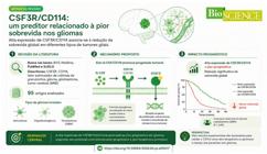
Introduction: Gliomas are the most frequent primary brain tumors, accounting for more than 80% of all primary neoplasms of the central nervous system. These tumors are classified into different histological subtypes, the main ones being astrocytoma, oligodendroglioma, and glioblastoma. Among them, glioblastoma stands out as the most common and aggressive subtype, associated with a poor prognosis, even in the face of multimodal therapeutic approaches that combine surgical resection, radiotherapy, and chemotherapy with temozolomide
Objective: To analyze the association of CD114 expression with survival in patients with glioma.
Method: The articles were selected by searching the following databases: Virtual Health Library (VHL), Medline, PubMed, and SciELO. The descriptors used were “CSF3R, CD114, colony-stimulating factor of granulocytes, glioma, glioblastoma, brain tumor”, performing advanced search by combining them with the Boolean operator AND.
Result: A total of 95 articles were analyzed.
Conclusion: There is an association between high expression of CSF3R/CD114 with worst prognosis of patients, reflected by the reduction in overall survival in different types of glial tumors.
KEYWORDS:
CSF3R; CD114; Granulocyte colony-stimulating factor; Glioma; Glioblastoma; Braintumor

RESUMO
Introdução: Os gliomas são os tumores cerebrais primários mais frequentes, representando mais de 80% de todas as neoplasias primárias do sistema nervoso central. Esses tumores são classificados em diferentes subtipos histológicos, sendo os principais o astrocitoma, o oligodendroglioma e o glioblastoma. Dentre eles, o glioblastoma destaca-se como o subtipo mais comum e agressivo, associado a prognóstico reservado, mesmo diante de abordagens terapêuticas multimodais que combinam ressecção cirúrgica, radioterapia e quimioterapia com temozolomida
Objetivo: Analisar a associação da expressão de CD114 com a sobrevida em pacientes com glioma.
Método: Os artigos foram selecionados por meio de busca nas seguintes bases de dados: Biblioteca Virtual em Saúde (BVS), Medline, PubMed e SciELO. Os descritores utilizados foram “CSF3R, CD114, fator estimulador de colônias de granulócitos, glioma, glioblastoma, tumor cerebral”, realizando busca avançada combinando-os com o operador booleano AND.
Resultado: Foram analisados 95 artigos.
Conclusão: Existe associação entre a alta expressão de CSF3R/CD114 e o pior prognóstico dos pacientes, refletido pela redução da sobrevida global em diferentes tipos de tumores gliais.
PALAVRAS-CHAVE:
CSF3R; CD114; Fator estimulador de colônias de granulócitos; Glioma; Glioblastoma; Tumor cerebral
INTRODUCTION
Gliomas are the most frequent primary brain tumors, accounting for more than 80% of all primary central nervous system (CNS) neoplasms. These are histologically heterogeneous neoplasms and classified by the World Health Organization in different degrees, according to their cellular characteristics and their capacity for tissue invasion. Gliomas are neoplasms derived from glial cells, including astrocytes, oligodendrocytes, and ependymal cells. These tumors are classified into different histological subtypes, the main ones being astrocytoma, oligodendroglioma, and glioblastoma (GBM), which, among them, stands out as the most common, aggressive subtype, with a poor prognosis even in the face of multimodal therapeutic approaches that combine surgical resection, radiotherapy, and chemotherapy with temozolomide. The presence or absence of mutation of the enzyme isocitrate dehydrogenase (IDH) allows GBM to be subdivided into three categories: 1) GBM wild-type HDI, which corresponds to about 90% of cases; 2) GBM with mutated HDI; 3) unspecified GBM (NOS), when the IDH mutation is not evaluated.1-5
The cell surface protein CD114 (Cluster of Differentiation 114), encoded by the CSF3R gene, acts as a receptor for granulocyte colony-stimulating factor (G-CSF).6,7 When stimulated by G-CSF, CD114 activation triggers the transcription activator-mediated signaling pathway 3 (STAT3), which regulates cellular processes such as cell proliferation, differentiation, and survival, and in abnormal situations can promote a cancer stem cell phenotype.8
CD114 has been proposed as a marker of cancer stem cells in tumors derived from the neural crest, such as neuroblastoma and melanoma.7,9,10 CD114 expression has also been identified in several other solid tumors, such as ovarian, cervical, bladder, and skin tumors.7,10-18 In addition, mutations in the CSF3R gene have been found in rare forms of leukemia.19.20
CD114-positive cancer cells demonstrated the ability to self-renew and generate offspring.9 Malignant brain neoplasms are capable of confiscating molecular and cellular mechanisms involved in neuronal plasticity.21-26 In this context, G-CSF acts as a growth factor, stimulating the survival and plasticity of neurons and neural stem cells27 and, in addition to stimulating neurogenesis,6 G-CSF acts synergistically with stem cell factor to induce the growth of cell projections, dendrites and axons, in cortical neurons.28
The combination of G-CSF and stem cell factor also protects against neurodegeneration and promotes the reorganization of the neurostructural network in a murine model of traumatic brain injury.29
In human gliomas, generalized expression of RNA and G-CSF protein was demonstrated, with increased proliferation and migration in CD114+ cells after exposure to the factor. Neutralization of G-CSF, in turn, inhibited tumor growth and cellular migratory capacity.11
Despite the existence of evidence, the role of CSF3R/CD114 as a possible prognostic biomarker in gliomas is not yet well elucidated and, if its role in pathogenesis is confirmed, CD114 could be used as a therapeutic target in the development of new drugs or even as a diagnostic and prognostic marker of the disease.30
This study aims to analyze the levels of CSF3R transcription in gliomas and its possible association with clinical prognosis, evaluated by means of overall survival, and to analyze the association of CD114 expression with survival in patients with glioma.
METHOD
The articles were selected through a search in the following databases: Virtual Health Library (VHL), International Literature in Health Sciences (Medline), PubMed and SciELO. The descriptors used were “CSF3R, CD114, colony-stimulating factor of granulocytes, glioma, glioblastoma, brain tumor”, performing an advanced search combining the descriptors with the Boolean operator AND. The inclusion criteria included freely available publications in full in English and Portuguese, specifically addressing glioma and its subtypes. Incomplete or repetitive articles, preliminary clinical trials in animals, or those involving other diseases unrelated to the tumors mentioned were excluded, totaling 95 articles.
DISCUSSION
Gliomas are the most common and lethal group of brain neoplasms, accounting for about 80% of malignant brain tumors. In addition, they, including low-grade ones, have highly variable clinical behavior and their survival cannot be adequately predicted by histological classification.31
The highly aggressive and invasive nature of gliomas poses significant challenges to their complete elimination using current therapeutic strategies, including surgery, radiotherapy, and chemotherapy. Residual tumour cells often lead to malignant recurrence and progression, with some gliomas tending to progress to glioblastoma (WHO grade IV glioma) in a short period of a few months, resulting in a poor prognosis.31
These tumors are classified into different types, according to the type of cell from which they originate or with which they share histological characteristics. Thus, gliomas are classified into ependymomas, astrocytomas, and oligodendrogliomas. Of these, astrocytoma is the most common type of glial tumors.32
Glioblastoma, classified as grade IV glioma by the World Health Organization, is the most common and aggressive type of glioma.1,4,33 In this sense, it presents excessive malignancy, being almost always lethal and leading to the death of the patient usually within 12-18 months after diagnosis,32 regardless of the treatment, and in cases of recurrence, it is quickly fatal.34 It differs from low-grade gliomas in that it presents necrosis and microvascular proliferation, with infiltrative behavior and rapid growth as characteristics. In addition, glioblastoma can arise as a primary or secondary tumor, and the secondary tumor is a malignant transformation of a low-grade brain tumor and/or with a mutation in the isocitrate dehydrogenase (IDH) gene35 and codeletion of chromosome 1p/19q.32
One of the most striking and challenging biological characteristics of gliomas, which are diffuse, is the extreme infiltration capacity of their tumor cells. These cells can spread over extensive regions beyond the primary focus, following structures of the neuropilus, which deals with the network of nerve fibers and glial cells of the brain, even crossing to the contralateral hemisphere and, in some cases, originating additional tumor foci, as observed in multiple glioblastoma. And it is this biological behavior that is one of the greatest challenges of treatment, considering that complete microscopic resection can never be achieved.34
Extracranial metastases are very rare events in glioblastoma, as it is the communication between neoplastic cells and the adjacent tumor stroma that has been shown to be a crucial factor for the invasive growth and dissemination of this type of glioma. Thus, the processes of invasion and migration by brain tissue represent the main mechanisms of disease propagation.36
In this context, three fundamental routes of dissemination are considered: 1) the collective invasive potential, in which tumor cells advance through the adjacent tissue through interstices of the extracellular matrix, expanding from the primary tumor without completely detaching; 2) the invasion mediated by inflammatory cells, characterized by the action of pro-tumor immune cells that secrete metalloproteinases and degrade the extracellular matrix, creating an imbalance with their physiological inhibitors and facilitating the advancement of malignant cells and it is, in this scenario, that tumor hypoxia stimulates the production of pro-inflammatory proteins and favors the activation of cancer stem cells, promoting gliomagenesis; and 3) via epithelial-mesenchymal transition, described in heterogeneous subpopulations of glioblastoma, where intercellular adhesion and senescence are compromised, which favor epigenetic alterations that culminate in cell dedifferentiation and acquisition of phenotypes similar to those of stem cells, expanding the invasive and adaptive capacity of the tumor.35
The epithelial-mesenchymal transition is a biological process in which cells with epithelial characteristics, that is, those with high adhesion to each other and little mobility, start to acquire characteristics of mesenchymal cells, with less adhesiveness and greater migratory and invasion capacity. This mechanism decisively confers the aggressiveness, plasticity and therapeutic resistance of glioblastomas. In them, there are subpopulations of heterogeneous cells with different phenotypes, so that they pass through similar pathways to the epithelial-mesenchymal transition, even though it is not a classic epithelium; This concept is expanded to tumors of the central nervous system.
Under natural conditions, cell adhesion limits proliferation. However, when it begins the process of senescence, this adhesion is impaired, this “natural brake” is lost, favoring tumor proliferation.
Thus, this pathway gives glioblastoma an infiltrative and adaptive phenotype, making surgical and pharmacological treatments difficult, and in clinical terms it is one of the reasons why glioblastoma has a high recurrence rate and poor prognosis.
The current standard treatment of glioblastomas consists of multimodal therapy involving surgical resection - which should be as broad and safe as possible - radiotherapy and chemotherapy. However, even with adequate treatment results, median survival is 12-15 months.37-39 In this core, even with aggressive treatment, in 75-90% of cases, recurrence occurs within 7-10 months after surgery.32
Pharmacological treatment options for these patients are very limited, making the development of new alternatives one of the greatest unmet demands of current medicine.40-43 There is also an urgent need to identify and validate new appropriate molecular biomarkers to improve diagnosis, as well as to guide treatment choices.44-46
Glioblastoma in the current classification of central nervous system tumors
The classification of central nervous system (CNS) tumors is traditionally based on histological characteristics and supported by immunohistochemical analyses.
Glioblastoma multiforme, which was named by Percival Bailey and Harvey Cushing in 192647, based on the histological appearance where it was evident that the tumor tissue would originate from precursors of glial cells and, in addition, because its configuration is very varied due to the presence of necrosis, hemorrhages and cysts, it was called “multiforme”.48 Currently, glioblastoma multiforme is called glioblastoma. Thus, it is considered an extremely heterogeneous disease, and this tumor heterogeneity is translated by histological patterns, genetic alterations and gene expression profiles.34
The status of the gene encoding the enzyme isocitrate dehydrogenase (IDH) determines the classification of glioblastomas into three types: 1) wild-type HDI, which represents about 90% of cases; 2) mutated HDI; and 3) unspecified.
IDH is an enzyme that stimulates the expression of proteins involved in cellular metabolism and energy production. It performs oxidative decarboxylation of isocitrate to form alpha-ketoglutarate, a crucial step in the Krebs cycle.49
In glioblastoma classified as IDH wild-type, the gene is normal, that is, it does not present a mutation and consequently produces the protein in its normal chemical structure. When the mutation occurs in the IDH1 or IDH2 genes, the enzyme gains a new function and starts to produce 2-hydroxyglutarate (2-HG), which is an oncometabolite that alters the methylation of DNA and chromatin, promoting epigenetic changes that affect cell differentiation and proliferation.50
In adults, primary glioblastomas are also defined as wild-type HDI, while secondary glioblastomas can be mutated HDI and intact 1p/19q, or mutated HDI and 1p/19q codeletion.32 Mutations in the IDH gene appear to be early events in gliomagenesis, followed by acquisition of mutations in the TP53 gene, and are markers of secondary glioblastoma.34
IDH-wild tumors typically occur in older patients, while those with IDH mutations, which correspond to about 10% of cases, affect younger people and originate from gliomas of lower grade in addition to presenting a better prognosis.2.4
More recently, molecular markers have been validated and incorporated, acquiring increasing importance, and are already represented in the most recent classification of the World Health Organization (WHO). The fifth edition of the international standard classification for brain and spinal cord tumors, established by the WHO and published in 20214 represents an update of the previous classification from 2016, incorporating advances in the molecular and genetic understanding of CNS tumors.
Thus, this classification determines that glioblastoma (wild-type HDI) must include molecular criteria, such as: mutation in the telomerase reverse transcriptase promoter (TERT), amplification of the gene encoding the epidermal growth factor receptor (EGFR), gain of chromosome 7, and loss of chromosome 10 [+7/−10].4
Current treatment of glioblastoma
The current standard treatment of glioblastoma in newly diagnosed patients consists of extensive surgical resection followed by radiotherapy and adjuvant chemotherapy with temozolomide.39,51-54 The fruitful work of Stupp et al. in 200539 established the multimodal treatment protocol for glioblastoma.39
Neurosurgical management
The current consensus indicates that maximal surgical resection correlates significantly with better prognosis as measured by survival in patients with glioblastoma.
Harvey Cushing, known as the father of modern neurosurgery, pioneered craniotomy for the resection of hundreds of glioblastomas from the beginning of the twentieth century.
In the 1950s and 1960s, the development of pneumoencephalography and angiography allowed important advances in the localization of tumors. The ability to visualize tumors underwent a revolution in the 1970s with the invention of computed tomography and, later, functional magnetic resonance imaging (MRI), allowing better surgical planning.5.55
In addition, MRI tractography is a complementary imaging method that allows visualizing, mapping, and reconstructing the direction of nerve fibers, i.e., the tracts, in their path in the white matter.56
Glioblastoma is a highly aggressive and invasive tumor. Early attempts at extensive resections, including removal of the entire affected cerebral hemisphere, often resulted in significant neurological deficits, without, however, preventing disease recurrence.
The trend from the following decades onwards became the search for a balance between the maximum increase in survival and the preservation of neurological function. In the 1980s and 1990s, there was an advance in the concept of “maximum safe surgical resection”, aiming at the removal of the largest possible volume of tumor mass with minimal functional impairment.
The introduction of craniotomies performed in awake patients allowed intraoperative functional mapping, with the identification and preservation of eloquent cortical areas involved in motor functions and language during surgery. Surgical precision has been constantly improved, with the inclusion, since the 2000s, of ultrasonic aspiration instruments, intraoperative electrophysiological monitoring, and neuronavigation systems using information obtained with CT or MRI in real time.57-59
Radiotherapy management
Radiotherapy protocols in the treatment of glioblastoma have advanced notably in terms of the establishment, fractionation, spacing and staggering of the doses used, in order to adjust the treatment to each patient individually. From a technological point of view, advances are made in the use of MRI-based techniques and postitron emission tomography (PET), including MRI-based linear accelerators. These new strategies aim to reduce treatment toxicity by refining the selectivity of exposure on tumor tissue, preserving healthy ones. Other areas of progress include particle, photon, and proton therapy techniques.52
Chemotherapy management
Temozolomide is an atypical alkylating agent for oral use that has emerged as the cytotoxic chemotherapy agent of choice for patients with glioblastoma. The addition of adjuvant temozolomide to treatment leads to significant prognostic improvements, with an increase in median overall survival compared with radiotherapy-based treatment.53,60 In addition, the introduction of temozolomide in multimodal therapy does not lead to significant increases in toxicity.61
The effectiveness of temozolomide treatment is influenced and importantly by the methylation status of the O6-methylguanine-DNA methyltransferase (MGMT) promoter. Methylation is associated with better response to temozolomide and can be used as a predictive biomarker, while unmethylated MGMT is related to temozolomide resistance, which is currently one of the biggest challenges in terms of limiting treatment efficacy.53.62
Molecular changes in glioblastoma
Tyrosine kinase receptors
Glioblastoma presents high biological heterogeneity among tumors, with significant variations in molecular and cellular abnormalities. Molecular and genetic alterations common to several tumors are observed in cell signaling pathways involved in proliferation, growth, survival, resistance to apoptosis, and invasion capacity in glioblastoma cells.63-65
Oncogenes are genes that, when activated by mutations, promote the development of cancer, transforming normal cells into cancerous ones. The amplification and activation by gain-of-function mutations of these oncogenes, including genes encoding receptor tyrosine kinase (receptor tyrosine kinases, RTKs), such as the epidermal growth factor receptor (EGFR), which is a protein present on the surface of cells and plays a fundamental role in cell growth and development. Mutations in this protein can trigger uncontrolled cell growth. Also called ErbB1 or HER1, EGFR is the prototypical member of the ErbB/EGFR family, which also includes ErbB2 (HER2/neu), ErbB3 (HER3), and ErbB4 (HER4).
EGFR is one of the most frequently altered oncogenes in glioblastoma, being observed in 57% of tumors that present modified amplification, mutation, rearrangement, or splicing.66,67 The increase in copy number and gain-of-function mutations of the EGFR gene or other RTKs, such as platelet-derived growth receptor (PDGFR) and HER2/neu and MET, which are two important receptors involved in cell growth and development and that determine the increased activation of intracellular signaling pathways mediated by protein kinases, such as phosphoinositide 3-kinase (PI3K)/AKT and mitogen-activated protein kinase (MAPK)/extracellular signal-regulated kinase (ERK). Activation of the PI3K pathway also occurs frequently in glioblastoma due to loss of function of the tumor suppressor gene PTEN.64.68-70
Apoptosis and cell cycle regulatory genes
Loss-of-function mutations in tumor suppressor genes that regulate the cell cycle and apoptosis are also commonly found in glioblastoma, e.g. loss of function of the p53 protein due to mutations in the TP53 gene or aberrant expression of MDM2 (Murine Double Minute 2), negative p53 inhibitor, and loss of function of p16INK4a and p14ARF due to deletion of the CDKN2A gene.71 The loss of p16INK4a leads to activation of cyclin-dependent kinase (CDK), resulting in phosphorylation of the retinoblastoma protein, which then releases the transcription factor E2F, allowing cell cycle progression. The p14ARF protein downregulates MDM2 and thus acts as a stabilizer of p53. The loss of these regulatory proteins promotes cell cycle progression and evasion of apoptosis, contributing to glioma tumorigenesis.64,69-72
Epigenetic mechanisms
Mutations that affect the process of DNA organization and compaction, within the cell nucleus and epigenetic regulation are also central features of gliomas.
Alterations in components of the epigenetic regulatory system include mutations and modified expression of histone deacetylases (HDACs), which regulate chromatin structure.67,73 Mutations in the IDH1 and IDH2 genes are common in low-grade and secondary gliomas, leading to the production of oncometalyte 2-hydroxyglutarate (2-HG), which inhibits α-ketoglutarate-dependent enzymes, such as histone demethylases and DNA demethylases of the TET (ten-eleven translocation) family. This results in alteration in the epigenome that contributes to cell dedifferentiation and malignant transformation.73.74
Tumor microenvironment
The tumor microenvironment is a concept that refers to the cellular and extracellular environment that permeates a tumor, which directly influences growth, progression, and response to treatment. The understanding is that the tumor microenvironment is a complex and dynamic ecosystem, where tumor cells interact with normal cells, blood vessels, stromal cells, extracellular matrix, and immune system cells.75
This concept evidences heterogeneous collection of infiltrating and resident host cells, secreted factors, and extracellular matrix, where tumor cells stimulate significant molecular, cellular, and physical changes in host tissues to sustain tumor growth and progression. The “tumor microenvironment is believed to be not just a silent bystander, but rather an active promoter of cancer progression.75.76
From the beginning of tumor growth, a dynamic and reciprocal relationship is established between cancer cells and the components of the tumor microenvironment, which favors survival, local invasion, and locoregional or metastatic dissemination of tumor cells.
In the face of a tumor microenvironment, cell proliferation determines a tissue state of hypoxia and acidity, causing the promotion of angiogenesis with the objective of restoring the supply of oxygen and nutrients, in addition to the removal of metabolic waste.
In the analysis of tumors, it is observed that they are infiltrated by several adaptive immune cells. The role of these cells in the tumor microenvironment (TME) can be both to suppress tumor formation (antitumor microenvironment) and to promote tumorigenesis (immunosuppressive microenvironment). Depending on the context and tumor type, immune cells can perform both pro-tumor and anti-tumor functions.75
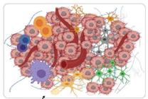
In this context, additional alterations that contribute to tumor progression include those that affect the tumor microenvironment. In the case of glioblastoma, this involves activation of angiogenesis, through overexpression of vascular endothelial growth factor (VEGF), and remodeling of the extracellular matrix (ECM) through the regulation of matrix metalloproteinases (MMPs), facilitating invasion. Gliomas also exhibit remarkable ability to evade the host immune response by expressing immunoinhibitory ligands such as PD-L1, recruiting immunosuppressive immune cells such as regulatory T cells (Tregs) and tumor-associated macrophages (TAMs), and releasing immunosuppressive cytokines such as TGF-β and IL-10.77.78 (Figure 1)
Tumor stem cells
The scientific conceptualization of tumor stem cells is based on their ability to differentiate and self-renew, having the same characteristics as normal stem cells, but also being able to initiate tumor proliferation composed of more heterogeneous cellularity of normal tissue.
It is believed that tumor stem cells promote tumorigenesis from the mechanism of differentiation, i.e., cNeural stem cells turning into glioma stem cells and driving gliomagenesis.79
It is identified that neural stem cells, which transform into glioma stem cells and are the purported precursors of glioma stem cells, are commonly found in the toothed swivel of the hippocampus, subcortical white matter, and subventricular zone.79
Intratumoral heterogeneity is a hallmark of glioblastoma, with genetic, epigenetic, phenotypic diversity, and distinct subpopulations of cells coexisting within the same tumor. Glioma tumor stem cells represent a subpopulation of cells with a high capacity for self-renewal, resistance to therapy, and potential to initiate tumors. Glioma tumor stem cells reside in specific niches, such as perivascular and hypoxic zones, and are regulated by signaling pathways such as Notch, Hedgehog, and Wnt, which are characteristic of the regulation of embryonic development.80,81
The Notch, Hedgehog and Wnt signaling pathways are the main signaling pathways involved in embryonic development, being essential for the transformation of the single-celled zygote into a highly specialized multicellular organism. These same pathways also regulate fundamental cellular processes such as cell proliferation, differentiation, and migration. It is important to highlight that its action is not limited to differentiated somatic cells, but also extends to adult stem cells.82
Given their central participation in essential biological processes, alterations or dysregulations in these pathways are often associated with several diseases, including cancer.
Molecular targets and experimental pharmacological therapies in glioblastoma
Examples of experimental targeted therapies already evaluated in clinical studies, some focusing on tumor angiogenesis, are studies of antiangiogenic inhibitors such as cediranib (Recentin®), vatalanib, sunitinib (Sutent®), sorafenib (Nexavar®, and vandetanib (Zactima®). Phase I/II studies of these agents administered as monotherapy or combined with cytotoxic chemotherapy and radiotherapy have demonstrated some promising initial results. The PDGFR inhibitor, imatinib mesylate (Gleevec®) has shown limited activity in patients with relapsed glioblastoma. Rapamycin analogues such as sirolimus (Rapamune®) have been tested as monotherapy or in combination with EGFR inhibitors. More recently, epigenetic modulators and immunotherapy are among the most promising candidate therapies.43,64,65
CD114 granulocyte colony-stimulating receptor and glioblastoma
CD114 as a regulator of brain development and neural plasticity
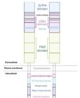
Brain tumors utilize the cellular and molecular processes of healthy CNS development and synaptic plasticity, hijacking and modifying these mechanisms.21,22,25,26 The CD114 cell surface protein, also called GCSFR or CSFR and encoded by the CSF3R gene, acts as a receptor for granulocyte colony stimulating factor (GCSF).6,7 Granulocyte colony-stimulating factor (GCSF) is a pleiotropic cytokine encoded by the CSF3 gene.83 The GCSF receptor, GCSFR, is an 813-amino acid protein encoded by the CSF3R gene and is a member of the cytokine receptor family. Its structure corresponds to the single transmembrane protein composed of several extracellular and intracellular domains CSF3 (Figure 2).83
By binding GCSF to the receptor, CD114 activation stimulates signal transducer and activator of transcription 3 (STAT3). GCSF/CD114 signaling promotes neuronal and neural stem cell survival and plasticity,27 in addition to stimulating neurogenesis6 and, together with stem cell factor (SCF), induces the growth of neurites in cortical neurons.28 The combination of GCSF and SCF has neuroprotective action and promotes the reorganization of neural networks in experimental models of neurodegeneration and brain trauma.29 Systemic administration of GCSF attenuates learning and memory impairments and dendritic spine density in the hippocampus in rats submitted to a model of cerebral ischemia.84 GCSF treatment also recovers synaptic plasticity as measured by long-term depression in hippocampal slices obtained from the APP/PS1 transgenic model of Alzheimer’s disease.85
CD114 in malignant neoplasms
Activation of STAT3 by GCSF/CD114 promotes a tumor stem cell phenotype, and CD114 is proposed as a biomarker of tumor stem cells in neural crest-derived cancers such as neuroblastoma and melanoma.7-10
CD114-positive cells isolated from neuroblastoma tumors were highly tumorigenic and capable of self-renewal and differentiation.7
Later studies have also demonstrated that CD114 expression is limited to a subpopulation of melanoma tumor cells that demonstrate altered growth and resistance to treatment, suggesting that CD114 expression may be a marker for identifying melanoma tumor cells that contribute to disease relapse and poor patient outcomes.10
CD114 expression is found in adult solid tumor types including ovarian, cervical, bladder, and skin cancers, although most previous studies have found no significant associations between CSF3R/CD114 expression and survival prognosis.7,10-18 In addition, mutations in the CSF3R gene have been described in an atypical type of chronic leukemia.19,20 In brain tumors, CD114 expression occurs in medulloblastoma, the most common type of malignant pediatric brain tumor, both in cell lines, mouse grafts, and primary tumor samples. The presence of CD114 appears to confer resistance to cytotoxic chemotherapy.17 In medulloblastoma samples obtained from patients, there is a difference in CSF3R gene expression between tumors of the Group 3 molecular subgroup compared to the other subgroups, as well as between the SHH molecular subtypes γ, Group 3 α, and Group 3 β.
However, no significant associations were found between CSF3R expression levels and overall survival of patients.18
A previous study in glioma described the widespread expression of GCSF, both at the mRNA and protein level, in tumor samples. Cell proliferation and migration were stimulated by GCSF treatment in CD114-expressing glioblastoma cells, and an antibody against GCSF produced opposite effects.11
Binding of GCSF to its CD114 receptor (CSF3R) activates the JAK-STAT signaling pathway, culminating in the phosphorylation and activation of signal transducer and transcription activator 3 (STAT3), which acts as a transcriptional regulator of genes involved in the proliferation, survival, invasion, and maintenance of tumor stem cells in glioblastoma.86
STAT3 regulates several cellular processes including cell growth, differentiation and apoptosis, and is often activated during tumor genesis. Thus, STAT3 signaling was considered important for the maintenance of tumor stem cell-like subpopulations, including glioblastoma.87
Specific STAT3 inhibitors are currently being developed and represent a new class of stimulant therapeutic agents likely to be effective against tumor stem cell subpopulations in these tumors.7
Thus, the volume of evidence regarding the possible role of CSF3R/CD114 as prognostic biomarkers or therapeutic targets in glioblastoma or other types of glioma is very limited. In this context, this study aimed to evaluate the expression of the CSF3R gene and its correlation with overall survival in tumors of patients with different subtypes of glioma, with a special focus on glioblastoma.
Conceptual aspects
Gliomas constitute the most frequent group of primary malignant tumors of the central nervous system in adults.88 the clinical course is heterogeneous, with median survival of 14-16 months in IDH-wildtype glioblastoma,89 and may exceed 8-10 years in diffuse IDH-mutant gliomas of lesser degree.90
These tumors are classified into different types, according to the type of cell from which they originate or with which they share histological characteristics, and are then classified into ependymomas, astrocytomas, and oligodendrogliomas. Of these, astrocytomas are the most common type of glial tumors.32
In gliomas, residual tumor cells often lead to malignant recurrence and progression, with some gliomas tending to progress to glioblastoma,31 a very aggressive neoplasm.
Although morphological classification based on histopathology provides important information for the diagnosis of glioblastoma, it has limitations, as it cannot reflect the heterogeneity of these tumors and, therefore, ends up being insufficient for patient management.70
The 2016 WHO classification of CNS tumors, therefore, restructured the classification of glioblastoma by incorporating molecular features into histopathological appearances and was recently updated, in 2021, by advances in the molecular and genetic understanding of CNS tumors,4 so that there was progress in the role of molecular diagnostics in the subclassification of glioblastoma.
In fact, glioblastoma is one of the first tumor types that have been systematically investigated by large international cancer projects, e.g., TCGA, due to its high incidence and in cohort , glioblastoma corresponded to 57.51% (n = 153) of the sample.
The CSF3R gene encodes the GCSFR protein. GCSFR presents differentiation giving rise to CD114, which is the granulocyte colony-stimulating factor (GCSF) receptor.
Granulocytic colony-stimulating factor (GCSF) was first identified in bone marrow cells and derivatives, and is a cytokine, i.e., a small protein that acts as a messenger of the immune system, encoded by the CSF3 gene that acts as a hematopoietic growth factor regulating the function of granulocytic precursors and neutrophils.6.7
The actions of the GCSF are mediated by the activation of its receptor, called GCSFR or CD114. Recombinant human GCSF is clinically used to prevent neutropenia, due to its effects on neutrophil mobilization and maturation.83 GCSF/CD114 signaling has also been investigated as a modulator of neuronal survival, synaptic plasticity6,28,29,84,85, and cancer.7,10-20
Specifically, CD114 has been proposed as a marker to identify cancer stem cell subpopulations associated with tumorigenicity, metastasis, and treatment resistance.7,9,10
In epithelial skin tumors, the presence of CD114 is significantly higher compared to normal skin, Bowen’s disease, or actinic keratosis, and has been associated with carcinogenesis. However, no association between CD114 protein expression and patient mortality was found.13
Similarly, different levels of CSF3R transcriptions occur between different tumor subgroups and medulloblastoma subtypes, but no significant association with patient survival has been established.18
A previous study in glioma looked at GCSF and CD114 RNA and protein expression in a set of 22 human gliomas (WHO grade II, III, and IV) and cell cultures derived from these tumors.
Although GCSF and CD114 expression, as well as that of granulocyte-macrophage colony-stimulating factor (GMCSF) and its receptor, has been found in all gliomas and cell cultures, joint expression of both factors and their receptors has been selectively observed in grade IV tumors (GBMs), and thus expression correlates with advanced tumor stage.11 This makes understanding CSF3R gene expression important to know the oncogenesis of each tumor type and, eventually, to seek ways to block it.
Transcription Activator Signal Transducer 3 (STAT3) regulates several cellular processes, including cell growth, differentiation, and apoptosis, and is frequently activated during tumor genesis, so STAT3 signaling has been found to be important for the maintenance of cancer stem cell-like subpopulations, including glioblastoma.87
Compared to non-tumor neural tissue (n = 8), significantly higher levels of CSF3R transcripts were found in astrocytoma, pilocytic astrocytoma, and glioblastoma.
The present transcription analyses indicate that a significant association between high CSF3R mRNA levels and worse prognosis measured through shorter overall survival was found in patients with gliomas.
Analyzing the 266 glioma samples from the GEO- (GSE 16011) section, higher expression of CSF3R/CD114 was observed, which was associated with a worse prognosis, based on shorter overall survival.
Similarly, there was a significant association between high CSF3R mRNA levels and shorter overall survival in GBM patients (n = 150; p < 0.05).
These initial in silicon findings suggest that further experimental studies should characterize the effects of GCSF/CD114 inhibition in experimental models of GBM.
IDH-wild tumors typically occur in older patients, while IDH-mutated tumors, which correspond to about 10% of cases, affect younger patients and originate from lower-grade gliomas, in addition to having a better prognosis.2,4
No significant impact of CSF3R mRNA expression on overall survival was found when GBM patients were divided according to HDI status (mutated vs. wild-type), possibly due to the limited number of samples available in each subgroup.
In addition, there was no significant difference in CSF3R levels between GBM with wild-type HDI and those with IDH-mutation. Some IDH1 mutations are considered prognostic markers, with patients with mutated tumors having better overall survival.90 Corroborating these data, worse overall survival was observed in patients with GBM IDH wild-type.
One study reported that in mutated wild-type GBM carrying mice, GCSF is secreted by GBM CSCs, and blocking GCSF accelerates tumor progression by acting on the myeloid cells that infiltrate the tumor.91
STAT3 is involved in mediating the cellular effects of CD114 activation. Several studies have indicated that STAT3 is an oncogene in GBM. Activation of STAT3 is associated with shorter, progression-free overall survival in patients with GBM,92 and STAT3 is required for the maintenance of a CSC phenotype in GBM cells.93 STAT3 can have a dual role in the GBM, promoting or suppressing the progression of the GBM.94,95
Future experiments should investigate the role of STAT3 downstream of CD114 in different types of glioma.
CONCLUSION
Exploring the role of CD114 in glioma cell lines, other primary tumors, and the tumor microenvironment in future studies may improve understanding of the role of GCSF and similar growth factors in the progression of CNS malignancies. Genomic analyses are currently the gold standard for categorizing tumor subtypes, still presenting some overlap between groups, but less than classical histology. Biomarkers are essential not only for diagnosis and prognosis but also for personalized therapeutic planning. The search for specific proteins and receptors in CNS cancer cells can determine the identification of molecular targets that, when inoperative, can inhibit the growth, proliferation, and spread of the neoplasm, as well as allow the development of therapies selectively targeted at gliomas. Several strategies for the search for molecularly directed therapies, or targeted therapies, have been developed based on the inhibition of cell signaling component in tumor cells or their microenvironment. Several of these experimental therapies have already been evaluated in clinical trials. Inhibition of growth factor receptors or their ligands is a therapeutic strategy that interrupts the signals that promote cell growth and proliferation, especially in the context of the treatment of gliomas, and although expected, the primary endpoint of increased survival has not yet been achieved.
References
-
1 Wen PY, Kesari S. Malignant gliomas in adults. N Engl J Med. 2008;359(5):492-507. doi: https://doi.org/10.1056/NEJMra0708126
» https://doi.org/10.1056/NEJMra0708126 -
2 Alexander BM, Cloughesy TF. Adult glioblastoma. J Clin Oncol. 2017;35(21):2402-9. doi: https://doi.org/10.1200/jco.2017.73.0119
» https://doi.org/10.1200/jco.2017.73.0119 -
3 Funakoshi Y, Hata N, Kuga D, Hatae R, Sangatsuda Y, Fujioka Y, et al. Pediatric glioma: An update of diagnosis, biology, and treatment. Cancers (Basel). 2021;13(4):758. doi: https://doi.org/10.3390/cancers13040758
» https://doi.org/10.3390/cancers13040758 -
4 Louis DN, Perry A, Wesseling P, Brat DJ, Cree IA, Figarella?Branger D, et al. The 2021 WHO Classification of Tumors of the Central Nervous System: A summary. Neuro Oncol. 2021;23(8):1231-51. doi: https://doi.org/10.1093/neuonc/noab106
» https://doi.org/10.1093/neuonc/noab106 -
5 Hardigan AA, Jackson JD, Patel AP. Surgical management and advances in the treatment of glioma. Semin Neurol. 2023;43(6):810-24. doi: https://doi.org/10.1055/s-0043-1776766
» https://doi.org/10.1055/s-0043-1776766 -
6 Schneider A, Kruger C, Steigleder T, Weber D, Pitzer C, Laage R, et al. The hematopoietic factor G?CSF is a neuronal ligand that counteracts programmed cell death and drives neurogenesis. J Clin Investig. 2005;115:2083?98. https://doi.org/10.1172/jci23559
» https://doi.org/10.1172/jci23559 -
7 Zage PE, Whittle SB, Shohet JM. CD114: a new member of the neural crest?derived cancer stem cell marker family. J Cell Biochem. 2017;118:221-31. doi: https://doi.org/10.1002/jcb.25656
» https://doi.org/10.1002/jcb.25656 -
8 Agarwal S, Lakoma A, Chen Z, Hicks J, Metelitsa LS, Kim ES, et al. G-CSF Promotes Neuroblastoma Tumorigenicity and Metastasis via STAT3-Dependent Cancer Stem Cell Activation. Cancer Res. 2015;75;(12):2566-79. doi: https://doi.org/10.1158/0008-5472.can-14-2946
» https://doi.org/10.1158/0008-5472.can-14-2946 -
9 Hsu DM, Agarwal S, Benham A, Coarfa C, Trahan DN, Chen Z, et al. G?CSF receptor positive neuroblastoma subpopulations are enriched in chemotherapy?resistant or relapsed tumors and are highly tumorigenic. Cancer Res. 2013;73(13):4134-46. https://doi.org/10.1158/0008-5472.can-12-4056
» https://doi.org/10.1158/0008-5472.can-12-4056 - 10 Zhang L, Agarwal S, Shohet JM, Zage PE. CD114 expression mediates melanoma tumor cell growth and treatment resistance. Anticancer Res. 2015;35(7):3787-92.
-
11 Mueller MM, Herold?Mende CC, Riede D, Lange M, Steiner HH, Fusenig NE. Autocrine growth regulation by granulocyte colony?stimulating factor and granulocyte?macrophage colony?stimulating factor in human gliomas with tumor progression. Am J Pathol. 1999;155(5):1557-67. doi: https://doi.org/10.1016/S0002-9440(10)65472-7
» https://doi.org/10.1016/S0002-9440(10)65472-7 -
12 Kyo S, Kanaya T, Takakura M, Inoue M. A case of cervical cancer with aggressive tumor growth: Possible autocrine growth stimulation by G?CSF and IL?6. Gynecol Oncol. 2000;78(Pt 1):383-7. doi: https://doi.org/10.1006/gyno.2000.5904
» https://doi.org/10.1006/gyno.2000.5904 -
13 Hirai K, Kumakiri M, Fujieda S, Sunaga H, Lao LM, Imamura Y, et al. Expression of granulocyte colony?stimulating factor and its receptor in epithelial skin tumors. J Dermatol Sci. 2001;25(3):179-88. https://doi.org/10.1016/s0923- 1811(00)00131-6
» https://doi.org/10.1016/s0923- -
14 Savarese TM, Mitchell K, McQuain C, Campbell CL, Guardiani R, Wuu J, et al. Coexpression of granulocyte colony stimulating factor and its receptor in primary ovarian carcinomas. Cancer Lett. 2001;162(1):105 15. https://doi.org/10.1016/s0304-3835(00)00623-6"
» https://doi.org/10.1016/s0304-3835(00)00623-6 -
15 Chakraborty A, Guha S. Granulocyte colony-stimulating factor/granulocyte colony-stimulating factor receptor biological axis promotes survival and growth of bladder cancer cells. Urology. 2007;69(6):1210-5. doi: https://doi.org/10.1016/j.urology.2007.02.035
» https://doi.org/10.1016/j.urology.2007.02.035 -
16 Hanumanthu VS, Pirruccello SJ. GCSF?R expression in myelodysplastic and myeloproliferative disorders and blast dysmaturation in CML. Am J Clin Pathol. 2013;140(2):155-64. doi: https://doi.org/10.1309/AJCPCLHZR5KUHUBM
» https://doi.org/10.1309/AJCPCLHZR5KUHUBM -
17 Paul SR, Huo Y, Liu A, Lesperance J, Garancher A, Wechsler-Reya RJ, et al. Characterization of G?CSF receptor expression in medulloblastoma. Neurooncol. 2020;2(1):vdaa062. https://doi.org/10.1093/noajnl/vdaa062
» https://doi.org/10.1093/noajnl/vdaa062 -
18 Monteiro JM, Reis Ramos JI, Teixeira e Sousa I, Bighetti?Trevisan RL, Ribas- Filho JM, Isolan GR. Identification of CD114 membrane receptors as a molecular target in medulloblastomas. Int J Mol Sci. 2023;24(6):5331. doi: https://doi.org/10.3390/ijms24065331
» https://doi.org/10.3390/ijms24065331 -
19 Maxson JE, Gotlib J, Pollyea DA, Fleischman AG, Agarwal A, Eide CA, et al. Oncogenic CSF3R mutations in chronic neutrophilic leukemia and atypical CML. N Engl J Med. 2013;368(19):1781-90. https://doi.org/10.1056/nejmoa1214514
» https://doi.org/10.1056/nejmoa1214514 -
20 Maxson JE, Ries RE, Wang YC, Gerbing RB, Kolb EA, Thompson SL, et al. CSF3R mutations have a high degree of overlap with CEBPA mutations in pediatric AML. Blood. 2016;127(24):3094-98. https://doi.org/10.1182/blood- 2016-04-709899
» https://doi.org/10.1182/blood- -
21 Venkatesh HS, Johung TB, Caretti V, Noll A, Tang Y, Nagaraja S, et al. Neuronal activity promotes glioma growth through neuroligin?3 secretion. Cell. 2015;161(4):803-16. doi: https://doi.org/10.1016/j.cell.2015.04.012
» https://doi.org/10.1016/j.cell.2015.04.012 -
22 Venkatesh HS, Morishita W, Geraghty AC, Silverbush D, Gillespie SM, Arzt M, et al. Electrical and synaptic integration of glioma into neural circuits. Nature. 2019;573(7775):539-45. doi: https://doi.org/10.1038/s41586-019-1563-y
» https://doi.org/10.1038/s41586-019-1563-y -
23 Pinheiro KV, Thomaz A, Souza BK, Metcalfe VA, Freire NH, Brunetto AT, de Farias CB, et al. Expression and pharmacological inhibition of TrkB and EGFR in glioblastoma. Mol Biol Rep. 2020;47:6817-28. https://doi.org/10.1007/s11033- 020-05739-2
» https://doi.org/10.1007/s11033- -
24 Thomaz A, Jarger M, Brunett AL, Brunetto AT, Gregianin L, de Farias CB, et al. Neurotrophin signaling in medulloblastoma. Cancers. 2020;12(9):2542. doi: https://doi.org/10.3390/cancers12102542
» https://doi.org/10.3390/cancers12102542 -
25 Krsna S, Choudhury A, Keough MB, Seo K, Ni L, Kakaizada S, et al. Glioblastoma remodelling of human neural circuits decreases survival. Nature. 2023;617(7961):599-607. doi: https://doi.org/10.1038/s41586-023-06036-1
» https://doi.org/10.1038/s41586-023-06036-1 -
26 Rodrigues EM, Giovanini AF, Ribas CAPM, Malafaia O, Roesler R, Isolan GR. The nervous system development regulator neuropilin?1 as a potential prognostic marker and therapeutic target in brain cancer. Cancers. 2023;15(20):4922. https://doi.org/10.3390/cancers15204922
» https://doi.org/10.3390/cancers15204922 -
27 Wallner S, Peters S, Pitzer C, Resch H, Bogdahn U, Schneider A. The granulocyte?colony stimulating factor has a dual role in neuronal and vascular plasticity. Front Cell Dev Biol. 2015;3:48. doi: https://doi.org/10.3389/fcell.2015.00048
» https://doi.org/10.3389/fcell.2015.00048 -
28 Su Y, Cui L, Piao C, Li B, Zhao LR. The effects of hematopoietic growth factors on neurite outgrowth. PLoS One. 2013;8(10):e75562. doi: https://doi.org/10.1371/journal.pone.0075562
» https://doi.org/10.1371/journal.pone.0075562 -
29 He J, Russell T, Qiu X, Hao F, Kyle M, Chin L, et al. The contribution of stem cell factor and granulocyte colony stimulating factor in reducing neurodegeneration and promoting neurostructure network reorganization after traumatic brain injury. Brain Res. 2020;1746:147000.doi: https://doi.org/10.1016/j.brainres.2020.147000"
» https://doi.org/10.1016/j.brainres.2020.147000 - 30 Beckhauser AP, Nisihara RM, Tonial AF, Nassif PAN, dos Santos CEO, Lages Neto M, et al. O papel da obesidade (e da perda de peso pela cirurgia bariátrica) nas doenças autoimunes. BioSCIENCE. 2024;82:e032
-
31 Hou L, Tian L, Li J, Zhang Z, Han X, Zhou H, et al. Systematic Analysis of an Immune-Related Gene Signature for Predicting Prognosis and Immune Characteristics in Primary Lower Grade Glioma. Biomed Res Int. 2025:2025:6180391. https://doi.org/10.1155/bmri/6180391
» https://doi.org/10.1155/bmri/6180391 -
32 Jov?evska I. Genetic secrets of long-term glioblastoma survivors. Bosn J Basic Med Sci. 2019;19(2):116-24. https://doi.org/10.17305/bjbms.2018.3717
» https://doi.org/10.17305/bjbms.2018.3717 -
33 Berger TR, Wen PY, Lang-Orsini M, Chukwueke UN. World Health Organization 2021 classification of central nervous system tumors and implications for therapy for adult-type gliomas: A review. JAMA Oncol. 2022;8(10):1493-501. doi: https://doi.org/10.1001/jamaoncol.2022.2844
» https://doi.org/10.1001/jamaoncol.2022.2844 -
34 Olar A, Aldape KD. Using the molecular classification of glioblastoma to inform personalized treatment. JPathol. 2014;232(2):165-77. https://doi.org/10.1002/path.4282
» https://doi.org/10.1002/path.4282 -
35 Noroxe DS, Poulsen HS, Lassen U. Hallmarks of glioblastoma: a systematic review. ESMO. 2016;1:e000144. http://doi.org/10.1136/esmoopen-2016-000144
» http://doi.org/10.1136/esmoopen-2016-000144 - 36 de Oliveira LBC, Malafaia O, Tabushi FS, Carvalhal SRS, Ioshii SO, Schuh R, et al. Análise de citocinas e CD68 na aorta: novos parâmetros para estudo da inflamação no diabete melito tipo 2. BioSCIENCE. 2025;83(S2):e00023
-
37 Roda D, Veiga P, Melo JB, Carreira IM, Ribeiro IP. Principles in the management of glioblastoma. Genes (Basel). 2024;15(4):501. doi: https://doi.org/10.3390/genes15040501
» https://doi.org/10.3390/genes15040501 -
38 Schwartzbaum JA, Fisher JL, Aldape KD, Wrensch M. Epidemiology and molecular pathology of glioma. Nat Clin Pract Neurol. 2006;2(9):494?503. https://doi.org/10.1038/ncpneuro0289
» https://doi.org/10.1038/ncpneuro0289 -
39 Stupp R, Mason WP, van den Bent MJ, Weller M, Fisher B, Taphoorn MJB, et al. Radiotherapy plus concomitant and adjuvant temozolomide for glioblastoma. N Engl J Med. 2005;352(10):987-96. doi: https://doi.org/10.1056/NEJMoa043330
» https://doi.org/10.1056/NEJMoa043330 -
40 Bao J, Sun R, Pan Z, Wei S. Current chemotherapy strategies for adults with IDH-wildtype glioblastoma. Front Oncol. 2024;14:1438905. doi: https://doi.org/10.3389/fonc.2024.1438905
» https://doi.org/10.3389/fonc.2024.1438905 -
41 Ippen FM, Scherm A, Kessler T, Hau P, Agkatsev S, Baurecht H, et al. Targeted agents in patients with progressive glioblastoma-a systematic meta-analysis of randomized clinical trials. Cancer Med. 2024;13(12):e7362. https://doi.org/10.1002/cam4.7362
» https://doi.org/10.1002/cam4.7362 -
42 Rich JN, Bigner DD. Development of novel targeted therapies in the treatment of malignant glioma. Nat Rev Drug Discov. 2004;3(5):430-46. https://doi.org/10.1038/nrd1380
» https://doi.org/10.1038/nrd1380 -
43 Tang J, Karbhari N, Campian JL. Therapeutic targets in glioblastoma: molecular pathways, emerging strategies, and future directions. Cells. 2025;14(7):494. doi: https://doi.org/10.3390/cells14070494
» https://doi.org/10.3390/cells14070494 -
44 Bae WH, Maraka S, Daher A. Challenges and advances in glioblastoma targeted therapy: the promise of drug repurposing and biomarker exploration. Front Oncol. 2024;14:1441460. doi: https://doi.org/10.3389/fonc.2024.1441460
» https://doi.org/10.3389/fonc.2024.1441460 -
45 Jacome MA, Wu Q, Pina Y, Erame AB. Evolution of molecular biomarkers and precision molecular therapeutic strategies in glioblastoma. Cancers (Basel). 2024;16(21):3635. https://doi.org/10.3390/cancers16213635
» https://doi.org/10.3390/cancers16213635 -
46 Tataranu LG, Turliuc S, Kamel A, Rizea RE, Dricu A, Staicu GA, et al. Glioblastoma tumor microenvironment: an important modulator for tumoral progression and therapy resistance. Curr Issues Mol Biol. 2024;46(9):9881-94. doi: https://doi.org/10.3390/cimb46090588
» https://doi.org/10.3390/cimb46090588 -
47 Stoyanov GS, Dzhenkov DL. On the Concepts and History of Glioblastoma Multiforme - Morphology, Genetics and Epigenetics. Folia Med (Plovdiv). 2018;60(1):48-66. https://doi.org/10.1515/folmed-2017-0069
» https://doi.org/10.1515/folmed-2017-0069 -
48 Martin?Villalba A, Okuducu AF, von Deimling A. The evolution of our understanding on glioma. Brain Pathol. 2008;18(3):455-63. https://doi.org/10.1111/j.1750-3639.2008.00136.x
» https://doi.org/10.1111/j.1750-3639.2008.00136.x -
49 Reiman ZJ, Yan H. Isocitrate dehydrogenase 1 and 2 mutations in cancer: alterations at a crossroads of cellular metabolism. J Natl Cancer Inst. 2010;102(13):932-41. https://doi.org/10.1093/jnci/djq187
» https://doi.org/10.1093/jnci/djq187 - 50 Sardeto GA, Boss LEC, Pacheco MJR. O impacto da obesidade nos resultados pós-operatórios de operações abdominais.BioSCIENCE. 2024;82:e042
-
51 Brandes AA, Franceschi E, Tosoni A, Benevento F, Scopece L, Mazzocchi V, et al. Temozolomide concomitant and adjuvant to radiotherapy in elderly patients with glioblastoma: correlation with MGMT promoter methylation status. Cancer. 2009;115(15):3512-8. doi: https://doi.org/10.1002/cncr.24406
» https://doi.org/10.1002/cncr.24406 -
52 Gal O, Mehta MP, Kotecha R. Radiotherapeutic advances in the management of glioblastoma.J Neurooncol. 2024;170(3):509-20. doi: https://doi.org/10.1007/s11060-024-04824-x"
» https://doi.org/10.1007/s11060-024-04824-x -
53 Inggas MAM, Patel U, Wijaya JH, Otinashvili N, Menon VR, Iyer AK, et al. The role of temozolomide as adjuvant therapy in glioblastoma management: a systematic review and meta-analysis. BMC Cancer. 2025;25(1):399. https://doi.org/10.1186/s12885-025-13757-1
» https://doi.org/10.1186/s12885-025-13757-1 -
54 Minniti G, de Sanctis V, Muni R, Filippone F, Bozzao A, Valeriani M, et al. Radiotherapy plus concomitant and adjuvant temozolomide for glioblastoma in elderly patients. JNeurooncol. 2008;88(1):97-103. https://doi.org/10.1007/s11060-008-9538-0"
» https://doi.org/10.1007/s11060-008-9538-0 -
55 Brown TJ, Brennan MC, Li M, Church EW, Brandmeir NJ, Rakszawski KL, et al. Association of the extent of resection with survival in glioblastoma: A systematic review and meta-analysis. JAMA Oncol. 2016;2(11):1460-9. doi: https://doi.org/10.1001/jamaoncol.2016.1373
» https://doi.org/10.1001/jamaoncol.2016.1373 -
56 Nagae LM, Pinho M da C, Funari MB de G. Tractography. Einstein. 2010; 8(2 Pt 1):252-3. https://doi.org/10.1590/S1679-45082010AI1112
» https://doi.org/10.1590/S1679-45082010AI1112 -
57 Gerritsen JKW, Broekman MLD, de Vleeschouwer S, Schucht P, Nahed BV, Berger MS, et al. Safe surgery for glioblastoma: Recent advances and modern challenges. Neurooncol Pract. 2022;9(5):364-79. doi: https://doi.org/10.1093/nop/npac019"
» https://doi.org/10.1093/nop/npac019 -
58 Revilla Pacheco F, Rodríguez-Salgado P, Barrera-Ramírez M, Morales-Ruiz MP, Loyo-Varela M, Rubalcava-Ortega J, et al. Extent of resection and survival in patients with glioblastoma multiforme: systematic review and meta analysis. Medicine (Baltimore).2021;100(25):e26432. https://doi.org/10.1097/md.0000000000026432
» https://doi.org/10.1097/md.0000000000026432 -
59 Sanai N, Polley MY, McDermott MW, Parsa AT, Berger MS. An extent of resection threshold for newly diagnosed glioblastomas. J Neurosurg. 2011;115(1):3?8. https://doi.org/10.3171/2011.2.jns10998
» https://doi.org/10.3171/2011.2.jns10998 -
60 Parisi S, Corsa P, Raguso A, Perrone A, Cossa S, Munafo T, et al. Temozolomide and radiotherapy versus radiotherapy alone in high grade gliomas: a very long term comparative study and literature review. Biomed Res Int. 2015;2015:620643. doi: https://doi.org/10.1155/2015/620643
» https://doi.org/10.1155/2015/620643 -
61 Zhao Y, Wu J, Shi L. Adjuvant temozolomide for the treatment of glioblastoma: a meta?analysis of randomized controlled studies. Clin Neuropharmacol. 2021;44(4):132-7. doi: https://doi.org/10.1097/WNF.0000000000000449
» https://doi.org/10.1097/WNF.0000000000000449 -
62 Singh N, Miner A, Hennis L, Mittal S. Mechanisms of temozolomide resistance in glioblastoma - a comprehensive review. Cancer Drug Resist. 2021;4(1):17-43. doi: https://doi.org/10.20517/cdr.2020.79
» https://doi.org/10.20517/cdr.2020.79 -
63 Cancer Genome Atlas Research Network. Comprehensive genomic characterization defines human glioblastoma genes and core pathways. Nature. 2008;455(7216):1061-8. doi: https://doi.org/10.1038/nature07385
» https://doi.org/10.1038/nature07385 -
64 Politis A, Stavrinou L, Kalyvas A, Boviatsis E, Piperi C, et al. Glioblastoma: molecular features, emerging molecular targets and novel therapeutic strategies. Crit Rev Oncol Hematol. 2025;104764. https://doi.org/10.1016/j.critrevonc.2025.104764"
» https://doi.org/10.1016/j.critrevonc.2025.104764 -
65 Roesler R, Brunetto AT, Abujamra AL, de Farias CB, Brunetto AL, Schwartsmann G. Current and emerging molecular targets in glioma. Expert Rev Anticancer Ther. 2010;10(11):1735?51. https://doi.org/10.1586/era.10.167
» https://doi.org/10.1586/era.10.167 -
66 An Z, Aksoy O, Zheng T, Fan QW, Weiss WA. Epidermal growth factor receptor and EGFRvIII in glioblastoma: signaling pathways and targeted therapies. Oncogene. 2018;37(12):1561-75. doi: https://doi.org/10.1038/s41388-017-0045- 7
» https://doi.org/10.1038/s41388-017-0045- -
67 Brennan CW, Verhaak RGW, McKenna A, Campos B, Noushmehr H, Salama SR, et al. The somatic genomic landscape of glioblastoma. Cell. 2013;155(2):462-77. doi: https://doi.org/10.1016/j.cell.2013.09.034
» https://doi.org/10.1016/j.cell.2013.09.034 -
68 Huse JT, Holland EC. Targeting brain cancer: advances in the molecular pathology of malignant glioma and medulloblastoma. Nat Rev Cancer. 2010;10(5):319-31. https://doi.org/10.1038/nrc2818
» https://doi.org/10.1038/nrc2818 -
69 Ismailov A, Spallone A, Belogurov-Jr A, Herbert A, Poptsova M. Molecular biology of the deadliest cancer ? glioblastoma: what do we know? Front Immunol. 2025;16:1530305. https://doi.org/10.3389/fimmu.2025.1530305
» https://doi.org/10.3389/fimmu.2025.1530305 -
70 Lan Z, Li X, Zhang X. Glioblastoma: an update in pathology, molecular mechanisms and biomarkers. Int J Mol Sci. 2024;25(2):3040. doi: https://doi.org/10.3390/ijms25023040
» https://doi.org/10.3390/ijms25023040 -
71 Zhang Y, Dube C, Gibert-Jr M, Cruickshanks N, Wang B, Coughlan M, et al. The p53 Pathway in Glioblastoma. Cancers (Basel). 2018;10(9):297. https://doi.org/10.3390/cancers10090297
» https://doi.org/10.3390/cancers10090297 -
72 Ushio Y, Tada K, Shiraishi S, Kamiryo T, Shinojima N, Kochi M, et al. Correlation of molecular genetic analysis of p53, MDM2, p16, PTEN, and EGFR and survival of patients with anaplastic astrocytoma and glioblastoma. Front Biosci. 2003;8:e281-8. https://doi.org/10.2741/865
» https://doi.org/10.2741/865 -
73 Minucci S, Pelicci PG. Histone deacetylase inhibitors and the promise of epigenetic (and more) treatments for cancer. Nat Rev Cancer. 2006;6(1):38-51. https://doi.org/10.1038/nrc1779
» https://doi.org/10.1038/nrc1779 -
74 Lucchini S, Constantinou M, Marino S. Unravelling the mosaic: Epigenetic diversity in glioblastoma. Mol Oncol. 2024;18(12):2871-89. doi: https://doi.org/10.1002/1878-0261.13706
» https://doi.org/10.1002/1878-0261.13706 -
75 Anderson NM, Simon MC. The tumor microenvironment. Curr Biol. 2020;30(16):R921-5. doi: https://doi.org/10.1016/j.cub.2020.06.081
» https://doi.org/10.1016/j.cub.2020.06.081 -
76 Truffi M, Sorrentino L, Corsi F. Fibroblasts in the tumor microenvironment. Adv Exp Med Biol. 2020;1234:15-29. doi: https://doi.org/10.1007/978-3-030-04414- 0_2
» https://doi.org/10.1007/978-3-030-04414- -
77 Robinson SD, Filippopoulou C, Besta S, Samuels M, Betrán AL, Ajamieh MA, et al. Spatial biology - unravelling complexity within the glioblastoma microenvironment. Trends Mol Med. 2025;25:S1471?4914(25):00014?0. doi: https://doi.org/10.1016/j.molmed.2025.01.014
» https://doi.org/10.1016/j.molmed.2025.01.014 -
78 Tataranu LG, Turliuc S, Rizea RE, Dricu A, Alexandru O, Staicu GA, et al. A synopsis of biomarkers in glioblastoma: past and present. Curr Issues Mol Biol. 2024;46(7):6903-39. doi: https://doi.org/10.3390/cimb46070412
» https://doi.org/10.3390/cimb46070412 -
79 Abdullah KG, Adamson C, Brem S. The molecular pathogenesis of glioblastoma. Glioblastoma. 2017:21-31. https://doi.org/10.1016/B978-0-323-47660-7.00003-3
» https://doi.org/10.1016/B978-0-323-47660-7.00003-3 -
80 Ghosh N, Chatterjee D, Datta A. Tumor heterogeneity and resistance in glioblastoma: the role of stem cells. Apoptosis. 2025. doi: https://doi.org/10.1007/s10495-025-02123-y
» https://doi.org/10.1007/s10495-025-02123-y -
81 Singh SK, Hawkins C, Clarke ID, Squire JA, Bayani J, Hide T, et al. Identification of human brain tumour initiating cells. Nature. 2004;432(7015):396-401. doi: https://doi.org/10.1038/nature03128
» https://doi.org/10.1038/nature03128 -
82 Kumar A, Vashishta M, Long L, Wu X, Lu JJ, Guha C, et al. The role of Notch, Hedgehog, and Wnt signaling pathways in the resistance of tumors to anticancer therapies. Front Cell Dev Biol. 2021;9:e650772. doi: https://doi.org/10.3389/fcell.2021.650772
» https://doi.org/10.3389/fcell.2021.650772 -
83 Park SD, Saunders AS, Reidy MA, Bender DE, Clifton S, Morris KT. A review of granulocyte colony?stimulating factor receptor signaling and regulation with implications for cancer. Front Oncol. 2022;12:932608. https://doi.org/10.3389/fonc.2022.932608
» https://doi.org/10.3389/fonc.2022.932608 -
84 Sarkala HB, Jahanshahi M, Dolatabadi LK, Namavar MR. G?CSF improved the memory and dendritic morphology impairments in the hippocampal CA1 pyramidal neurons after brain ischemia in male rats. Metab Brain Dis. 2023;38(8):2573?81. https://doi.org/10.1007/s11011-023-01286-4
» https://doi.org/10.1007/s11011-023-01286-4 -
85 Song S, Wang X, Sava V, Weeber EJ, Sanchez-Ramos J. In vivo administration of granulocyte colony?stimulating factor restores long?term depression in hippocampal slices prepared from transgenic APP/PS1 mice. J Neurosci Res. 2014;92(8):975-80. https://doi.org/10.1002/jnr.23378
» https://doi.org/10.1002/jnr.23378 -
86 Ahsan H, Asghar M, Malik SI. Potential diagnostic and drug target markers in glioblastoma. Sci Rep. 2024;14(1):7292. https://doi.org/10.1038/s41598-024- 57752-1
» https://doi.org/10.1038/s41598-024- -
87 Sherry MM, Reeves A, Wu JK, Cochran BH. STAT3 is required for proliferation and maintenance of multipotency in glioblastoma stem cells. Stem Cells. 2009;27(10):2383-92. https://doi.org/10.1002/stem.185
» https://doi.org/10.1002/stem.185 -
88 Ostrom QT, Price M, Neff C, Cioffi G, Waite KA, Kruchko C, et al. CBTRUS Statistical Report: Primary Brain and Other Central Nervous System Tumors Diagnosed in the United States in 2015-2019. Neuro Oncol. 2022;24(Suppl 5):v1- v95. https://doi.org/10.1093/neuonc/noac202
» https://doi.org/10.1093/neuonc/noac202 -
89 Stupp R, Hegi ME, Mason WP, van den Bent MJ, Taphoorn MJB, Janzer RC, et al. Effects of radiotherapy with concomitant and adjuvant temozolomide versus radiotherapy alone on survival in glioblastoma in a randomised phase III study: 5-year analysis of the EORTC-NCIC trial. Lancet Oncol. 2009 May;10(5):459-66. https://doi.org/10.1016/s1470-2045(09)70025-7
» https://doi.org/10.1016/s1470-2045(09)70025-7 -
90 Sareen H, Ma Y, Becker TM, Roberts TL, de Souza P, Powter B. Molecular biomarkers in glioblastoma: a systematic review and meta?analysis. Int J Mol Sci. 2022;23(16):8835. https://doi.org/10.3390/ijms23168835
» https://doi.org/10.3390/ijms23168835 -
91 Alghamri MS, McClellan BL, Avvari RP, Thalla R, Carney S, Hartlage CS, et al. G-CSF secreted by mutant IDH1 glioma stem cells abolishes myeloid cell immunosuppression and enhances the efficacy of immunotherapy. Sci Adv. 2021;7(40):eabh3243. doi: https://doi.org/10.1126/sciadv.abh3243
» https://doi.org/10.1126/sciadv.abh3243 -
92 Masliantsev K, Pinel B, Balbous A, Guichet PO, Tachon G, Milin S, et al. Impact of STAT3 phosphorylation in glioblastoma stem cells radiosensitization and patient outcome. Oncotarget. 2017;9(3):3968-79.https://doi.org/10.18632/oncotarget.23374
» https://doi.org/10.18632/oncotarget.23374 -
93 Im CN, Yun HH, Song B, Youn DY, Cui MN, Kim HS, et al. BIS?mediated STAT3 stabilization regulates glioblastoma stem cell?like phenotypes. Oncotarget. 2016;7(23):35056-70. doi: https://doi.org/10.18632/oncotarget.9039
» https://doi.org/10.18632/oncotarget.9039 -
94 De la Iglesia N, Konopka G, Lim KL, Nutt CL, Bromberg JF, Frank DA, et al. Deregulation of a STAT3-interleukin 8 signaling pathway promotes human glioblastoma cell proliferation and invasiveness. J Neurosci. 2008;28(23):5870-8. doi: https://doi.org/10.1523/JNEUROSCI.5387-07.2008
» https://doi.org/10.1523/JNEUROSCI.5387-07.2008 -
95 Luwor RB, Stylli SS, Kaye AH. The role of Stat3 in glioblastoma multiforme. J Clin Neurosci. 2013;20(7):907-11. https://doi.org/10.1016/j.jocn.2013.03.006
» https://doi.org/10.1016/j.jocn.2013.03.006


 Source: Tonan (2025)
 Source: Park et al.