Dose-dependent hematological effects of cadmium in Wistar rats: peripheral blood indices as early biomarkers of toxicity
Abstract
Cadmium is a known environmental toxicant that affects various physiological systems, including hematopoiesis. This study aimed to assess the dose-dependent effects of cadmium on peripheral blood parameters in Wistar rats. Twenty four animals were assigned to four groups: control, permissible exposure limit (PEL), subtoxic and acute exposure. Cadmium was administered via drinking water over 28 days. Hematological analysis showed no statistically significant changes in the PEL group. However, rats exposed to subtoxic and acute doses exhibited marked reductions in red blood cell count, hemoglobin level, hematocrit and erythrocyte indices, demonstrating the development of microcytic hypochromic anemia. A biphasic pattern in leukocyte count was observed: leukocytosis in the PEL group and leukopenia in higher dose groups, suggesting immune modulation depending on dose. A significant decline in platelet counts was recorded under high-dose exposure, reflecting impaired thrombopoiesis. The results confirm that cadmium causes hematological alterations at doses exceeding environmental limits. Peripheral blood indices, particularly erythrocyte and platelet parameters, proved to be sensitive indicators of cadmium – induced toxicity. These findings emphasize the importance of including hematological screening in toxicological evaluations and ecological monitoring systems.
Keywords:
cadmium; hematotoxicity; Wistar rats; biomarker; environmental toxicology
Resumo
O cádmio é um poluente ambiental conhecido por afetar diversos sistemas fisiológicos, incluindo a hematopoiese. Este estudo teve como objetivo avaliar os efeitos dose-dependentes do cádmio nos parâmetros sanguíneos periféricos em ratos Wistar. Vinte e quatro animais foram distribuídos em quatro grupos: controle, limite de exposição permissível (LEP), subtóxicio e exposição aguda. O cádmio foi administrado via água potável durante 28 dias. A análise hematológica não revelou alterações estatisticamente significativas no grupo LEP. No entanto, os ratos expostos a doses subtóxicas e agudas apresentaram reduções acentuadas na contagem de eritrócitos, nos níveis de hemoglobina, no hematócrito e nos índices eritrocitários, demonstrando o desenvolvimento de anemia microcítica hipocrômica. Observou-se um padrão bifásico na contagem de leucócitos: leucocitose no grupo LEP e leucopenia nos grupos de maior dose, sugerindo modulação imunológica dependente da dose. Uma diminuição significativa na contagem de plaquetas foi registrada sob exposição a altas doses, refletindo comprometimento da trombopoiese. Os resultados confirmam que o cádmio causa alterações hematológicas em doses que excedem os limites ambientais. Os índices sanguíneos periféricos, particularmente os parâmetros eritrocitários e plaquetários, mostraram-se indicadores sensíveis da toxicidade induzida por cádmio. Esses achados reforçam a importância da inclusão de triagem hematológica em avaliações toxicológicas e em sistemas de monitoramento ecológico.
Palavras-chave:
cádmio; hematotoxicidade; ratos Wistar; biomarcador; toxicologia ambiental
1. Introduction
Cadmium (Сd) is a non-essential and highly toxic heavy metal widely distributed in the environment through industrial emissions, agricultural fertilizers, and mining activities. Even at trace concentrations, cadmium poses a significant public health risk due to its long biological half-life, bioaccumulation, and ability to interfere with essential metal metabolism (Genchi et al., 2020; Charkiewicz et al., 2023; Harsh et al., 2024). Chronic exposure has been associated with anemia, renal and hepatic dysfunction, osteoporosis, and hematopoietic suppression (Xiong et al., 2022; Iqbal et al., 2022; Lee et al., 2022; Andjelkovic et al., 2019; Zhao et al., 2022b).
Although the hematotoxic effects of cadmium have been studied for decades, most investigations have used acute or high-dose models that do not accurately represent environmentally relevant exposure levels. Consequently, there is still limited understanding of how subtoxic or permissible concentrations affect blood formation and function under chronic exposure conditions.
In Kazakhstan, particularly in industrial areas such as Almaty and East Kazakhstan regions, cadmium levels in soil and water have been reported to exceed permissible limits (Cherednichenko et al., 2021; Woszczyk et al., 2018; Krupa et al., 2020). These findings highlight the need to evaluate cadmium’s hematological impact at doses reflecting real environmental exposure. In this study, three concentrations of cadmium chloride (0.001, 0.01, and 0.1 mg/L) were selected to simulate the permissible, subtoxic, and acute exposure levels, respectively. Unlike previous studies that predominantly employed high experimental doses or focused primarily on biochemical and histopathological endpoints, the present study specifically examines hematological alterations induced by environmentally relevant cadmium concentrations. Particular emphasis is placed on assessing whether routinely measured peripheral blood indices can serve as simple and accessible biomarkers for the early detection of cadmium toxicity.
The present study aimed to characterize the dose-dependent hematological effects of chronic cadmium chloride exposure in adult male Wistar rats using environmentally relevant and experimentally elevated concentrations (0.001, 0.01 and 0.1 mg/L). We hypothesized that progressive cadmium exposure would produce dose-related alterations in erythrocyte, leukocyte, and platelet parameters before the onset of overt systemic toxicity. Accordingly, routine hematological indices (RBC, HGB, HCT, MCV, MCH, MCHC, WBC, and PLT) were evaluated to assess their potential as early biomarkers of cadmium-induced hematotoxicity.
2. Materials and Methods
2.1. Experimental animals and design
A total of 24 adult male Wistar rats, each weighing between 200 and 220 g, were used in this experiment. The animals were supplied by the certified vivarium of Asfendiyarov Kazakh National Medical University (Almaty, Kazakhstan). Before the start of the trial, the rats were kept for an acclimatization period under standard laboratory conditions: room temperature maintained at 22±2 °C, relative humidity 50-60%, and a 12 h light/dark cycle. Food and water were provided ad libitum throughout. All experimental manipulations complied with institutional animal welfare regulations and were reviewed and approved by the local Ethics Committee.
Rats were randomly assigned to four equal groups (n=6 per group):
- Group I (control): received tap water;
- Group II (PEL dose): received CdCl2 at a concentration equal to the maximum permissible level in water (e.g., 0.001 mg/L);
- Group III (subtoxic dose): received CdCl2 at 0.01 mg/L;
- Group IV (acute dose): received CdCl2 at 0.1 mg/L.
The sample size (n=6) was determined based on previous toxicological and hematological studies demonstrating that a group size of 5-8 animals provides adequate sensitivity to detect biologically relevant differences in hematological parameters with an effect size of Cohen’s d=1.2 at a power (1-β) of 0.8 and a significance level of α=0.05 (Faul et al., 2009; Andjelkovic et al., 2019).
A priori power analysis was conducted using G*Power version 3.1.9.7 (University of Kiel, Germany), which confirmed that a minimum of six animals per group is sufficient to detect ≥20% differences in mean blood indices (e.g., Hb, RBC, WBC, PLT) between control and treated groups under one-way ANOVA design. This sample size also aligns with recommendations from the OECD Guidelines for the Testing of Chemicals (OECD, 2018) for subchronic toxicity studies and minimizes animal use in accordance with the 3R principle (Replacement, Reduction, Refinement).
Cadmium chloride (CdCl2, ≥99% purity, Sigma-Aldrich, USA) was dissolved in distilled water to prepare three concentrations: 0.001 mg/L, 0.01 mg/L, and 0.1 mg/L, corresponding to environmentally permissible, subtoxic, and acute exposure levels, respectively. The dosing solutions were renewed every two days to maintain stability.
Rats received CdCl2 orally by stainless-steel gavage once daily for 28 consecutive days. The administered volume was standardized at 10 mL/kg body weight, ensuring consistent exposure among individuals (average body weight 200-220 g). The estimated daily cadmium intake per rat was approximately 0.000002, 0.00002, and 0.0002 mg Cd/kg/day for the low, medium, and high doses, respectively.
The selection of cadmium chloride doses in this study was based on environmentally and toxicologically relevant exposure ranges reported in previous research and international safety guidelines. The lowest dose (0.001 mg/L) corresponds approximately to the maximum permissible concentration (MPC) for cadmium in drinking water, as recommended by the World Health Organization (WHO, 2011) and adopted in Kazakhstan’s Sanitary Rules and Norms (Republic of Kazakhstan, 2015). This concentration reflects chronic low-level environmental exposure that may occur in industrially impacted regions.
The intermediate concentration (0.01 mg/L) represents a subtoxic level, simulating cadmium contamination found in polluted surface waters and agricultural soils near metallurgical and mining industries (Cherednichenko et al., 2021; Woszczyk et al., 2018; Krupa et al., 2020). This dose was selected to evaluate potential hematological alterations under prolonged but non-lethal exposure conditions.
The highest dose (0.1 mg/L) reflects an acute exposure level used in toxicological models to elicit measurable hematological and oxidative stress responses (Andjelkovic et al., 2019; Zhao et al., 2022a). Although such concentrations exceed environmental norms, they are critical for identifying threshold levels beyond which cadmium causes significant systemic toxicity.
Thus, the chosen dose range enables a comparative evaluation of hematological changes under environmentally realistic and experimentally controlled exposure conditions, linking laboratory findings to potential public health risks.
2.2. Hematological analysis
On day 29, rats were anesthetized and blood samples were collected via cardiac puncture into EDTA tubes for hematological analysis. The complete blood count (CBC) was performed using an automated veterinary hematology analyzer Mindray BC-2800 Vet (Mindray Bio-Medical Electronics Co., Shenzhen, China). Parameters measured included hemoglobin (HGB), red blood cells (RBC), white blood cells (WBC), hematocrit (HCT), platelet count (PLT), mean corpuscular volume (MCV), mean corpuscular hemoglobin (MCH), mean corpuscular hemoglobin concentration (MCHC).
2.3. Statistical analysis
Results are presented as mean ± standard error of the mean (SEM). Data were analyzed using one-way ANOVA followed by Tukey’s post-hoc test. A p-value of <0,05 was considered statistically significant.
3. Results
The present study investigated the hematological responses of Wistar rats following a 28-day exposure to cadmium chloride at environmentally relevant and higher doses. A dose-dependent pattern of alterations was evident across all major blood parameters.
3.1. Red blood cell count (RBC), hemoglobin (HGB), and hematocrit (HCT)
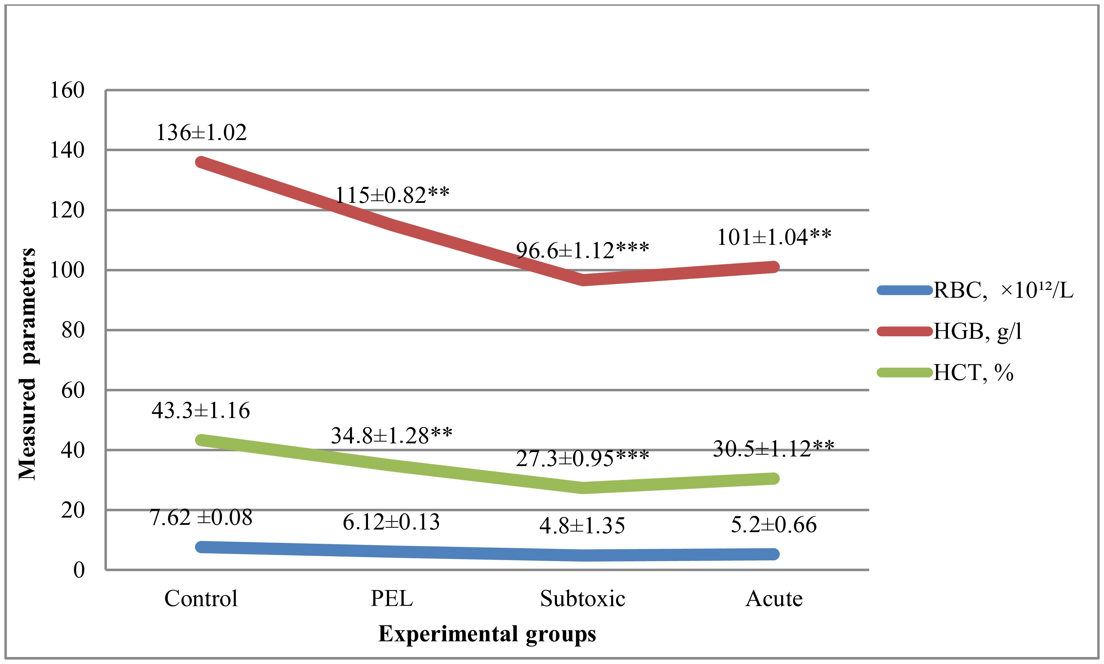
Cadmium exposure caused a consistent reduction in erythrocyte indices across all treated groups. The decrease in RBC count, hemoglobin concentration, and hematocrit was most evident in the subtoxic and acute dose groups, demonstrating a progressive anemic response. These alterations confirm impaired erythropoiesis and reduced oxygen-carrying capacity associated with cadmium toxicity (Figure 1).

Dose-dependent changes in RBC, HGB, and HCT levels in Wistar rats following cadmium exposure. RBC = red blood cell count; HGB = hemoglobin; HCT = hematocrit; PEL = permissible exposure limit. Data are expressed as Mean ± SEM (n = 6). *p < 0.05, **p < 0.01, ***p<0.001.
3.2. Mean corpuscular volume (MCV), mean corpuscular hemoglobin (MCH), and mean corpuscular hemoglobin concentration (MCHC)
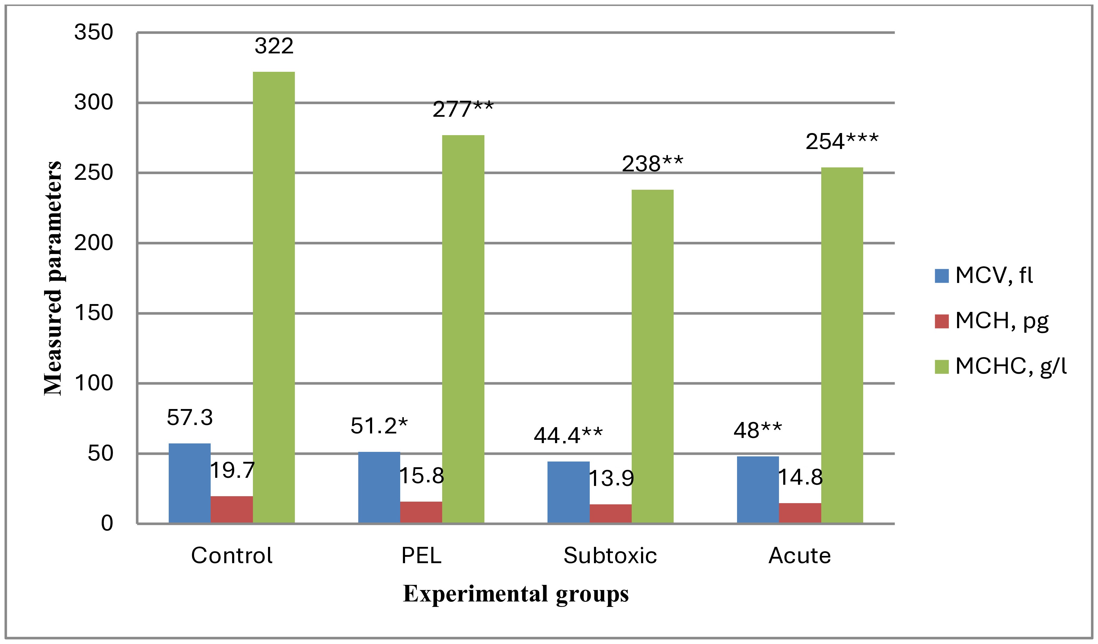
In line with the above findings, all three erythrocyte indices – MCV, MCH, and MCHC – decreased progressively with increasing cadmium concentration (Figure 2). The decline in MCV and MCHC indicates a shift toward microcytic hypochromic anemia, consistent with cadmium’s interference in iron metabolism and hemoglobin synthesis.

Effects of cadmium chloride on erythrocyte indices (MCV, MCH, MCHC) in Wistar rats: MCV = mean corpuscular volume; MCH = mean corpuscular hemoglobin; MCHC = mean corpuscular hemoglobin concentration; PEL = permissible exposure limit. *p < 0.05, **p < 0.01, *** p<0.001.
3.3. White blood cell count (WBC)
Interestingly, WBC counts showed a biphasic response (Figure 3). The PEL group exhibited a modest leukocytosis, likely reflecting an adaptive immune activation. In contrast, subtoxic and acute doses led to a marked leukopenia (p < 0.05), suggesting immunosuppressive effects due to bone marrow suppression or oxidative damage to leukocytes. These results highlight dose-dependent alterations in leukocyte dynamics, indicating differential immunological responses depending on cadmium exposure level.

White blood cell (WBC) response to graded cadmium exposure in Wistar rats. PEL – permissible exposure limit. *p < 0.05.
3.4. Platelet count (PLT)
A clear dose-dependent decline was also observed in platelet counts (Figure 4). The reduction was particularly pronounced in the acute-dose group, pointing to thrombocytopenia likely caused by cadmium-induced inhibition of megakaryopoiesis. This trend underscores the vulnerability of platelet-forming cells to heavy metal exposure.

Platelet counts (PLT) in rats after 28 days of cadmium chloride administration. PEL – permissible exposure limit. *p<0.001.
Overall, these findings demonstrate that even subtoxic cadmium concentrations can disturb hematological balance, affecting oxygen transport, immune regulation, and hemostasis in a graded manner depending on exposure level.
4. Discussion
Cadmium chloride exposure caused clear, dose-dependent hematological alterations in Wistar rats, characterized by anemia, leukopenia, and thrombocytopenia. These hematological impairments suggest that the hematopoietic system is one of the primary targets of cadmium toxicity.
Among the mechanisms involved, oxidative stress appears to play a central role. Cadmium does not directly generate reactive oxygen species (ROS), but it indirectly promotes oxidative damage by depleting antioxidants such as glutathione and inhibiting enzymes like catalase and superoxide dismutase (Takiguchi et al., 2003; Chen et al., 2022). The accumulation of ROS induces lipid peroxidation of erythrocyte membranes, resulting in structural fragility, premature hemolysis, and reduced red blood cell lifespan (Iqbal et al., 2022). These mechanisms explain the observed decreases in RBC count, hemoglobin, and hematocrit, as well as the microcytic and hypochromic pattern noted in erythrocyte indices (MCV, MCH, MCHC).
Another major mechanism is metal ion competition, where cadmium interferes with essential trace elements such as iron and zinc, crucial for heme synthesis and erythrocyte maturation (Horiguchi et al., 2004). Cadmium competitively inhibits the activity of δ-aminolevulinic acid dehydratase (ALAD), a key enzyme in the heme biosynthetic pathway, thereby reducing hemoglobin production (Cirovic and Cirovic, 2022; Zivancevic et al., 2024). This disruption leads to iron dysregulation and ineffective erythropoiesis, which are classical hallmarks of cadmium-induced anemia (Sun et al., 2015).
The bone marrow suppressive effects of cadmium were also evident in this study, reflected in reduced leukocyte and platelet counts. High-dose exposure may lead to bone marrow hypoplasia, apoptosis of hematopoietic stem cells, and altered cytokine signaling, resulting in decreased production of mature blood cells (Gökdemir, 2025; Suljevic et al., 2025). Moreover, inhibition of thrombopoietin pathways and endothelial injury contribute to the observed thrombocytopenia (Zhao et al., 2022b; Haidar et al., 2023).
The biphasic leukocyte response observed here-mild leukocytosis at low doses followed by leukopenia at higher doses – may reflect a transition from adaptive to toxic response. Initial immune activation can be attributed to cadmium-induced inflammation, whereas sustained oxidative damage and mitochondrial dysfunction ultimately suppress leukopoiesis (Das and Al-Naemi, 2019; Lee et al., 2022). This pattern underscores cadmium’s dual impact on immune regulation, which may have implications for increased infection risk in chronically exposed populations.
Field and experimental data futher support these findings. Studies on wild rats and other small mammals from cadmium-contaminated regions have documented reductions in hemoglobin, erythrocyte, and leukocyte counts, along with bone marrow atrophy and organ pathology (Wakeel et al., 2020; Chwalba et al., 2023). Laboratory investigations using Wistar rats similarly confirmed bone marrow hypoplasia and the emergence of immature progenitors, reflecting cadmium’s suppression of hematopoiesis (Gökdemir, 2025).
Although this experiment was performed in rats, the dose-dependent hematological effects observed here direct implications for human populations chronically exposed to cadmium. In many industrial regions worldwide-including parts of Kazakhstan, China, Poland, and Japan – cadmium concentrations in drinking water and food products have been reportedat levels comparable to the lower doses used in this study (Cherednichenko et al., 2021; Woszczyk et al., 2018; Wieczorek et al., 2018; WHO, 2011). Long-term dietary intake is considered the main route of exposure for the general population, whereas occupational exposure remains a major risk for workers in metallurgy, mining, and battery production industries.
The early onset of microcytic hypochromic anemia and immune suppression at relatively low exposure levels in our experiment suggests that similar subclinical changes may develop in exposed humans long before more severe manifestations such as kidney dysfunction, osteoporosis, or malignancy appear. Such silent hematological alterations may contribute to fatigue, increased susceptibilty to innfections, and impaired physical performance-symptoms commonly reported in individuals from cadmium-polluted regions.
Furthermore, populations with additional risk factors-such as iron deficiency, malnutrition, or chronic inflammatory conditions - may experience greater hematotoxic sensitivity, even at permissible exposure limits. Vulnerable groups, including children and pregnant women, are of particular concern because cadmium can cross the placenta and accumulate for decades in the body.
These findings highlight the need for regular monitoring of hematological profiles as part of surveillance programs for communities residing near industrial zones or consuming cadmium-contaminated water and food. Early detection via routine blood testing could help prevent the progression of cadmium-related health disorders and targeted public-health interventions aimed at reducing exposure.
5. Conclusion
This study demonstrates that chronic exposure to cadmium chloride, even at concentrations reflecting environmentally relevant levels, leads to marked dose-dependent hematological disturbances in Wistar rats. The significant decline in RBC, HGB, HCT, as well as reductions in MCV, MCH, and MCHC confirm the development of microcytic hypochromic anemia driven by impaired iron metabolism and oxidative damage to erythrocytes. Suppressive effects on leukocyte and platelet counts further indicate cadmium-induced bone marrow dysfunction and compromised immune regulation.
Importantly, the observed alterations in peripheral blood parameters proved highly sensitive to cadmium exposure across all doses. These results highlight the novel contribution of this work – showing that basic hematological indices can serve as early, practical, and accessible biomarkers for detecting cadmium-related toxicity before more severe systemic impairments emerge.
Given ongoing environmental and occupational cadmium exposure risks, especially in industrial regions, integrating hematological monitoring into routine ecological surveillance and public-health assessment may significantly improve early detection and prevention strategies.
Data Availability Statement
The datasets generated during the study are available from the corresponding author on reasonable request.
References
-
ANDJELKOVIC, M., BUHA DJORDJEVIC, A., ANTONIJEVIC, E., BUHA, A., MILOVANOVIC, V., JOVANOVIC, V. and MATOVIC, V., 2019. Toxic effect of acute cadmium and lead exposure in rat blood, liver, and kidney. International Journal of Environmental Research and Public Health, vol. 16, no. 2, pp. 274. https://doi.org/10.3390/ijerph16020274 PMid:30669347.
» https://doi.org/10.3390/ijerph16020274 -
CHARKIEWICZ, A.E., OMELJANIUK, W.J., NOWAK, K., GARLEY, M. and NIKLINSKI, J., 2023. Cadmium toxicity and health effects: a brief summary. Molecules, vol. 28, no. 18, pp. 6620. https://doi.org/10.3390/molecules28186620 PMid:37764397.
» https://doi.org/10.3390/molecules28186620 -
CHEN, X., BI, M., YANG, J., CAI, J., ZHANG, H., ZHU, Y., LI, J. and WANG, C., 2022. Cadmium exposure triggers oxidative stress, necroptosis, Th1/Th2 imbalance and promotes inflammation through the TNF-α/NF-κB pathway in swine small intestine. Journal of Hazardous Materials, vol. 421, pp. 126704. https://doi.org/10.1016/j.jhazmat.2021.126704 PMid:34325292.
» https://doi.org/10.1016/j.jhazmat.2021.126704 -
CHEREDNICHENKO, V.S., CHEREDNICHENKO, A.V., CHEREDNICHENKO, A.I.V., ZHEKSENBAEVA, A.K. and MADIBEKOV, A.S., 2021. Heavy metal deposition through precipitation in Kazakhstan. Heliyon, vol. 7, no. 1, e05844. https://doi.org/10.1016/j.heliyon.2020.e05844 PMid:33506121.
» https://doi.org/10.1016/j.heliyon.2020.e05844 -
CHWALBA, A., ORŁOWSKA, J., SŁOTA, M., JEZIORSKA, M., FILIPECKA, K., BELLANTI, F., DOBRAKOWSKI, M., KASPERCZYK, A., ZALEJSKA-FIOLKA, J. and KASPERCZYK, S., 2023. Effect of cadmium on oxidative stress indices and vitamin D concentrations in children. Journal of Clinical Medicine, vol. 12, no. 4, pp. 1572. https://doi.org/10.3390/jcm12041572 PMid:36836105.
» https://doi.org/10.3390/jcm12041572 -
CIROVIC, A. and CIROVIC, A., 2022. Iron deficiency as promoter of heavy metals-induced acute myeloid leukemia. Leukemia Research, vol. 112, pp. 106755. https://doi.org/10.1016/j.leukres.2021.106755 PMid:34844140.
» https://doi.org/10.1016/j.leukres.2021.106755 -
DAS, S. and AL-NAEMI, H., 2019. Cadmium toxicity: oxidative stress, inflammation and tissue injury. Occupational Diseases and Environmental Medicine, vol. 7, no. 4, pp. 144-163. https://doi.org/10.4236/odem.2019.74012
» https://doi.org/10.4236/odem.2019.74012 -
FAUL, F., ERDFELDER, E., LANG, A.G. and BUCHNER, A., 2009. Statistical power analyses using GPower 3.1: tests for correlation and regression analyses. Behavior Research Methods, vol. 41, no. 4, pp. 1149-1160. https://doi.org/10.3758/BRM.41.4.1149 PMid:19897823.
» https://doi.org/10.3758/BRM.41.4.1149 -
GENCHI, G., SINICROPI, M.S., LAURIA, G., CAROCCI, A. and CATALANO, A., 2020. The effects of cadmium toxicity. International Journal of Environmental Research and Public Health, vol. 17, no. 11, pp. 3782. https://doi.org/10.3390/ijerph17113782 PMid:32466586.
» https://doi.org/10.3390/ijerph17113782 -
GÖKDEMIR, G.Ş., 2025. Morphological changes in blood cells in a rat model of cadmium poisoning. Journal of Medical and Dental Investigations, vol. 6, e250165. https://doi.org/10.5577/jomdi.e250165
» https://doi.org/10.5577/jomdi.e250165 -
HAIDAR, Z., FATEMA, K., SHOILY, S.S. and SAJIB, A.A., 2023. Disease-associated metabolic pathways affected by heavy metals and metalloid. Toxicology Reports, vol. 10, pp. 554-570. https://doi.org/10.1016/j.toxrep.2023.04.010 PMid:37396849.
» https://doi.org/10.1016/j.toxrep.2023.04.010 -
HARSH, S., VANDANA, S., VAISHALI, K., GUPTA, R., MEHTA, N., PATEL, D. and SHARMA, M., 2024. Toxicological and bioremediation profiling of nonessential heavy metals (mercury, chromium, cadmium, aluminium) and their impact on human health: a review. Toxicologie Analytique et Clinique, vol. 36, no. 3, pp. 205-234. https://doi.org/10.1016/j.toxac.2024.03.096
» https://doi.org/10.1016/j.toxac.2024.03.096 -
HORIGUCHI, H., TERANISHI, H., NIIYA, K., AOSHIMA, K., KATOH, T., SAKURAGI, S. and KASUYA, M., 2004. Comprehensive study of the effects of age, iron deficiency, diabetes mellitus, and cadmium burden on dietary cadmium absorption in cadmium-exposed female Japanese farmers. Toxicology and Applied Pharmacology, vol. 196, no. 1, pp. 114-123. https://doi.org/10.1016/j.taap.2003.11.024 PMid:15050413.
» https://doi.org/10.1016/j.taap.2003.11.024 -
IQBAL, Z., QUDS, R. and MAHMOOD, R., 2022. Cadmium chloride generates cytotoxic reactive species that cause oxidative damage and morphological changes in human erythrocytes. Biochemistry and Cell Biology, vol. 100, no. 6, pp. 485-498. https://doi.org/10.1139/bcb-2022-0188 PMid:36288609.
» https://doi.org/10.1139/bcb-2022-0188 -
KRUPA, E., BARINOVA, S., ROMANOVA, S., AUBAKIROVA, M. and AINABAEVA, N., 2020. Heavy metals in fresh waters of Kazakhstan and methodological approaches to developing a regional water quality classification. Central Asian Journal of Water Research, vol. 6, no. 2, pp. 19-41. https://doi.org/10.29258/CAJWR/2020-R1.v6-2/19-41.eng
» https://doi.org/10.29258/CAJWR/2020-R1.v6-2/19-41.eng -
LEE, D.C., CHOI, Y.J. and KIM, J.H., 2022. Toxic effects of waterborne cadmium exposure on hematological parameters, oxidative stress, neurotoxicity, and heat shock protein 70 in juvenile olive flounder, Paralichthys olivaceus. Fish & Shellfish Immunology, vol. 122, pp. 476-483. https://doi.org/10.1016/j.fsi.2022.02.022 PMid:35176470.
» https://doi.org/10.1016/j.fsi.2022.02.022 - ORGANISATION FOR ECONOMIC CO-OPERATION AND DEVELOPMENT – OECD, 2018. Guidelines for the testing of chemicals, section 4: health effects Paris: OECD Publishing.
- REPUBLIC OF KAZAKHSTAN. Ministry of Health, 2015. SanPiN 2.1.4.1116-02: hygienic requirements for the quality of water of centralized drinking water supply. Astana.
-
SULJEVIĆ, D., MILANOVIC, V., MITRAŠINOVIC-BRULIC, M., JOVANOVIC, D., MARKOVIC, N. and STOJANOVIC, T., 2025. Association of different doses and routes of acute cadmium exposure with bone marrow hypoplasia, cellular and biochemical alterations in male Wistar rats. Biological Trace Element Research, vol. 203, no. 11, pp. 5624-5632. https://doi.org/10.1007/s12011-025-04591-4 PMid:40146489.
» https://doi.org/10.1007/s12011-025-04591-4 -
SUN, L., WANG, L., WANG, Z., HE, W., ZHANG, S., GUO, W., QIAN, Y., JI, H., RONG, H. and LIU, S., 2015. Cadmium depletes cellular iron availability through enhancing ferroportin translation via iron responsive element. Molecular Medicine Reports, vol. 11, no. 4, pp. 3129-3133. https://doi.org/10.3892/mmr.2014.3015 PMid:25435269.
» https://doi.org/10.3892/mmr.2014.3015 -
TAKIGUCHI, M., ACHANZAR, W., QU, W., LI, G. and WAALKES, M., 2003. Effects of cadmium on DNA-(Cytosine-5) methyltransferase activity and DNA methylation status during cadmium-induced cellular transformation. Experimental Cell Research, vol. 286, no. 2, pp. 355-365. https://doi.org/10.1016/S0014-4827(03)00062-4 PMid:12749863.
» https://doi.org/10.1016/S0014-4827(03)00062-4 -
WAKEEL, J., EHSAN, N., AKHTAR, R.W. and HUSSAIN SHAH, S.A., 2020. Morphology, histopathology and hematology as biomarkers of cadmium toxicity in field rats. Iranian Journal of Toxicology, vol. 14, no. 1, pp. 33-42. https://doi.org/10.32598/ijt.14.1.33
» https://doi.org/10.32598/ijt.14.1.33 -
WIECZOREK, J., BARAN, A., URBAŃSKI, K., MAZUREK, R. and KLIMOWICZ-PAWLAS, A., 2018. Assessment of the pollution and ecological risk of lead and cadmium in soils. Environmental Geochemistry and Health, vol. 40, no. 6, pp. 2325-2342. https://doi.org/10.1007/s10653-018-0100-5 PMid:29589150.
» https://doi.org/10.1007/s10653-018-0100-5 - WORLD HEALTH ORGANIZATION – WHO, 2011. Guidelines for drinking-water quality 4th ed. Geneva; WHO.
-
WOSZCZYK, M., SPYCHALSKI, W. and BOLUSPAEVA, L., 2018. Trace metal (Cd, Cu, Pb, Zn) fractionation in urban-industrial soils of Ust-Kamenogorsk (Oskemen), Kazakhstan: implications for the assessment of environmental quality. Environmental Monitoring and Assessment, vol. 190, no. 6, pp. 362. https://doi.org/10.1007/s10661-018-6733-0 PMid:29802453.
» https://doi.org/10.1007/s10661-018-6733-0 -
XIONG, L., FAN, C., SONG, J., ZHANG, H., LI, Y., CHEN, Z. and WANG, X., 2022. Associations of long-term cadmium exposure with peripheral white blood cell subtype counts and indices in residents of cadmium-polluted areas. Chemosphere, vol. 308, pp. 135946. https://doi.org/10.1016/j.chemosphere.2022.135946
» https://doi.org/10.1016/j.chemosphere.2022.135946 -
ZHAO, Y., HE, J., ZHU, T., ZHANG, Y., ZHAI, Y., XUE, P., LI, C. and WANG, X., 2022a. Cadmium exposure reprograms energy metabolism of hematopoietic stem cells to promote myelopoiesis at the expense of lymphopoiesis in mice. Ecotoxicology and Environmental Safety, vol. 231, pp. 113208. https://doi.org/10.1016/j.ecoenv.2022.113208 PMid:35051759.
» https://doi.org/10.1016/j.ecoenv.2022.113208 -
ZHAO, Y., ZHANG, Y., HE, J., WANG, X., LIU, P. and ZHAI, Y., 2022b. Cadmium suppresses bone marrow thrombopoietin production and impairs megakaryocytopoiesis in mice. Toxicological Sciences: An Official Journal of the Society of Toxicology, vol. 186, no. 2, pp. 309-322. https://doi.org/10.1093/toxsci/kfac010 PMid:35134237.
» https://doi.org/10.1093/toxsci/kfac010 -
ZIVANCEVIC, K., ZIVANOVIC, J., BARALIC, K., BOZIC, D., MARIC, D., VUKELIC, D. and BULAT, Z., 2024. Integrative investigation of hematotoxic effects induced by low doses of lead, cadmium, mercury and arsenic mixture: in vivo and in silico approach. The Science of the Total Environment, vol. 930, pp. 172608. https://doi.org/10.1016/j.scitotenv.2024.172608 PMid:38653421.
» https://doi.org/10.1016/j.scitotenv.2024.172608
Edited by
-
Editor:
Marcelo A.M. Esquisatto