Morphological features, genetic analysis and life history of the exotic cladoceran Moina macrocopa (Straus, 1820) in Southeastern Brazil
Abstract
The spreading of the exotic cladoceran Moina macrocopa within the South American continent highlights the requirement for more detailed studies on this species. To date, morphological approaches in South America have identified specimens as Moina macrocopa macrocopa (Straus, 1820). However, no genetic study has confirmed this identification, and there are no available reports on the biology of this exotic species in the South American continent. Therefore, this research elucidates, through morphological and genetic analyses, the taxonomy of M. macrocopa found in southeastern Brazil. Furthermore, life history traits of cultivated parthenogenetic females were followed. Female morphological features and ephippium were in accordance with the description for M. macrocopa. The obtained haplotype of the mitochondrial COI gene and one haplotype of 18S rDNA gene revealed that the studied specimens represent an Asian sub-clade within the M. macrocopa macrocopa clade. Parthenogenetic females of the clone and population studied matured in 5.1 ± 1.1 days, with the first offspring released between 6 and 7 days, and juvenile production occurring throughout their lives. The average lifespan was 21.3 ± 4.3 days, generating 38.8 ± 9.9 neonates during the entire life. Compared to literature data on the native species Moina cf. micrura, the average size of neonates, primiparous females, and adults of M. macrocopa were larger, as was the number of offspring per brood. It is suggested that these features may confer hypothetically a competitive advantage to M. macrocopa over native species in temporary ponds.
Keywords:
life table; Moinidae; tropical cladocerans; bioinvasion; Brazil; COI; 18S rDNA
Resumo
A expansão da ocorrência do cladócero exótico Moina macrocopa na América do Sul destaca a necessidade de estudos mais detalhados sobre a espécie. Até o momento, abordagens morfológicas identificaram os espécimes encontrados como Moina macrocopa macrocopa (Straus, 1820). Contudo, nenhum estudo genético confirmou essa identificação e inexistem relatos disponíveis sobre a biologia dessa espécie exótica no continente sul-americano. Diante disso, esta pesquisa elucida, por meio de análises morfológicas e genéticas, a taxonomia de M. macrocopa encontrada no sudeste do Brasil. Além disso, foi acompanhado o ciclo de vida de fêmeas partenogenéticas em cultivo. As características morfológicas das fêmeas estudadas provenientes de um mesmo clone e efípio estão de acordo com a forma típica de M. macrocopa. O haplótipo obtido do gene mitocondrial COI e um haplótipo do gene 18S rDNA revelaram que os espécimes estudados são um subclado asiático dentro do clado M. macrocopa macrocopa. Fêmeas partenogenéticas apresentaram maturação em 5,1 ± 1,1 dias, com a primeira prole liberada entre 6 e 7 dias e havendo a produção de juvenis ao longo de toda a vida. A expectativa de vida média foi de 21,3 ± 4,3 dias, gerando 38,8 ± 9,9 neonatos durante todo o ciclo de vida. Em comparação com dados da literatura sobre a espécie nativa Moina cf. micrura, o tamanho médio dos neonatos, primíparas e adultos de M. macrocopa foi maior, assim como o número de neonatos por ninhada. Sugere-se que essas características podem conferir hipoteticamente à M. macrocopa vantagem competitiva sobre as espécies nativas em lagoas temporárias.
Palavras-chave:
tabela de vida; Moinidae; cladóceros tropicais; bioinvasão; Brasil; COI; rDNA 18S
1. Introduction
Cladocerans are one of the main components of the freshwater zooplankton and play an important role in the aquatic food chain (Azuraidi et al., 2013), whilst being the preferred prey of other invertebrates and by both larvae and adult fish (Monakov, 2003). Their size, nutritional composition and ease of cultivation make them an important food source in aquaculture and laboratory experiments. Among moinids, the genus Moina has been widely employed in aquaculture and laboratory research including protocols for toxicity tests in Ecological Risk Assessment. Among the various species of this genus, Moina macrocopa has been highlighted as an easily cultured organism and resistant to management in experimental trials. Valued for its nutritional content, this cladoceran has been exploited as live food for fish and crustaceans and presents the potential to replace Artemia (Rasdi et al., 2021). Additionally, due to its sensitivity to various pollutants, M. macrocopa serves as a model for several toxicity assessments of chemical contaminants (Samarakoon and Fujino, 2024; Manríquez-Guzmán et al., 2025).
Taxonomically, two subspecies that present differing geographical distributions are recognized in Moina macrocopa. The Moina macrocopa macrocopa (Straus, 1820) is restricted to the Old World with records in Africa, Europe, Russia, Middle East, and Southeast Asia (Goulden, 1968; Bhanushali et al., 2021). The other, Moina macrocopa americana Goulden, 1968 is confined to the North American continent on the northern side of the Rio Bravo (Elías-Gutiérrez et al., 1999). More recently, Montoliu-Elena et al. (2019) re-described Moina macrocopa americana Goulden, 1968 and considered it to be a distinct species, and not a subspecies, of Moina macrocopa s.l., and expanded the distribution of the species to the Central Plateau of Mexico.
In South America, the first report of M. macrocopa introduction came from Peru in 1988 and was linked to aquaculture activities. At that time, the species had yet to be detected in natural aquatic ecosystems (Valdívia-Villar, 1988). The first detection of M. macrocopa in South American natural environments was recorded in a lake in Bolivia (Elías-Gutiérrez and Zamuriano-Claros, 1994), the second in Lake Valencia Basin in Venezuela (Zoppi de Roa & López, 2008) and the third in a small roadside ephemeral pond in Argentina (Paggi, 1997). In the latter country, the species was then discovered in natural Pampean lakes (Vignatti et al., 2013). In Brazil, M. macrocopa was detected in a temporary pond located in downtown Rio de Janeiro city (Elmoor-Loureiro et al., 2010), and in a eutrophic urban reservoir of another Brazilian city (Belo Horizonte) (Rietzler et al., 2014). More recently, M. macrocopa was detected in a permanent high-altitude tropical shallow lake in Serra do Cipó National Park (Eskinazi-Sant’Anna et al., 2020) in the Brazilian countryside. The morphological features of the Argentinean and Brazilian specimens coincided with those reported by Paggi (1997) and Elmoor-Loureiro et al. (2010). However, the ephippium of the M. macrocopa from Rio de Janeiro was more similar to that of M. macrocopa americana (see Elmoor-Loureiro et al., 2010). Besides association with aquaculture activities, the invasion of Moina macrocopa has also been related with other anthropogenic activities such as laboratory experiments, including standard protocol for toxicological bioassays. Notwithstanding by ways of introduction, a notable aspect in the South American records of M. macrocopa is the presence in both temporary and natural permanent ponds (Paggi, 1997; Elmoor-Loureiro et al., 2010; Eskinazi-Sant’Anna et al., 2020) and in eutrophic and hypertrophic lakes and reservoirs (Vignatti et al., 2013; Rietzler et al., 2014).
Macêdo et al. (2022) predicted the range expansion of M. macrocopa (Straus, 1820) in South America, identifying new areas at risk for invasion due to rising temperatures and decreasing winter precipitation. One important issue addressed by those authors was the looming potential competition of this invader with congeneric native species. In the Neotropical Region, this can pose a threat, as smaller cladocerans such as Moina are frequently ubiquitous in freshwater ecosystems, including coastal lakes (Branco et al., 2007), river-associated ponds (Neves et al., 2003), reservoirs (Sampaio et al., 2002; Brito et al., 2013) and estuary (Paranaguá et al., 2005). Moina cf. micrura was recognized as being the most found cladoceran species in Brazil by Castilho-Noll et al. (2023), hence being detected in shallow lakes, coastal lagoons, reservoirs, and fish farming systems (Santangelo et al., 2008, 2011; Loureiro et al., 2018).
Given the lack of more in-depth studies on Moina macrocopa within South America, this study aims to characterize morphological and genetic features of a clone originating from a river in Rio de Janeiro city, in southeastern Brazil (22o43’44” S, 43o16’03” W). Genetic information on this exotic cladoceran will further enhance future ecological and biogeographical studies on its spread in South America. A second target was to investigate the life cycle of parthenogenetic females of this clone, which was cultivated in laboratory under controlled conditions. The life history traits of invasive cladocerans outside their natural range are notably scarce, thus highlighting a significant gap in the degree of our understanding of strategies employed by these invaders under new environmental conditions and the prospect that looms for their spread and colonization.
2. Material and Methods
2.1. Culture of Moina macrocopa
The Environmental Health Assessment and Promotion Laboratory at Oswaldo Cruz Institute in Rio de Janeiro provided the original inoculum of M. macrocopa that were 20 individuals, from the same clone, which was isolated from the polluted Iguaçu River by Dr. Mauro Vilar. This river comprises the Iguaçu-Sarapuí River basin, which receives direct inputs of raw sewage and waste from agricultural and industrial activities (Coelho, 2020).
Moina macrocopa was cultivated in commercial mineral water (Minalba® brand, Brazil) using Raphidocelis subcapitata as the unique food source (Steinberg et al., 2010; Suhett et al., 2011). This chlorophyte has sickle-shaped capricorn-shaped cells, arcuated, normally found in unicellular form (length between 8-15µm and width 1.9-4µm). R. subcapitata was cultivated following the Brazilian NBR 13373 standard methods for aquatic ecotoxicological tests (ABNT, 2017).
The number of algae supplied to the cladocerans followed the method NBR 13373 (ABNT, 2017) for Ceriodaphnia spp. culture for aquatic ecotoxicology and chronic toxicity tests that corresponds to 2 x 10⁵ - 5 x 10⁵ cells of algae per cladoceran. The algal density was estimated using a hematocytometer. The medium of the M. macrocopa cultures was changed once a week. All algae and cladoceran cultures were kept at 24 ± 1 °C in a 12:12 light:dark photoperiod in a growth chamber LT 320 TFP-I (Limatec Laboratory Equipment) during the experiments. Both cultures were kept at the Laboratory of Culture and Experiments of the Institute of Bioscience at the Federal University of the State of Rio de Janeiro (Brazil).
2.2. General morphological features
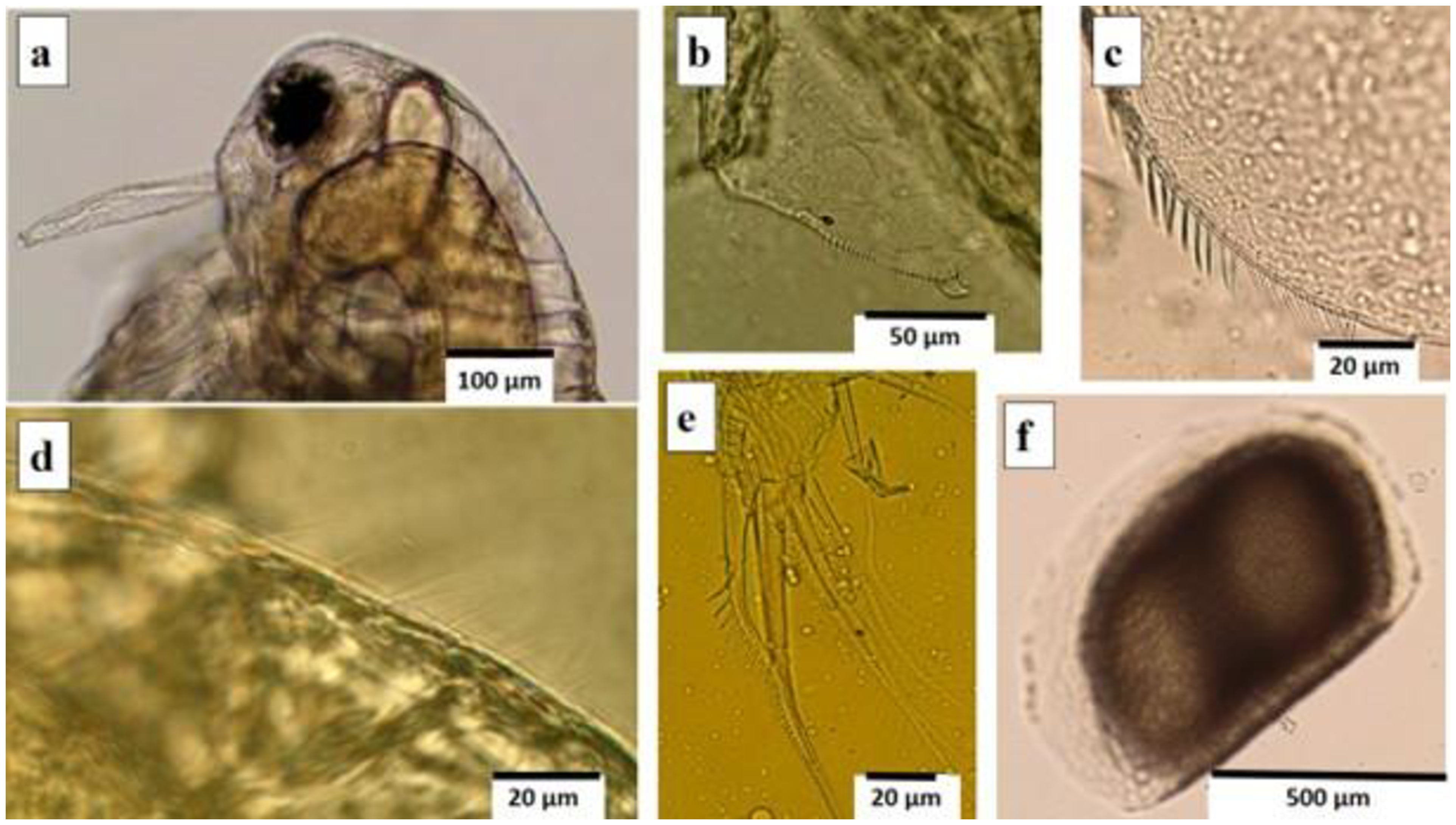
Ephippium and parthenogenetic females of the main culture were dissected and photographed under a microscope Olympus BX-51 with digital camera and image analysis software ToupView 3.7. Taxonomically important morphological aspects were studied as discussed by Paggi (1997), Elmoor-Loureiro et al. (2010) and Montoliu-Elena et al. (2019). The features of the surface of ephippium and its number of eggs were analysed since in Moina macrocopa macrocopa the ephippium is covered with thick rounded cells being sub-rectangular in lateral view and contains two eggs. Other features analysed were the shape of the head, insertion of sensory seta in the antennule, setae of the posterior ventral part of the carapace, setae of dorsal shell rim (fine spinules), toothed seta of first leg (L1), and hook at the posterior end of the carapace.
2.3. Genetic analyses and DNA sequencing
Genomic DNA from four M. macrocopa individuals from the same clone stored in 70% ethanol was extracted at the Molecular Biology Laboratory at the University of Bialystok (Poland) using the DNeasy Blood & Tissue Kit (Qiagen, Hilden, Germany), following the manufacturer’s instructions. The mitochondrial cytochrome c oxidase subunit I (COI) gene and the nuclear 18S small subunit ribosomal RNA gene (18S rDNA) were targeted for molecular sequencing. The polymerase chain reaction (PCR) was used to amplify about 630 bp of the COI gene fragment using primers jgLCO1490 and jgHCO2198, which are a universal tool for molecular identification of marine invertebrate species designed by Geller et al. (2013). The PCR was also used to amplify about 680 bp 18S rDNA gene fragment using primers Merm forward and Merm reverse designed by Kobylinski et al. (2012). PCR amplification of COI and 18S rDNA genes were conducted in a Labcycler Gradient (SensoQuest, Göttingen, Germany) in 5 μL volumes. The reaction mixtures consisted of ~ 25 ng extracted genomic DNA as a template, 1.7 μL of Qiagen Multiplex PCR Master Mix (1×), 0.3 μL mix of primers (for COI and 18S rDNA, respectively) and 1 μL of Qiagen nuclease-free water. Thermocycler program consisted of initial denaturation at 95 °C for 15 min followed by 40 cycles of denaturation at 94 °C for 30 s, annealing at 42 °C (for COI gene) or 57 °C (for 18S rDNA gene) for 90 s, extension at 72 °C for 60 s, and final elongation step of 30 min at 60 °C. The presence of amplification products of COI and 18S rDNA gene fragments were then verified by electrophoresis on 1.5% agarose gels.
All amplification products were purified with the EPPiC Fast mixture (A&A Biotechnology, Gdańsk, Poland), which contained two enzymes that effectively degrade dNTPs and primer left-overs from previous PCR mixtures, while leaving the double stranded DNA PCR products untouched. Purification of PCR products were carried out in a thermal cycler following the manufacturer’s recommendations: 37 °C for 5 min, 80 °C for 1 min. Then the sequence reaction using both forward and reverse primers for analysed genetic markers was carried out with a BigDye™ Terminator Cycle Sequencing Kit v.3.1 (Applied Biosystems, Foster City, CA, USA). Unincorporated dideoxynucleotides were then eliminated from the sequencing reaction using the ExTerminator Kit (A&A Biotechnology). In the end, the sequencing products of COI and 18S rDNA were run on an automated capillary sequencer ABI 3130 (Applied Biosystems, Foster City, CA, USA). Sequencing results were revised and aligned manually using BioEdit v.7.0.5.3 (Hall, 1999).
2.4. Phylogenetic analyses
Phylogenetic analyses were carried out using COI haplotype obtained in this study, combined with representative Moina sp. sequences retrieved from the GenBank database. The most suitable nucleotide substitution model was selected based on the Akaike Information Criterion (AIC) using jModelTest v. 0.1.1 (Posada, 2008). According to this approach, the GTR+I+G model was chosen for subsequent phylogenetic analyses. A phylogenetic tree was constructed using the Maximum Likelihood (ML) method to infer evolutionary relationships among Moina sp. haplotype derived from the mitochondrial cytochrome c oxidase subunit I (COI) gene obtained in this study, as well as sequences downloaded from GenBank. The analysis was performed in Mega v.11 (Tamura et al., 2021), applying 1000 bootstrap replicates to evaluate the statistical support for the inferred tree nodes. The selection of sequences aimed to represent the main lineages within the Moina macrocopa complex rather than to exhaustively include all available records.
2.5. Life table
For the life history study, fifteen M. macrocopa neonates (with less than 24h) from the same clone were separated in different beakers (one neonate per beaker) with 100 mL of the medium (with 2 x 10⁶ cells of R. subcapitata per cladoceran). The medium of the 15 replicates was completely changed every two days. All cladocerans were followed up until their death. Maturation time was considered to be when eggs appear in the incubation chamber, and the age of primipara was considered to be when the neonates are born. The size of all females was measured every two days under microscope (Olympus BX-51, 40X) with the ToupView software version 3.7. Every day, we recorded offspring, molts, number of offspring per birth, size of the offspring, production of resting eggs, absence or presence of males, and life span.
3. Results
3.1. General morphological features
Our female specimens were characterized by a relatively large and rounded head, without a supraocular depression (Figure 1a). The dorsal part of the head with very thin hairs restricted to the posterior two-thirds of the dorsum-lateral surface of the head (Figure 1d). The antennules are large and robust, with sensory setae inserted in the middle of the antennule (Figure 1a). The ventral rim of the shell with 68 to 73 setae, which are long and densely spaced (setae length twice as long as the distance between setae) (Figure 1c). The first leg (L1) is very distinct because the anterior seta has long and stout teeth (Figure 1e). Ephippium contained two eggs and presented a surface covered with thick rounded cells, which are sub-rectangular in lateral view (Figure 1f). All these morphological characteristics are consistent with the description for M. macrocopa.
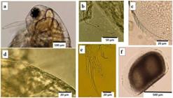
Morphological features of the female Moina macrocopa. (a) Sensory setae inserted in the middle of the antennule; (b) hook at the end of the carapace; (c) setae at the ventral margin of the carapace; (d) thin hairs at the dorsum-lateral surface of the head; (e) L1 with toothed setae; (f) ephippium contained two eggs.
3.2. Genetic analyses, DNA sequencing, and phylogenetic analyses
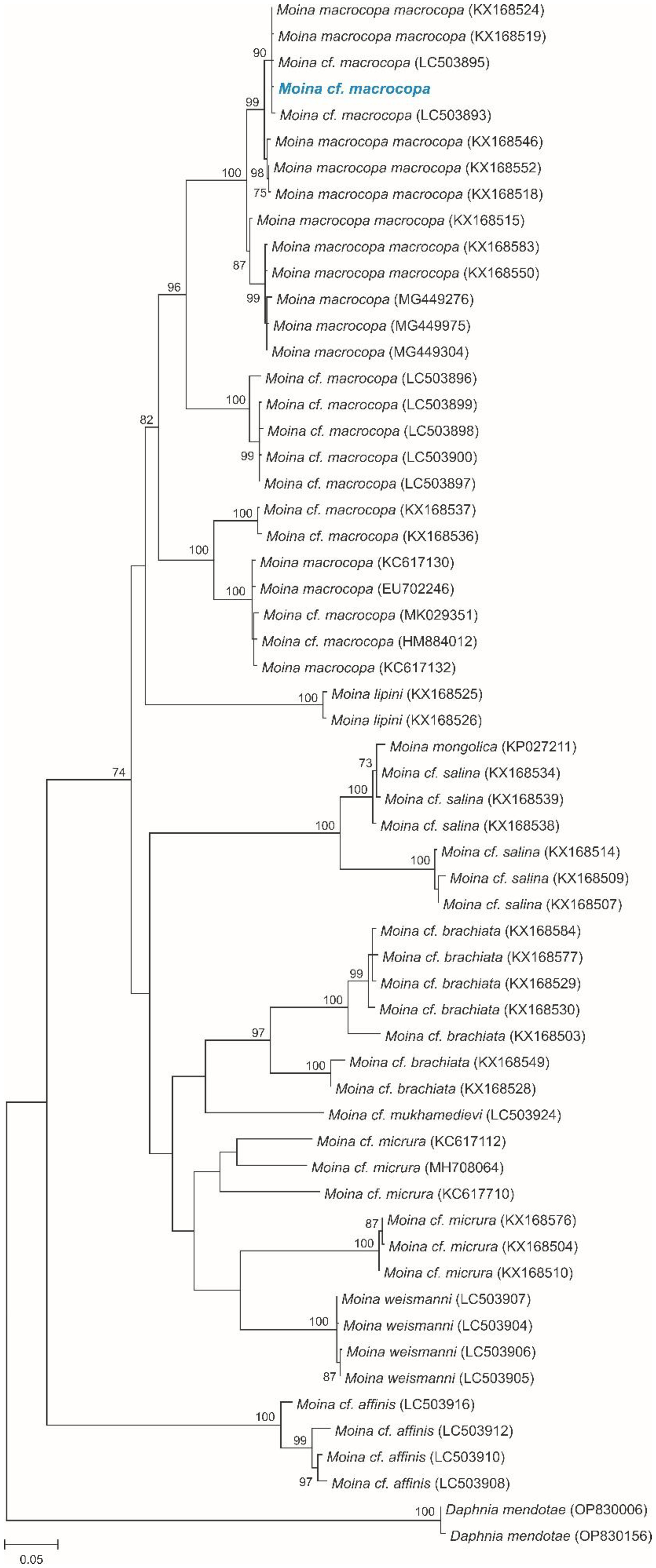
The obtained sequences from four individuals yielded one haplotype of the mitochondrial COI gene fragment and one haplotype of the 18S rDNA gene fragment. Haplotypes obtained in this study were combined with those for Moina sp. available in GenBank to corroborate the identity of our sequences. Our haplotype of the mitochondrial COI gene fragment was identical to the haplotype of M. macrocopa from Japan described by Makino et al. (2020) (GenBank accession no. LC503892) and Hiki et al. (2023) (GenBank accession no. LC705056), and from Russia described by Bekker et al. (2016) (GenBank accession no. KX168523). The haplotype revealed by Bekker et al. (2016) represents an Asian sub-clade within the M. macrocopa macrocopa clade. On the phylogenetic ML tree, we illustrated the position of the mtDNA COI haplotype identified in our analyses (Figure 2). 18S rDNA haplotype obtained in this study was identical to the sequence of M. macrocopa found in Japan by Makino et al. (2020) (GenBank accession no. LC503929). Importantly, sequences corresponding to the North American lineage traditionally referred to as Moina macrocopa americana (e.g., HM884012) formed a separate and well-supported clade in our analysis, clearly distinct from the clade containing our haplotype and other Eurasian populations.
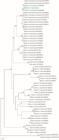
Maximum likelihood (ML) phylogenetic tree of Moina, illustrating relationships inferred from haplotypes of the mitochondrial cytochrome c oxidase subunit I (COI) gene. The haplotype identified in this study is indicated in blue. Numbers following species names represent GenBank accession numbers of the sequences used. The tree was reconstructed under the GTR+I+G model of nucleotide substitution. Node values correspond to bootstrap support percentages calculated from 1000 replicates.
3.3. Life table
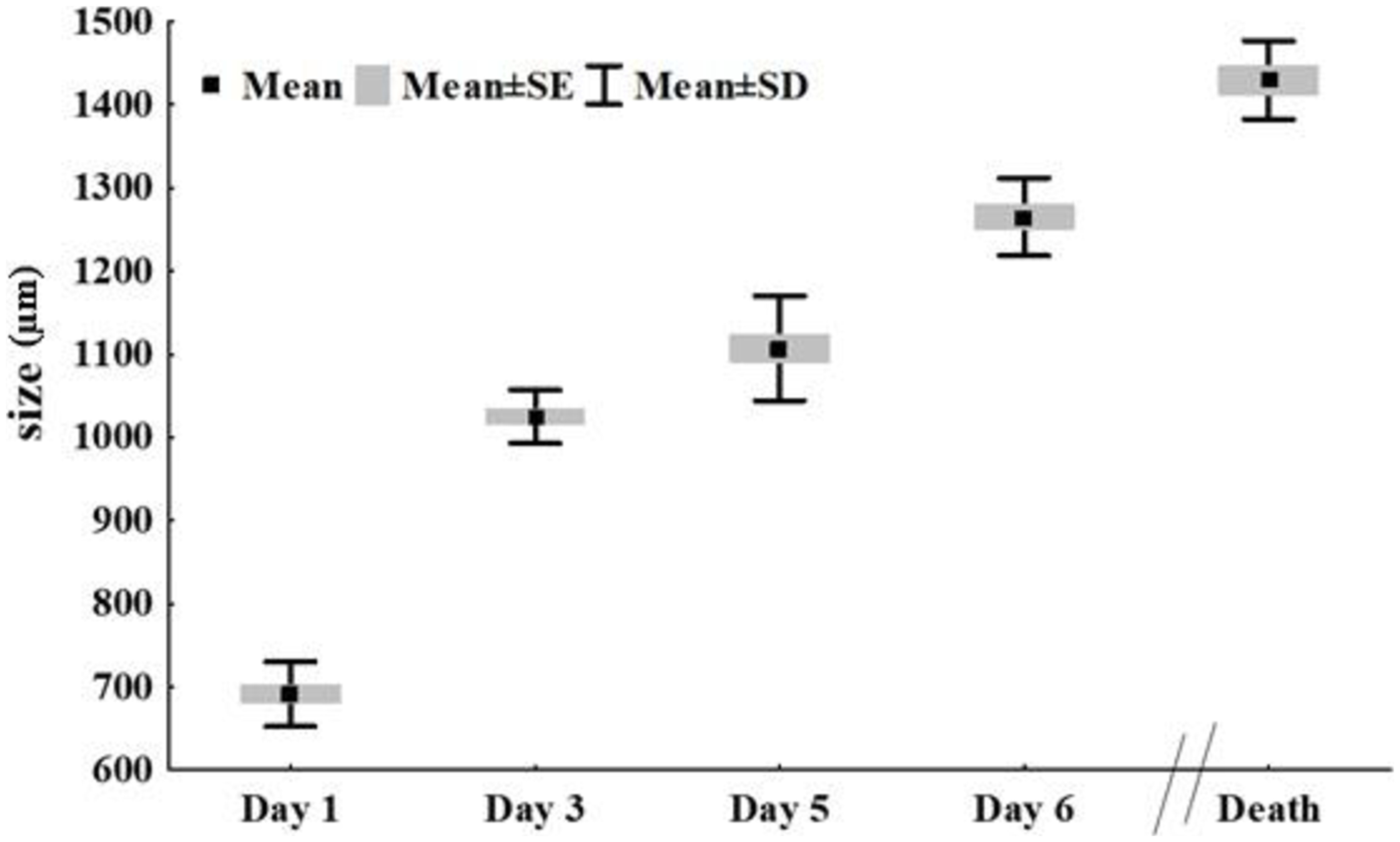
According to our results, the average length of neonates was 583µm, parthenogenetic females attained 1,025µm on day three, and the average maximum size of adults was 1,429.9 ± 47.2 µm (Figure 3; Table 1). From birth to the fifth day, the total length increases fast (57%, percentage of length increase), and continued to increase throughout life, although at a lower percentage. From the second to the 5th day (juvenile stage), molting happened three times, characterizing three juvenile stages.
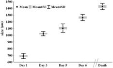
Growth in size of Moina macrocopa (n=15) at days 1, 3, 5, 6, and at the time of death (size in µm).
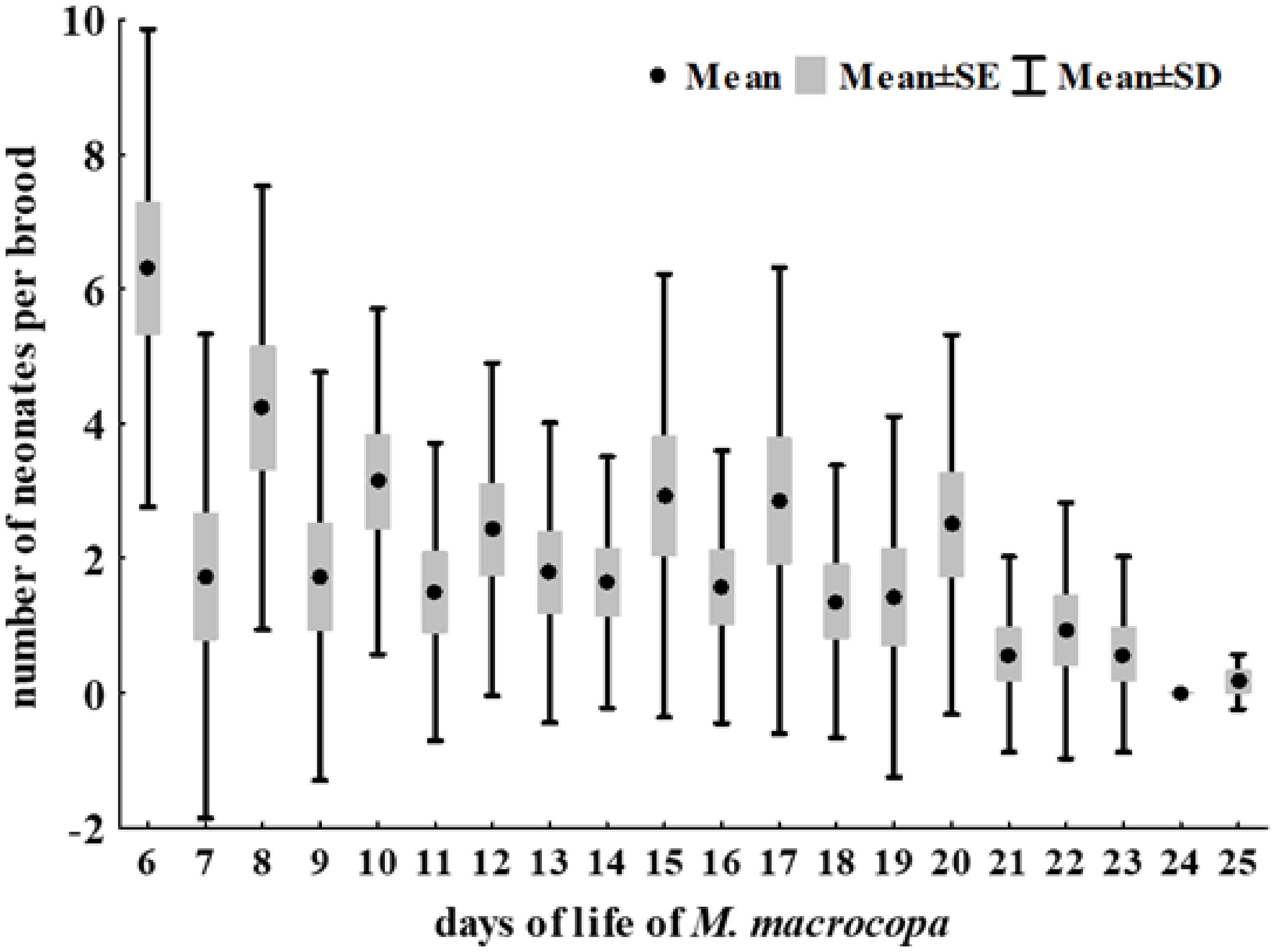
Females matured within 5 days. On the 5th day, first eggs were found and, on the 6th or 7th day, almost all the cladocerans had their first offspring (Figure 4). After the first offspring, molting occurred only after each hatching, with an average of eight molts per female. However, after the first offspring, the total length increases by only 13% until the end of the life.
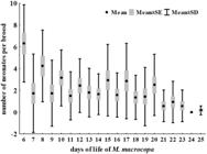
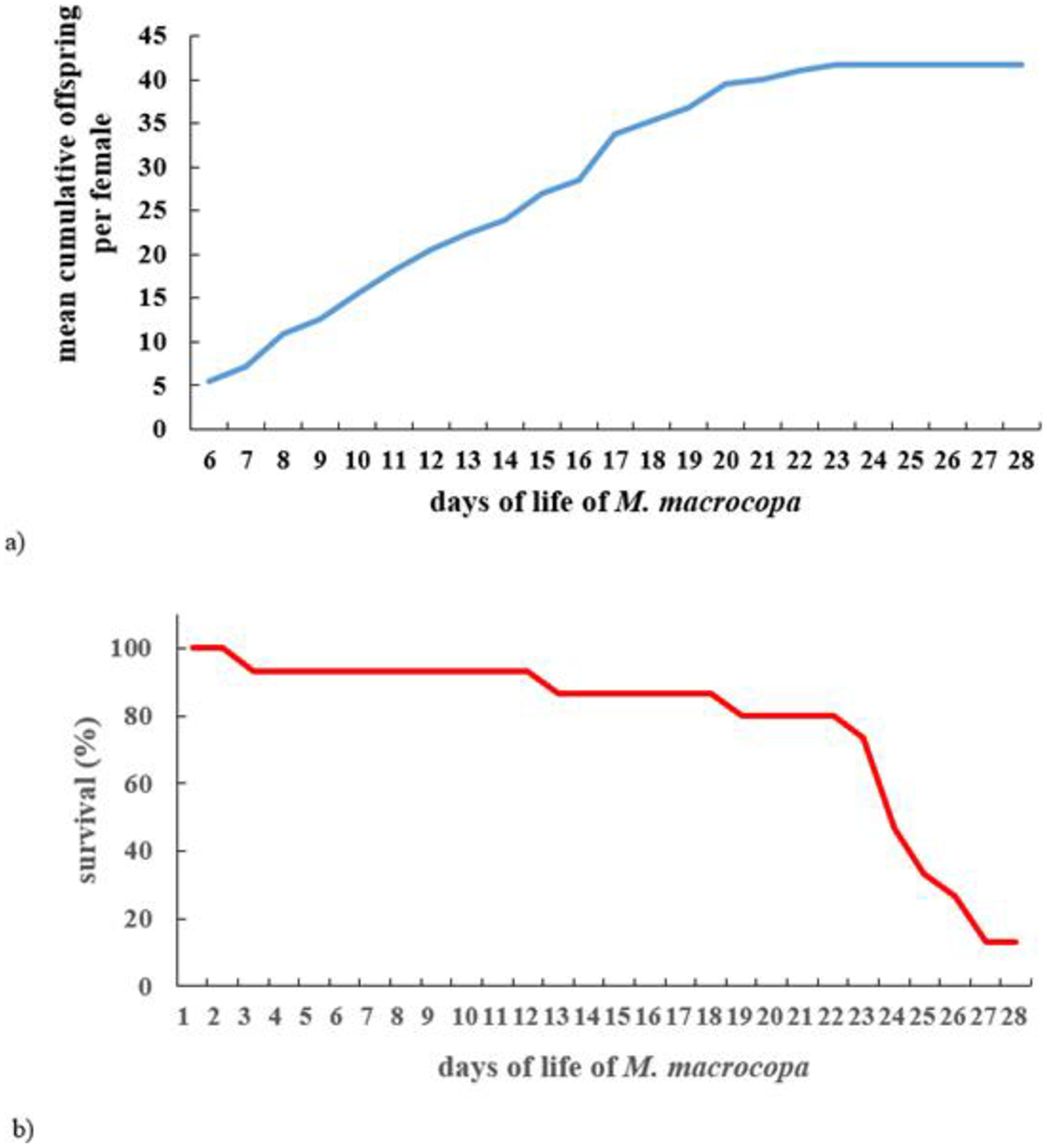
After the first brood, broods occurred at each one, two or three days and the number of neonates per brood varied between 1 and 10, having an average of 5.1 ± 1.6. Considering the whole life span of the M. macrocopa, females had an average of 7.8 ± 2.0 broods with the first one having a greater number of neonates and the latter with a smaller number. Mean cumulative offspring per female was 38.8 ± 9.9, with most newborns being produced within half of their lifespan (Figure 5a).
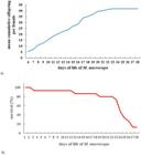
(a) Mean cumulative offspring per female of Moina macrocopa across the whole life; (b) Surviving curve for Moina macrocopa (n=15).
Individuals lived on average 21.3 ± 4.3 days, comprising a minimum of 3 days and a maximum of 28 days (Figure 5b; Table 1). The mortality increased after the 24th day of life, and only 13.3% of the individuals lived for 28 days. No males were born during the experiment, and no resting eggs were found either.
4. Discussion
4.1. Morphological features, genetic analyses, and DNA sequencing
In the first detailed report on taxonomic morphological features of M. macrocopa in South America, Paggi (1997) highlighted that M. macrocopa macrocopa could be differentiated from M. macrocopa americana by the distribution of setae along the posterior border of the shells and by the ornamentation of the ephippium. The parthenogenetic females of our study showed variation in the number of setae at the ventral margin, that ranged from 68 to 73, which were close to the reported by Paggi (1997), who detected 73 to 76 setae, and by Elmoor-Loureiro et al. (2010) that found about 75 setae. These results were higher than the 55 to 65 reported by Goulden (1968) for M. macrocopa macrocopa in the Old World. Previously, Elias-Gutierrez and Zamuriano-Claros (1994) had reported only 45 to 56 setae for the M. macrocopa from Bolivia, and after, Vignatti et al. (2013) found 42 to 74 setae, which number was different among lakes. This last observation is important, and only a greater number of studies can elucidate whether the number of setae along the border of the shell would be a phenotypic aspect related to the local environment.
In the redescription of Moina macrocopa americana Goulden, 1968 conducted by Montoliu-Elena et al. (2019), this taxon was elevated to species rank. Other morphological differences between M. macrocopa and M. americana were listed, such as features of the sensory seta in the antennule (A1), type of spinules of the setae of posterior shell rim, and feature of the teeth of the toothed seta of L1. In the specimens of our study, all these morphological details are in accordance with the description for M. macrocopa (see Montoliu-Elena et al., 2019). In addition, characteristics of the ephippium found in the main culture corroborated M. macrocopa, such as the presence of two eggs, and a surface covered with thick rounded cells, which are sub-rectangular in lateral view. Despite the similarities, it must be stressed that morphological identification has significant limitations since phenotypic plasticity and genetic variability may lead to difficulty in differentiating between morphologically cryptic taxa and therefore lead to incorrect identification of species (Bhanushali et al., 2021; Karpowicz et al., 2024). These limitations can be overcome by using a micro-genomic identification, as we did.
The genomic analysis of the present study proves that the specimens collected in the polluted river in Rio de Janeiro city are of M. macrocopa macrocopa. We can suggest that the specimens from that temporary pond in Rio de Janeiro studied by other authors (Elmoor-Loureiro et al., 2010; Steinberg et al., 2010; Suhett et al., 2011; Hofmann et al., 2012; Santangelo et al., 2018) could also be this species. However, it is important to note the potential morphological variations in M. macrocopa macrocopa, particularly regarding the number of setae on the ventral margin of the carapace. We should also point out that our study has limitations regarding other possible intraspecific morphological variations, since it is based on a population originating from the same clone.
The genetic analyses showed similarities between the clade collected in Rio de Janeiro and one from Japan. Both the haplotype of the mitochondrial COI gene fragment and the 18S rDNA haplotype obtained were identical to the sequence of the Japanese M. macrocopa (Makino et al., 2020). Paggi (1997) suggested that specimens of M. macrocopa imported from Japan were cultured as food for fish larvae in the Pampa region in Argentina. The connection between the information on the M. macrocopa imported from Japan by Paggi (1997) and the present finding is attractive but can be merely speculative. Notwithstanding, any discussion on the possible dispersion of the same original clone from Argentina to Brazil could be interesting with respect to the potential dispersion of non-native cladoceran species in South America. This route south-southeast across South America was already verified for invasive protists and other small-sized zooplankters (Macêdo et al., 2021; Pereboev et al., 2025). It should be noted that some sequences available in public databases (e.g., HM884012), although sometimes labelled as Moina cf. macrocopa, correspond to the North American lineage currently recognized as Moina americana. In our phylogenetic reconstruction, these sequences formed a distinct clade, separate from the Eurasian M. macrocopa macrocopa lineage, which further supports the identification of our material as belonging to the latter group. However, a detailed resolution of relationships within the M. macrocopa species complex was beyond the scope of the present study.
4.2. Life table
The life cycle of Moina spp. is typical of many species of Cladocera, being reproduction primarily by parthenogenesis, and males and sexual females appearing in the populations under changes in food availability or quality, crowding, and temperature conditions (D’Abramo, 1980; Nandini and Sarma, 2019). Since we did not find males or resting eggs in our experiment, which comprised 28 days, we can consider the conditions used to evaluate the reproduction of M. macrocopa in the present study as “not stressful” for the cladocerans. The experimental conditions included a greater quantity of algae per individual, corresponding to 2 x 10⁶ cells.ind-1 and a larger volume of culture medium (100mL) for each individual when compared with the culture conditions. This procedure was important since the growth of M. macrocopa depends on the algal concentrations, and the culture volume was considered as a determinant factor for the number of newborns in M. macrocopa (Benider et al., 2002; Bouchnak and Steinberg, 2014).
Clones from M. macrocopa originating from the temporary pond in Rio de Janeiro (Rio) (Elmoor-Loureiro et al., 2010) were used in several laboratory trials, with different experimental conditions regarding temperature and concentration of algae (Suhett et al., 2011; Hofmann et al., 2012; Santangelo et al., 2018) (Table 2). Given the different approach used in these experimental studies with M. macrocopa from Rio and their main targets, we considered data related to the individuals’ control or to the most similar conditions to proceed with comparisons.
Regarding the neonates, their average length in our study was larger than that reported by literature, and from birth to the third day of life, the neonates’ sizes almost doubled (75.7%). A similar result of fast growth in the first two days was reported by Rodmongkoldee et al. (2020) for Moina spp. (M. cf. micrura, M. macrocopa and M. siamensis), and by Benider et al. (2002) for M. macrocopa. Increases in the body length of cladocerans mainly occur between molts, and newborns grow rapidly (Smirnov, 2014). From the second to the 5th day (juvenile stage), we observed molting happening three times, probably associated with fast growth. This showed M. macrocopa as having three juvenile stages, and after sexual maturity, the females present molting after each clutch release and a slower growth rate as reported by Benider et al. (2002). After the first offspring (sixth – seventh day), the growth decreased, but continued to happen throughout life, although at a lower rate.
Maturation occurs extremely early (± 3 days) in the genus Moina (Santangelo et al., 2018). However, the age of primipara in our study (6.2 ± 0.4 days) was higher, including of that observed by Suhett et al. (2011) (3 days) and by Santangelo et al. (2018) (3.5 days). Under experimental conditions with food testing, this time could be as early as day 2 of life (Loh et al., 2013; Rodmongkoldee et al., 2020).
On the other hand, the size of the primiparous (1,265µm) in the present study was like the one reported by Suhett et al. (2011) (1,110µm) and by Santangelo et al. (2018) (1,200 µm). The accelerated increase in size in the first days of life observed for M. macrocopa is probably related to the strategy of having larger primiparous females. This could be a tactic performed by several animals, from invertebrates to vertebrates, when females delay reproduction while storing material for future offspring (e.g., Wise and Jaeger, 2021; Gergely and Tökölyi 2023). However, it seems more likely that M. macrocopa may delay reproduction until reaches the optimal foraging size and then begins to reproduce as reported for the cladoceran Simocephalus acutirostratus (García and Pereira, 2000).
In cladocerans, after maturity under non-limiting food conditions and constant temperature, the energy channelled toward reproduction is higher than in body growth (Díaz-Castro and Hardy, 1998). This pattern was observed by our study since M. macrocopa reached maturity in 5 days, showing a smaller growth after, possibly because of their reproduction effort. Other aspects of reproduction in Moina spp. such as the number of offspring per brood (clutch size), number of broods (clutches) per female, and total offspring can be variable under laboratory conditions, depending on food density and type (Sipaúba-Tavares and Bachion, 2002), culture volume (Martínez-Jerónimo et al., 2007), and temperature (Chen et al., 2015). In our study, the number of offspring per brood in M. macrocopa varied from 2 to 10, which was less than that observed by Santangelo et al. (2018). These last authors found from the first to the third brood a clutch-size between 12 and 18, but as they used different concentrations and species of algae (105 cells mL-1 of Scenedesmus sp.) and culture temperature (20 °C) it is difficult to make a comparison. The number of broods per female appears to be more homogeneous among the different studies, including ours. This number is from six to eight, except for one study (Rodmongkoldee et al., 2020) that reported 3.9 broods per female. In our study, M. macrocopa produced eggs and had juveniles throughout its whole life, even in the latest days of life, as reported by Hofmann et al. (2012).
As mentioned before, although life histories are difficult to compare due to different cultivation conditions, the potential for producing offspring by M. macrocopa must be discussed. The average number of neonates per female was 38.8, with a minimum of 15 and maximum of 50, which points to an important variation within the same cladoceran clone under the same culture conditions. Nevertheless, the variation in newborns production was related to the length of lifespan. Despite the suggestion regarding a longer lifespan associated with reduced offspring in some cladocerans, this was not observed in our study. The longest-lived females (26-28 days of life) produced more offspring (45-50) compared to the others. Suhett et al. (2011) and Hofmann et al. (2012) found higher maximum of 60 and 75 neonates for M. macrocopa from Rio. However, the shorter lifespan they reported combined with a higher number of neonates per brood may explain this difference.
Food, population density, geographic origin, clone features, and the temperature of cultures can influence the size of cladocerans (Goulden, 1968; Benider et al., 2002; Sipaúba-Tavares and Bachion, 2002; Azuraidi et al., 2013). In our life table experiment, parthenogenetic females of M. macrocopa had a mean size of 1,429.9 µm, ranging from 1,353 to 1,479 µm. Similar mean sizes were reported for M. macrocopa from Rio in experimental controls (Hofmann et al., 2012; Santangelo et al., 2018). The parthenogenetic females of M. macrocopa reported from aquatic ecosystem in South America presented sizes that range from 680 µm to 1,780 μm (Paggi, 1997; Elmoor-Loureiro et al., 2010; Vignatti et al., 2013; Rietzler et al., 2014). Only future studies will be able to better elucidate the relationship between the size of individuals kept in the laboratory and those found in natural environments. Nevertheless, the body size of planktonic animals appears to be strongly linked to environmental features, tending to increase under more favourable conditions (Karpowicz et al., 2020).
The results of the life table discussed above show the potential of M. macrocopa for expansion across South America (Macêdo et al., 2022) as an invasive cladoceran in temporary waters. Several laboratory trials have pointed to a better competition of Moina macrocopa and Moina macrocopa americana facing other Moina spp. (Steinberg et al., 2010; Nandini and Sarma, 2019; Rodmongkoldee et al., 2020). Literature data on life table of the native M. cf. micrura and M. macrocopa (Table 2) show some important similarities and differences between the two congeneric species. The age of primipara can vary in both species as the total number of offspring per female; lifespan tends to be between 10 and 20 days, and number of broods per female between 6 and 8. However, the number of offspring per brood and the number of neonates at the first brood seems to be higher in M. macrocopa. These latter differences, together with the comparatively larger size of neonates, primiparous, and adults may enhance the exotic cladoceran competition fitness in both temporary and permanent environments when compared with the native one. However, this can only be clarified through direct competition experiments and comparison under identical conditions between the native M.micrura and the exotic M. macrocopa.
5. Conclusions
The elucidation of taxonomic aspects via morphological and genetic analyses carried out here provides a basis for future studies on the presence of Moina macrocopa in South American waters. The pathways to exotic species spreading, especially of microscopic organisms, are often obscured and hampered by taxonomic difficulties. Long lifespan and survivorship, and reproduction throughout life confirm the experimental conditions (100mL per individual, 2 x 10⁶ cells of R. subcapitata per cladoceran, 24°C ± 1°C) as suitable for the research on life history of M. macrocopa. Therefore, an important aspect for future studies on the biology of M. macrocopa is the standardization of morphological analysis, cultivation, and experimental methodologies. This will help to compare specific characteristics of M. macrocopa clones from native and invaded areas and to provide clues to this potential invader' competitiveness over native congeneric ones. Features of M. macrocopa showed by our study, such as large average sizes of neonates, primiparous and adults, and greater numbers of neonates especially in the first brood may confer important advantages in colonizing temporary ponds and in spreading across the South American freshwaters.
Acknowledgements
The authors are grateful to Aloysio Ferrão-Filho (Environmental Health Assessment and Promotion Laboratory at Oswaldo Cruz Institute), and to Mauro Vilar for providing the original clone of Moina macrocopa for the culture used in this study. The authors thank all people who contributed to the maintenance of the long-term Moina macrocopa culture, in special Dr Adriana Lammana Puga, and Rayssa dos Santos Nitzsche. The authors are grateful to the National Council for Scientific and Technological Development of Brazil (CNPq) for the scholarships for BRDR.
Data availability Statement
The data of this study are available from the corresponding author upon reasonable request.
References
- ANDREOTTI, C. and GAGNETEN, A.M., 2006. Efecto ecotoxicológicos del sedimento del río Salado inferior (Argentina) en la supervivencia y reproducción de Moina micrura (Crustacea, Clodocera). Reviews in Toxicology, vol. 23, pp. 146-150.
-
ASSOCIAÇÃO BRASILEIRA DE NORMAS TÉCNICAS – ABNT, 2017 [viewed 1 February 2026]. ABNT 13373: Ecotoxicologia aquática - Toxicidade crônica - Método de ensaio com Ceriodaphnia spp (Crustacea, Cladocera) [online]. Rio Janeiro. Available from: https://www.normas.com.br/visualizar/abnt-nbr-nm/9890/abnt-nbr13373-ecotoxicologia-aquatica-toxicidade-cronica-metodo-de-ensaio-com-ceriodaphnia-spp-crustacea-cladocera
» https://www.normas.com.br/visualizar/abnt-nbr-nm/9890/abnt-nbr13373-ecotoxicologia-aquatica-toxicidade-cronica-metodo-de-ensaio-com-ceriodaphnia-spp-crustacea-cladocera -
AZURAIDI, O.M., YUSOFF, F.M., SHAMSUDIN, M.N., RAHA, R.A., ALEKSEEV, V.R. and MATIAS-PERALTA, H.M., 2013. Effect of food density on male appearance and ephippia production in a tropical cladoceran, Moina micrura Kurz, 1874. Aquaculture, vol. 412, pp. 131-135. https://doi.org/10.1016/j.aquaculture.2013.06.034
» https://doi.org/10.1016/j.aquaculture.2013.06.034 -
BEKKER, E.I., KARABANOV, D.P., GALIMOV, Y.R. and KOTOV, A.A., 2016. DNA barcoding reveals high cryptic diversity in the North Eurasian Moina species (Crustacea: Cladocera). PLoS One, vol. 11, no. 8, e0161737. https://doi.org/10.1371/journal.pone.0161737 PMid:27556403.
» https://doi.org/10.1371/journal.pone.0161737 -
BENIDER, A., TIFNOUTI, A. and POURRIOT, R., 2002. Growth of Moina macrocopa (Straus 1820) (Crustacea, Cladocera): influence of trophic conditions, population density and temperature. Hydrobiologia, vol. 468, no. 1-3, pp. 1-11. https://doi.org/10.1023/A:1015214530485
» https://doi.org/10.1023/A:1015214530485 -
BHANUSHALI, S., KATTI, K., RAMCHANDANI, J. and SEN, S., 2021. A cost-effective DNA isolation strategy from crustaceans enables the first molecular phylogenetic identification of Moina macrocopa from India. Genetics of Aquatic Organisms, vol. 5, no. 2, pp. 77-85. https://doi.org/10.4194/2459-1831-v5_2_04
» https://doi.org/10.4194/2459-1831-v5_2_04 -
BOUCHNAK, R. and STEINBERG, C.E.W., 2014. Algal diets and natural xenobiotics impact energy allocation in cladocerans. II. Moina macrocopa and M. micrura. Limnologica, vol. 44, pp. 23-31. https://doi.org/10.1016/j.limno.2013.06.002
» https://doi.org/10.1016/j.limno.2013.06.002 -
BRANCO, C.W.C., KOSLOWSKY-SUZUKI, B. and ESTEVES, F.A., 2007. Environmental changes and zooplankton temporal and spatial variation in a disturbed Brazilian coastal lagoon. Brazilian Journal of Biology, vol. 67, no. 2, pp. 251-262. https://doi.org/10.1590/S1519-69842007000200010 PMid:17876435.
» https://doi.org/10.1590/S1519-69842007000200010 -
BRITO, S.L., MAIA-BARBOSA, P. and PINTO-COELHO, R., 2013. Length-weight relationships and biomass of the main microcrustacean species of two large tropical reservoirs in Brazil. Brazilian Journal of Biology, vol. 73, no. 3, pp. 593-604. https://doi.org/10.1590/S1519-69842013000300017 PMid:24212700.
» https://doi.org/10.1590/S1519-69842013000300017 -
BUNIOTO, T.C. and ARCIFA, M.S., 2007. Effects of food limitation and temperature on cladocerans from a tropical Brazilian lake. Aquatic Ecology, vol. 41, no. 4, pp. 569-578. https://doi.org/10.1007/s10452-007-9114-2
» https://doi.org/10.1007/s10452-007-9114-2 -
CASTILHO-NOLL, M.S.M., PERBICHE-NEVES, G., DOS SANTOS, N.G., SCHWIND, L.T.F., LANSAC-TÔHA, F., SILVA, A.C.S., MEIRA, B.R., JOKO, C.Y., MORAIS-JÚNIOR, C.S. and SILVA, E.C., 2023. A review of 121 years of studies on the freshwater zooplankton of Brazil. Limnologica, vol. 100, pp. 126057. https://doi.org/10.1016/j.limno.2023.126057
» https://doi.org/10.1016/j.limno.2023.126057 -
CHEN, R., XU, N., ZHAO, F., WU, Y., HUANG, Y. and YANG, Z., 2015. Temperature-dependent effect of food size on the reproductive performances of the small-sized cladoceran Moina micrura. Biochemical Systematics and Ecology, vol. 59, pp. 297-301. https://doi.org/10.1016/j.bse.2015.02.013
» https://doi.org/10.1016/j.bse.2015.02.013 - COELHO, A.L.S., 2020. Problemas socioambientais e deficiência no saneamento básico: bacia Rio Iguaçu-Sarapuí. Revista Tecnologia & Cultura, vol. 31, pp. 36-44.
-
D’ABRAMO, L.R., 1980. Ingestion rate decrease as the stimulus for sexuality in populations of Moina macrocopa. Limnology and Oceanography, vol. 25, no. 3, pp. 422-429. https://doi.org/10.4319/lo.1980.25.3.0422
» https://doi.org/10.4319/lo.1980.25.3.0422 -
DÍAZ-CASTRO, J.G. and HARDY, E., 1998 [viewed 1 February 2026]. Life history of Moina micrura (Kurz) fed with three algae species, in the laboratory. Amazoniana [online], vol. 15, pp. 25-34. Available from: https://repositorio.inpa.gov.br/handle/1/19261
» https://repositorio.inpa.gov.br/handle/1/19261 -
ELÍAS-GUTIÉRREZ, M., CIROS-PÉREZ, J., SUÁREZ-MORALES, E. and SILVA-BRIANO, M., 1999. The Freshwater Cladocera (Orders Ctenopoda and Anomopoda) of Mexico, with Comments on Selected Taxa. Crustaceana, vol. 72, no. 2, pp. 171-186. https://doi.org/10.1163/156854099503267
» https://doi.org/10.1163/156854099503267 - ELÍAS-GUTIÉRREZ, M. and ZAMURIANO-CLAROS, R., 1994. Primer registro de Moina macrocopa (Daphhniiformes: Moinidae) en Bolivia. Revista de Biología Tropical, vol. 42, pp. 385.
-
ELMOOR-LOUREIRO, L.M.A., SANTANGELO, J.M., LOPES, P.M. and BOZELLI, R.L., 2010. A new report of Moina macrocopa (Straus, 1820) (Cladocera, Anomopoda) in South America. Brazilian Journal of Biology, vol. 70, no. 1, pp. 225-226. https://doi.org/10.1590/S1519-69842010000100031 PMid:20231982.
» https://doi.org/10.1590/S1519-69842010000100031 -
ESKINAZI-SANT’ANNA, E.M., SANTOS, G.S., ALVES, N.J.S., BRITO, L.A.F. and LEITE, M.G.P., 2020. The relative importance of regional and local factors in shaping zooplankton diversity in high-altitude tropical shallow lakes. Journal of Freshwater Ecology, vol. 35, no. 1, pp. 203-221. https://doi.org/10.1080/02705060.2020.1770874
» https://doi.org/10.1080/02705060.2020.1770874 -
FERRÃO-FILHO, A.S., ARCIFA, M.S. and FILETO, C., 2003. Resource limitation and food quality for cladocerans in a tropical Brazilian lake. Hydrobiologia, vol. 491, no. 1-3, pp. 201-210. https://doi.org/10.1023/A:1024496611829
» https://doi.org/10.1023/A:1024496611829 -
FERRÃO-FILHO, A.S., GALVÃO, L.E.C. and MAGALHÃES, V.F., 2014. Differential susceptibility of cladoceran species to a saxitoxin-producer strain of Cylindrospermopsis raciborskii (cyanobacteria). Ecotoxicology and Environmental Contamination, vol. 9, no. 1, pp. 33-41. https://doi.org/10.5132/eec.2014.01.005
» https://doi.org/10.5132/eec.2014.01.005 -
FERRÃO-FILHO, A.S., DIAS, T.M., PEREIRA, U.J. and KOZLOWSKY-SUZUKI, B., 2019. Nutritional and toxicity constraints of phytoplankton from a Brazilian reservoir to the fitness of cladoceran species. Environmental Science and Pollution Research International, vol. 26, no. 13, pp. 12881-12893. https://doi.org/10.1007/s11356-019-04851-6 PMid:30887454.
» https://doi.org/10.1007/s11356-019-04851-6 -
FILETO, C., ARCIFA, M.S., FERRÃO-FILHO, A.S. and SILVA, L.H.S., 2004. Influence of phytoplankton fractions on growth and reproduction of tropical cladocerans. Aquatic Ecology, vol. 38, pp. 503-514. https://doi.org/10.1007/s10452-004-4087-x
» https://doi.org/10.1007/s10452-004-4087-x -
FILETO, C., ARCIFA, M.S., HENRY, R. and FERREIRA, R.A.R., 2010. Effects of temperature, sestonic algae features, and seston mineral content on cladocerans of a tropical lake. Annales de Limnologie - International Journal of Limnology, vol. 46, no. 2, pp. 135-147. https://doi.org/10.1051/limn/2010007
» https://doi.org/10.1051/limn/2010007 -
GARCÍA, J.V. and PEREIRA, S., 2000. Divergent life history traits in two closely related species of Simocephalus (Cladocera: Daphnidae) inhabiting lentic environments in Venezuela. Hydrobiologia, vol. 428, no. 1, pp. 129-133. https://doi.org/10.1023/A:1003990314907
» https://doi.org/10.1023/A:1003990314907 -
GELLER, J., MEYER, C., PARKER, M. and HAWK, H., 2013. Redesign of PCR primers for mitochondrial cytochrome c oxidase subunit I for marine invertebrates and application in all‐taxa biotic surveys. Molecular Ecology Resources, vol. 13, no. 5, pp. 851-861. https://doi.org/10.1111/1755-0998.12138 PMid:23848937.
» https://doi.org/10.1111/1755-0998.12138 -
GERGELY, R. and TÖKÖLYI, J., 2023. Resource availability modulates the effect of body size on reproductive development. Ecology and Evolution, vol. 13, no. 1, e9722. https://doi.org/10.1002/ece3.9722 PMid:36620418.
» https://doi.org/10.1002/ece3.9722 -
GOULDEN, C.E., 1968. The systematics and evolution of the Moinidae. Transactions of the American Philosophical Society, vol. 58, no. 6, pp. 1-101. https://doi.org/10.2307/1006102
» https://doi.org/10.2307/1006102 - HALL, T.A. 1999. BioEdit: a user-friendly biological sequence alignment editor and analysis program for Windows 95/98/NT. Nucleic Acids Symposium Series, vol. 41, pp. 95-98.
-
HIKI, K., OKA, K., NAKAJIMA, N., WATANABE, H., YAMAMOTO, H. and YAMAGISHI, T., 2023. The complete mitochondrial genome of water flea Ceriodaphnia dubia (Crustacea: Cladocera) NIES strain. Mitochondrial DNA. Part B, Resources, vol. 8, no. 8, pp. 831-835. https://doi.org/10.1080/23802359.2023.2241663 PMid:37560176.
» https://doi.org/10.1080/23802359.2023.2241663 -
HOFMANN, S., TIMOFEYEV, M.A., PUTSCHEW, A., SAUL, N., MENZEL, R. and STEINBERG, C.E., 2012. Leaf litter leachates have the potential to increase lifespan, body size, and offspring numbers in a clone of Moina macrocopa. Chemosphere, vol. 86, no. 9, pp. 883-890. https://doi.org/10.1016/j.chemosphere.2011.10.041 PMid:22115468.
» https://doi.org/10.1016/j.chemosphere.2011.10.041 -
KARPOWICZ, M., KARABANOV, D., ŚWISŁOCKA-CUTTER, M., SŁUGOCKI, Ł., WHITMORE-STOLAR, E.A., CONNOLLY, J.K., WATKINS, J.M. and KOTOV, A.A., 2024. A rapid and parallel Late Pleistocene/Holocene morphological radiation in a predaceous planktonic water flea: the case of Bythotrephes (Cladocera: Cercopagididae). Zoological Journal of the Linnean Society, vol. 202, no. 3, pp. zlae132. https://doi.org/10.1093/zoolinnean/zlae132
» https://doi.org/10.1093/zoolinnean/zlae132 -
KARPOWICZ, M., SŁUGOCKI, Ł., KOZŁOWSKA, J., OCHOCKA, A. and LÓPEZ, C., 2020. Body size of Daphnia cucullata as an indicator of the ecological status of temperate lakes. Ecological Indicators, vol. 17, pp. 106585. https://doi.org/10.1016/j.ecolind.2020.106585
» https://doi.org/10.1016/j.ecolind.2020.106585 -
KOBYLINSKI, K.C., SYLLA, M., BLACK IV, W. and FOY, B.D., 2012. Mermithid nematodes found in adult Anopheles from southeastern Senegal. Parasites & Vectors, vol. 5, no. 1, pp. 1-7. https://doi.org/10.1186/1756-3305-5-131 PMid:22741946.
» https://doi.org/10.1186/1756-3305-5-131 -
LOH, J.Y., ONG, A., HII, Y., SMITH, T., LOCK, M. and KHOO, G., 2013 [viewed 1 February 2026]. Impact of potential food sources on the life table of the Cladoceran, Moina macrocopa. Israeli Journal of Aquaculture - Bamidgeh [online], vol. 65, pp. 1-8. Available from: http://hdl.handle.net/10524/32469
» http://hdl.handle.net/10524/32469 - LOUREIRO, B.R., COSTA, S.M., MACEDO, C.F., HUSZAR, V.L. and BRANCO, C.W.C., 2018. Comunidades zooplanctônicas em sistemas de criação de peixes. Boletim do Instituto de Pesca, vol. 37, pp. 47-60.
-
MACÊDO, R.L., RUSSO, P., CORRÊA, R.F., ROCHA, O., SANTOS, L.N. and BRANCO, C.W.C., 2021. The drifting dinoflagellate Ceratium furcoides (Levander) Langhans 1925: fundamental niche shift during global invasion. Hydrobiologia, vol. 848, no. 9, pp. 2105-2117. https://doi.org/10.1007/s10750-020-04495-5
» https://doi.org/10.1007/s10750-020-04495-5 -
MACÊDO, R.L., SOUSA, F.D.R., DUMONT, H.J., RIETZLER, A.C., ROCHA, O. and ELMOOR‑LOUREIRO, L.M., 2022. Climate change and niche unfilling tend to favor range expansion of Moina macrocopa Straus 1820: a potentially invasive cladoceran in temporary waters. Hydrobiologia, vol. 849, no. 17-18, pp. 4015-4027. https://doi.org/10.1007/s10750-022-04835-7 PMid:35342194.
» https://doi.org/10.1007/s10750-022-04835-7 -
MAKINO, W., MACHIDA, R.J., OKITSU, J. and USIO, N., 2020. Underestimated species diversity and hidden habitat preference in Moina (Crustacea, Cladocera) revealed by integrative taxonomy. Hydrobiologia, vol. 847, no. 3, pp. 857-878. https://doi.org/10.1007/s10750-019-04147-3
» https://doi.org/10.1007/s10750-019-04147-3 -
MANRÍQUEZ-GUZMÁN, D.L., CHAPARRO-HERRERA, D.J., RAMÍREZ-GARCÍA, P. and ZAMORA-BARRIOS, C.A., 2025. Effect of Acrylonitrile Butadiene Styrene (ABS) secondary microplastics on the demography of Moina macrocopa (Cladocera). Biology, vol. 14, no. 5, pp. 555. https://doi.org/10.3390/biology14050555 PMid:40427744.
» https://doi.org/10.3390/biology14050555 -
MARTÍNEZ-JERÓNIMO, F., RODRÍGUEZ-ESTRADA, J. and VILLASEÑOR-CÓRDOVA, R., 2007. Effect of culture density and volumen on Moina micrura (Kurz, 1874) reproduction, and sex ratio in the progeny. Hydrobiologia, vol. 594, no. 1, pp. 69-73. https://doi.org/10.1007/s10750-007-9081-6
» https://doi.org/10.1007/s10750-007-9081-6 - MONAKOV, A.B., 2003. Feeding of freshwater Invertebrates. Ghent: Kenobi Productions.
-
MONTOLIU-ELENA, L., ELIAS-GUTIERREZ, M. and SILVA-BRIANO, M., 2019. Moina macrocopa (Straus, 1820): a species complex of a common Cladocera, highlighted by morphology and DNA barcodes. Limnetica, vol. 38, no. 1, pp. 253-277. https://doi.org/10.23818/limn.38.19
» https://doi.org/10.23818/limn.38.19 -
NANDINI, S. and SARMA, S.S.S., 2019. Reproductive strategies of Moina (Cladocera) in relation to their habitat. Limnetica, vol. 38, no. 1, pp. 137-145. https://doi.org/10.23818/limn.38.15
» https://doi.org/10.23818/limn.38.15 -
NEVES, I.F., ROCHA, O., ROCHE, K.F. and PINTO, A.A., 2003. Zooplankton community structure of two marginal lakes of the River Cuiabá (Mato Grosso, Brazil) with analysis of Rotifera and Cladocera diversity. Brazilian Journal of Biology, vol. 63, no. 2, pp. 329-343. https://doi.org/10.1590/S1519-69842003000200018 PMid:14509855.
» https://doi.org/10.1590/S1519-69842003000200018 -
PAGGI, J.C., 1997. Moina macrocopa (Straus, 1820) (Branchiopoda, Anomopoda) in South America: another case of species introduction? Crustaceana, vol. 70, no. 8, pp. 886-893. https://doi.org/10.1163/156854097X00500
» https://doi.org/10.1163/156854097X00500 -
PARANAGUÁ, M.N., NEUMANN-LEITÃO, S., NOGUEIRA-PARANHOS, J.D., SILVA, T.A. and MATSUMURA-TUNDISI, T., 2005. Cladocerans (Branciopoda) of a tropical estuary in Brazil. Brazilian Journal of Biology, vol. 65, no. 1, pp. 107-115. https://doi.org/10.1590/S1519-69842005000100015 PMid:16025910.
» https://doi.org/10.1590/S1519-69842005000100015 -
PEREBOEV, D.D., KARPOWICZ, M. and KOTOV, A.A., 2025. Unexpected arrival of Daphnia magna Straus, 1820 (Crustacea: Cladocera) in high mountain Lake Titicaca. Biological Invasions, vol. 27, no. 8, pp. 182. https://doi.org/10.1007/s10530-025-03645-x
» https://doi.org/10.1007/s10530-025-03645-x -
POSADA, D., 2008. jModeltest: phylogenetic model averaging. Molecular Biology and Evolution, vol. 25, no. 7, pp. 1253-1256. https://doi.org/10.1093/molbev/msn083 PMid:18397919.
» https://doi.org/10.1093/molbev/msn083 -
RASDI, N.W., YUSLAN, A., SUHAIMI, H., QIN, J.G., NASEER, N.M., IKHWANUDDIN, M., SUNG, Y.Y., YUSOFF, N. and HAGIWARA, A., 2021. The impact of feeding algae and canola oil on the growth, survival and reproduction of Moina sp. Songklanakarin Journal of Science and Technology, vol. 43, pp. 864-870. https://doi.org/10.14456/sjst-psu.2021.114
» https://doi.org/10.14456/sjst-psu.2021.114 -
RIETZLER, A.C., MAIA‐BARBOSA, P.M., RIBEIRO, M.M. and MENÉNDEZ, R., 2014. On the first record of the exotic Moina macrocopa (Straus, 1820) in Minas Gerais State, Brazil. Brazilian Journal of Biology, vol. 74, no. 2, pp. 518-520. https://doi.org/10.1590/1519-6984.14113 PMid:25166341.
» https://doi.org/10.1590/1519-6984.14113 - RODMONGKOLDEE, M., TAPARHUDEE, W. and SAENGPHAN, N., 2020. Laboratory study on life history of three water flea species (Cladocera: Moinidae) in Thailand. Burapha Science Journal, vol. 25, pp. 129-140.
-
SAMARAKOON, T. and FUJINO, T., 2024. Toxicity of triclosan, an antimicrobial agent, to a non-target freshwater zooplankton species Moina macrocopa. Environmental Toxicology, vol. 39, no. 1, pp. 314-328. https://doi.org/10.1002/tox.23950 PMid:37705231.
» https://doi.org/10.1002/tox.23950 -
SAMPAIO, E.V., ROCHA, O., MATSUMURA-TUNDISI, T. and TUNDISI, J.G., 2002. Composition and abundance of zooplankton in the limnetic zone of seven reservoirs of the Parapanema River, Brazil. Brazilian Journal of Biology, vol. 62, no. 3, pp. 525-545. https://doi.org/10.1590/S1519-69842002000300018 PMid:12530191.
» https://doi.org/10.1590/S1519-69842002000300018 -
SANTANGELO, J.M., BOZELLI, R.L., ROCHA, A.D.M. and ESTEVES, F.A., 2008. Effects of slight salinity increases on Moina micrura (Cladocera) populations: field and laboratory observations. Marine and Freshwater Research, vol. 59, no. 9, pp. 808-816. https://doi.org/10.1071/MF08026
» https://doi.org/10.1071/MF08026 -
SANTANGELO, J.M., DINIZ, F., ESTEVES, F.A., TOLLRIAN, R. and BOZELLI, R., 2011. A small-bodied cladoceran (Moina micrura) reacts more strongly to vertebrate than invertebrate predators: a transgenerational life-table approach. Journal of Plankton Research, vol. 33, no. 11, pp. 1767-1772. https://doi.org/10.1093/plankt/fbr063
» https://doi.org/10.1093/plankt/fbr063 -
SANTANGELO, J.M., SOARES, B.N., PAES, T., MAIA-BARBOSA, P., TOLLRIAN, R. and BOZELLI, R., 2018. Effects of vertebrate and invertebrate predators on the life history of Daphnia similis and Moina macrocopa (Crustacea: Cladocera). Annales de Limnologie - International Journal of Limnology, vol. 54, pp. 25. https://doi.org/10.1051/limn/2018015
» https://doi.org/10.1051/limn/2018015 -
SIPAÚBA-TAVARES, L.H. and BACHION, M.A., 2002. Population growth and development of two species of Cladocera, Moina micrura and Diaphanosoma birgei in laboratory. Brazilian Journal of Biology, vol. 62, no. 4A, pp. 701-711. https://doi.org/10.1590/S1519-69842002000400018 PMid:12659020.
» https://doi.org/10.1590/S1519-69842002000400018 - SMIRNOV, N.N., 2014. Physiology of the Cladocera. 1st ed. London: Academic Press.
-
STEINBERG, C.E.W., VIĆENTIĆ, L., RAUCH, R., BOUCHNAK, R., SUHETT, A.L. and MENZEL, R., 2010. Exposure to humic material modulates life history traits of the cladocerans Moina macrocopa and M. micrura. Chemistry and Ecology, vol. 26, no. sup2, pp. 135-143. https://doi.org/10.1080/02757540.2010.494156
» https://doi.org/10.1080/02757540.2010.494156 -
SUHETT, A.L., STEINBERG, C.E.W., SANTANGELO, J.M., BOZELLI, R.L. and FARJALLA, V.F., 2011. Natural dissolved humic substances increase the lifespan and promote transgenerational resistance to salt stress in the cladoceran Moina macrocopa. Environmental Science and Pollution Research International, vol. 18, no. 6, pp. 1004-1014. https://doi.org/10.1007/s11356-011-0455-y PMid:21301977.
» https://doi.org/10.1007/s11356-011-0455-y -
TAMURA, K., STECHER, G. and KUMAR, S., 2021. MEGA11: molecular evolutionary genetics analysis version 11. Molecular Biology and Evolution, vol. 38, no. 7, pp. 3022-3027. https://doi.org/10.1093/molbev/msab120 PMid:33892491.
» https://doi.org/10.1093/molbev/msab120 - VALDIVIA-VILLAR, R.S., 1988. Checklist of freshwater Cladocera from Perú. Amazoniana, vol. 10, pp. 283-297.
-
VIGNATTI, A.M., CABRERA, G.C. and ECHANIZ, A.S., 2013. Distribution and biological aspects of the introduced species Moina macrocopa (Straus, 1820) (Crustacea, Cladocera) in the semi-arid central region of Argentina. Biota Neotropica, vol. 13, no. 3, pp. 86-92. https://doi.org/10.1590/S1676-06032013000300011
» https://doi.org/10.1590/S1676-06032013000300011 -
WISE, S.E. and JAEGER, R.G., 2021. Maternal body size and condition predict measures of reproductive success and future reproductive allocation in territorial Eastern red-backed salamanders. Ichthyology & Herpetology, vol. 109, no. 1, pp. 55-63. https://doi.org/10.1643/h2019323
» https://doi.org/10.1643/h2019323 - ZOPPI DE ROA, E. and LÓPEZ, C., 2008. An updated checklist of inland Cladocera (Crustacea: Orders Ctenopoda and Anomopoda) from Venezuela. Zootaxa, vol. 45, no. 57, pp. 2008.
Edited by
-
Editor:
Takako Matsumura Tundisi