Obesity exacerbates giardiasis: metabolic, intestinal, and immune response in high-fat diet-fed gerbils
Abstract
Obesity has emerged as a critical factor influencing host-pathogen interactions and infection outcomes. However, its role in protozoan infections such as giardiasis remains poorly understood. In this study, we investigated the effects of a high-calorie, high-fat diet on metabolic, intestinal, and immune parameters in gerbils infected with Giardia lamblia. Our results demonstrated that the high-fat diet significantly increased body weight and induced metabolic disturbances, including dyslipidemia, hepatic fat accumulation, and altered adipokine production. Moreover, Giardia infection exacerbated these effects, leading to intensified intestinal inflammation and epithelial damage. Notably, obese animals exhibited a compromised immune response to infection, characterized by reduced production of inflammatory cytokines and increased parasitic load after two weeks. These findings indicated that obesity weakened the immune system's ability to control Giardia infection, thereby exacerbating the severity of giardiasis. Additionally, the combination of obesity and infection resulted in morphological alterations in intestinal tissue, further amplifying inflammation and impairing immune responses. Our study underscores the importance of obesity management to mitigate the inflammatory state that worsens infectious diseases, such as giardiasis. This is particularly relevant in vulnerable populations, such as children, who are at higher risk of infection and its associated complications.
Key words:
Obesity; Experimental giardiasis; High-fat diet; Immune response
Introduction
The World Health Organization (WHO) defines obesity as excessive fat accumulation that harms health (1), and its prevalence has been steadily increasing worldwide, reaching epidemic proportions (2,3). Beyond well-established metabolic and cardiovascular consequences, obesity affects immune function, promoting chronic low-grade inflammation through the release of adipokines and pro-inflammatory mediators (4- 6). These immune and inflammatory changes increase susceptibility to infections (7).
Giardia lamblia, a protozoan parasite responsible for giardiasis, is a major global health concern, particularly in children, with around 280 million symptomatic cases annually (8,9). Due to its high prevalence, giardiasis was included in the WHO's Neglected Diseases Initiative in 2004 (10). The immune system plays a critical role in the host's response to giardiasis, with both innate and adaptive mechanisms acting at the intestinal mucosa, the primary interface between the parasite and the host (11-13).
Gerbils are valuable experimental models for giardiasis because of their susceptibility to infection and similarity to human disease patterns (14). Despite extensive research on nutrition and Giardia infections (15- 17), the impact of obesity induced by high-fat and simple carbohydrate diets on giardiasis remains poorly understood. This study investigated the metabolic, inflammatory, and histopathological alterations in the duodenum of obese gerbils infected with G. lamblia, addressing a relevant gap in the understanding of host-parasite interactions under obese conditions.
Material and Methods
Animals and experimental design
Forty-two adult male (average age of 25 weeks) gerbils (Meriones unguiculatus) were obtained and kept in the animal facility of the Department of Parasitology of Federal University of Minas Gerais (UFMG), under controlled temperature and lighting, with free access to filtered water and diet. The number of samples included was based on previous comparable studies (18) and practical considerations related to animal welfare. The experiments were approved by the Ethics Committee for Animal Trials, Federal University of Minas Gerais (CEUA/UFMG, protocol number 136/13).
After a week of acclimatization, animals were evenly divided according to body weight into two groups of 21 animals each: control diet (CD), treated with a diet containing 4.15 kcal/g, and high-fat diet (HFD), containing 5.86 kcal/g (Table 1). After 10 weeks, when obesity was already established in the HFD group, each group was randomly divided into three subgroups with 7 animals each: control (CD), control infected for 1 week (CD+G1w), control infected for 2 weeks (CD+G2w), high-fat diet (HFD), high-fat diet infected for 1 week (HFD+G1w), and high-fat diet infected for 2 weeks (HFD+G2w).
At the 10th week of the experiment, animals from CD+G1w, CD+G2w, HFD+G1w, and HFD+G2w groups were infected by gavage with 8×10⁵ trophozoites (strain GS/M clone H7 - ATCC 50581) contained in 0.8 mL phosphate-buffered saline (PBS), while the CD and HFD groups received 0.8 mL of PBS. The experiment lasted 11 weeks for the CD, CD+G1w, HFD, and HFD+G1w groups and 12 weeks for the CD+G2w and HFD+G2w groups. The experimental design is illustrated in Figure 1.

Experimental design. Gerbils were fed a control diet (CD, n=21), containing 4.15 kcal/g, or a high-fat diet (HFD, n=21), containing 5.86 kcal/g for 10 weeks, when animals were divided by chance into three groups with 7 animals each: CD, Control infected for 1 week (CD+G1w), Control infected for 2 weeks (CD+G2w), HFD, HFD infected for 1 week (HFD+G1w), and HFD infected for 2 weeks (HFD+G2w). Animals were infected by gavage with 8×10⁵ trophozoites. The experiment lasted 11 weeks for CD, CD+G1w, HFD, and HFD+G1w groups and 12 weeks for CD+G2w and HFD+G2w groups.
After 11 or 12 weeks of experimentation and 12 h of fasting, the gerbils were anesthetized (100 mg/kg ketamine and 12 mg/kg xylazine, ip). Blood was collected from the axillary artery and the serum was separated by centrifugation (1500 g, 10 min) at room temperature (22-24°C) and frozen at −80°C for subsequent biochemical and histological analyses. A proximal portion of the small intestine (approximately 5 cm) was collected for histopathological analysis and fixed in Bouin's solution for 24 h and 10% buffered formalin solution. The middle part of the intestine (±0.5 cm) was separated for cytokine determination and the distal portion (±18 cm) for trophozoite counting. Adipose tissues (perirenal, epididymal, and mesenteric) were weighed, and a portion of the epididymal adipose tissue (±1 cm) was fixed in 10% formaldehyde solution for histological analysis and cytokine measurement. The liver was weighed and small fractions of the right lobe (±1 cm) were separated for lipid extraction and analysis.
Assessment of body weight, adiposity, and food consumption
Body weight and food consumption were recorded weekly. Energy consumption (kcal) and the amounts of ingested saturated, monounsaturated, and polyunsaturated fatty acids were calculated. At the end of the experiment, the relative weights of liver and adipose tissue were obtained (organ weight/final body weight × 100).
Biochemical parameters and lipid profile analysis
Serum levels of total protein, albumin, total cholesterol, glucose, and triglycerides were evaluated using an enzymatic colorimetric assay (Labtest Diagnóstica S.A., Brazil) as recommended by the manufacturer's protocols.
The determination of a hepatic lipid profile was performed after extraction of the total lipids in organic solvent, as described by Folch et al. (19), using 100 mg of liver tissue. Lipid extracts were resuspended in 500 µL of isopropanol to determine total cholesterol and triglycerides.
Assessments carried out in the small intestine
Count of trophozoites detached from the intestinal epithelium
The final portion of the intestine was macerated and transferred to conical tubes containing 6 mL of PBS (pH 7.2 at 4°C) for approximately 15 min, and then concentrated by centrifugation at 1000 g at room temperature for 5 min. The number of trophozoites released in the supernatant was determined using a Neubauer chamber (hemocytometer).
Extraction and determination of cytokines
Fragments of the small intestine were collected and homogenized in a protease inhibitor solution (0.4 M NaCl, 0.5% bovine serum albumin, 0.5% Tween 20, 0.1 mM phenylmethylsulfonyl fluoride, 0.1 nM benzethonium chloride, 10 nM EDTA, 20 IU aprotinin) and centrifuged at 1000 g for 10 min at 4°C. The supernatant was collected and used for measuring interleukin (IL)-2, IL-4, IL-5, IL-6, IL-13 concentrations by ELISA (R&D Systems, USA), according to the manufacturer's instructions. The results are reported in ng/mL.
Morphometric analysis
Fragments of the proximal portion of the small intestine were fixed in 10% buffered formalin (pH 7.2), embedded in paraffin, sliced to a thickness of 4 µm, and then stained with hematoxylin and eosin (HE). After processing, 30 villi and crypts were randomly scanned with a JVC TK-1270/RGB micro-camera (Japan), using up to 10× magnification to analyze villus height and crypt depth. These measurements were calculated by KS400 software on a Carl Zeiss image analyzer (Germany) and data were used to calculate the villus/crypt ratio. Results are reported in μm.
Assessments performed on epididymal adipose tissue
Extraction and determination of cytokines
Extraction was performed as described for intestinal tissue, and the supernatant was collected for measurement of IL-6, IL-10, tumor necrosis factor (TNF)-α, leptin, and adiponectin by ELISA, using a commercial kit (R&D Systems), according to the manufacturer's instructions. The results are reported in ng/mL.
Assessments performed on the liver
Histopathology and morphometry of hepatic steatosis
Liver fragments were collected and washed with a saline solution. Tissues were then fixed in 10% buffered formalin, embedded in paraffin, and then sliced to a thickness of 4 µm and stained with HE. Representative areas of 30 random images of liver tissue were calculated by the KS400 software on a Carl Zeiss image analyzer to evaluate hepatic steatosis.
Statistical analysis
All data are reported as means±SE and analyzed using GraphPad Prism (version 9.0, GraphPad Software Inc., USA). Shapiro-Wilk test was used to verify the normality of the dataset, followed by Grubbs to identify outliers. Comparisons between three or more experimental groups were done by ordinary one-way ANOVA. To evaluate two groups, a two-tailed Student's t-test was used. Probability values below 0.05 were considered statistically significant.
Results
Body weight and adiposity
To analyze the effect of G. lamblia infection in the context of different diet feeding, gerbils were first fed with CD (n=21) or HFD (n=21) for 10 weeks. At this time, animals fed with a HFD displayed a significantly increased body weight compared to the control group (P<0.0001) (Figure 2A). All gerbils from the infected groups (CD+G1w, CD+G2w, HFD+G1w, and HFD+G2w) were positive for Giardia.

Influence of obesity on growth parameters and serum leptin and adiponectin levels of gerbils infected or not with Giardia lamblia. Gerbils were fed a control diet (CD, n=21) or a high-fat diet (HFD, n=21) and were divided into the following groups: CD, Control infected for 1 week (CD+G1w), Control infected for 2 weeks (CD+G2w), HFD, HFD infected for 1 week (HFD+G1w), and HFD infected for 2 weeks (HFD+G2w). A, Total body weight after 10 weeks; B, final body weight; C, final body weight of high-fat diet (HFD) groups; D, Relative weight of adipose tissue; E, Relative weight of adipose tissue of HFD groups; F, Relative liver weight; G, Relative liver weight of CD groups; H, Leptin levels; I, Adiponectin levels; J, Adiponectin levels of control diet (CD) groups. Data are reported as means±SE; n=7 per group. *P<0.05, **P<0.01, ***P<0.001, ****P<0.0001. Different letters indicate P<0.05. T-test was used to compare CD and HFD groups, and one-way ANOVA was used to compare differences among several groups.
After 1 to 2 weeks of infection, all animals on a high-fat diet (HFD, HFD+G1w, and HFD+G2w groups) showed higher body weight (Figure 2B) and increased relative adipose tissue weight (Figure 2D) compared to their respective CD group counterparts. In animals fed a HFD, a significant reduction in body weight (Figure 2C) and in relative adipose tissue weight (Figure 2E) was observed after two weeks of infection. Additionally, an increase in relative liver weight was observed in the CD+G2w and HFD+G2w groups compared to their respective uninfected counterparts (CD and HFD groups) (Figure 2F and G). However, after one week of infection, no significant difference was observed.
Leptin secretion was not changed (Figure 2H), and adiponectin secretion was reduced in obese groups (Figure 2I and J) and in infected animals fed with a control diet (CD+G1w and CD+G2w) compared to the uninfected CD group.
Metabolic parameters
Animals fed a HFD had a 22% higher average caloric intake than animals fed a control diet. Interestingly, in the first week of infection, there was a 5% reduction regardless of diet, while in the second week of infection, there was a 34% increase in the group fed a control diet and a 25% increase in calorie consumption in the group fed a HFD. The intake of saturated and monounsaturated fatty acids was significantly higher in animals fed the hypercaloric diets regardless of infection.
The HFD increased serum glucose levels in non-infected animals (Figure 3A) and, regardless of the infection, increased serum and hepatic triglycerides (Figure 3B and F) and hepatic total lipids and cholesterol (Figure 3D and H). After one week of G. lamblia infection, animals fed a HFD showed increased serum cholesterol (Figure 3C), while animals fed a control diet had higher levels of hepatic total lipids and triglycerides (Figure 3E and G) and lower hepatic cholesterol (Figure 3I) compared to non-infected animals fed with the same diet.

Influence of obesity on metabolic parameters of gerbils infected or not with Giardia lamblia. Gerbils were fed a control diet (CD, n=21) or a high-fat diet (HFD, n=21) and were divided into the following groups: CD, Control infected for 1 week (CD+G1w), Control infected for 2 weeks (CD+G2w), HFD, HFD infected for 1 week (HFD+G1w), and HFD infected for 2 weeks (HFD+G2w). Serum levels of (A) glucose, (B) triglycerides, and (C) cholesterol; hepatic levels of (D) total lipids of all groups and (E) of CD groups; (F) triglycerides of all groups (G) and of CD groups; (H) cholesterol of all groups and (I) of CD groups. Data are reported as means±SE; n=7 per group. *P<0.05, **P<0.01, ***P<0.001, ****P<0.0001. Different letters indicate P<0.05. T-test was used to compare CD and HFD groups, and one-way ANOVA was used to compare differences between groups that received the same diet.
Immune response in the gut
After one week of infection, there was a substantial increase in the production of IL-2 (Figure 4A-C) and IL6 (Figure 4D-F) in the CD+G1w and HFD+G1w groups compared to their respective uninfected counterparts (CD and HFD), independently of diet. This increase was reversed after two weeks of infection only in the HFD+G2w group (Figure 4C and F). Although the HFD decreased IL-4 production in non-infected animals (Figure 4G), giardiasis overcame this reduction and increased this cytokine level in the HFD+G1w group compared to the uninfected HFD group (Figure 4H). The two-week infection decreased the secretion of IL-13 in animals fed a HFD (Figure 4I and J) and increased IL-5 levels in the CD+G1w and CD+G2w groups compared to the uninfected CD group (Figure 4K and L).

Influence of obesity on intestinal inflammatory profile of gerbils infected or not with Giardia lamblia. Gerbils were fed a control diet (CD, n=21) or a high-fat diet (HFD, n=21), and were divided into the following groups: CD, Control infected for 1 week (CD+G1w), Control infected for 2 weeks (CD+G2w), HFD, HFD infected for 1 week (HFD+G1w), and HFD infected for 2 weeks (HFD+G2w). A, Interleukin (IL)-12 of all groups, (B) CD groups, and (C) HFD groups; D, IL-6 of all groups, (E) CD groups, and (F) HFD groups; G, IL-4 of all groups and (H) HFD groups; I, IL-13 of all groups and (J) HFD groups; K, IL-5 of all groups and (L) CD groups. Data are reported as means±SE; n=7 per group. *P<0.05, **P<0.01. Different letters indicate P<0.05. T-test was used to compare CD and HFD groups, and one-way ANOVA was used to compare differences between groups that received the same diet.
Inflammatory response in adipose tissue
In the adipose tissue, the HFD+G1w group showed increased IL-6 and IL-10 levels compared to the uninfected HFD group (Figure 5A, B, and G), and the HFD+G1w and HFD+G2w groups showed reduced TNF-α levels compared to the uninfected HFD group (Figure 5D). The HFD increased the release of TNF-α and reduced IL-10 (Figure 5C and F) compared to the control diet, and the same results were found with infected animals fed the control diet (Figure 5E and H).

Influence of obesity on adipose tissue inflammatory profile of gerbils infected or not with Giardia lamblia. Gerbils were fed a control diet (CD, n=21) or a high-fat diet (HFD, n=21) and were divided into the following groups: CD, Control infected for 1 week (CD+G1w), Control infected for 2 weeks (CD+G2w), HFD, HFD infected for 1 week (HFD+G1w), and HFD infected for 2 weeks (HFD+G2w). A, Interleukin (IL)-6 of all groups and (B) HFD groups; C, tumor necrosis factor (TNF)-α of all groups, (D) HFD groups, and (E) CD groups; F, IL-10 of all groups, (G) HFD groups, and (H) CD groups. Data are reported as means±SE; n=7 per group. *P<0.05, **P<0.01, ***P<0.001. Different letters indicate P<0.05. T-test was used to compare CD and HFD groups, and one-way ANOVA was used to compare differences between groups that received the same diet.
Histological analysis of the intestine
The morphometric analysis of the intestinal mucous membrane revealed that after 2 weeks of infection, the villus:crypt ratio was significantly reduced in obese animals compared to the control group (Figure 6A). This reduction was observed in infected animals independently of diet, but the decrease was exacerbated by the association between HFD and infection (Figure 6B and C). Lastly, the number of goblet cells was increased by the HFD compared with the control diet (Figure 6D) and was increased in the CD+G1w and CD+G2w groups compared to the uninfected CD group (Figure 6E).

Influence of obesity on intestinal histological measurements of gerbils infected or not with Giardia lamblia. Gerbils were fed a control diet (CD, n=21) or a high-fat diet (HFD, n=21) and were divided into the following groups: CD, Control infected for 1 week (CD+G1w), Control infected for 2 weeks (CD+G2w), HFD, HFD infected for 1 week (HFD+G1w), and HFD infected for 2 weeks (HFD+G2w). A, Villus:crypt ratio of all groups, (B) CD groups, and (C) HFD groups; D, Number of goblet cells number of all groups and (E) CD groups. Data are reported as means±SE; n=7 per group. *P<0.05, **P<0.01. Different letters indicate P<0.05. One-way ANOVA was used to compare differences between groups that received the same diet.
The duodenum of gerbils from both the CD and HFD groups showed histological features consistent with normal morphology (Figure 7A-C and Figure 8A-C). In the CD+G1W group, moderate to intense parasitism (Figure 7F) and a predominantly mononuclear inflammatory infiltrate (Figure 7E and G) were observed. However, in the CD+G2W group (Figure 7H), there was a reduction in both inflammation (Figure 7I and J) and parasitism (Figure 7K).

Duodenum of gerbils from the control groups: uninfected (CD), infected for 1 week (CD+G1W), and infected for 2 weeks (CD+G2W). A, CD group showing duodenum with normal histological appearance. Scale bar: 200 µm. B, Higher magnification of panel A. Scale bar: 100 µm. C, Detail of panel B showing the lamina propria and intestinal crypts with normal cellularity. Scale bar: 50 µm. D, CD+G1W group: shortening of intestinal villi. Scale bar: 200 µm. E, Expansion of the lamina propria due to intense inflammatory infiltrate (*) and edema. Hyperplastic intestinal crypts (arrows). Scale bar: 100 µm. F, Detail of panel E showing Giardia lamblia trophozoites in the intestinal lumen (arrowhead). Scale bar: 50 µm. G, Higher magnification of panel F showing a predominantly mononuclear inflammatory infiltrate and part of the hyperplastic crypts. Scale bar: 50 µm. H, CD+G2W group: presence of some shortened intestinal villi. Scale bar: 200 µm. I, Reduction of inflammatory infiltrate in the lamina propria compared to the CD+G1W group. Hyperplastic crypt (cr). Scale bar: 100 µm. J, Predominantly mononuclear inflammatory infiltrate (*). K, Detail of the panel J showing fewer trophozoites (arrowheads) in the intestinal lumen compared to the CD+G1W group. Scale bar: 50 µm. Intestinal villus (V); Muscularis mucosae (mm); lamina propria (la); intestinal crypt (cr). Hematoxylin and eosin.
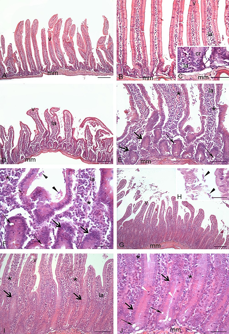
In the HFD+G1W group (Figure 8D), approximately half of the animals exhibited mild parasitism associated with mild to moderate inflammation (Figure 8E and F), while the other half showed moderate to intense parasitism and severe inflammation. In contrast, the HFD+G2W group (Figure 8G) displayed a significant increase in both parasitic intensity and inflammation compared to the HFD+G1W group (Figure 8H and I).

Duodenum of gerbils from the obese groups: uninfected (high-fat diet (HFD)), infected for 1 week (HFD+G1W), and infected for 2 weeks (HFD+G2W). A, HFD group. Duodenum with normal histological appearance. Scale bar: 200 µm. B, Higher magnification of panel A. Scale bar: 100 µm. C, Detail of panel B showing the lamina propria and intestinal crypts with normal cellularity. Scale bar: 50 µm. D, HFD+G1W group. Note the shortening of intestinal villi. Scale bar: 200 µm. E, Inflammatory infiltrate (*) and edema in the lamina propria. Hyperplastic intestinal crypts (thick arrows). Goblet cell hyperplasia (thin arrows). Scale bar: 100 µm. F, Higher magnification of panel E showing a predominantly mononuclear inflammatory infiltrate (*), crypt hyperplasia (thick arrows), and goblet cell hyperplasia (thin arrows). Scattered Giardia lamblia trophozoites in the intestinal lumen (arrowheads). Scale bar: 50 µm. G, HFD+G2W group. Greater number of shortened intestinal villi compared to the HFD+G1W group. Scale bar: 200 µm. H, Detail of panel G showing numerous Giardia lamblia trophozoites in the intestinal lumen (arrowheads). Scale bar: 50 µm. I, Higher magnification of panel H displaying shortened villi, markedly hyperplastic crypts (thick arrows), and a more intense inflammatory infiltrate than in the HFD+G1W group (*). Scale bar: 100 µm. J, Higher magnification of panel I showing hyperplastic crypts (thick arrows), mitotic figures in the crypts (white arrowheads), goblet cell hyperplasia (thin arrows), and a predominantly mononuclear inflammatory infiltrate (*). Scale bar: 50 µm. Intestinal villus (V); Muscularis mucosae (mm); lamina propria (la); intestinal crypt (cr). Hematoxylin and eosin.
Qualitatively, in the gerbils of the HFD+G2W group, the presence of many shortened villi was observed compared to the CD infected groups (Figure 7D and H and Figure 8G). The intestinal crypts of the gerbils in the CD+G1W, CD+G2W, HFD+G1W, and especially the HFD+G2W groups appeared hyperplastic, with a visible increase in depth, number of mitotic figures, and goblet cells (Figures 7E, G, I, and K and 8E and I).
In the CD+G1W group, shortening of intestinal villi (Figure 7D), expansion of the lamina propria with intense inflammatory infiltrate and crypt hyperplasia (Figure 7E), as well as moderate to intense parasitism (Figure 7F) and a predominantly mononuclear inflammatory infiltrate (Figure 7G) were observed.
Discussion
As expected, our results showed that the obese groups consumed more calories than the control groups, but, surprisingly, there was a restriction in caloric intake in the first week of infection, which was reversed in the second week, regardless of the diet. It is well described that immune function activation is energetically expensive due to the costs of generating fever, protein synthesis, and producing other components (20). The total energy cost of the entire immune system is about 20% of the resting metabolic rate but can increase 25-60% when activated (21). One benefit of reduced calorie intake during infection is that the consequent reduction of diet-induced thermogenesis increases the metabolic resources available to the immune system (20), suggesting that the host's behavioral response to infection offers a potential immunological benefit.
The changes resulting from obesity, such as hypertriglyceridemia, increased cytokines that induce lipolysis, increased local inflammation, and decreased adipokines that regulate inflammation, in addition to hepatic steatosis, were presented and well discussed by us (18). However, as the main objective of this study was to evaluate the influence of parasitic infection on this metabolic and immunological profile, the results must be analyzed in their entirety.
During infection, the first line of defense against invading pathogens is the innate immune response, which detects and limits infection. In this study, we observed that G. lamblia infection had different impacts depending on the nutritional status of the animals. In animals fed the control or high-fat diet, in the first week after inoculation, the infection caused deleterious effects on local tissue, confirmed by an increase in crypt depth and, consequently, a decrease in the villus:crypt ratio by more than 30%. Even though it is mostly an asymptomatic infection (22), giardiasis can, in some individuals, cause a series of mucosal damage such as apoptosis of enterocytes, disruption of tight junctions, followed by shortening of the villi concomitant with crypt hyperplasia (12).
The finding that a high-fat diet exacerbated intestinal damage aligns with growing evidence in the field. For instance, a high-fat diet in mice significantly increases the severity of Giardia infection, leading to a higher trophozoite burden and greater mucosal infiltration by inflammatory cells (23). This suggests that HFD-induced metabolic changes create an intestinal environment that is not only less resistant to physical damage but also more conducive to parasite survival and replication.
Diffuse shortening of epithelial microvilli may lead to reduced secretion of disaccharidases contributing to impaired lipid absorption and digestion and other clinical manifestations of the disease (24). In this study, however, we did not observe changes resulting from poor nutrient absorption, such as weight loss or excretion of lipids in feces in control animals infected for one or two weeks.
Animals receiving the control diet after one week of parasitic infection demonstrated systemic changes such as the deposition of fat microvesicles in the liver and a pro-inflammatory profile in adipose tissue, with increased TNF-α and decreased IL-10 and adiponectin compared to the uninfected CD group. The adipose tissue is composed of numerous cell types including monocytes, dendritic cells, mast cells, eosinophils, B cells, regulatory T cells, and CD8+ T cells, which are known as the vascular stroma fraction of adipose tissue (25). Considering that the majority are cells of the immune system, adipose tissue can also play an important role in the immune response, either through the direct effect of resident immune cells or indirectly by modulating the immune response in an endocrine and/or paracrine manner (26,27).
Despite the relevant changes triggered in the host organism after one week of infection, the animals in the control group were able to trigger a robust inflammatory response for more than one week, characterized mainly by increased production of the cytokines IL-2, IL-5, and IL-6 in the CD+G1w group compared to the uninfected CD group. Data regarding the host immune response to G. lamblia infection are extremely varied and controversial since studies use different experimental models and trophozoite strains. In our study, we observed that all animals in the control or obese infected groups presented increased intestinal concentrations of IL-6 at the initial moments of infection by G. lamblia (CD+G1w and HFD+G1w groups). The fundamental role of IL-6 in the induction of innate cellular responses and also in mediating the production of specific antibodies is already well described in giardiasis (27), and mice deficient in IL-6 are not able to eliminate the parasite (28).
The essential role of IL-5 in intestinal helminth infections is well described. IL-5 acts by inducing the activation of eosinophils that participate in the elimination of parasites through cytotoxic effects (29). In humans, it has also been demonstrated that eosinophilia is not limited to helminth infections but also acts in infections by protozoa, such as G. lamblia (30), and its association with allergic manifestations has been long-standing (31). We found, in addition to increased IL-5, eosinophils in the inflammatory infiltrate of the lamina propria, which may also have contributed to the suppression of the infection. Other cells, such as mast cells, may be relevant in this phase of the immune defense, given the importance of mast cells in the innate defense against giardiasis (13). It can be inferred that the presence of IL-5 at the beginning of the infection could also originate from these cells.
During immune response, IL-2 is rapidly produced, mainly by activated CD4+ T lymphocytes after antigen presentation by dendritic cells in secondary lymphoid organs, such as lymph nodes. In the present study, control animals after the first week of infection showed a greater secretion of this cytokine, which reduced after the second week of infection due to the possible activation of specific cells. The role of dendritic cells as antigen-presenting cells in giardiasis has been previously reported (32) in addition to their importance in the production of IL-6 (33). From the results, we can assume that the role of these cells in Giardia infection is not limited to the production of IL-6, but also of IL-2, considering the rapid increase in this cytokine observed in infected animals.
In contrast, the response to infection was impaired in the obese animals. The exact mechanism responsible for changes in the immune system of obese patients is unknown, but it may be related to the negative effects that nutritional, metabolic, and endocrine changes cause in the functions of different cells of the immune system (34). Obese animals were unable to establish an effective response to giardiasis seven days after infection. After the second week, parasitism increased markedly, as did the inflammatory infiltrate in the duodenal mucosa and submucosa, making epithelial lesions even more severe. This observation highlights that the low-grade, chronic inflammation associated with obesity appears to preemptively compromise the host's ability to mount a robust and sustained acute immune response, as suggested by the lack of sustained increases of IL-2 and IL-6. The resulting state of “premature exhaustion” leads to unchecked parasite replication and greater tissue pathology, thereby establishing a detrimental metabolic-immunological axis. After the first week of infection, there was no change in IL-5 and IL-13, and although IL-6 and IL-2 increased in the intestine, these increases were not sustained into the following week, nor was the antioxidant response observed in these animals. This suggests that the immune response in obese animals may enter a state of premature exhaustion and is unable to combat parasitism. Thus, after the second week of infection, obese animals presented greater damage such as increased parasitism and abundant inflammatory infiltrate, with consequent villus shortening and crypt hyperplasia, further increasing the villus:crypt ratio, which exceeded a 50% reduction in relation to the uninfected control.
In adipose tissue, giardiasis momentarily altered the inflammatory profile characteristic of obesity, significantly decreasing TNF-α levels in both infected groups (HFD+G1w and HFD+G2w) compared to the uninfected HFD group (consistent with Figure 5D), in addition to increasing the concentration of IL-10 in the HFD+G1w group compared to the uninfected HFD group. However, these animals were unable to reestablish normal adiponectin levels. Some studies report that obese individuals are more prone to infections, which tend to be more severe, with worse outcomes and increased post-surgical complications (35). Although obesity-related metabolic and inflammatory changes in adipose tissue are linked to disease development, it remains unclear whether adipose tissue directly contributes to host defense, despite containing hematopoietic lineage cells (36).
Leptin plays a key role in immune defense, as leptin-deficient (ob/ob) and leptin receptor-deficient (db/db) mice show increased susceptibility to bacterial, viral, and parasitic infections, and pneumonia due to impaired cytokine production (37). Intraperitoneal leptin injections prior to Streptococcus pneumoniae infection improved survival in ob/ob mice, but not in wild-type controls (37). Conversely, ob/ob mice infected with Plasmodium berghei did not develop cerebral malaria, likely due to the absence of leptin-mediated inflammatory responses (38).
Although many studies have shown that obesity impairs host defense against infections, the exact mechanisms remain unclear. We suggest that obesity may compromise the innate response to Giardia infection, preventing effective disease control. Additionally, the interaction between obesity and giardiasis may alter macrophage phenotype in adipose tissue, evidenced by reduced TNF-α and increased IL-10, a shift that could potentially contribute to weakened systemic immune defense. However, more research is needed to clarify how excess adipose tissue and metabolic changes affect immune function and contribute to greater susceptibility to infections like giardiasis.
The 14-day follow-up period adopted in this study was designed to encompass the critical phases of G. lamblia infection in Mongolian gerbils. The most significant immunological alterations occurred one week post-infection, coinciding with the peak parasite burden, and began to normalize by the second week. This observation window was therefore adequate to capture both the acute response and the early recovery stage, aligning with reports describing the temporal progression and resolution of Giardia infection in gerbils (39). Future studies should extend the follow-up period to explore chronic and recovery-associated outcomes.
Conclusions
While obesity did not affect weight gain or food intake, both of which were mainly influenced by the hypercaloric diet, our study demonstrated that it significantly worsened the impact of giardiasis. The combination of these conditions led to profound metabolic, immunological, and structural alterations in the small intestine and liver, including a detrimental shift in the adipose tissue cytokine profile (increased TNF-α and decreased IL-10), increased hepatic lipid accumulation and fecal fat excretion, and significant impairment in the host's ability to control G. lamblia infection, characterized by parasite persistence and severe tissue damage.
The main contribution of this study is the unprecedented finding that the obesity-associated inflammatory state not only coincided with but also actively compromised the innate immune response against Giardia lamblia at the intestinal level, creating a cycle of exacerbated disease progression.
Taken together, these data highlight the importance of controlling obesity to reduce the systemic inflammatory state that worsens the progression of infectious diseases, such as giardiasis. We believe this study can contribute to new public health initiatives, emphasizing the need for more effective policies for the prevention and control of obesity, especially in children, who are most affected by giardiasis, an infection that can cause serious cognitive and physical deficits in this age group. Future studies should focus on identifying the specific cellular and molecular targets in the adipose tissue that mediate this immune dysfunction, extending the follow-up period to explore chronic effects, and investigating whether pharmacological reversal of the inflammatory profile could restore effective parasite clearance.
Data Availability Statement
The data that support the findings of this study are available in the article. Further inquiries can be directed to the corresponding author.
-
Funding
This work was supported by Fundação de Apoio è Pesquisa do Estado de Minas Gerais - FAPEMIG (APQ 03545-18).
References
-
1 WHO (World Health Organization). Obesity and overweight. Updated 1 March 2024. http://www.who.int/mediacentre/factsheets/fs311/en/ Accessed July 2025.
» http://www.who.int/mediacentre/factsheets/fs311/en/ -
2 Badhwar R, Kaur G, Popli H, Yadav D, Buttar HS. Pathophysiology of obesity-related non-communicable chronic diseases and advancements in preventive strategies. In: Tappia P, Ramjiawan B, Dhalla N (Editors), Pathophysiology of obesity-induced health complications. Advances in biochemistry in health and disease, Springer; 2020, doi: 10.1007/978-3-030-35358-2_19.
» https://doi.org/10.1007/978-3-030-35358-2_19 -
3 Ling J, Chen S, Zahry NR, Kao TSA. Economic burden of childhood overweight and obesity: a systematic review and meta-analysis. Obes Rev. 2023;24(2):e13535, doi: 10.1111/obr.13535.
» https://doi.org/10.1111/obr.13535 -
4 Auger C, Kajimura S. Adipose tissue remodeling in pathophysiology. Annu Rev Pathol Mech Dis. 2023;18:71-93, doi: 10.1146/annurev-pathol-042220-023633.
» https://doi.org/10.1146/annurev-pathol-042220-023633 -
5 García-Rodríguez S, Espinosa-Cabello JM, García-González A, González-Jiménez E, Aguilar-Cordero MJ, Castellano JM, et al. Interplay of postprandial triglyceride-rich lipoprotein composition and adipokines in obese adolescents. Int J Mol Sci. 2024;25(2):1112, doi: 10.3390/ijms25021112.
» https://doi.org/10.3390/ijms25021112 -
6 Le Lay S, Simard G, Martinez MC, Andriantsitohaina R. Oxidative stress and metabolic pathologies: from an adipocentric point of view. Oxid Med Cell Longev. 2014; 2014:908539, doi: 10.1155/2014/908539.
» https://doi.org/10.1155/2014/908539 -
7 Zheng Y, Wang S, Wu J, Wang Y. Mitochondrial metabolic dysfunction and non-alcoholic fatty liver disease: new insights from pathogenic mechanisms to clinically targeted therapy. J Transl Med. 2023;21(1):510, doi: 10.1186/s12967-023-04367-1.
» https://doi.org/10.1186/s12967-023-04367-1 -
8 Pugliese G, Liccardi A, Graziadio C, Barrea L, Muscogiuri G, Colao A. Obesity and infectious diseases: pathophysiology and epidemiology of a double pandemic condition. Int J Obes (Lond). 2022;46(3):449-65, doi: 10.1038/s41366-021-01035-6.
» https://doi.org/10.1038/s41366-021-01035-6 -
9 Vicente B, De Freitas A, Freitas M, Midlej V. Systematic Review of diagnostic approaches for human giardiasis: unveiling optimal strategies. Diagnostics (Basel). 2024;14(4):364, doi: 10.3390/diagnostics14040364.
» https://doi.org/10.3390/diagnostics14040364 -
10 Savioli L, Smith H, Thompson A. Giardia and Cryptosporidium join the ‘neglected diseases initiative'. Trends Parasitol. 2006;22:203-8, doi: 10.1016/j.pt.2006.02.015.
» https://doi.org/10.1016/j.pt.2006.02.015 -
11 Singer SM, Angelova VV, DeLeon H, Miskovsky E. What's eating you? An update on Giardia, the microbiome and the immune response. Curr Opin Microbiol. 2020;58:87-92, doi: 10.1016/j.mib.2020.09.006.
» https://doi.org/10.1016/j.mib.2020.09.006 -
12 Solaymani-Mohammadi S. Mucosal defense against Giardia at the intestinal epithelial cell interface. Front Immunol. 2022;13:817468, doi: 10.3389/fimmu.2022.817468.
» https://doi.org/10.3389/fimmu.2022.817468 -
13 Katsoulis-Dimitriou K, Kotrba J, Voss M, Dudeck J, Dudeck A. Mast cell functions linking innate sensing to adaptive immunity. Cells. 2020;9(12):2538, doi: 10.3390/cells9122538.
» https://doi.org/10.3390/cells9122538 -
14 Araújo NS, Mundim MJS, Gomes MA, Amorim RMR, Viana JCV, Queiroz RP, et al. Giardia duodenalis: pathological alterations in gerbils, Meriones unguiculatus, infected with different dosages of trophozoites. Exp Parasitol. 2008;118(4):449-57, doi: 10.1016/j.exppara.2007.10.007.
» https://doi.org/10.1016/j.exppara.2007.10.007 -
15 Ribeiro MRS, Oliveira DR, Caliari MV, Machado DCC, Andrade MER, Cardoso VN, et al. Saccharomyces boulardii as therapeutic alternative in experimental giardiasis. J Appl Microbiol. 2021;131(1):460-9, doi: 10.1111/jam.14941.
» https://doi.org/10.1111/jam.14941 -
16 Gomes MA, Oliveira DR, de Freitas SE, Viana MP, Borges EL. Effect of giardiasis combined with low-protein diet on intestinal absorption of glucose and electrolytes in gerbils. Exp Parasitol. 2012;131(4):448-51, doi: 10.1016/j.exppara.2012.04.016.
» https://doi.org/10.1016/j.exppara.2012.04.016 -
17 Shukla G, Kamboj S, Sharma B. Comparative analysis of antigiardial potential of heat inactivated and probiotic protein of probiotic Lactobacillus rhamnosus GG in murine giardiasis. Probiotics Antimicrob Proteins. 2020;12(1):271-9, doi: 10.1007/s12602-018-9506-8.
» https://doi.org/10.1007/s12602-018-9506-8 -
18 Ventura LLA, Fortes NCL, Santiago HC, Caliari MV, Gomes MA, Oliveira DR. Obesity-induced diet leads to weight gain, systemic metabolic alterations, adipose tissue inflammation, hepatic steatosis, and oxidative stress in gerbils (Meriones unguiculatus). PeerJ. 2017;5:e2967, doi: 10.7717/peerj.2967.
» https://doi.org/10.7717/peerj.2967 -
19 Folch J, Lees M, Stanley S. A simple method for the isolation and purification of total lipids from animal tissues. J Biol Chem. 1957;226:497-509, doi: 10.1016/S0021-9258(18)64849-5.
» https://doi.org/10.1016/S0021-9258(18)64849-5 -
20 Schrock JM, Snodgrass JJ, Sugiyama LS. Lassitude: the emotion of being sick. Evol Hum Behav. 2020;41(1):44-57, doi: 10.1016/j.evolhumbehav.2019.09.002.
» https://doi.org/10.1016/j.evolhumbehav.2019.09.002 -
21 Surazakov A, Klassen A, Gizinger O. The bioenergetics of COVID-19 immunopathology and the therapeutic potential of biophysical radiances. J Photochem Photobiol B. 2020;213:112083, doi: 10.1016/j.jphotobiol.2020.112083.
» https://doi.org/10.1016/j.jphotobiol.2020.112083 -
22 Adam RD. Giardia duodenalis: Biology and pathogenesis. Clin Microbiol Rev. 2021;34(4):e0002419, doi: 10.1128/CMR.00024-19.
» https://doi.org/10.1128/CMR.00024-19 -
23 Klimczak S, Packi K, Rudek A, Wenclewska S, Kurowski M, Kurczabińska D, et al. The influence of the protozoan Giardia lamblia on the modulation of the immune system and alterations in host glucose and lipid metabolism. Int J Mol Sci. 2024;25(16):8627, doi: 10.3390/ijms25168627.
» https://doi.org/10.3390/ijms25168627 -
24 Koot BGP, ten Kate FJW, Juffrie M, Rosalina I, Taminiau JJAM, Benninga MA. Does Giardia lamblia cause villous atrophy in children? A retrospective cohort study of the histological abnormalities in giardiasis. J Pediatr Gastroenterol Nutr. 2009;49(3):304-8, doi: 10.1097/MPG.0b013e31818de3c4.
» https://doi.org/10.1097/MPG.0b013e31818de3c4 -
25 Corvera S. Cellular heterogeneity in adipose tissues. Annu Rev Physiol. 2021;83:257-78, doi: 10.1146/annurev-physiol-031620-095446.
» https://doi.org/10.1146/annurev-physiol-031620-095446 -
26 Sinton MC, Kajimura S. From fat storage to immune hubs: the emerging role of adipocytes in coordinating the immune response to infection. FEBS J. 2025;292(8):1868-83, doi: 10.1111/febs.17302.
» https://doi.org/10.1111/febs.17302 -
27 Serradell MC, Gargantini PR, Saura A, Oms SR, Rupil LL, Berod L, et al. Cytokines, antibodies, and histopathological profiles during Giardia infection and variant-specific surface protein-based vaccination. Infect Immun. 2018;86(6):e00773-17, doi: 10.1128/IAI.00773-17.
» https://doi.org/10.1128/IAI.00773-17 -
28 Singer SM, Fink MY, Angelova VV. Recent insights into innate and adaptive immune responses to Giardia Adv Parasitol. 2019;106:171-208, doi: 10.1016/bs.apar.2019.07.004.
» https://doi.org/10.1016/bs.apar.2019.07.004 -
29 Gazzinelli-Guimaraes PH, Jones SM, Voehringer D, Mayer-Barber KD, Samarasinghe AE. Eosinophils as modulators of host defense during parasitic, fungal, bacterial, and viral infections. J Leukoc Biol. 2024;116(6):1301-23, doi: 10.1093/jleuko/qiae173.
» https://doi.org/10.1093/jleuko/qiae173 -
30 Ustun S, Turgay N, Delibas SB, Ertabaklar H. Interleukin (IL) 5 levels and eosinophilia in patients with intestinal parasitic diseases. World J Gastroenterol. 2004;10(24):3643-6, doi: 10.3748/wjg.v10.i24.3643.
» https://doi.org/10.3748/wjg.v10.i24.3643 -
31 Di Prisco MC, Hagel I, Lynch NR, Jimenez JC, Rojas R, Gil M, et al. Association between giardiasis and allergy. Ann Allergy Asthma Immunol. 1998;81(3):261-5, doi: 10.1016/S1081-1206(10)62823-2.
» https://doi.org/10.1016/S1081-1206(10)62823-2 -
32 Castro-Sánchez P, Martín-Villa JM. Gut immune system and oral tolerance. Br J Nutr. 2013;109(Suppl 2):S3-S11, doi: 10.1017/S0007114512005223.
» https://doi.org/10.1017/S0007114512005223 -
33 Lee HY, Kim J, Noh HJ, Kim HP, Park SJ. Giardia lamblia binding immunoglobulin protein triggers maturation of dendritic cells via activation of TLR4-MyD88-p38, and ERK1/2 MAPKs. Parasite Immunol. 2014;36(12):627-46, doi: 10.1111/pim.12119.
» https://doi.org/10.1111/pim.12119 -
34 Moulin CM, Marguti I, Peron JPS, Rizzo LV, Halpern A. Impact of adiposity on immunological parameters. Arq Bras Endocrinol Metabol. 2009;53(2):183-9, doi: 10.1590/S0004-27302009000200010.
» https://doi.org/10.1590/S0004-27302009000200010 -
35 Kwong JC, Campitelli MA, Rosella LC. Obesity and respiratory hospitalizations during influenza seasons in Ontario, Canada: a cohort study. Clin Infect Dis. 2011;53(5):413-21, doi: 10.1093/cid/cir442.
» https://doi.org/10.1093/cid/cir442 -
36 Falagas ME, Kompoti M. Obesity and infection. Lancet Infect Dis. 2006;6(7):438-46, doi: 10.1016/S1473-3099(06)70523-0.
» https://doi.org/10.1016/S1473-3099(06)70523-0 -
37 Hsu A, Aronoff DM, Phipps J, Goel D, Mancuso P. Leptin improves pulmonary bacterial clearance and survival in ob/ob mice during pneumococcal pneumonia. Clin Exp Immunol. 2007;150(2):332-9, doi: 10.1111/j.1365-2249.2007.03491.x.
» https://doi.org/10.1111/j.1365-2249.2007.03491.x -
38 Robert V, Bourgouin C, Depoix D, Thouvenot C, Lombard MN, Grellier P. Malaria and obesity: obese mice are resistant to cerebral malaria. Malar J. 2008;7:81, doi: 10.1186/1475-2875-7-81.
» https://doi.org/10.1186/1475-2875-7-81 -
39 Gil FF, Ventura LLA, Fonseca JF, Saniago HC, Busatti H, Santos JFG, et al. Hematological profile in natural progression of giardiasis: kinetics of experimental infection in gerbils. J Infect Dev Ctries. 2018;12(6):492-498, doi: 10.3855/jidc.9626.
» https://doi.org/10.3855/jidc.9626
Edited by
-
Editor
Roberto César P. Lima Júniorhttps://orcid.org/0000-0002-7033-655X and Section Editor Pedro R.T. Romãohttps://orcid.org/0000-0002-1039-2509