Aerobic capacity evaluation of Sprague Dawley rats in treadmill running: comparison between protocols
Abstract
The objective of the present study was to compare and test the applicability of different protocols for accessing aerobic capacity in Sprague Dawley rats using treadmill running. Fifteen 70-day-old adult Sprague Dawley rats (270-290 g) were used. After 5 days of adaptation to the treadmill, the animals underwent 7 days of evaluations with a 48-h interval between each protocol. On the first two days, they underwent, in random order, a graded exercise test, with (GXT2) or without (GXT1) blood sample collections to determine blood lactate concentrations and the anaerobic threshold. In the subsequent 4 days, they underwent continuous 30-min efforts to determine the maximal lactate steady state (MLSS) with the intensity prescribed in percentages of the maximum speed (MaxS) obtained in GXT1, and on the last day they underwent the minimum lactate (ML) protocol. The MaxS obtained in GXT2 was higher than in GXT1, and there was a moderate correlation (r=0.614, P=0.011) between them. In many cases, lactate and glucose blood concentrations did not show the expected kinetics, making aerobic capacity determination impossible using these protocols. MLSS showed a higher success rate compared to other protocols (MLSS=80%; GXT2=47%; ML=60%). In conclusion, with the MLSS protocol, it is only possible to measure time to exhaustion at each intensity, which does not exactly reflect aerobic capacity, and the use of blood lactate and glucose concentrations to evaluate the aerobic capacity of rats in incremental and ML treadmill running protocols is still discouraged.
Exercise; Animal; Performance; Test; Aerobic
Introduction
The use of animal models is common in basic research and in exercise physiology studies when investigating the mechanisms induced by physical effort. The most commonly used animal models are rodents, rats, and mice, as they are small and easy to handle and the controlled conditions lead to low variation within the experimental groups and thus, enable the detection of small changes (1,2). The most commonly used physical exercise protocols are treadmill running and swimming, with the latter being the most used, both in studies that investigate the acute or chronic effects of physical exercise (3,4) and in protocols to determine effort intensity and exercise intensity in rats (1,5,6). The development of physical training protocols for animal models, both in swimming and running, is commonly based on parameters of aerobic capacity.
The most well-accepted protocol for aerobic capacity assessment, both in humans (7) and in animal models (8), is the maximal lactate steady state (MLSS), proposed by Beneke (9). The physiological basis of MLSS is comparable to the anaerobic threshold (AnT) theory (10), with MLSS characterized by the highest lactate blood concentration [La] that can be maintained in a steady state during submaximal, prolonged, and constant load exercise (10- 12). The intensity corresponding to MLSS represents the highest submaximal exercise intensity that can be maintained in a steady state, in which blood lactate ([La]) remains stable during exercise (10,11). However, several days of assessment are required to determine this intensity, which can be considered a disadvantage of this protocol.
Another evaluation protocol also widely used to access aerobic capacity in animal models is the minimum lactate (ML) protocol proposed by Tegtbur et al. (13), which consists of three distinct phases. First, high-intensity efforts are carried out to induce hyperlactacidemia (phase 1), and then an 8-min recovery is required for the lactate to be transferred from the muscle to the bloodstream, so it can be quantified (phase 2). After recovery, an incremental test begins until exhaustion (phase 3). Blood lactate is expected to decrease during the first stages of the incremental test and to decrease in subsequent stages, giving the lactate kinetics curve a “U” shape. The intensity corresponding to the lowest point of this curve represents the ML intensity, which is an estimate of the equilibrium between lactate production and removal, similar to the MLSS (13,14). However, caution is necessary when applying an evaluation protocol that uses lactacidemia as a parameter, as it is known that manipulation of the variables of an incremental test in humans can alter both maximum speed (MaxS) (15) and the metabolic thresholds (16). The same effect could be expected in rodents, but this is not yet clear.
There are other protocols for determining aerobic capacity that, unlike MLSS and ML, require only one evaluation session. Among these, the graded exercise tests (GXT, in which there are progressive intensity increases) are widely used to determine the AnT (17). Typically, in these protocols, an exponential increase in lactate is observed with increasing effort intensity, and AnT is obtained from the inflection point of the [La]/intensity relationship (17,18). In addition to AnT, a GXT can provide another widely used parameter, the MaxS, which is the highest running intensity obtained in an incremental treadmill test and is commonly used to prescribe the intensity of running exercise sessions for animal models (19). In studies carried out in humans, in addition to [La], glycemia is also commonly measured for assessing aerobic capacity, since blood glucose has a “U“-shape kinetics both in incremental protocols (20) and ML protocols with an increase in intensity (21).
Although protocols to assess aerobic capacity are established, they are difficult to apply as they require lactate concentration quantification. In an attempt to offer a viable alternative for exercise intensity evaluation and monitoring in animal models, the objective of the present study was to compare and test the applicability of different protocols for assessing aerobic capacity in Sprague Dawley rats using treadmill running.
Material and Methods
Animals
To calculate the appropriate sample size, an a priori power analysis was conducted using G*Power (version 3.1.9.7, Germany). An effect size of 0.53 was used based on the previous results from Manchado-Gobatto et al. (22), which showed a significant effect on [La] at four distinct intensities. With a target power of 95% and an alpha level of P<0.05, 11 rats were required. However, considering a possible sample loss, which in previous studies was 25% on average, we decided to use fifteen seven-week-old adult Sprague Dawley rats (270-290 g), obtained from the Central Animal Facility of USP (Ribeirão Preto campus). Throughout the experiment, the animals were kept in collective cages, with four rats per cage and on a 12-h light/dark cycle. The experiments were carried out in the morning by the same researchers who maintained the animals in the cages. All animals received commercial food (Nuvilab, Brazil) and water ad libitum. The animals' food intake was controlled by weighing them daily before each procedure. All procedures in the present study were submitted and approved by the Ethics and Research Committee for the Use of Animals of the School of Physical Education and Sports of Ribeirão Preto (protocol No. 2016.1.462.90.7).
Experimental design
All assessments were carried out on a treadmill (Gesam, Brazil), which has individual bays measuring 60×10×6.5 cm. The animals were adapted to the exercise for 5 days by running 10 to 60 min/day (Table 1). Seventy-two hours after adaptation, the animals were subjected to incremental evaluation protocols (GXT1 and GXT2) to determine AnT and MaxS, separated by 48 h. The order in which these protocols were carried out was randomized. Forty-eight hours after the second incremental protocol, the animals were subjected to 4 continuous efforts with constant loads separated by 48 h to determine the MLSS. Forty-eight hours after the last continuous effort, the animals were subjected to the ML test (Figure 1). For the GXT2, MLSS, and ML protocols, the success rate was calculated by dividing the number of tests that could be used to determine aerobic capacity by the number of tests in each protocol.
Protocol for adapting animals to treadmill exercise. The graded exercise test was applied on day 5.

Chronological diagram of procedures. GXT: graded exercise test; MLSS: maximal lactate steady state; ML: minimum lactate test; MaxS: maximum speed.
Collection and analysis of blood samples
Blood samples (25 μL) were collected by calibrated glass capillaries containing heparin from the distal end of the animals' tails and placed in microtubes containing 50 μL of sodium fluoride (1% NaF), homogenized, and kept at -4°C. [La] and glucose were determined using a Yellow Spring Instruments (YSI) Electrochemical Lactimeter, model 2300 Stat (Hong Kong).
Graded exercise test 1 (GXT1) to determine the maximum running speed (MaxS)
Before the incremental test and exercise stimuli, the animals remained for 10 min in their respective treadmill stall for acclimatization. MaxS was determined during an incremental test with an initial speed of 8 m/min and increments of 3 m/min every 3 min until exhaustion. The exhaustion criterion used was the loss of the running pattern (i.e., interruption of the locomotion pattern, even with the application of stimuli). This same criterion was also adopted in the other evaluations. The maximum speed was determined from the equation:
where CS is the speed of the last completed stage, I is the velocity increments between stages, time is the time in the stage in which exhaustion occurred in seconds, and the constant 180 refers to the duration (in seconds) of the test stages (23).
Graded exercise test 2 (GXT2) to determine the anaerobic threshold (AnT)
To determine AnT, we used the same protocol used in GXT1, but with blood collections at rest and at the end of each stage. AnT was determined using the bi-segmented method (24- 26) by two experienced evaluators. In case of discrepancy, a third evaluator made the determination. To determine the glycemic threshold, a second-order polynomial adjustment was applied to the relationship between glucose and GXT intensity, and the glucose minimum was considered as the minimum point or zero derivative of the curve formed.
Maximal lactate steady state (MLSS)
To determine the MLSS, the animals were subjected to 4 continuous efforts of 30 min each with intervals of 48 h between efforts at intensities corresponding to 60, 70, 80, and 90% of the MaxS obtained at GXT1, since the starting point for the first evaluation session is closer to real MLSS intensity than the determination of the intensities. With this strategy, fewer sessions are required. We based the choice of these intensities on the study by Ferreira et al. (19), which showed that MLSS intensity corresponded to 60% of MaxS, obtained in a protocol similar to GXT1. The order in which these efforts were carried out was randomized. Blood collections were performed every 10 min. The intensity corresponding to MLSS was considered the highest effort intensity at which the difference in [La] between the 30th and 10th was less than 1 mM (5).
Minimum lactate (ML) test
We used the hyperlactacidemia induction phase of the ML test following the protocol used by de Araújo et al. (1), which consists of two efforts, the first lasting 30 s and the second after 30 s of passive recovery until exhaustion. The intensity of these efforts was fixed at 20 m/min. This speed represents 121, 106, and 108% of the MaxS of these animals in the GXT1, GXT2, and ML protocols, respectively. After the induction phase, the animals remained in passive recovery for 8 min, and blood samples were collected in the 3rd, 5th, and 7th min of recovery. Immediately after 8 min, the animals were subjected to an incremental protocol similar to GXT2. From both the [La] and glucose values, a second-order polynomial adjustment was applied to the relationship of these variables and the intensity of the GXT and the ML point was considered to be the minimum point or zero derivative of the formed curve.
Statistical analysis
Data are reported as means±SD. Data normality was measured and confirmed using the Shapiro-Wilk test. To compare the methods, one-way repeated measures ANOVA and the Pearson correlation coefficient (r) test were used to assess possible correlations. The significance level was set at P<0.05.
Results
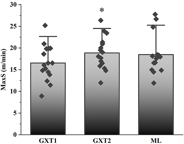
The MaxS achieved in the incremental tests was 16.5±4.1, 18.9±3.8, and 18.5±4.5 m/min, and the time to exhaustion was 11.2±3.9, 13.8±3.7, and 13.5±4.4 min in the GXT1, GXT2, and ML protocols, respectively. The MaxS in GXT2 was higher than in GXT1 (P<0.05) (Figure 2). Furthermore, a moderate positive correlation was found between GXT1 and GXT2 (P<0.05) (Table 2).
Maximum speed (MaxS) reached in incremental protocols. Data are reported as means and SD. *P<0.05 compared to GST1; ANOVA. GXT1: incremental test without pauses for blood collections; GXT2: incremental test with pauses for blood collections; ML: minimum lactate test.
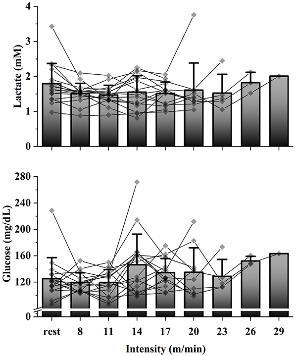
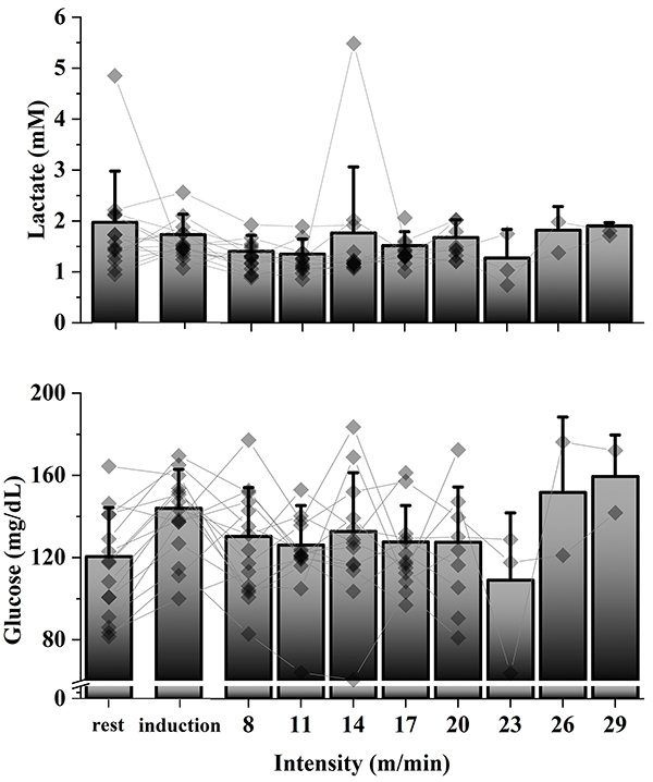
Figures 3 and 4 show the individual values and average at each point of lactate and glucose in the GXT2 and ML protocols, respectively. By a visual inspection of these two variables, we obtained a low success rate in accessing the aerobic capacity in these protocols (Table 3).
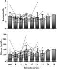
Mean and individual lactate and glucose values from the graded exercise test 2. Each line represents an individual animal.
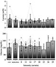
Average and individual lactate and glucose values from the minimum lactate test. Each line represents an individual animal.
Discussion
The methods used in the present study sought to define the relationships between aerobic capacity assessment protocols and the use of GXT without blood collection to determine lactacidemia as a reliable method for evaluating and controlling the intensity through these protocols in an animal model based on its relationship with gold standard tests (i.e., MLSS). Our results point to a large variation between the results of the different protocols, mainly due to the low success rate in determining the aerobic capacity and the fact that [La] did not present the expected behavior.
Variables that represent aerobic capacity and power from assessment protocols are extremely important for individual prescription and quantification of training loads. The most accurate method for assessing the aerobic capacity is MLSS, so this protocol can be used for this purpose as well as for other experimental protocol standardization. However, this method has a limiting factor, which is the relatively high number of assessments required, which makes its use unfeasible for prescription, monitoring, and measuring protocols. In the present study, a satisfactory success rate (80%) was observed for the MLSS protocol based on the pre-established criteria, showing that this protocol could be used for assessing aerobic capacity in rats. Based on the [La] found in the other protocols, it is not certain that increases in intensity reflect [La] increases as well, although other studies point to the effectiveness of this protocol in an animal model in running exercise (22,27) and in swimming (6).
The most commonly used alternative protocols to assess aerobic capacity (e.g., incremental protocol and minimum lactate) require moments of high-intensity effort and blood collections to measure lactate and glucose. Behavioral studies indicate that these conditions can increase stress levels and, consequently, lead to inflammatory conditions in animals such as serum high-sensitive C-reactive protein, tumor necrosis factor α, and adiponectin (28). However, these protocols have the main advantage of requiring only one session, as well as the possibility of identifying a maximum effort parameter in the same test. We tested two incremental protocols and the ML protocol to determine the AnT, and from these tests we also obtained MaxS, a parameter commonly used to prescribe exercise in animal models. We used a protocol similar to that used by Ferreira et al. (19) in mice at GXT1, but we decided to use the initial speed of 8 m/min, following a recent standardized evaluation protocol for rats (29).
We did not observe any difference in the MaxS obtained in the ML protocol compared to the incremental protocols. However, the MaxS was lower for GXT1 than for GXT2. It is logical to consider that this difference was due to the fact that at GXT2 there were small breaks (considered recovery breaks) for blood collection, which may have contributed to the better performance. Even so, the values obtained in GXT1 and GXT2 showed a moderate correlation. Ferreira et al. (19) showed that MLSS in mice is on average 60% of MaxS, while in the present study, the intensity corresponding to MLSS was 74% of MaxS. Due to the low success rate in assessing aerobic capacity in GXT1 and ML, the percentage of their corresponding intensities may not represent the reality for this group of animals.
We sought to identify the intensities corresponding to AnT using the GXT2 and ML tests, in which an exponential increase in lactate and a U-shaped kinetics, respectively, were expected, as occurs in humans (30- 32) or even in rats in swimming (33). However, blood lactate did not show the expected kinetics in these protocols, making it impossible to analyze and identify the intensity corresponding to AnT in many cases of the present study using these methods. Langfort et al. (34) and Soya et al. (35) successfully identified the expected kinetics of lactate increase in an incremental running protocol in Wistar rats. However, Carvalho et al. (36) found [La] kinetics similar to that of the present study in female Wistar rats during treadmill running. Some studies were successful in assessing aerobic capacity using the minimum lactate test in swimming in rats (1,37) and mice (5), but there are no studies that had the same success when running. Therefore, caution is needed when choosing the type of physical exercise in which these protocols will be applied, since lactate may not present the expected kinetics during running. We can speculate that running exercise may cause some additional physiological stress to the animals, so that lactate analysis is impaired. A possible reason for this could be the exhaustion caused by the exercise-induced changes in the core body temperature of rats (38,39).
In the present study, blood glucose, as lactate, also did not show the expected kinetics, making aerobic capacity assessment impossible based on this variable. There are no studies that measured blood glucose in rodents for this purpose. Therefore, based on the results of the present study, the use of glucose for the assessment of aerobic capacity is discouraged.
Some possible limitations regarding the present study are: i) the GXT protocols vary widely in the literature, which limits the comparison amongst studies; ii) the fact there were no breaks between stages in GXT1 made its comparison with GXT2 difficult. Considering the difficulties found in the present study, future studies are needed to find practical and reliable protocols for aerobic capacity assessment in running models in rats.
Conclusion
In conclusion, with the MLSS protocol, it is only possible to measure time to exhaustion at each intensity, which does not exactly reflect aerobic capacity. The use of [La] and glucose for aerobic capacity assessment in incremental and ML protocols is still discouraged, but the MaxS obtained in these protocols is an interesting parameter that can be used in evaluation protocols and subsequent prescriptions for running intensity in rats.
Acknowledgments
We want to thank Professor Claudia Maria Padovan, supervisor of the Laboratory of Neurobiology of Stress and Depression - FFCLRP/USP, for technical support.
-
Funding
This study was financed in part by the Coordenação de Aperfeiçoamento de Pessoal de Nível Superior - Brasil (CAPES, Finance Code 001) and by Fundação de Amparo è Pesquisa do Estado de São Paulo (FAPESP grant #2017/27042-6).
References
-
1 de Araújo GG, Papoti M, Manchado FB, de Mello MAR, Gobatto CA. Protocols for hyperlactatemia induction in the lactate minimum test adapted to swimming rats. Comp Biochem Physiol A Mol Integr Physiol. 2007 Dec;148(4):888-892, doi: 10.1016/j.cbpa.2007.09.002.
» https://doi.org/10.1016/j.cbpa.2007.09.002 -
2 Wang Y, Wisloff U, Kemi OJ. Animal models in the study of exercise-induced cardiac hypertrophy. Physiol Res. 2010;59(5):633-644, doi: 10.33549/physiolres.931928.
» https://doi.org/10.33549/physiolres.931928 -
3 de Araujo GG, Papoti M, Delbin MA, Zanesco A, Gobatto CA. Physiological adaptations during endurance training below anaerobic threshold in rats. Eur J Appl Physiol. 2013 Jul;113(7):1859-1870, doi: 10.1007/s00421-013-2616-9.
» https://doi.org/10.1007/s00421-013-2616-9 -
4 Freitas JS, Carneiro-Junior MA, Franco FSC, Rezende LS, dos Santos AS, Maia HO, et al. Treinamento aeróbio em natação melhora a resposta de parâmetros metabólicos de ratos durante teste de esforço [in Portuguese]. Rev Bras Med Esporte. 2010;16(2):134-138, doi: 10.1590/S1517-86922010000200012.
» https://doi.org/10.1590/S1517-86922010000200012 -
5 Rodrigues NA, Torsoni AS, Fante T, Dos Reis IGM, Gobatto CA, Manchado-Gobatto FB. Lactate minimum underestimates the maximal lactate steady-state in swimming mice. Appl Physiol Nutr Metab. 2017 Jan;42(1):46-52, doi: 10.1139/apnm-2016-0198.
» https://doi.org/10.1139/apnm-2016-0198 -
6 Gobatto CA, de Mello MAR, Sibuya CY, de Azevedo JRM, dos Santos LA, Kokubun E. Maximal lactate steady state in rats submitted to swimming exercise. Comp Biochem Physiol A Mol Integr Physiol. 2001 Aug;130(1):21-27, doi: 10.1016/S1095-6433(01)00362-2.
» https://doi.org/10.1016/S1095-6433(01)00362-2 -
7 Iannetta D, Ingram CP, Keir DA, Murias JM. Methodological reconciliation of CP and MLSS and their agreement with the maximal metabolic steady state. Med Sci Sports Exerc. 2022 Apr 1;54(4):622-632, doi: 10.1249/MSS.0000000000002831.
» https://doi.org/10.1249/MSS.0000000000002831 -
8 Almeida JA, Petriz BA, Gomes CP, Rocha LAO, Pereira RW, Franco OL. Determination of the maximal lactate steady state in obese Zucker rats. Int J Sports Med. 2013 Mar;34(3):214-217, doi: 10.1055/s-0032-1316360.
» https://doi.org/10.1055/s-0032-1316360 -
9 Beneke R. Anaerobic threshold, individual anaerobic threshold, and maximal lactate steady state in rowing. Med Sci Sports Exerc. 1995 Jun;27(6):863-867, doi: 10.1249/00005768-199506000-00010.
» https://doi.org/10.1249/00005768-199506000-00010 -
10 Beneke R, Hutler M, Von Duvillard SP, Sellens M, Leithäuser RM. Effect of test interruptions on blood lactate during constant workload testing. Med Sci Sports Exerc. 2003;35(9):1626-1630, doi: 10.1249/01.MSS.0000084520.80451.D5.
» https://doi.org/10.1249/01.MSS.0000084520.80451.D5 - 11 Baron B, Dekerle J, Depretz S, Lefevre T, Pelayo T. Self selected speed and maximal lactate steady state speed in swimming. J Sports Med Phys Fitness. 2005 Mar;45(1):1-6.
-
12 Loures JP, Chamari K, Ferreira EC, Campos EZ, Zagatto AM, Milioni F, et al. Specific determination of maximal lactate steady state in soccer players. J Strength Cond Res. 2015 Jan;29(1):101-106, doi: 10.1519/JSC.0000000000000621.
» https://doi.org/10.1519/JSC.0000000000000621 - 13 Tegtbur U, Busse MW, Braumann KM. Estimation of an individual equilibrium between lactate production and catabolism during exercise. Vol. 25, Med Sci Sports Exerc. 1993 May;25(5):620-627.
-
14 Messias LHD, Gobatto CA, Beck WR, Manchado-Gobatto FB. The lactate minimum test: concept, methodological aspects and insights for future investigations in human and animal models. Front Physiol. 2017 Jun 8;8:389, doi: 10.3389/fphys.2017.00389.
» https://doi.org/10.3389/fphys.2017.00389 -
15 Jamnick NA, Botella J, Pyne DB, Bishop DJ. Manipulating graded exercise test variables affects the validity of the lactate threshold and [Formula: see text]. PLoS One. 2018 Jul 30;13(7):e0199794, doi: 10.1371/journal.pone.0199794.
» https://doi.org/10.1371/journal.pone.0199794 -
16 Iannetta D, Azevedo RA, Keir DA, Murias JM. Establishing the V̇O2 versus constant-work-rate relationship from ramp-incremental exercise: simple strategies for an unsolved problem. J Appl Physiol (1985). 2019 Dec 1;127(6):1519-1527, doi: 10.1152/japplphysiol.00508.2019.
» https://doi.org/10.1152/japplphysiol.00508.2019 -
17 Faude O, Kindermann W, Meyer T. Lactate threshold concepts: how valid are they? Sports Med. 2009;39(6):469-490, doi: 10.2165/00007256-200939060-00003.
» https://doi.org/10.2165/00007256-200939060-00003 -
18 Binder RK, Wonisch M, Corra U, Cohen-Solal A, Vanhees L, Saner H, et al. Methodological approach to the first and second lactate threshold in incremental cardiopulmonary exercise testing. Eur J Prev Cardiol. 2008 Dec;15(6):726-734, doi: 10.1097/HJR.0b013e328304fed4.
» https://doi.org/10.1097/HJR.0b013e328304fed4 -
19 Ferreira JCB, Rolim NPL, Bartholomeu JB, Gobatto CA, Kokubun E, Brum PC. Maximal lactate steady state in running mice: effect of exercise training. Clin Exp Pharmacol Physiol. 2007 Aug;34(8):760-765, doi: 10.1111/j.1440-1681.2007.04635.x.
» https://doi.org/10.1111/j.1440-1681.2007.04635.x -
20 Webster T, Harber V, Bell R, Bell G. Hormonal responses associated with the nadir in blood glucose during graded cycling exercise. J Exerc Sci Fitness. 2013;11(1):6-11, doi: 10.1016/j.jesf.2012.12.002.
» https://doi.org/10.1016/j.jesf.2012.12.002 -
21 Junior PB, Neiva CM, Denadai BS. Effect of an acute 13-adrenergic blockade on the blood glucose response during lactate minimum test. J Sci Med Sport. 2001 Sep;4(3):257-265, doi: 10.1016/S1440-2440(01)80035-9.
» https://doi.org/10.1016/S1440-2440(01)80035-9 - 22 Manchado-Gobatto FB, Gobatto CA, Contarteze RVL, Mello MAR. Non-exhaustive test for aerobic capacity determination in running rats. Indian J Exp Biol. 2011 Oct;49(10):781-785.
-
23 Kuipers H, Verstappen FTJ, Keizer HA, Geurten P, van Kranenburg G. Variability of aerobic performance in the laboratory and its physiologic correlates. Int J Sports Med. 1985 Aug;6(4):197-201, doi: 10.1055/s-2008-1025839.
» https://doi.org/10.1055/s-2008-1025839 -
24 Papoti M, Vitório R, de Araújo GG, DA Silva ASR, Santhiago V, Martins LEB, et al. Determination of force corresponding to maximal lactate steady state in tethered swimming. Int J Exerc Sci. 2009 Oct 15;2(4):269-279, doi: 10.70252/JJCI8675.
» https://doi.org/10.70252/JJCI8675 -
25 Lima MCS, Balikian P, Gobatto CA, Garcia JR, Ribeiro LFP. Proposta de teste incremental baseado na percepção subjetiva de esforço para determinação de limiares metabólicos e parâmetros mecânicos do nado livre [in Portguese]. Rev Bras Med Esporte. 2006;12(5):268-274, doi: 10.1590/S1517-86922006000500009.
» https://doi.org/10.1590/S1517-86922006000500009 - 26 Kalva-Filho CA. Validação de um teste específico para determinação do máximo déficit acumulado de oxigênio (MAOD) e da máxima fase estável de lactato em nado atado. PhD Thesis. Universidade Estadual Paulista; 2013.
-
27 De Manchado FB, Gobatto CA, Contarteze RVL, Papoti M, Rostom De Mello MA. Maximal lactate steady state in running rats. J Exerc Physiol Online. 2005;8(5):29-35, doi: 10.1111/j.1440-1681.2007.04635.x.
» https://doi.org/10.1111/j.1440-1681.2007.04635.x -
28 de Lemos ET, Pinto R, Oliveira J, Garrido P, Sereno J, Mascarenhas-Melo F, et al. Differential effects of acute (extenuating) and chronic (training) exercise on inflammation and oxidative stress status in an animal model of type 2 diabetes mellitus. Mediators Inflamm. 2011;2011:253061, doi: 10.1155/2011/253061.
» https://doi.org/10.1155/2011/253061 -
29 de Carvalho CD, Kalva-Filho CA, Da Rocha AL, Silva ASR, Papoti M. Glycogen kinetics of wistar rats: different exercise intensities and tissue analyzed influence. Int J Exerc Sci. 2022 Feb 1;15(2):289-299, doi: 10.70252/DAEM9945.
» https://doi.org/10.70252/DAEM9945 -
30 Jones AM, Doust JH. The validity of the lactate minimum test for determination of the maximal lactate steady state. Med Sci Sports Exerc. 1998 Aug;30(8):1304-1313, doi: 10.1097/00005768-199808000-00020.
» https://doi.org/10.1097/00005768-199808000-00020 -
31 Podolin DA, Munger PA, Mazzeo RS. Plasma catecholamine and lactate response during graded exercise with varied glycogen conditions. J Appl Physiol (1985). 1991 Oct;71(4):1427-1433, doi: 10.1152/jappl.1991.71.4.1427.
» https://doi.org/10.1152/jappl.1991.71.4.1427 -
32 Buono MJ, Clancy TR, Cook JR. Blood lactate and ammonium ion accumulation during graded exercise in humans. J Appl Physiol Respir Environ Exerc Physiol. 1984 Jul;57(1):135-139, doi: 10.1152/jappl.1984.57.1.135.
» https://doi.org/10.1152/jappl.1984.57.1.135 -
33 Voltarelli FA, Gobatto CA, de Mello MAR. Determination of anaerobic threshold in rats using the lactate minimum test. Braz J Med Biol Res. 2002 Nov;35(11):1389-1394, doi: 10.1590/S0100-879X2002001100018.
» https://doi.org/10.1590/S0100-879X2002001100018 -
34 Langfort J, Zarzeczny R, Wiestaw P, Kaciuba-Uściłko H, Nazar K, Porta S. Effect of sustained hiperadrenalinemia on execise performance and lactate threshlod in rats. Comp Biochem Physiol A Physiol. 1996 May;114(1):51-55, doi: 10.1016/0300-9629(95)02087-X.
» https://doi.org/10.1016/0300-9629(95)02087-X -
35 Soya H, Mukai A, Deocaris CC, Ohiwa N, Chang H, Nishijima T, et al. Threshold-like pattern of neuronal activation in the hypothalamus during treadmill running: establishment of a minimum running stress (MRS) rat model. Neurosci Res. 2007 Aug;58(4):341-348, doi: 10.1016/j.neures.2007.04.004.
» https://doi.org/10.1016/j.neures.2007.04.004 -
36 Carvalho JF, Masuda MO, Pompeu FAMS. Method for diagnosis and control of aerobic training in rats based on lactate threshold. Comp Biochem Physiol A Mol Integr Physiol. 2005 Apr;140(4):409-413, doi: 10.1016/j.cbpb.2004.12.002.
» https://doi.org/10.1016/j.cbpb.2004.12.002 -
37 Beck WR, de Araujo GG, Gobatto CA. Methods of exercise intensity and lactataemia determination of lactate minimum test in rats. Comp Exerc Physiol. 2012;8(2):113-116, doi: 10.3920/CEP12004.
» https://doi.org/10.3920/CEP12004 -
38 Andrade MT, Barbosa NHS, Souza-Junior RCS, Fonseca CG, Damasceno WC, Regina-Oliveira K, et al. Determinants of body core temperatures at fatigue in rats subjected to incremental-speed exercise: The prominent roles of ambient temperature, distance traveled, initial core temperature, and measurement site. Int J Biometeorol. 2023 May;67(5):761-775, doi: 10.1007/s00484-023-02453-z.
» https://doi.org/10.1007/s00484-023-02453-z - 39 Andrade MT, Goulart KNO, Barbosa NHS, Soares DD, Andrade AGP, Gonçalves DAP, et al. Core body temperatures of rats subjected to treadmill exercise to fatigue or exhaustion: the journal Temperature toolbox. Temperature (Austin). 2023 Sep 1;10(3):287-312.