Antibiofilm activity of silver nanoparticles against Candida sp. isolated from the oral cavity
Abstract
Candida biofilm is difficult to control due to the poor penetration and unspecificity of antifungal drugs against the microorganisms associated with this structure. Nanoparticles have been investigated for their antimicrobial potential. In this context, the present study evaluated the antibiofilm activity of silver nanoparticle colloids (AgNPs) against candida isolated from the oral cavity. AgNPs were prepared in two different ethanolic syntheses - without and with ammonium hydroxide: AgNP-1 and AgNP-2, respectively. AgNPs were characterized by atomic absorption spectroscopy (AAS), UV-visible (UV-vis) absorption electron spectroscopy, zeta potential, dynamic light scattering (DLS), and transmission electron microscopy (TEM). Subsequently, they were evaluated against biofilm formation by clinical isolates of Candida albicans and Candida krusei by determining the minimum inhibitory concentration (MIC), minimum fungicidal concentration (MFC), and minimum biofilm formation inhibitory concentration (MBIC50). AgNPs inhibited biofilm formation of tested C. albicans and C. krusei and showed antibiofilm activity at subinhibitory concentrations against all yeasts. AgNP-1 inhibited biofilm formation at concentrations between 7.55 and 60.46 µg/mL and AgNP-2 between 7.71 and 30.80 µg/mL. The characterization of AgNP showed that AgNP-1 and AgNP-2 differ mainly in size and dispersion, with AgNP-2 being monodisperse, indicating that these characteristics could be related to the activity against the formation of C. albicans and C. krusei biofilm, and the silver nanoparticles may represent innovative and complementary alternatives to the available antifungal arsenal.
Key words:
Biofilm; Yeast; Colloidal dispersion; Candida krusei; Candida albicans; Antifungal
Introduction
Currently, around 150 species of candida have been described, and although infections caused by C. albicans constitute the majority of cases, diseases caused by other species are becoming increasingly frequent (1). Candida coexists in the normal microbiota of human mucous membranes and skin. However, due to certain conditions, they increase their affinity for mucous membranes and behave as pathogens in the oral cavity, gastrointestinal, respiratory, and urogenital tracts (2,3).
Candida species have several virulence factors, including metabolic adaptability, the ability to alter morphology, expression of surface molecules, secretion of hydrolytic enzymes, and biofilm formation (1). The ability to form biofilms is a relevant factor in Candida's virulence. It protects the fungus from the host's immune responses, limits the penetration of substances through the matrix, and consequently increases resistance to antifungal drugs (4). Species of the genus Candida are among the yeasts of greatest medical interest and have the greatest capacity to form biofilms, which make them responsible for around 90% of systemic mycoses and are the fourth most common cause of blood infections in the hospital environment (4).
Biofilm is a structured microbial community interlocked by a protective extracellular matrix and attached to a biotic or abiotic surface. The phenomenon of Candida spp. biofilm formation has been observed on surfaces such as mucous membranes, blood vessels, and medical devices that come into direct contact with the patient's body, such as catheters, endotracheal tubes, and implants (4,5).
As resistance to conventional antifungals is inherent in Candida spp. biofilms, the treatment of fungal infections associated with this structure, is particularly challenging. The mechanisms of antifungal resistance are varied and complex, but they are attributed to the biofilm's three-dimensional structure, efflux pumps, and persistent cells associated with the biofilm (5).
Silver nanoparticle colloids (AgNPs) are active against yeasts resistant to conventional antifungals, have shown broad-spectrum activity, low resistance induction potential, and have received attention as antibiofilm materials (6,7). The addition of nanomaterials with antibiofilm activity to dental surfaces is another interesting line of research (8). AgNPs can also act synergistically and potentiate active compounds against biofilms, allowing for more efficient use of antimicrobial agents (9).
AgNPs are promising microbial agents in the treatment and control of infectious diseases, as they are active against yeasts due to the release of ions and oxidative stress, damage to the cell membrane and wall, inhibition of enzymatic activity, regulation of gene expression, reduction of ATP, DNA, and protein content, and mitochondrial dysfunction (10,11).
Fungal infections caused by Candida species represent a clinical challenge due to their ability to form biofilm that blocks the action of antifungals and increases the cases of recurrent infections. AgNPs have been studied for their antimicrobial and antifungal potential in combating resistant microorganisms. In this context, it is necessary to investigate the activity of AgNPs against yeasts of the genus Candida, particularly the inhibition of biofilm formation and the correlation of inhibitory activity with the characteristics of the synthesized nanoparticles.
Material and Methods
This quantitative experimental study used a total of seven yeasts isolated from the oral cavity of volunteers (UEG Research Ethics Committee, No. 3.604.978); four were identified as Candida krusei, two as Candida albicans, and the standard strain Candida albicans ATCC 10231 all from the microorganism collection of the Bioassay Laboratory of the Research and Postgraduate Center (LabBio-CPPG) of the State University of Goiás.
Synthesis and characterization of AgNPs
AgNPs were synthesized through the alcoholic reduction of silver using ethanol as a reducing agent and polyvinylpyrrolidone (PVP) as a stabilizer according to the methodology described by Lee and Oh (12). Briefly, silver nitrate (89.5 mmol/L) and PVP (89.5 mmol/L, Mw 10,000) were dissolved separately in 50 mL of ethanol (pa). The two solutions (silver nitrate and PVP) were combined and kept under reflux and constant magnetic stirring for four hours at 80°C, forming a stable AgNP-1. For the synthesis of AgNP-2, 0.3 mL of 28% (v/v) ammonium hydroxide was added to the reaction system when the temperature of 80°C was reached, aiming to increase the conversion of Ag+ ions into Ag° atoms, accelerating the ethanol oxidation reaction and influencing the size and distribution of silver nanoparticles.
The synthesized AgNPs were characterized by determining the concentration of silver ions using atomic absorption apectroscopy (AAS) with an AAnalyst 400 spectrophotometer (Perkin Elmer, USA). UV-visible (UV-Vis) spectra were obtained on a 5100 ultraviolet/visible spectrophotometer (Metash, China). The hydrodynamic size (by dynamic light scattering (DLS) and zeta potential were determined using a Zetasizer Pro Blue unit (Malvern Panalytical, UK). Transmission electron microscopy (TEM) and selected area electron diffraction (SAED) studies were performed using a JEOL JEM-2010 200 kV ultra-high-resolution analytical electron microscope (Japan).
The results were organized in the Google Sheets application (Google, USA), which allowed the construction of graphs and statistical analyses in OriginPro 8.5 (Originlab, USA). The images obtained from TEM were analyzed with ImageJ (National Institutes of Health, USA).
Biofilm formation assay
Biofilm formation was assessed by detecting total biomass using the crystal violet method described by Zayed et al. (13), with some modifications, and by counting the number of yeasts associated with the biofilm. Yeast inocula were obtained by suspending colonies grown in a sterile physiological solution (SPS) and adjusting the turbidity using the 0.5 McFarland scale. Then, 500 µL of the inocula was transferred to tubes containing 4500 µL of bovine heart and brain infusion broth with 2% sucrose (BHIS), and 200 µL of the inocula were aseptically transferred to 96-well polystyrene microplates with a flat bottom (Cralplast, Cral, Brazil), which were incubated at 35.5°C for 48 h.
After incubation, the microplates were processed by removing the broth with total growth, washing the wells, fixing the biofilms with 96° ethanol, and staining with 0.1% crystal violet. The absorbance (620 nm) of the adhered biofilms was then read on a microplate spectrophotometer (Multiskan FC, Thermo Scientific, USA). The average absorbance value obtained in the non-inoculated control wells was used as a reference for classifying biofilm formation as strong, moderate, weak, or non-forming. All the yeasts were tested in independent triplicates totaling 24 wells, and the results obtained were organized as means and standard deviations.
The quantification of viable yeasts associated with the biofilms was determined by incubating 100 µL of the yeast inocula in Eppendorf microtubes with 900 µL of BHIS broth at 35.5°C for 48 h. The microtubes were then processed by aseptically removing the grown broth after 48 h of incubation, washing 3 times in SPS, and detaching the adhered cells with sonication for 5 min in a 40 KHz ultrasonic bath (Ultronique Q3. 8/40A, Brazil) for 5 min at room temperature. The solution was then homogenized in a vortex for 30 s, diluted, and plated so that the number of yeasts associated with the mature biofilm could be counted. All the yeasts were tested in independent triplicates, and the results obtained were organized as means and standard deviations (13).
Antifungal activity of AgNPs
The yeasts were subjected to the broth microdilution test to determine the minimum inhibitory concentration (MIC) and the minimum fungicidal concentration (MFC) of the nanoparticles, adapting the method recommended by the Clinical and Laboratory Standard Institute for antimicrobial susceptibility tests (14). AgNPs were diluted in round-bottomed microplates (Olen, China) at 1548 to 12.10 μg/mL for AgNP-1 and 1580 to 12.34 μg/mL for AgNP-2.
The yeast inocula were prepared as previously described for the biofilm formation assay by dissolving typically isolated colonies in SPS. The density of the inocula was adjusted using the 0.5 McFarland scale. After adjustment, a 1:10 dilution was made in BHIS broth, and then 20 µL of inocula were transferred to the wells of microplates.
The MIC was determined by visually reading the lowest concentration of the extract capable of inhibiting yeast growth after incubation at 35.5°C for 48 h. MFC was defined as the lowest concentration of the extracts capable of inhibiting the recovery of viable yeasts after transferring 100 µL of BHIS broth from the wells without detectable turbidity to plates containing CPS agar (Biomérieux, Brazil) after incubation at 35.5°C for 48 h. All the tests were carried out in independent triplicates, and the MIC and MFC values were obtained.
Activity of AgNPs against biofilm formation
The activity of AgNPs against yeast biofilm formation was evaluated as previously described in a total biofilm biomass assay (13). The concentrations of 967.50 to 7.55 μg/mL of AgNP-1 and 987.50 to 7.71 μg/mL of AgNP-2 were tested in flat-bottomed microplates (Cralplast, Cral), and the absorbance of the biofilms formed in the presence of the nanoparticle concentrations were compared with untreated controls. The lowest concentrations that inhibited biofilm formation by at least 50% were considered the minimum biofilm formation inhibitory concentration (MBIC50) (15).
Results
Characterization of AgNPs
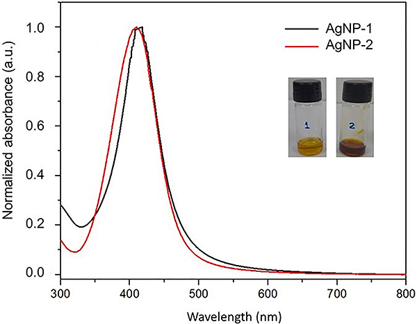
The tests for the characterization of AgNPs demonstrated that the concentration of silver ions was 3.870 μg/mL for AgNP-1 and 3.950 μg/mL for AgNP-2. The strong plasmonic bands centered at 418 nm for AgNP-1 and 407 nm for AgNP-2, observed by UV-vis, suggest that the silver nanoparticles are spherical and confirm the yellow coloration (Figure 1).
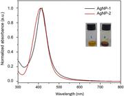
UV-visible absorbance spectrum of silver nanoparticles without ammonium hydroxide (AgNP-1) and silver nanoparticles with ammonium hydroxide (AgNP-2).
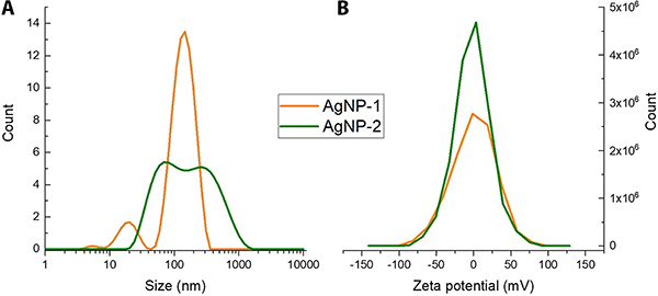
Figure 2A presents the zeta potential values obtained for AgNPs. The values for determining the zeta potential were close to 0 mV (-0.201 mV for AgNP-1 and -1.784 mV for AgNP-2). These suggest that the colloidal stability of the samples occurs mainly through steric hindrance (steric repulsion) caused by the presence of PVP, which acts as a stabilizer. In this case, colloidal stability occurred due to the PVP layer formed in the ethanolic medium.
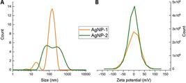
A, Hydrodynamic diameter (nm) and B, zeta potential (mV) of silver nanoparticle without ammonium hydroxide (AgNP-1) and silver nanoparticle with ammonium hydroxide (AgNP-2).
The size distribution detected by DLS (Figure 2B) demonstrated that AgNP-1 had a size of 154.2 nm, AgNP-2 had a size of 108.3 nm, and that aggregates could surround the AgNPs. The dispersion indices of the hydrodynamic diameters (DIH) were 0.32 and 0.45 nm for AgNP-1 and AgNP-2, respectively. These results demonstrated that the hydrodynamic diameters were not homogeneous.
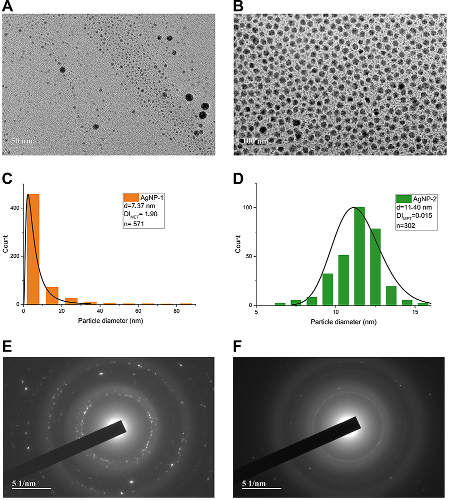
TEM analysis showed a size of 7.37 nm for AgNP-1 and 11.40 nm for AgNP-2 (Figure 3A and B). Therefore, spherical AgNPs were obtained. Based on the size distribution histograms (Figure 3C and D), the dispersion index (DITEM) was calculated as 1.90 for AgNP-1, indicating pronounced polydispersity, while 0.015 for AgNP-2 suggested monodispersity. AgNPs with monodispersity have a single and primary population size, as occurs with AgNP-2, and are therefore homogeneous.
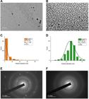
Transmission electron micrograph (TEM) of (A) silver nanoparticle without ammonium hydroxide (AgNP-1) and (B) silver nanoparticle with ammonium hydroxide (AgNP-2); scale bars 50 and 100 μm. Size distribution histogram of nanoparticles (C) AgNP-1 and (D) AgNP-2. Selected area electron diffraction (SAED) of AgNP-1 (E) and AgNP-2 (F); scale bar 51 nm.
Through SAED analyses (Figure 3E and F), the face-centered cubic crystalline phases of the nanoparticles were determined, with no mixture of crystalline phases confirmed by the diffraction rings of the crystalline planes. Crystallographic planes 111, 200, 220, 222, and 422 were found for AgNP-1 and crystallographic planes 200, 321, 422, and 433 were found for AgNP-2 (according to ICSD crystallographic chart No. 00-004-0783).
Biofilm formation
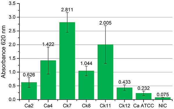
All yeasts formed biofilm under the test conditions. The yeasts C. krusei Ck7, C. krusei Ck11, C. albicans Ca4, and C. krusei Ck8 obtained the highest absorbance readings and were classified as strong producers. C. albicans Ca2 and C. krusei Ck12 were classified as moderate producers, and C. albicans ATCC 10231 was identified as a weak producer (Figure 4).
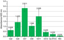
Absorbance (620 nm) readings of yeast biofilms detected by the crystal violet method. Ca: C. albicans; Ck: C. krusei; ATCC: American Type Culture Collection; NIC: non-inoculated control.
The isolates with the highest counts of yeasts associated with biofilms were C. krusei Ck8 (2.48×10⁶ cfu/mL), C. krusei Ck7 (2.39×10⁶ cfu/mL), C. krusei Ck11 (9.37×10⁵ cfu/mL), C. albicans Ca2 (4.70×10⁵ cfu/mL), and C. krusei Ck12 (3.95×10⁵ cfu/mL). On the other hand, the yeasts with the lowest results were C. albicans Ca4 (8.90×10⁴ cfu/mL) and C. albicans ATCC 10231 (2.43×10⁴ cfu/mL). The correlation between the total biomass detected by the crystal violet method and the count of viable cells associated with the biofilm was observed for all the yeasts except for C. albicans Ca4.
Antifungal activity of AgNPs
The results of the antifungal activity of the nanoparticles are shown in Table 1. The nanoparticles inhibited all the yeasts. C. krusei was the most sensitive, inhibited by AgNP-1 with MIC=15.11 µg/mL and AgNP-2 with MIC=15.40 µg/mL, while C. albicans were most resistant, with MIC=60.46 µg/mL for AgNP-1 and MIC=61.70 µg/mL for AgNP-2.
Regarding fungicidal activity, both nanoparticles (AgNP-1 and AgNP-2) required concentrations 8× higher than the MICs against C. krusei. For C. albicans, the MFC values were identical to the MICs for most yeasts and double for C. albicans Ca4.
Antibiofilm activity of AgNPs
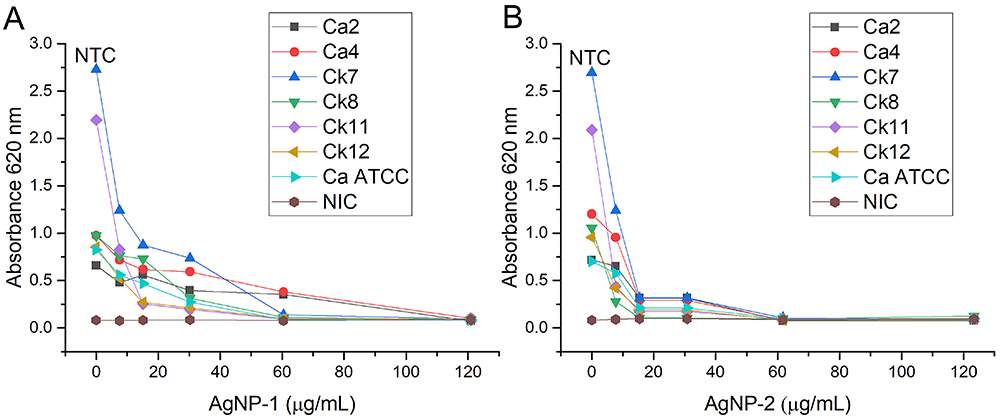
Figure 5A and B show the impact of AgNPs concentrations on yeast biofilm formation. The inhibitory activity of the nanoparticles on biofilm formation was concentration-dependent. The highest activity of AgNP-1 (Figure 5A) was detected at 60.46 µg/mL, except for C. albicans Ca2 and C. albicans Ca4. AgNP-2 (Figure 5B) showed the highest activity at 61.70 µg/mL against all yeasts, but from 15.40 µg/mL, it inhibited biofilm formation.
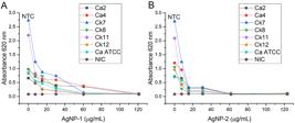
Absorbance (620 nm) readings of yeast biofilms in the presence of silver nanoparticle without ammonium hydroxide (AgNP-1) (A) and with ammonium hydroxide (AgNP-2) (B) at different concentrations. NTC: Non-treated control; Ca: C. albicans; Ck: C. krusei; ATCC: American Type Culture Collection; NIC: Non-inoculated control.
The inhibition of biofilm formation by AgNP-1 ranged from 7.55 to 60.46 µg/mL. The most sensitive yeasts were C. krusei Ck7 and Ck11, with MBIC50=7.55 µg/mL. C. albicans Ca2 and Ca4 were the most resistant, with MBIC50=60.46 µg/mL. The antibiofilm activity of AgNP-2 ranged from 7.71 to 30.80 µg/mL. C. krusei was the most sensitive with MBIC50=7.71 µg/mL, and C. albicans Ca4 was the most resistant with MBIC50=30.80 µg/mL (Table 2).
Both nanoparticles (AgNP-1 and AgNP-2) inhibited biofilm formation by the yeasts analyzed. Although the concentrations of silver ions were similar between the two nanoparticles, factors such as the particle sizes, the size distribution of the agglomerates, and the hydrodynamic radius may have influenced the results.
However, AgNP-1 showed yeast-dependent relationships between MBIC50 and MIC. While for the yeasts Ck7, Ck11, and Ca ATCC 10231 biofilm formation was inhibited by subinhibitory concentrations (1/2 MIC), the same effect was not observed against C. krusei Ck8, as antibiofilm activity was observed at 2× MIC.
AgNP-2 showed a more homogeneous MBIC50/MIC ratio among the yeasts, as biofilm formation was inhibited by 1/2 MIC for Ca4, Ck7, Ck8, Ck11, and Ck12, and by 1/4 MIC for Ca2 and Ca ATCC 10231.
Although the inhibitory activity of both nanoparticles was more intense against C. krusei than C. albicans, the inhibition of biofilm formation by AgNP-2 was more homogeneous among the yeasts tested, which can be explained by its monodispersion of 0.15 per IDMET.
Discussion
Characterization of AgNPs
Our results indicated that the nanoparticles were spherical, similar in size, and with almost zero surface charges but with different dispersivity indices (Figure 3A and B). The data obtained indicated that AgNP-1 is polydisperse. AgNP-2, synthesized in an ammonium hydroxide reaction medium, is monodisperse, i.e., it presents a homogeneous size distribution (Figure 3C and D). It is known that interactions between AgNPs and microorganisms depend on their physicochemical characteristics, such as hydrodynamic size, surface charge, aggregation (DITEM), and availability of ionic silver release rate (16).
Biofilm formation
As shown in Figure 4, all colonies formed biofilm under the test conditions. It is known that the total biomass detected by crystal violet is the amount of exopolysaccharides present in the microbial biofilm. Biofilms comprise a population of microbial cells that are adhered to each other and to solid surfaces but are embedded in the self-produced viscous extracellular matrix (17).
Previously, C. albicans ATCC 10231 was identified as a strong biofilm producer (18). In another study, the same yeast showed absorbance of 0.175 and viable cell counts of 2.30×10⁸ cfu/mL (19). However, the same strain had the lowest biofilm formation in our study compared to the yeasts isolated from the oral cavity. This difference between the results may be due to the different methodological conditions used in the studies.
The biofilm formation profile is a determining factor in the prognosis of candida infections. This ability may be related to increased mortality, impacting yeast resistance. Yeast infection processes associated with biofilms have a mortality rate almost twice as high as planktonic infections (20).
Candida biofilms are formed by a mixture of extracellular material, yeast cells, and hyphae, which are essential for their development. The transition from yeast to hyphae contributes to the overall virulence of Candida cells and may constitute a significant target for developing antifungal drugs (21).
The ability of Candida yeasts to proliferate and establish biofilms is also influenced by their interaction with host homeostasis and variations, such as mucosal pH changes or nutritional alterations, and by the state of the host immune system (22).
Antifungal activity of AgNPs
The antifungal activity of the nanoparticles did not differ significantly between AgNP-1 and AgNP-2. However, when we analyzed the difference between the inhibitory and fungicidal concentrations, we found that, for C. krusei, the concentrations needed to reach the MFC were eight times higher than the MIC. The same phenomenon was not observed for C. albicans (Table 1).
A possible explanation for the fungicidal effect may be related to the spherical shape and average diameter of the AgNPs synthesized in our study (Figure 3A and B). In a previous work, spherical silver nanoparticles with diameters of 2.7 to 12.2 nm were active against planktonic and biofilm-associated C. albicans and C. dubliniensis with MICs of 7.8 to 15.6 μg/mL and MFCs of 31.2 to 62.5 μg/mL (23). AgNPs showed significant antifungal activity against C. albicans with MIC from 62 to 250 μg/mL and MFC from 125 to 500 μg/mL (24), similar to our results.
The fungistatic and fungicidal action of AgNPs is dependent on their concentration (25,26). Nevertheless, the antimicrobial efficiency of nanoparticles also depends on the synthesis methods and the size and shape of the nanoparticles formed (27). AgNPs prepared by different methods show different levels of antifungal activity depending on their size, morphology, and surface modification (28). In addition, the release rate of metallic nanoparticles is related to their chemical composition, structure, metallic core, surface coating, and environmental conditions, such as pH and ionic strength (29).
Metal nanoparticles have a high surface-to-volume ratio, which guides their ability to interact with the membrane of microorganisms and exert their action. The two most commonly accepted theories are death by direct contact due to damage to the cell membrane and protein deactivation caused by the release of Ag+ ions (28).
The antifungal activity of silver nanoparticles is based on the fact that they produce reactive oxygen species (ROS) that act on the cell wall and interrupt DNA replication (26,30). In addition, they may be associated with the induction of apoptosis in C. albicans cells due to increased oxidative stress and mitochondrial dysfunction (31). A previous study showed that, in the presence of AgNPs, even at a subinhibitory concentration (5 µg/mL), ROS increased by around 73% in yeasts (29).
Antibiofilm activities of AgNPs
The AgNPs analyzed in our study showed differences in their biofilm inhibitory activities (Table 2). While AgNP-1 inhibited biofilm formation at concentrations similar to the inhibitory concentrations (MIC), AgNP-2 showed MBIC50 lower than the inhibitory concentrations, which may indicate a better performance in inhibiting biofilm formation, even at sub-inhibitory concentrations. This observation may be related to the monodispersity of AgNP-2 (Figure 3B and D).
Candida biofilms are notoriously difficult to treat due to their multifactorial resistance, requiring higher concentrations of antifungal drugs compared to the treatment of planktonic cells (6). Candida krusei belongs to the group of etiological agents of candidiasis. Although it is not isolated as frequently as other Candida species, its infection is relevant in the clinical environment due to its intrinsic resistance to fluconazole (32). AgNPs disrupt biofilm integrity by interacting with the extracellular polymeric matrix, DNA, lipids, and proteins of biofilms. Such interactions depend on colloidal forces and dynamic biophysical-chemical interactions (33).
The cell wall of C. albicans is a dynamic structure modulated to generate elliptical and tubular shapes that define a wide range of morphologies in which this fungus can grow (34). Its chemical composition, although not fully clarified, is composed of an amorphous inner skeletal layer of β(1,3)- and β(1,6)-glucan and chitin and another outer fibrillary layer, which is believed to be dominated by highly mannosylated cell wall proteins of sizes that can vary between 40-60 nm (35).
The morphological changes in the surface of fungal cells may be due to cell wall and membrane damage. The effects exerted by AgNPs on the cell wall and cell membrane may alter the physical state and, therefore, the fluidity of the membrane. The depletion of ergosterol after AgNPs treatment is also thought to be responsible for the sensitivity of fungal cells, the integrity of the membrane, and its changes in the cellular microenvironment (26).
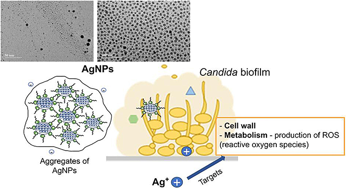
The mechanisms by which AgNPs interact with biofilms determine their activity against the microorganisms associated with this structure. AgNPs cause oxidative stress, protein dysfunction, membrane rupture, and DNA damage, leading to microbial death; they can also alter cell adhesion to prevent biofilm formation (36). In addition, AgNPs exert an inhibitory effect against fungal biofilms by damaging the cell wall, mainly through distortion and rupture of the outer surface of the fungal cell wall. This distortion and rupture cause an increase in ROS and the production of hydroxyl radicals, which also contributes to damage the cell membrane (37). Recent studies suggest that the production and delivery process of the extracellular matrix provides a useful therapeutic target for biofilms of various fungal species (Figure 6) (38).
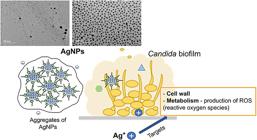
Schematic representation of the action of silver nanoparticles (AgNPs) on Candida biofilm. Ag+: silver ions
The physicochemical properties of AgNPs are considered critical factors for their biological applications (11). The activity of AgNPs against yeasts was verified in a previous study, in which AgNPs with sizes ranging from 43.4 to 120.6 nm, a negative charge, and a surface coating showed antifungal activity with MICs ranging from 1.25 to 40 µM against clinical isolates of Candida albicans and Candida krusei, among other yeasts (39).
Smaller nanoparticles tend to have higher microbicidal effects due to the greater surface area in contact with the microorganism (30). The small size of AgNPs plays a crucial role in their more effective penetration into the cell, influencing the selective permeability of the membrane. Once in contact with the intracellular environment, AgNPs act to reduce certain metabolic functions, slow down vital activities, and lead to cell damage (6).
The interactions between AgNPs and the biofilm can be described by the sequential mechanisms of transport of the nanoparticles to the biofilm-fluid interface, attachment to the biofilm surface, and migration into the biofilm (40).
The type of synthesis, the size and charge of the nanoparticles formed, and the specific interactions of the candida species that make up the biofilm interfere with the interactions with yeast biofilms. The AgNP-induced alteration observed in cellular targets can potentially affect drug resistance, pathogenesis, and virulence of fungal pathogens (26).
The size of the AgNPs mainly controls the initial penetration step into the extracellular polymeric substance (EPS) of the biofilm. At the same time, the surface properties of the AgNPs, such as charge and functional groups, affect the interactions with components of the EPS matrix. When AgNPs come into contact with an environment containing organic molecules, a corona-like coating is formed on the surface of the AgNPs, and the nature of this corona influences AgNP-biofilm interactions (40).
Conclusion
The results of this study highlight the role of size homogeneity in colloidal dispersions and nanoparticle synthesis in optimizing antimicrobial performance. Incorporating ammonium hydroxide in the synthesis of AgNP-2 improved nanoparticle size homogeneity and enhanced biofilm inhibition, highlighting the importance of designing tailored nanomaterials.
Future studies should explore in vivo applications, long-term toxicity, and synergistic effects with conventional antifungals in order to translate these results into clinical practice. In conclusion, AgNP-2 is a promising candidate for combatting Candida biofilms, offering a nanotechnology-based strategy to address the growing challenge of antifungal resistance in mucosal and device-associated infections.
-
Funding
The authors thank the Goiás State University for funding this work by Edital PrP/UEG No. 005/2021. We also thank the Coordination for the Improvement of Higher Education Personnel (CAPES) for granting a scholarship (Social Demand Program) to M.J. Barbaresco, J.S.S.C. da Silva, and F.N. de Souza Neto.
References
-
1 Izadi A, Paknia F, Roostaee M, Mousavi SAA, Barani M. Advancements in nanoparticle-based therapies for multidrug-resistant candidiasis infections: a comprehensive review. Nanotechnology. 2024;35:1-16, doi: 10.1088/1361-6528/ad4bed.
» https://doi.org/10.1088/1361-6528/ad4bed -
2 Marak MB, Dhanashree B. Antifungal susceptibility and biofilm production of Candida spp. Isolated from clinical samples. Int J Microbiol. 2018;2018:7495218, doi: 10.1155/2018/7495218.
» https://doi.org/10.1155/2018/7495218 -
3 Sahal G, Bilkay IS. Distribution of clinical isolates of Candida spp. and antifungal susceptibility of high biofilm-forming Candida isolates. Rev Soc Bras Med Trop. 2018;51:644-50, doi: 10.1590/0037-8682-0136-2018.
» https://doi.org/10.1590/0037-8682-0136-2018 -
4 Malinovská Z, Čonková E, Váczi P. Biofilm formation in medically important Candida species. J Fungi (Basel). 2023;9:955, doi: 10.3390/jof9100955.
» https://doi.org/10.3390/jof9100955 -
5 Kaur J, Nobile CJ. Antifungal drug-resistance mechanisms in Candida biofilms. Curr Opin Microbiol. 2023;71:102237, doi: 10.1016/j.mib.2022.102237.
» https://doi.org/10.1016/j.mib.2022.102237 -
6 Yin IX, Zhang J, Zhao IS, Mei ML, Li Q, Chu CH. The antibacterial mechanism of silver nanoparticles and its application in dentistry. Int J Nanomedicine. 2020;15:2555-62, doi: 10.2147/IJN.S246764.
» https://doi.org/10.2147/IJN.S246764 -
7 Santos LM, Rodrigues DM, Alves BVB, Kalil MA, Azevedo V, Barh D, et al. Activity of biogenic silver nanoparticles in planktonic and biofilm-associated Corynebacterium pseudotuberculosis Peer J. 2024;12:e16751, doi: 10.7717/peerj.16751.
» https://doi.org/10.7717/peerj.16751 -
8 Dorgham RA, Al Moaty MNA, Chong KP, Elwakil BH. Molasses-silver nanoparticles: synthesis, optimization, characterization, and antibiofilm activity. Int J Mol Sci. 2022;23:10243, doi: 10.3390/ijms231810243.
» https://doi.org/10.3390/ijms231810243 -
9 Mohanta YK, Chakrabartty I, Mishra AK, Chopra H, Mahanta S, Avula SK, et al. Nanotechnology in combating biofilm: a smart and promising therapeutic strategy. Front Microbiol. 2022;13: 1028086, doi: 10.3389/fmicb.2022.1028086.
» https://doi.org/10.3389/fmicb.2022.1028086 -
10 Tăbăran AF, Matea CT, Mocan T, Tăbăran A, Mihaiu M, Iancu C, et al. Silver nanoparticles for the therapy of tuberculosis. Int J Nanomedicine. 2020;15:2231-58, doi: 10.2147/IJN.S241183.
» https://doi.org/10.2147/IJN.S241183 -
11 do Carmo PHF, Garcia MT, Figueiredo-Godoi LMA, Lage ACP, Silva NSD, Junqueira JC. Metal nanoparticles to combat Candida albicans infections: an update. Microorganisms. 2021;11:138, doi: 10.3390/microorganisms11010138.
» https://doi.org/10.3390/microorganisms11010138 -
12 Lee Y, Oh SG. Ostwald ripening and control of Ag ion reduction degree by ammonium hydroxide in alcohol reduction process. J Ind Eng Chem. 2015;21:768-71, doi: 10.1016/j.jiec.2014.04.010.
» https://doi.org/10.1016/j.jiec.2014.04.010 -
13 Zayed SM, Aboulwafa MM, Hashem AM, Saleh SE. Biofilm formation by Streptococcus mutans and its inhibition by green tea extracts. AMB Express. 2021;11:73, doi: 10.1186/s13568-021-01232-6.
» https://doi.org/10.1186/s13568-021-01232-6 -
14 Wayne PA. Clinical and laboratory standards institute: performance standards for antimicrobial susceptibility testing: informational supplement, M100. Clinical and Laboratory Standards Institute (CLSI). 2018. https://clsi.org/media/1930/m100ed28_sample.pdf
» https://clsi.org/media/1930/m100ed28_sample.pdf -
15 Martínez A, Manrique-Moreno M, Klaiss-Luna MC, Stashenko E, Zafra G, Ortiz G. Effect of essential oils on growth inhibition, biofilm formation and membrane integrity of Escherichia coli and Staphylococcus aureus Antibiotics (Basel). 2021;10:1474, doi: 10.3390/antibiotics10121474.
» https://doi.org/10.3390/antibiotics10121474 -
16 Vazquez-Muãoz R, Bogdanchikova N, Huerta-Saquero A. Beyond the nanomaterials approach: influence of culture conditions on the stability and antimicrobial activity of silver nanoparticles. ACS Omega. 2020;5:28441-51, doi: 10.1021/acsomega.0c02007.
» https://doi.org/10.1021/acsomega.0c02007 -
17 Dave SR, Vaishnav AM, Upadhyay KH, Tipre DR. Microbial exopolysaccharide-an inevitable product for living beings and environment. J Bacteriol Mycol Open Access. 2016;2:109-11, doi: 10.15406/jbmoa.2016.02.00034.
» https://doi.org/10.15406/jbmoa.2016.02.00034 -
18 Sachivkina N, Karamyan A, Semenova V, Ignatiev A, Abdurasulov A, Muratova R, et al. The effects of Angelica ternata extract from Kyrgyzstan on the formation of Candida albicans ATCC 10231 biofilms. App Sci. 2023;13: 12042, doi: 10.3390/app132112042.
» https://doi.org/10.3390/app132112042 -
19 Atiencia-Carrera MB, Cabezas-Mera FS, Vizuete K, Debut A, Tejera E, Machado A. Evaluation of the biofilm life cycle between Candida albicans and Candida tropicalis Front Cell Infect Microbiol. 2022;12:953168, doi: 10.3389/fcimb.2022.953168.
» https://doi.org/10.3389/fcimb.2022.953168 -
20 Tsay SV, Mu Y, Williams S, Epson E, Nadle J, Bamberg WM, et al. Burden of Candidemia in the United States, 2017. Clin Infect Dis. 2020;71:e449-53, doi: 10.1093/cid/ciaa193.
» https://doi.org/10.1093/cid/ciaa193 -
21 Shareck J, Belhumeur P. Modulation of morphogenesis in Candida albicans by various small molecules. Eukaryot Cell. 2011;10:1004-12, doi: 10.1128/EC.05030-11.
» https://doi.org/10.1128/EC.05030-11 -
22 Chandra J, Mukherjee PK. Candida biofilms: development, architecture, and resistance. Microbiol Spectr. 2015;3:10.1128/microbiolspec.MB-0020-2015, doi: 10.1128/microbiolspec.MB-0020-2015.
» https://doi.org/10.1128/microbiolspec.MB-0020-2015 -
23 Narayanan KB, Park HH. Unnatural amino acid-mediated synthesis of silver nanoparticles and their antifungal activity against Candida species. J Nanopart Res. 2014;16:2523, doi: 10.1007/s11051-014-2523-y.
» https://doi.org/10.1007/s11051-014-2523-y -
24 Bahey MG, Gabr BM, Gabr AM, Abo Hagar AM, Hegazy EE. Effect of silver nanoparticles on different Candida species isolated from patients with oral candidiasis. Microbes Infect Dis. 2024;5:1642-53, doi: 10.21608/mid.2024.295446.1980.
» https://doi.org/10.21608/mid.2024.295446.1980 -
25 Kim KJ, Sung WS, Suh BK, Moon SK, Choi JS, Kim JG, et al. Antifungal activity and mode of action of silver nano-particles on Candida albicans Biometals. 2009;22:235-42, doi: 10.1007/s10534-008-9159-2.
» https://doi.org/10.1007/s10534-008-9159-2 -
26 Radhakrishnan VS, Mudiam MKR, Kumar M, Dwivedi SP, Singh SP, Prasad T. Silver nanoparticles induced alterations in multiple cellular targets, which are critical for drug susceptibilities and pathogenicity in fungal pathogen (Candida albicans). Int J Nanomedicine. 2017;13:2647-63, doi: 10.2147/IJN.S150648.
» https://doi.org/10.2147/IJN.S150648 -
27 Köroğlu A, Şahin O, Kürkçüoğlu I, Dede DÖ, Özdemir T, Hazer B. Silver nanoparticle incorporation effect on mechanical and thermal properties of denture base acrylic resins. J App Oral Sci. 2016;24:590-6, doi: 10.1590/1678-775720160185.
» https://doi.org/10.1590/1678-775720160185 -
28 de Souza AO. Overview of nanomaterials and cellular interactions. Biointerface Res Appl Chem. 2022;13:367, doi: 10.33263/BRIAC134.367.
» https://doi.org/10.33263/BRIAC134.367 -
29 Balhaddad AA, Garcia IM, Ibrahim MS, Rolim JPML, Gomes EAB, Martinho FC, et al. Prospects on nano-based platforms for antimicrobial photodynamic therapy against oral biofilms. Photobiomodul Photomed Laser Surg. 2020;38:481-96, doi: 10.1089/photob.2020.4815.
» https://doi.org/10.1089/photob.2020.4815 - 30 Durán N, Rolin WR, Durán M, Fávaro WJ, Seabra AB. Nanotoxicology of silver nanoparticles: toxicity in aninals and humans. Quim Nova. 2019;42:206-13.
-
31 Hwang IS, Lee J, Hwang JH, Kim KJ, Lee DG. Silver nanoparticles induce apoptotic cell death in Candida albicans through the increase of hydroxyl radicals. FEBS J. 2012;279:1327-38, doi: 10.1111/j.1742-4658.2012.08527.x.
» https://doi.org/10.1111/j.1742-4658.2012.08527.x -
32 Gómez-Gaviria M, Mora-Montes HM. Current aspects in the biology, pathogeny, and treatment of Candida krusei, a neglected fungal pathogen. Infect Drug Resist. 2020;13:1673-89, doi: 10.2147/IDR.S247944.
» https://doi.org/10.2147/IDR.S247944 -
33 Nel AE, Mädler L, Velegol D, Xia T, Hoek EMV, Somasundaran P, et al. Understanding biophysicochemical interactions at the nano-bio interface. Nat Mater. 2009;8:543-57, doi: 10.1038/nmat2442.
» https://doi.org/10.1038/nmat2442 -
34 Dantas AS, Lee KK, Raziunaite I, Schaefer K, Wagener J, Yadav B, et al. Cell biology of Candida albicans-host interactions. Curr Opin Microbiol. 2016;34:111-8, doi: 10.1016/j.mib.2016.08.006.
» https://doi.org/10.1016/j.mib.2016.08.006 -
35 Lenardon MD, Sood P, Dorfmueller HC, Brown AJP, Gow NAR. Scalar nanostructure of the Candida albicans cell wall; a molecular, cellular and ultrastructural analysis and interpretation. Cell Surf. 2020;6:1000047, doi: 10.1016/j.tcsw.2020.100047.
» https://doi.org/10.1016/j.tcsw.2020.100047 -
36 More PR, Pandit S, Filippis A, Franci G, Mijakovic I, Galdiero M. Silver nanoparticles: bactericidal and mechanistic approach against drug resistant pathogens. Microorganisms. 2023;1:369, doi: 10.3390/microorganisms11020369.
» https://doi.org/10.3390/microorganisms11020369 -
37 Atriwal T, Azeem K, Husain FM, Hussain A, Khan MN, Alajmi MF, et al. Mechanistic understanding of Candida albicans biofilm formation and approaches for its inhibition. Front Microbiol. 2021;12:638609, doi: 10.3389/fmicb.2021.638609.
» https://doi.org/10.3389/fmicb.2021.638609 -
38 Massey J, Zarnowski R, Andes D. Role of the extracellular matrix in Candida biofilm antifungal resistance. FEMS Microbiol Rev. 2023;47: fuad059, doi: 10.1093/femsre/fuad059.
» https://doi.org/10.1093/femsre/fuad059 -
39 Kaiser KG, Delattre V, Frost VJ, Buck GW, Phu JV, Fernandez TG, et al. Nanosilver: an old antibacterial agent with great promise in the fight against antibiotic resistance. Antibiotics (Basel). 2023;12:1264, doi: 10.3390/antibiotics12081264.
» https://doi.org/10.3390/antibiotics12081264 -
40 Fulaz S, Vitale S, Quinn L, Casei E. Nanoparticle-biofilm interactions: The role of the EPS matrix. Trends Microbiol. 2019;27:915-26, doi: 10.1016/j.tim.2019.07.004.
» https://doi.org/10.1016/j.tim.2019.07.004
Edited by
-
Editor
João Agostinho Machado-Netohttps://orcid.org/0000-0002-2937-8109