
 New discoveries of the action of L-ascorbic acid (vitamin C) - Enhanced efficacy in formulations
Abstract
Skin oxidation can impair physiological functions and induce skin diseases, such as photoaging and cancer. L-ascorbic acid (L-AA), or vitamin C, is commonly used in cosmetics because it is a potent antioxidant, inhibits melanogenesis, and promotes collagen and elastin synthesis in the skin. This study developed strategies to improve the stability of L-AA in its pure form with or without caffeic acid (CA) and evaluated its clinical efficacy using an ex vivo method. Oil/water emulsions were prepared with antioxidants and normal stability tests were conducted (various temperatures for 360 days). Antioxidant activity was assessed using a DPPH assay, and L-AA content was quantified by high-performance liquid chromatography. The thiobarbituric acid reactive substances method characterized the inhibition of lipid peroxides in the stratum corneum ex vivo. The formulation F1 (base + 10.0% L-AA) exhibited better L-AA stability over 360 days. The formulations F1 and F2 (base + 10.0% L-AA + 0.2% CA) increased the production of lipid peroxides when applied to the stratum corneum ex vivo and irradiated; however, when not irradiated, they inhibited the production of reactive oxygen species. For greater clinical efficacy of vitamin C on the skin, nighttime use is suggested as well as storage at low temperatures.
Keywords:
Ascorbic acid; Caffeic acid; Lipid peroxidation; Cosmetic stability; Tiobarbituric acid; Antioxidants
INTRODUCTION
Skin aging is classified as either intrinsic/chronological or extrinsic (Ramos et al., 2013). Intrinsic is inevitable and occurs naturally throughout the body, whereas extrinsic involves the exposure of the skin to free radicals, including environmental factors (pollution), lifestyle (tobacco, alcohol, poor diet, and sedentary lifestyle), and exposure to ultraviolet (UV) radiation, which consequently results in redox imbalance. This imbalance can affect the skin’s barrier function, which under normal conditions protects against moisture loss as well as chemical, physical, and mechanical injuries (Niki, 2015).
UV radiation produces reactive oxygen species (ROS), which damages lipid-rich membranes and causes epidermal hyperplasia, decreased collagen production, dermal matrix degradation, and erythema (Fonseca, 2010). Chronic exposure can predispose individuals to both photocarcinogenesis and photoaging of the skin because of the accumulation of oxidation products, including proteins, DNA, and lipids generated by free radicals (Niki, 2015). Approximately 80% of visible signs of aging are caused by exposure to UV rays (Peres, 2015; Oliveira et al., 2018). Antioxidants are the primary defense mechanism induced when the skin is exposed to free radicals (Fries, Frasson, 2010). Thus, the topical application of antioxidants has been routinely prescribed in dermatological clinics and desired in cosmetic formulations.
L-ascorbic acid (L-AA) is the chemically active form of vitamin C and is considered the least toxic and most potent among natural antioxidants. It functions by neutralizing and removing oxidants produced by skin interactions with environmental pollutants or after exposure to UV light, especially in the epidermis, where vitamin C is the most concentrated (Oliveira et al., 2018; Susana, Pisano, 2023). As a multifunctional agent, it has been incorporated into cosmetics to promote skin health by inhibiting and preventing the formation of wrinkles because of its high regenerative power and it functions as a biological protector in the synthesis of collagen and elastin. Furthermore, it has depigmenting and skinlightening properties via tyrosinase enzyme inhibition and anti-inflammatory activities in reducing erythema (Stamford, 2012; Wang et al., 2018).
The bioavailability of AA in most topical formulations is low, and the ineffective distribution of active substances into the deeper layers of the epidermis or dermis is challenging and may produce poor results (Jarros-Sajda, Budzisz, Erkiert-Polguj, 2024). Scientists have reported that the permeability of vitamin C can be improved by increasing its concentration (Wang et al., 2022). In most cases, for a product to have biological importance, it requires a concentration greater than 8%. Reputable products that are currently available are in the range of 10 to 20% (Al-Niaimi, Chiang, 2017); however, concentrations above these values have been known to irritate sensitive skin and do not provide an increase in activity (Lazar et al., 2023).
The main challenge when developing a formulation is to ensure the stability of the topical preparation, as ascorbic acid in its pure form is chemically active and unstable in an aqueous medium. The presence of oxygen and other oxidizing agents, high pH or temperature, and the amount of metal ions increase the rate of degradation or decomposition of AA, making its penetration into the skin weak because of the hydrophobic nature of the stratum corneum. For effective penetration of the epidermal barrier, aqueous AA formulations must have a pH (2.0-3.5) lower than the pKa (4.2) (Stamford, 2012; Ravetti et al., 2019; Susana, Pisano, 2023).
For these reasons, current strategies adopted by the cosmetic industry consist of the encapsulation of AA (water-soluble), including spray drying, spray cooling, fluidized bed coating, liposomes, and extrusion to maintain its stability and improve its delivery to the target site (Susana, Pisano, 2023). In addition, they also include a low pH and the inclusion of derivatives such as magnesium ascorbyl phosphate (MAP), sodium ascorbyl phosphate, ascorbyl 2-glucoside, 3-O-ethyl ascorbate (EAC), and tetrahexyldecyl ascorbate (THDA) (Sheraz et al., 2014; Ravetti et al., 2019). These derivatives are more stable and easier to formulate; however, they do not have the same biological activity as AA, and although all products may be marketed for the same use, their effects can be greatly altered by the addition of compounds in the formula, as well by increasing the formulation cost (Sheraz et al., 2014; Lazar et al., 2023). Furthermore, AA is the most biologically active and effective form, as it does not need to be converted or released to function, unlike its derivatives (Al-Niaimi, Chiang, 2017; Lazar et al., 2023).
Studies on the topical application of L-AA demonstrated a 20 times greater efficacy (15.0%) at pH 3.2 when compared to the application of MAP (12.0%) and ascorbyl-6-palmitate (10.0%) to the skin (Pinnell et al., 2001). According to Stamford (2012), although the stability of ascorbyl phosphate salts is considerably greater than that of AA, they are not antioxidant agents. Previous studies have shown that sodium ascorbyl phosphate was poorly absorbed into the epidermis and that MAP did not improve skin barrier function in vivo, which suggests a difference in skin penetration rate compared to AA. Moreover, there is no data suggesting that ascorbyl-6-palmitate inhibits melanogenesis through tyrosine inhibition (Stamford, 2012; Lazar et al., 2023), whereas AA indirectly inhibits tyrosinase activity because of its antioxidant capacity, thereby reducing melanogenesis (Wang et al., 2018).
Caffeic acid (3,4-dihydroxycinnamic acid) (CA) is another potent antioxidant (Spagnol et al., 2017). It is superior to p-coumaric, sinapic, and ferulic acids, can inhibit the oxidation of 2-deoxyribose (2-DR), and chelates pro-oxidant metal ions, mainly Fe (II). Furthermore, CA has antimicrobial activity and has shown promise in the treatment of dermal diseases (Magnani et al., 2014). Topical application thereof reduces erythema caused by UVB radiation and prevents premature skin aging (Hallan et al., 2021).
As the cutaneous antioxidant system is complex and not fully elucidated, this study aimed to contribute to the dermatological field, which constantly pursues the development of stable topical antioxidants for the prevention and treatment of various skin conditions (shortand long-term), mainly caused by UV radiation exposure. Here, we also discuss clinical treatment strategies on how to correctly apply vitamin C, which have become of interest medical, chemist and pharmacist.
MATERIAL AND METHODS
Material
Reagents: 1.1.3.3-Tetramethoxypropane (Sigma-Aldrich); 2.2-difenil-2-picrilhidrazila (Sigma-Aldrich); acetonitrile (Merck); metaphosphoric acid (Dinâmica); and methanol (Merck).
Secondary reference standard: 6-hydroxyl-2,5,7,8-tetramethylchromane-2-carboxylic acid (97% purity); L-AA (99% purity); and CA (99% purity) (Sigma-Aldrich).
Formulations
Oil-in-water (O/W) emulsions were formulated according to Table I. The aqueous and oil phase components were heated to 75.0°C, followed by mechanical stirring at 1,000 rpm during the cooling step. The other components were added at 35°C. Only AA was dispersed in propylene glycol and sodium metabisulfite after 24 h and added to the emulsified system. The pH value was corrected to 3.5 with citric acid.
Sufficient quantities were packaged to perform tests for 360 days. The formulations were named as F1 = base + 10.0% (w/w) L-AA; F2 = base + 10.0% L-AA + 0.2% (w/w) CA; and F3 = base + 0.2% (w/w) CA.
Normal stability test
Determination of organoleptic characteristics
The F1, F2, and F3 samples were stored in a refrigerator (5.0 ± 2.0ºC), at room temperature (25.0 ± 2.0ºC) protected from light, and in an oven (45.0 ± 2.0ºC) (Brazil, 2004). They were evaluated at t0 (48 h after preparation) and then on days 7, 15, 30, 60, 90, 120, and 360. The formulation bottles, stored under stated conditions and not previously analyzed, were opened on their respective days to avoid contamination. The parameters evaluated included changes in aspect, color, and odor.
pH
The samples were diluted in reverse osmosis water (1:10), and the pH was measured in triplicate using a pH meter and a combined direct immersion electrode. The initial pH of the formulations (t0) was equal to 3.5 and was monitored for 360 days. A variation of ≤10.0% was acceptable.
Determination of antioxidants by DPPH radical scavenging assay
The antioxidant potential of the formulations was evaluated by removing the free radicals generated by 2,2-diphenyl-1-picrylhydrazyl (DPPH). A 100 µM DPPH solution in methanol was prepared, and 250.0 mg of each formulation was weighed for each storage condition, which was diluted to 48 μg/mL of L-AA. Thereafter, 2.5 mL of 100 µM DPPH solution was added to the 0.5 mL aliquots in triplicate, incubated at 22.0 ± 2.0ºC for 30 min in the dark, and absorbance at 517 nm was measured using a spectrophotometer. The decay of sample absorbance (O1) was measured relative to the control (O2), resulting in the percentage of free radical scavenging (% SRLM) (Al-Niaimi, Chiang, 2017; Dario, 2016), according to Equation 1:
where O 1 is the absorbance of the negative control and O 2 is the absorbance of the test solution.
Chromatographic conditions and analytical validation method
In vitro test: chemical stability of AA in proposed formulations
Equivalent concentrations of 7.5 µg/mL of L-AA in each formulation were analyzed in triplicate using high-performance liquid chromatography (HPLC)-Shimadzu® LC-20AD/T. The mobile phase was composed of 2.0% metaphosphoric acid in water, methanol, and acetonitrile (90:8:2), 20 µL of the sample as injected, a flow rate of 1 mL/min, detection at 254 nm, a run time of 6 min, and retention for 3.47 ± 0.24 min. All analytical data were previously validated by Oliveira (2021).
Malondialdehyde quantification and analytical validation
Ex vivo test: calibration curve
Quantification of the lipid peroxides generated in the skin was performed using the thiobarbituric acid reactive substances (TBARS) assay, which measures malondialdehyde (MDA), a product derived from an endoperoxide of unsaturated fatty acids as a result of lipid substrate oxidation (Kumar et al., 2018). MDA was obtained by reacting 22 µL of 1,1,3,3-tetramethoxypropane in 10 mL of solution with 1.0% sulfuric acid. This methodology associated with Tape stripping is innovative in the available scientific literature for the evaluation of AA (vitamin C) as an ex vivo model.
Methodology data: C-18 reverse column (4.6 mm × 25.0 cm); 35:65 mobile phase (methanol:potassium phosphate buffer, pH 7.0); flow rate 1.0 mL/min; 532 nm for detection of the MDA-TBA adduct formed at 30.0 ± 1.0°C; and a run time of 10 min.
Tape stripping and TBARS
A novel Tape stripping methodology was proposed for the ex vivo, non-invasive evaluation of the influence of AA on lipid peroxidation. This method aimed to extract the outermost layers of the stratum corneum of the middle volar forearm (from 10 volunteers) using transparent Tape Scotch® 3M adhesive tape. The skin was cleaned with purified water from three areas (2.0 × 5.0 cm2) demarcated on the right and left forearms of the volunteers.
Formulations F1 and F2 were applied to the determined areas (2.5 mg/cm2) and the third area (control) had no emulsion applied (Gonçalves, 2019; Oliveira, 2021; Peres, 2015). After 2 h of contact, four consecutive samples were collected from the volunteers’ right forearm and irradiated for 2 h in a solar simulator chamber (Suntest® CPS+) at 2,753 KJ/m2. Samples collected from the left arm were not irradiated, but they were diluted in methanol and subjected to an ultrasonic bath-Unique® UltraCleaner 1600A-for 10 min to extract the lipids. After mixing in a vortex 3 Ika® and filtering, 1 mL of each sample was added to 144 µL of 0.2% butylated hydroxytoluene (BHT) and 400 µL of H3PO4 (0.44 M). Thereafter, 600 µL of 0.6% thiobarbituric acid (TBA) solution in H3PO4 was added, stirred for 1 min, and transferred to a water bath at 90°C for 45 min. After cooling to room temperature, 600 µL of n-butanol was added and centrifuged at 3,000 rpm for 10 min. The pink chromogens (TBARS) generated by the MDA-TBA reaction and heated in an acid solution were filtered, transferred to amber vials, and analyzed by HPLC.
Study population and general approach
The participants in this research were provided with the necessary information and clarification regarding the trial through oral and written informed consent. This project was reviewed and approved by the Ethics Committee of the Faculty of Pharmaceutical Sciences (FCF) of the University of São Paulo (USP) (number: 11101219.2.0000.0067). Participants included 10 women, aged 50 to 70 years old, with healthy skin and phototype II-III (Fitzpatrick classification). The studies were conducted in the Cosmetology laboratory at FCF/USP. Participants were instructed not to apply cosmetic products to the area to be tested for 24 h before the experiment.
Statistical analysis
The software Minitab®19 was used to perform all statistical analyses in this study, and analysis of variance (ANOVA, α = 0.05) was used to determine statistical significance.
RESULTS AND DISCUSSION
Organoleptic characteristics, pH, and antioxidant activity
Normal Stability Studies aim to guide the development of cosmetic preparations, as well as estimate their duration of viability and produce information on the product’s reliability and safety, even after exposure to extreme conditions (Ferreira, 2012).
Figure 1 shows the degradation of L-AA stored in a refrigerator (5.0 ± 2.0°C), room temperature (25.0 ± 2.0°C), and oven (45.0 ± 2.0°C) in closed containers.

Organoleptic characteristics of emulsions with L-ascorbic acid (L-AA) and caffeic acid (CA).
At low temperatures (5.0 ± 2.0°C), sample F1 (base + 10.0% L-AA) did not change in aspect, color, or odor. The pH (3.47 ± 0.03) and antioxidant activity (95.29 ± 0.33 SRLM) were maintained for 360 days. At room temperature, the pH (3.57 ± 0.01) and antioxidant activity (94.59 ± 0.38% SRLM) remained stable without significant statistical differences, with only a slight change in color after reaching 360 days.
In studies by Santos et al. (2019), changes occurred in formulations with L-AA that were maintained at room temperature after 60 days. Furthermore, formulations evaluated by Aquino, Felipe (2014) showed changes in color, odor, and appearance when stored at room temperature after 90 days.
Formulations containing AA at pH 3.5 have been shown to provide greater skin permeation and greater stability (Al-Niaimi, Chiang, 2017). This supports the good performance achieved by the F1 formulation. According to Ahmad et al. (2011), in emulsions containing L-AA, maximum photostability was maintained up to pH 4.0; however, above this value, there was an increase in the redox potential of L-AA, which enhanced the possibility of oxidation and decomposition of the molecule, thereby reducing the clinical efficacy.
Human cells are exposed to more than 100 oxidative events a day from hydroxyl radicals and other reactive species (Brudzyńska et al., 2022). The antioxidant activity present in the formulation can contribute to strengthening the endogenous antioxidant capacity of the skin and supporting the neutralization of the ROS formation process under the influence of external and internal factors.
In the formulations stored in the oven, there was a change in color (slightly yellowish) and odor after 60 days, a 62% reduction (36.98 ± 2.33 SRLM) in antioxidant activity after 120 days, and an increase in pH by ± 13% (3.95 ± 0.03) after 360 days.
F2 (base + 10.0% L-AA + 0.2% CA), stored in the refrigerator, showed no changes in aspect, color, odor, pH, or antioxidant activity. However, at room temperature, upon reaching 360 days, it underwent changes in color (Figure 1) and odor, showed a 12% increase in pH (3.92 ± 0.02), and an 8% decrease (89.97 ± 0.08) in antioxidant activity. In the oven, there were exacerbated changes in color (brown), a characteristic sulfur odor, an increase in pH of 22.0% (4.28 ± 0.00), and a 71% decrease (28,459 ± 1.25% SRLM) in antioxidant activity after 120 days.
In F3 (base + 0.2% CA), the antioxidant activity was limited in all tested conditions within 7 days; specifically, it reduced the antioxidant activity by 65% (33.12 ± 0.19 SRLM) in the refrigerator, by 67% (31.31 ± 0.35% SRLM) at room temperature, and by 69% (29.40 ± 0.09% SRLM) in the oven. The pH was increased by ±20.0% in all stored conditions (360 days), as well as presenting with greater fluidity, a slightly pearly appearance, and a reduction in odor intensity, thus demonstrating the high instability of the emulsion in the presence of CA in all studied conditions.
Formulations F2 and F3 had limited effects. The results showed that the addition of CA accelerated the degradation of AA. Synergism between these antioxidants was desired to maintain the antioxidant activity of the product for longer periods because weak activity does not provide greater effectiveness when applied to the skin. Therefore, it may be necessary to develop an oil/water/ oil (O/W/O) emulsion, add it to a phosphate buffer, or encapsulate the CA to provide better results, especially when incorporating the active ingredient into L-AA (Yin et al., 2022).
Determination of L-AA content in HPLC preparations
This study reports the methodology developed and validated to quantify the concentration of L-AA in emulsions during the stability study, as described by Oliveira (2021), which exhibited simple, fast, accurate, reproducible, and reliable characteristics.
The standard curve revealed a good linear relationship (R2 = 0.9998), detection, and quantification limit of 0.075 µg/mL, which corresponds with 10% w/w of L-AA in the emulsions evaluated. Recoveries were 97.51-98.12% with a relative standard deviation of 0.21-2.70%. There were no interference peaks between L-AA and CA, indicating that the method is suitable for vitamin C stability studies.
We emphasize the high specificity of this evaluation method, as shown in Figure 2.

The initial concentration of L-AA in the formulations was 100.62 ± 0.06 µg/mL under all storage conditions. The F1 emulsion stored at 5.0 ± 2.0ºC (refrigerator) had a 10.0% reduction in L-AA content compared to the initial concentration after twelve months (Figure 3A). This value is within the pharmacopeial standards of the US Pharmacopoeia (90-110%) (USP 41, 2018). Storage at room temperature (25.0 ± 2.0ºC) exhibited a 15 and 18% decline, after 6 and 12 months, respectively. In a study conducted by Krambeck (2011), at the same temperature, a 40% reduction of AA was observed after 90 days, a 23% degradation was observed after 7 days in a study by Yin et al. (2022), and a 14% reduction after 4 months in a study reported by Caritá et al. (2021).
When stored in an oven, faster degradation occurred, where 80% of the AA concentration (20.0 μg/mL) was degraded within 120 days, thus confirming that the increase in temperature is a determining factor in the AA degradation process (Caritá et al., 2021). This degradation changed the color of the formulation, possibly forming CO2, which gives the product an airy appearance, corroborating the results obtained in the previous item. Continued monitoring of this storage condition at further time points was discontinued because of the drastic reduction in the concentration of the active ingredient.
The decomposition rate of AA was faster in F2 compared to F1, which was reduced by 16% in the refrigerator and 40% at room temperature (Figure 3B) after 360 days. In the oven, a much greater decomposition rate of 82% occurred after 120 days (Figure 3C). It is possible that the formed degradation products may or may not be toxic, thus reducing the expected viable duration of the formulation and making it unsuitable for use (Sheraz et al., 2014).
The addition of CA accelerated the degradation of L-AA. This was unexpected because CA acts as a primary and secondary (mixed) antioxidant, which occasionally has synergistic action and stimulates the inactivation and/or removal of free radicals through the donation of a hydrogen atom from a hydroxyl group of its aromatic structure (Magnani et al., 2014; Espíndola et al., 2019). CA can support the unpaired electron by moving it throughout the electron system of the molecule and, in some cases, it can chelate transition metals (Cu1+ and Fe2+) at the beginning and also during the propagation of the oxidative process (Espíndola et al., 2019).
Therefore, to further extend the viable duration of vitamin C, we must assess new antioxidant assets. However, the results show the great effect of the O/W emulsion in the F1 formulation, because common formulations with this active ingredient do not always achieve long periods of stability. The emulsion minimized the degradation of the active ingredient, maintaining its stability under the various storage conditions studied. The careful selection of excipients in the development of the cosmetic base appears to be promising. According to Ahmad et al. (2011), the higher the viscosity of the medium, the lower the degradation of L-AA; therefore, we added glycerin, which increased the viscosity in addition to its humectant action. Furthermore, EDTA (chelating agent) was added to the formulation in synergy with sodium metabisulfite (preservative) because, in other studies, their combination exhibited good results in preventing L-AA degradation (Sheraz et al., 2014; Caritá et al., 2021; Yin et al., 2022).
Finally, the results suggest that F1 is a good cosmetic formulation for maintaining skin health, being indicated in medical prescriptions for dermocosmetics because of its antioxidant and antiaging effects. In compounding pharmacies, the maximum recommended stability is almost always around four months; however, this formulation allows for a higher stability of around twelve months.
Lipid peroxidation in ex vivo
MDA is a secondary product of lipid peroxidation and is used as a marker of oxidative stress to estimate the damage caused by ROS (Grotto et al., 2009). The TBARS assay is the most commonly used method to evaluate the production of MDA (Siddique et al., 2012).
To prevent the harmful action of ROS generated by UV radiation on the epidermis (stratum corneum as the first barrier) and dermis, the topical application of antioxidant(s) must penetrate the skin (Sauce, 2020). However, the difficulties in obtaining human skin samples and the worldwide ban on testing cosmetics on animals have led researchers to develop alternatives to evaluate skin penetration and permeation, such as the Tape stripping technique (Cândido et al., 2018). This is considered ex vivo, as it is based on the removal of the stratum corneum using adhesive tape, and the collected samples can be applied to multiple types of tests, such as irradiation or chemical reactions (Sauce, 2020).
In this study, the Tape stripping technique and the TBARS assay were used to evaluate the ability of the developed formulations to inhibit lipid peroxidation in the skin (stratum corneum) of volunteers under the action of UV radiation. The advantage of combining these two methodologies is that it allows for a non-invasive and effective evaluation of the effect of peroxidation on human skin.
The analytical curve presented a linear regression equation of y = 917637x + 1842 and a linear correlation coefficient (R 2) = 0.9998 for MDA-TBA estimation.
Non-irradiated skin was used as a control group and it was observed that the lower the level of MDA formed, the greater the antioxidant activity. Figure 4 reveals the evident increase in MDA production after all samples were exposed to the photostability chamber and irradiated at 2,753 KJ/m2. The irradiated skin (57%) and F1 (55%) samples were considered statistically equal.

The skin samples treated with F2 and then irradiated showed a significant increase in the production of lipid peroxides, with an approximate increase of 97%. These results demonstrate that ROS were generated even under the application of an antioxidant formulation. It is well established that antioxidants inhibit the oxidation process; however, the application of AA and CA was not sufficient to inhibit lipid peroxidation under irradiation exposure, even with high antioxidant activity, as they were applied to the skin of volunteers one day after being formulated and at t0, where the antioxidant activity for F1 was 95.23% and F2 was 97.17%.
In a study by Alonso et al. (2009), when a topical formulation (vitamin A (0.1%), vitamin E (1%), and vitamin C (5%)) was subjected to an irradiation intensity of 182.7 J/cm2, lipid peroxidation was inhibited; however, when the UV irradiation intensity was 365.4 J/cm2, it promoted the high formation of lipid peroxides in the corneal extract. It appears that the antioxidant formulation had a less protective effect against lipid peroxidation when high UV irradiation was used. However, we receive 1,361/1,400 J/s/m2 (1 Watt = 1 J/s) from the sun (Platnick, 2016). As observed by Sauce (2020), when they tested sunscreen with and without ferulic acid on irradiated skin, there was a significant increase in lipoperoxidation, which was different from samples that were not irradiated. According to the author, “It was expected that ferulic acid, as an antioxidant, could act to neutralize lipid peroxidation, thus reducing its value in comparison to sunscreen without it.”
In vivo evidence of the effects of the topical application of vitamin C against lipid peroxidation is scarce. In vitro methods do not reflect the actual biological processes in skin cells, and results obtained using cell lines, although similar, should be interpreted with caution. Therefore, the methodology applied in this study can be used to successfully evaluate other antioxidants and their behavior when exposed to UV radiation.
When comparing non-irradiated F1 and F2 samples with the control group (non-irradiated skin), there was an evident decrease in MDA levels after the formulation application, with an approximate reduction of 10 and 13% in the concentration of lipid peroxides, respectively. This demonstrates the efficacy and importance of its application in protecting the skin. These results and data interpretation are important for formulators and the dermatological medical profession, as they guide the prescription, optimal application time, and response of the skin to the application of the product. In addition to understanding whether the same product can improve or worsen the skin condition and inhibit or enhance the production of free radicals, the result is dependent on the mode of use.
Through this study, it was also possible to verify that there was no presence of erythema after stripping the skin with adhesive tape for F1 and F2, which suggests possible protection via the antioxidant action of AA. However, erythema was observed in the control demarcations (skin, without the addition of emulsions) on the forearms of all volunteers, which further suggests the possible anti-erythematosus protection by AA.
CONCLUSIONS
The evaluations performed in this research revealed that the formulations follow different behaviors. The cosmetic base was balanced and allowed for successful harmonization of the ingredients to incorporate pure L-AA. The F1 (base + 10.0% L-AA) formulation exhibited greater stability and potential to be prescribed and manipulated, unlike the F2 (base + 10.0% L-AA + 0.2% CA) and F3 (base + 0.2% CA) formulations, which showed limited stability.
When lipid peroxides generated in the stratum corneum were quantified using the TBARS method in an ex vivo model, it was observed that the F1 and F2 formulations had antioxidant potential when the samples were not irradiated, resulting in a 10 and 13% reduction of lipid peroxides, respectively. However, when the same formulations were exposed to UV radiation, the effect was the opposite; they became pro-oxidants, with a significant increase in lipoperoxidation because of MDA production compared to the control group (non-irradiated skin), with an increase of 55% and 97%, respectively. This study recommends the prescription and topical use of vitamin C or AA at night to optimize the antiaging action and inhibit the pro-oxidant effects that UV radiation can cause. How to correctly apply vitamin C is of interest to many dermatologists and formulators, considering that these results showed that the antioxidant effects depend on how the formulations are used, and information from ex vivo studies is scarce in the available scientific literature.
In addition, these results suggest storing the formulations under cool conditions or at most at room temperature (approximately 22ºC), for greater stability and dermatological efficacy.
ACKNOWLEDGMENTS
This work was supported by the Coordenação de Aperfeiçoamento de Pessoal de Nível Superior (CAPES) (grant number: 130013/2019-6), Brazil.
REFERENCES
- Ahmad I, Sheraz MA, Ahmed S, Shaikh RH, Vaid FH, ur Rehman Khattak S, Ansari SA. Photostability and interaction of ascorbic acid in cream formulations. AAPS Pharm Sci Tech. 2011;12(3):917-23.
- Al-Niaimi FA, Chiang NYZ. Topical vitamin Cand the skin: mechanisms of action and clinical application. J Clin Aesthet Dermatol. 2017;10(1):14-7.
-
Alonso C, Barba C, Rubio L, Scott S, Kilimnik A, Coderch L, Notario J, Parra JL. An ex vivo methodology to assess the lipid peroxidation in stratum corneum. J Photochem Photobiol B. 2009;9;97(2):71-6. doi: 10.1016/j.jphotobiol.2009.08.003.
» https://doi.org/10.1016/j.jphotobiol.2009.08.003. - Aquino JS, Felipe DF. Evaluation of accelerated stabilit y of different formulations with vitamin C. Rev Saúde Pesq. 2014;7(1):119-128.
- Brudzyńska P, Kurzawa M, Sionkowska A, Grisel M. Antioxidant Activity of Plant-Derived Colorants for Potential Cosmetic Application. Cosmetics. 2022;9(1):81.
- Cândido TM, de Oliveira CA, Ariede MB, Velasco MVR, Rosado C, Baby AR, Safety and Antioxidant Efficacy Profiles of Rutin-Loaded Ethosomes for Topical Application. AAPS Pharm Sci Tech . 2018;19(4):1773-1780.
- Caritá AC, Azevedo JR, Buri MV, Bolzinger MA, Chevalier Y, Riske KA, Leonardi GR. Stabilization of vitamin C in emulsions of liquid crystalline structures. Int J Pharmac. 2021;592(1):120092.
- Dario MF. Desenvolvimento e avaliação de eficácia de nanoemulsão catiônica bioativa na proteção capilar aos danos foto-oxidativos. [Master´s dissertation]. São Paulo: Faculdade de Ciências Farmacêuticas, USP; 2016.
- Espíndola KMM, Ferreira RG, Narvaez LEM, Rosario ACRS, Silva AHM, Silva AGB, Vieira APO, Monteiro MC. Chemical and Pharmacological Aspects of Caffeic Acid and Its Activity in Hepatocarcinoma. Front Oncol. 2019;541(1):1-9.
- Ferreira GA. Desenvolvimento de sistemas para veiculação de vitamina C, avaliação da estabilidade química, permeação e retenção cutânea. [dissertação]. São Paulo: Universidade Estadual Paulista-Júlio de Mesquita Filho; 2012.
- Fonseca BL. Efeitos neuroprotetores cutâneos de um extrato vegetal após radiação ultravioleta. [dissertação]. Porto Alegre: Pontifícia Universidade Católica do Rio Grande do Sul, 2010.
- Fries AT, frasson APZ. Avaliação da atividade antioxidante de cosméticos anti-idade. Rev Cont Saúde. 2010;10(19):17-23.
- Gonçalves PV. Avaliação ex vivo da inibição da peroxidação lipídica do estrato córneo promovida por filtro UVB. [dissertação]. São Paulo: Universidade de São Paulo, Faculdade de Ciências Farmacêuticas, 2019.
- Grotto D, Maria LS, Valentini J, Paniz C, Schmitt G, Garcia SC, Pomblum VJ, Rocha JBT, Farina M. Importance of the lipid peroxidation biomarkers and methodological aspects FOR malondialdehyde quantification. Quím Nova. 2009;32(1): 169-174.
- Hallan SS, Sguizzato M, Drechsler M, Mariani P, Montesi L, Cortesi R, et al. The Potential of Caffeic Acid Lipid Nanoparticulate Systems for Skin Application: In Vitro Assays to Assess Delivery and Antioxidant Effect. Nanomaterials. 2021;12;11 (1):171.
- Jarros-Sajda A, Budzisz E, Erkiert-Polguj A. Ascorbic Acid Treatments as Effective and Safe Anti-Aging Therapies for Sensitive Skin. Antiox. 2024;13(2):174.
- Krambeck K. Desenvolvimento de Preparações Cosméticas contendo Vitamina C. [dissertação]. Portugal: Universidade do Porto, Faculdade de Farmácia, 2011.
- Kumar S, Krishna Chaitanya R, Preedy VR. Assessment of antioxidant potential of dietary components, In: Preedy VR, Watson RR, editores. HIV/AIDS. Acad Press; 2018. pp.239-253.
- Lazar M, Rajanala S, De La Garza H, Vashi N. Consumer Preferences of Topical Vitamin C Products: A Comparative Study. Cureus. 2023;15(9):e45414.
- Magnani C, Isaac VLB, Correa MA, Salgado HRN. Caffeic acid: a review of its potential use in medications and cosmetics. Anal Methods. 2014;6:3203-3210.
- Niki E. Lipid oxidation in the skin. Free Radic Res. 2015;49(7):827-34.
- Oliveira AC, Pinto CASO, Baby AR, Bedin V, Velasco MVR. Effects of ascorbic acid against the skin aging. BWS Journal. 2018;1, e18040015: 1-7.
- Oliveira AC. Ácido ascórbico associado ao ácido cafeico em formulações tópicas: avaliação da ação antioxidante e da peroxidação lipídica. [dissertação]. São Paulo: Universidade de São Paulo, Faculdade de Ciências Farmacêuticas, 2021.
- Peres DD. Ácido ferúlico em protetores solares: desenvolvimento e eficácia multifuncional in vitro, ex vivo e in vivo. [Master´s dissertation]. São Paulo: Faculdade de Ciências Farmacêuticas, USP; 2015.
- Pinnell SR, Yang H, Omar H, Monteiro-Riviere N, Debuys HV, Walker LC, et al. Topical L-ascorbic acid: percutaneous absorption studies. Dermatol Surg. 2001;27(1):137-142.
- Platnick, S. The Earth Observer. NASA. 2016;28(1):1-44.
- Ramos SM, Celem LR, Ramos SS, Fucci CAP. Anti-aging cosmetics: facts and controversies. Clin Dermatol. 2013;6(1):750.
- Ravetti S, Clemente C, Brignone S, Hergert L, Alemandi D, Palma S. Ascorbic Acid in Skin Health. Cosmetics . 2019;6(4):58.
- Santos ACD, Araújo AM, Rocha BO, Chaves MFS, Coelho AG. Study of the stability of topical use formulations containing vitamin C manipulated in pharmacies of the city of Teresina-Pi. Braz J Health Rev. 2019;2(2):756-767.
- Sauce RS. Antilipoperoxidative and anti-inflammatory efficacy of multifunctional sunscreens containing ferulic acid. [Master´s dissertation]. São Paulo: Faculdade de Ciências Farmacêuticas, USP; 2020.
- Sheraz MA. Khan MF, Ahmed S, Kazi SH, Khattak SR, Ahmad I. Factors affecting formulation characteristics and stability of ascorbic acid in water-in-oil creams. Int J Cosm Scie. 2014;36(5), 494-504.
- Siddique YH, Ara G, Afzal M. Estimation of lipid peroxidation induced by hydrogen peroxide in cultured human lymphocytes. Dose Response. 2012;10(1):1-10.
- Spagnol CM, Filippo LD, Isaac VLB, Correa MA, Salgado HRN. Caffeic Acid in Dermatological Formulations: In Vitro Release Profile and Skin Absorption. Comb Chem High Throughput Screen. 2017;20(8):675-681.
- Stamford NPJ. Stability, transdermal penetration, and cutaneous effects of ascorbic acid and its derivatives. J Cosm Dermatol. 2012;11(1):310-317, 2012.
- Susana F, Pisano R. Advances in Ascorbic Acid (Vitamin C) Manufacturing: Green Extraction Techniques from Natural Sources. Processes. 2023;11(11):3167.
- USP 41: The United States Pharmacopeial Convention, 2018. The United States Pharmacopeia and National Formulary (USP 41 - NF 36). Rockville: United States Pharmacopeial Convention, 2018.4.
- Wang K, Jiang H, Li W, Qiang M, Dong TX, Li H. Role of Vitamin C in Skin Diseases. Front Physiol. 2018;9(819):1-9.
- Wang M, Lu W, Ge X, Lu Y, Jia X, Li H, Liu Q. Study on the Efficacy of Vitamin C Lotion on Skin: Permeable and Anti-Aging. J Cosm Dermatol og Sci Applic. 2022;12(1):67-82.
- Yin X, Chen K, Cheng H, Chen X, Feng S, Song Y, Liang L. Chemical Stability of Ascorbic Acid Integrated into Commercial Products: A Review on Bioactivity and Delivery Technology. Antiox . 2022;1(1):1-20.

 Legend:OVE (oven): 45.0 ± 2.0°C; REF (refrigerator): 5.0 ± 2.0°C; and R.T (room temperature): 25.0 ± 2.0°C; F1: base + 10.0% (w/w) L-AA; F2: base + 0.2% CA + 10.0% (w/w) L-AA; F3: base + 0.2% (w/w) CA; and T: Time.
 Legend: ascorbic acid (L-AA) as a secondary reference standard at 10 µg/mL; F1: base + 10.0% (w/w) L-AA; F2: base + 0.2% caffeic acid (CA) + 10.0% (w/w) L-AA; and F3: base + 0.2% (w/w) CA.
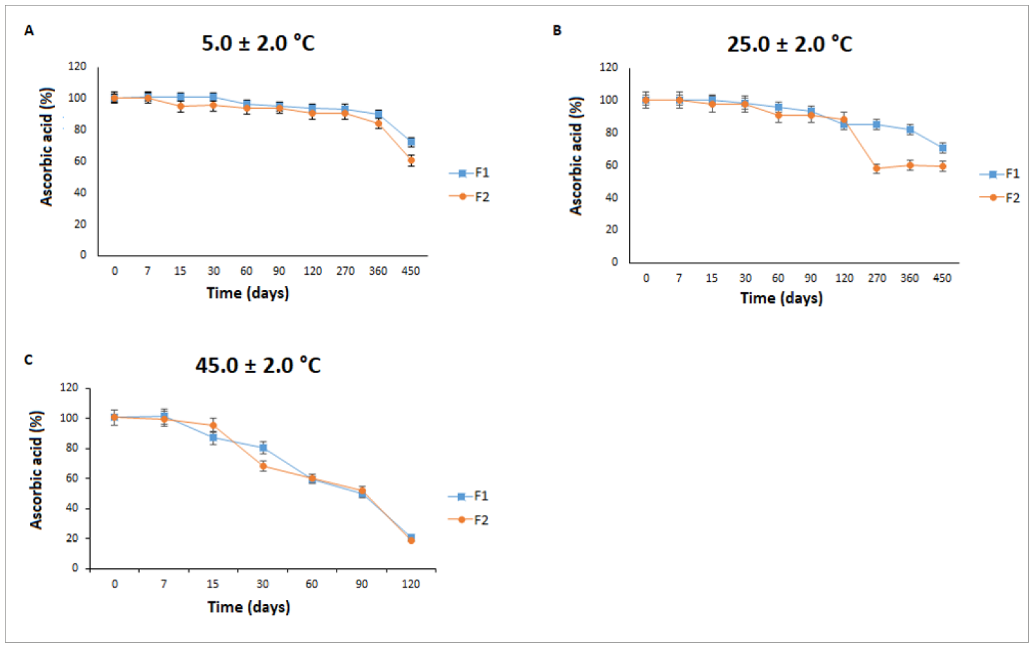 Legend: A: refrigerator; B: room temperature; C: oven; F1: base + 10.0% w/w L-AA; and F2: base + 10.0% L-AA + 0.2% w/w caffeic acid (CA). Statistical analysis was performed using analysis of variance (ANOVA). Values are presented as the mean ± standard deviation of measurements performed in triplicate (n = 3).
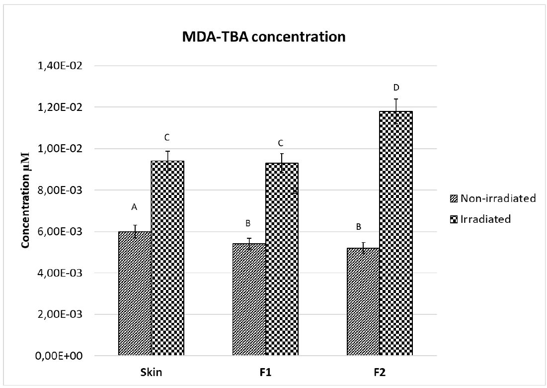 Legend:MDA: malonaldehyde; TBA: thiobarbituric acid; MDA-TBA: lipid peroxidation marker; F1: base + 10.0% (w/w) L-ascorbic acid (L-AA); and F2: base + 10.0% L-AA + 0.2% (w/w) caffeic acid (CA). Different letters for the same parameter indicate statistically significant differences between samples as determined using one-way analysis of variance.