 Quality of commercialized dry extracts of blood orange juice in Brazilian compounding pharmacies: Dry extracts of blood orange juice
Abstract
The juice of blood oranges, especially those of the Citrus sinensis variety Moro, cultivated in the region of Sicily (Italy), is an increasingly popular drink due to its beneficial health properties, such as its ability to reduce abdominal fat, related to its anthocyanin constituents, especially cyanidin-3-O-glycoside (C3G), as demonstrated in pre-clinical and clinical studies. However, the dry extract of C. sinensis juice currently available at compounding pharmacies in Brazil includes samples from various countries, some of which may not have adequate climatic conditions for the production of anthocyanins. In this work, we investigated samples from the three major suppliers (reference, A1, and A2). The composition of the samples was analyzed by LC-UV and LC-MS, total anthocyanin content (TAC), antioxidant activity (DPPH assay), and in vitro anti-inflammatory effect, by NO production in macrophages. C3G was detected only in the reference sample (1.6%), the TAC values were 1.45%, 0.1% and 0.01% in the reference, A1, and A2, respectively. The reference and A1 showed similar antioxidant activity (EC50 of 45.6 and 62.4 µg/mL, respectively), while A2 showed lower activity (EC50 315.1 µg/mL). Only the reference sample showed significant inhibition of NO release, demonstrating the need for quality control of these commercialized samples.
Keywords:
Anthocyanins; Citrus sinensis; Antioxidant; Anti-inflammatory
INTRODUCTION
Orange juice is one of the most widely consumed fruit juices around the world, and also one of the food commodities that is most subjected to adulteration and fraud (Pan et al., 2023). Sweet oranges (Citrus sinensis (L.) Osbeck) represent the largest citrus cultivar group grown worldwide, accounting for around 70% of the total annual production of Citrus species (Favela-Hernández et al., 2016).
C. sinensis is dived into two major groups, depending on the color of the pulp: white or pale oranges, and blood oranges. Blood oranges are a specific group of the sweet orange species, consisting of three main varieties: “Tarocco”, “Moro” (both native to Italy) and “Sanguinello” (native to Spain) (Reuther, Webber, 1967). These varieties are characterized by the unique color of their flesh and peel, which is due to the presence of red pigments belonging to the anthocyanin class; the major anthocyanin of the juice is cyanidin 3-O-glucoside (C3G) (Maccarone et al., 1998). Another peculiar characteristic of blood orange is its high concentration of vitamin C, flavanones (mainly hesperidin), and hydroxycinnnamic acids (Maccarone et al., 1998).
The amount and composition of anthocyanins varies, depending on the variety, maturity, and region of cultivation, among other environmental conditions. Most blood orange varieties require a wide day-night thermal range to maximize color formation, and cultivation in countries with a cooler climate is more suitable (Lo Piero, 2015). The biosynthesis of anthocyanin through postharvest management has been demonstrated through storage in a cold chamber (Magalhães et al, 2019). Therefore, the place of cultivation is important, and the fruit has been traditionally cultivated in Italy, Malta and Sicily for many centuries (Maccarone, Maccarone, Rapisarda, 1985).
It has been suggested that anthocyanins provide protection against oxidative stress, coronary heart diseases, certain cancers, and other age-related diseases (Amorini et al., 2003; Shu et al., 2023). Dry extract of Moro orange juice is widely used as a food supplement for disease prevention and obesity control, as investigated by in vivo (Titta et al., 2010) and clinical studies (Cardile, Graziano, Venditti, 2015; Kegele et al., 2019). Its possible anti-inflammatory activity, and its potential to improve endothelial function and reduce inflammation in nondiabetic subjects with increased cardiovascular risk, have also been demonstrated (Buscemi et al., 2012). Many authors who have evaluated the effects of blood orange juice attribute these effects not only to anthocyanins, but also to the synergistic effect between the components of the juice, such as flavonoids, carotenoids, ascorbic acid and hydroxycinnamic (Grosso et al., 2013; Magalhães et al., 2021).
A dry extract of the C. sinensis juice of Moro variety was patented by the company Bionap (Belpasso, Catania, Italy). This product is registered with the Brazilian Health Surveillance Agency (ANVISA) as a novel food and is marketed by compounding pharmacies in the form of hard capsules. Due to the high demand for this extract as a dietary supplement for weight loss and control, other two suppliers of this product have emerged in this commercial segment. “Moro extract” capsules are also widely sold on the Internet, but some of these products are of questionable quality. Notably, the national quality control guidelines for compounding pharmacies do not require authenticity analysis of plant extracts when the supplier is qualified (Brasil, 2007). In view of this scenario, this work aims to contribute to the quality control of these products by analyzing the major suppliers of C. sinensis dry extracts sold in compounding pharmacies, comparing their chromatographic profiles and determining the presence of C3G, total anthocyanin and lycopene contents, as well as their antioxidant and in vitro anti-inflammatory activities.
MATERIAL AND METHODS
Chemicals
Cyanidin-3-O-glycoside (C3G) (PubChem CID: 197081) was purchased from Sigma-6 Aldrich (batch 89616). Silica gel plates (Alugram® Xtra SIL G/ UV254), from Macherey-Nagel, Duren, Germany). Folin-Ciocalteu reagent, gallic acid (PubChem CID: 370), 2,2-diphenyl-1-picrylhydrazyl (DPPH) (PubChem CID: 29737978), 2,2-,3-(4,5-dimethylthiazol-2-yl)-2,5-diphenyltetrazolium bromide (MTT) (PubChem CID: 66611649), Lipopolysaccharide (LPS) and Oyster Glycogen were all purchased from Sigma (St. Louis, USA); Dulbecco’s Modified Eagle Medium (DMEM) was obtained from Vitrocell (Campinas, SP, Brazil); dimethyl sulfoxide (DMSO) from Synth (Diadema, SP, Brazil); phosphate buffer saline (PBS) and bovine serum albumin (BSA) from Gibco (Carlsbad, CA, USA); bovine fetal serum (BFS) from Invitrogen (Carlsbad, CA, USA); trichloroacetic acid (TCA) (PubChem CID: 6421) from Vetec (Rio de Janeiro, RJ, Brazil). For LC analysis, all solvents used were LC grade (Tedia, Fairfield, Ohio, USA). The water was purified using a Milli-Q system (Millipore, Massachusetts, USA). All solutions were filtered through RC 0.45 µm membrane (Macherey-Nagel, D age, Germany). All other solvents were of analytical grade.
Samples
The three commercialized dried extracts of C. sinensis juices used in the assays were purchased from compounding pharmacies in the Brazilian state of Santa Catarina. The reference sample (M) was of Italian origin, sample A1 was of Brazilian origin, and sample A2 was of Chinese origin, according to the quality certificate. The reported part of plant was the juice for M, while for A1 and A2 it was the fruit. Only the supplier of M stated the Brazilian Common Denomination (DCB), which 10820. The suppliers of samples M and A1 reported the variety Moro, but this information was absent in A2. As regards appearance, the supplier of M reported it as a “purple red power”, A1 as a “hygroscopic powder with a homogeneous appearance, purple-red in color, with an acidic taste and a characteristic odor” and A2 as a “fine yellow-green powder”. The assay of substances in M is described as: anthocyanins C3G (0.8%); ascorbic acid (4.50%), hydroxycinnamic acids (0.9%) and flavones (2.20%). For A1, these are reported as: anthocyanins C3G (3.0%), ascorbic acid (4.50%), hydroxycinnamic acids (1.39%) and flavonones (4.42%), while for A2, only bioflavonoids (90.3%) are reported.
Chemical analyses
Total anthocyanin and Lycopene analysis
The determination of total anthocyanin was performed using the differential pH method, according to Giusti and Wrolstad (2001). The results were calculated using the molecular weight and the absorptivity molar coefficient of C3G of 449.2 g/mol and 26,900 L/mol.cm, respectively. The total anthocyanin content was expressed in g of anthocyanins per 100 g. The lycopene analysis was performed according to Perkins-Veazie et al. (2001).
HPLC-UV analysis
High performance liquid chromatography was performed, coupled with an ultraviolet detection (HPLC-UV) system, using a Shimadzu LC-10AD LC (Shimadzu, Tokyo, Japan) consisting of a binary pump (LC-10ADvp), column oven (CTO-10Avp), automatic injector (SIL-10AF) and Shimadzu SPD-M10A photo diode array detector. The injections (20 µL) were carried out on a Phenomenex® (Torrance, California, USA) Luna C18 5 µm (250 x 4.6 mm), attached to a pre-column (C18, 4.6 mm) at 40 °C, with detection at 285 and 520 nm. The mobile phase at 0.8 mL/min consisted of a gradient of acetonitrile (A): methanol (B): 0.1% formic acid in water, pH 2.5 (C) of 0.5:0.5:99 (A:B:C) (0-2 min); 5:5:90 (2-3 min); 12.5:12.5:75 (3-6 min); 40:40:20 (6-12 min); 45:45:10 (12-15 min); 0.5:0.5:99 (15-20 min).
The samples were extracted in methanol, at a concentration of 2 mg/mL, sonicating for 20 minutes, filtering the sample through a regenerated cellulose membrane with a pore size of 0.45 μm and injecting 20 μL, in triplicate.
UPLC-PDA-ESI-HRMS/MS analysis
For the anthocyanin analysis, an ultra-performance liquid chromatography system (Acquity-UPLC™) was coupled with a photodiode detector (PDA) and a high-resolution mass spectrometer (Xevo® G2 QTof model - Waters®) equipped with an electrospray ionization source (ESI) operated in positive mode.
The LC-PDA-MS/MS analyses were performed on an Acquity UPLC system class H (Waters) composed of a PDA detector, sample manager and a quaternary solvent manager. Separation was achieved using a SynergiTM C18 column, 150 nm, 2.0 mm, particle size 4 μm (Phenomenex®) and temperatures of 40oC and 20oC for the column and the sample tray, respectively. The gradient elution consisted of a 2% aqueous solution of formic acid (A) and 1% of formic acid in acetonitrile (B): 0-14 min: 5-37.5% solvent B, 14-16 min 37.5-5% solvent B, at 0.4 mL/min. Sample solutions were prepared as described for the HPLC-UV analysis, at 1 mg/mL. A volume of 5 µL of samples was injected, after filtration through a regenerated cellulose membrane with a pore size of 0.22 μm.
Mass spectrometry detection was performed using a full scan, which ranged from 50 - 1500 m/z, with a scan time of 1 s. Data acquisition and processing was performed using MassLynx v 4.1 software. In the MS/ MS analysis, the collision energy varied from 10, 20 and 30 eV of argon gas, according to the compound being analyzed. The capillary voltage was 3.5 kV at 150°C, the desolvation temperature was 500°C; the gas flow was 1000 L/h and the cone voltage was 40 V, controlled by MassLynx v.4.1 software, for data analysis and processing. All the analyses were performed in triplicate.
Phenolic compound analysis was performed using a Waters BEH C18 column, 1.7 µm, 50 x 2.1 mm at 40°C and the injection volume was 5 µL. The gradient elution was performed with ultrapure water (solvent A) and acetonitrile (solvent B): 0-18 min, 5-45% (B); and 18-20 min, 45-5%, at 0.4 mL/min. Mass spectrum detection was performed using a full scan, which ranged from 50 - 1500 m/z, with a scan time of 1 s. Data acquisition and processing was performed using MassLynx v4.1 software. In the MS/MS analysis, collision energy varying from 10, 20, and 30 eV of argon gas was used, according to the analyzed compound. The capillary voltage was 3.5 kV with a temperature of 90°C, the desolvation temperature was 400°C, at 900 L/h; the gas flow was 30 L/h and the cone voltage was 40 V. All the analyses were performed in triplicate.
Antioxidant analysis
The antioxidant activity was determined through the capture of DPPH (2,2-diphenyl-1-picrylhydrazyl) free radicals, and the results were expressed as the percentage of free radical sequestration. A methanolic solution of DPPH at 1 mM was used to elaborate an analytical curve from 0.01-1 mM, at 515 nm, using methanol as blank. First, 1.0 mL of the DPPH solution was added to the test tubes, followed by 1.0 mL of each sample, diluted in methanol, at different concentrations. After 15 min, readings were performed at 515 nm. The EC50 was calculated in µg/mL (Rufino et al., 2007).
Anti-inflammatory activity
To evaluate the in vitro anti-inflammatory activity of the extract, the macrophage cell line RAW 264.7 (ATCC) was used, which was acquired from the Rio de Janeiro cell bank.
To exclude possible cytotoxic effects of the extracts on macrophages (RAW 264.7), cell viability was accessed by the MTT-assay (3-(4,5-dimethylthiazol-2-yl)-2,5-diphenyltetrazolium bromide). For this, macrophages (5x10⁴ cells) were kept in culture for 21 h in DMEM high glucose supplemented with 10% FBS and incubated with C. sinensis extracts, in a CO2 incubator, at 37.0 ºC and 5.0% CO2. Next, a MTT solution (5 mg/mL) was added to the wells and incubated for 3 h. The formazan crystals thus formed were dissolved by adding DMSO, and the absorbance was determined at 570 nm. Absorbance of cells that did not receive any treatment was considered 100% viability, and DMSO was used as a positive control of cytotoxicity (Denizot, Lang, 1986).
NO was indirectly quantified by the formation of its metabolites nitrate (NO -) and nitrite (NO -), using the Griess reaction (Green et al., 1982). For indirect quantification of NO production, macrophages were kept in culture with simultaneous incubation of C. sinensis extracts (0.1, 1, 10 or 100 μg/mL) stimulated or not with LPS (5 μg/mL) for 18 h, at 37.0°C in a 5.0% CO2 atmosphere. The supernatant was then collected for subsequent analysis of nitrite levels (NO2).
The data are expressed as mean ± standard error of the mean (S.E.M.). Statistical differences between groups were assessed by the Student’s t test or one-way analysis of variance (ANOVA) followed by Tukey’s post-hoc test, as appropriate. Values of p < 0.05 were considered significant.
RESULTS AND DISCUSSION
Due to the commercial interest in natural sources used for body management, the Brazilian compounding market has been targeted by three major suppliers of blood orange juice extracts, using similar marketing claims based on preclinical and clinical studies performed with C. sinensis dried juice extract of the Moro variety, that correlate this effect with anthocyanin content.
Initially, the colour of the three citrus samples, identified as M (reference sample),A1, and A2, was compared. M is violet, while A1 is red, and A2 is slightly yellow-green in colour (Table SI), suggesting the absence or very low levels of anthocyanin pigments in the latter. The reddish colour of anthocyanins, in its basic structure (flavylium cation), predominates at a pH lower than 3.0 (Mattioli et al., 2020). As the certificate of analysis of the three samples indicates that the pH of the samples must be between 2.5-3.5, at this pH, the anthocyanin pigments must be reddish in colour, as observed in a previous work of our research group (de Meneses et al., 2023).
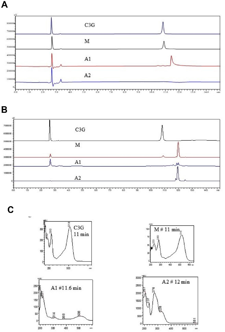
The total anthocyanin content (TAC) expressed in C3G was 1.47, 0.1, and 0.01% for samples M, A1, and A2, respectively. However, this methodology could suffer interference. Thus, chromatographical analysis were carried out. The LC-UV analysis also revealed the presence of C3G in the reference sample (M), but not in samples A1 or A2 (Figure 1A-B). This methodology aimed to separate C3G from the other components of C. sinensis juice extract. At visible radiation, 520 nm, the C3G showed a major peak at 10.8 min (Figure 1A), with a characteristic UV absorption profile (Figure 1C). Sample M showed the same chromatographical profile and UV absorption profile of C3G, while samples A1, and A2 showed a distinct composition (Figure 1A-B). Sample A1 showed a peak at 11.6 min, with a different UV absorption profile, compared with C3G (Figure 1C). Sample A2 did not show peaks at 520 nm (Figure 1A). On the other hand, at 285 nm, A2 showed more hydrophobic compounds, eluting at about 12 min, with a UV absorption profile characteristic of phenolic compounds (Figure 1B). These peaks were also visualized in the reference sample.

Chromatographical profile by LC-UV of C3G and samples at 2 mg/mL: M, A1, and A2, at 520 nm (A) and at 285 nm (B); UV absorption profiles of the major peaks (C).
Anthocyanins such as C3G absorb in the visible region, between 496 and 550 nm, as shown in Figure 1, whereas flavonoids absorb well between 350 and 380 nm (Fedenko, Shemet, Landi, 2016). Phenolic compounds present one or two UV absorption bands, with band 1 at 305-390 nm and band II at 230-300 nm (Spacil, Novakova, Solich, 2008).
The C3G peak showed good symmetry, and the RSD% of the area at 10 µg/mL was 0.59% (standard solution, n = 5). The concentration of C3G in the analysed samples was calculated by external standardization, based on the analytical curve of C3G at 1-100 µg/mL (r2 > 0.999, y= 27760 x + 1526). The C3G concentration of the reference was 1.6% and C3G was absent in A1 and A2. These results are similar to those found in the TAC analysis by spectrophotometry, as reported above, but different from those reported in the quality certificates (0.8% and 0.92% of C3G for the reference sample and A1, respectively). This difference may have been due to the origin of the fruits of C. sinensis, as the reference sample came from Italy, while sample A1 originated from Brazil, where climatic conditions are less favourable for the production of anthocyanins in the fruits (Latado et al., 2008). Studies with just one variety of blood orange, grown at different temperatures, showed that the accumulation of anthocyanins in fruits is positively influenced by the cultivation of plants in conditions of lower temperatures (Meredith, Young, 1969). Also Bitters (1961) verified that the cultivation of blood oranges in regions with a climate of hot days and cold nights favoured fruit production with more intensely coloured pulp. The discrepant results may also be due to the methodologies used by the suppliers, as the Brazilian supplier does not use the pH differential to measure anthocyanins, as informed by the technical support. A simple and fast test is proposed for compounding pharmacies to verify the presence of anthocyanins in these sample (de Meneses et al., 2023).
The precision of the analytical method was adequate, with a RSD of 0.97% using the sample M, by means of independent weighing in sextuplicate, and injections in triplicate, of each of these samples. According to the AOAC (2023), the RSD can be up to 2.7%.
Besides LC-UV, the samples were also analysed by LC-MS (Table I). It was observed that C3G was present in trace amounts in A1 and in a higher proportion in M, in agreement with the LC-UV and TAC analysis. Again, C3G was not detected in A2.
The amount of C3G was previously quantified by HPLC for three C. sinensis varieties (Sanguinello, Tarocco Rosso, and Tarocco Ippolito), and showed 7.95, 2.95 and 5.11 µg/100 g per edible portion of oranges, respectively (Cebadera-Miranda et al., 2019). Sommella et al. (2017) quantified C3G in two C. sinensis varieties juices, Torocco Lempso and Sanguinello reporting 0.92 and 0.11 µg/100 mL of C3G, respectively. These authors also reported the presence of other anthocyanins, especially Cyanidin-3-O-(6″-Malonyl) Glucoside. In addition, anthocyanins were not detected in C. reticulata species.
The phenolic compounds present in the samples were also evaluated using LC-MS; the results are summarized in Table I. A total of 24 phenolic compounds were identified in the present study. All three samples showed the presence of narirutin, hesperidin, neohesperidin, rutin, didymin, nobiletin, prunin, and tangeretin. In the literature, the isolation of naringin and hesperidin in high concentrations is reported for Moro orange (Sommella et al., 2017; Legua et al., 2022). In our study, naringin was found only in the A1 extract. Legua et al. (2022) observed the presence of vicenin-2, isorhamnetin, naringin, rutin, and hesperidin in 11 Citrus varieties, while Sommella et al. (2017) did not find isorhamnetin in the Sanguinello variety. Polymethoxyflavones nobiletin and sinensetin (Li et al., 2021, Cafeo et al., 2023) and tangeretin (Li et al., 2021) were also found in orange juices, not specifically including blood oranges.
Handmade juices showed different amounts of polymethoxyflavones, such as tangeretin, suggesting that these compounds are derived from flavedo and that their presence in industrial juices is a consequence of the industrial process itself (Leuzzi et al., 2000). Among the flavanone glycosides, handmade juice is mainly rich in hesperidin and narirutin, and among the flavones, higher concentrations of vicenin-2 (6,8-di-C-Glu-Apigenin) are also reported. In our investigation, vicenin-2 was only identified in samples A1 and A2, while tangeretin was found in all the extracts.
The reference extract (M) did not show cyanidin-3-O-(6″-malonyl glucoside), vicenin-2 and naringin, while A1 did not show cyanidin-3-O-(6″-malonyl glucoside) and only traces of C3G. Finally, A2 did not show any anthocyanin (Table I).
Antioxidant analysis by DPPH radical showed a similar EC50 value for the reference and A1 of 45.6 and 62.4 µg/mL, respectively, while A2 showed lower activity (EC50 315.1 µg/mL). An EC50 value ≤ 50 μg/ mL corresponds to high antioxidant activity; 50 μg/mL < EC50 ≤ 100 μg/mL to moderate activity; and EC50 > 200 μg/mL to no relevant antioxidant activity (Brito et al., 2014). Therefore, the reference and A1 presented moderate antioxidant capacity, while A2 showed no relevant antioxidant activity. Thus, sample A2 differed significantly from the other two extracts, with a different colour and chromatographic profile, the absence of anthocyanins (Figure 1), and lower antioxidant activity.
Some authors suggest that the anthocyanin content is the main factor that influences the antioxidant activity of blood orange. However, the content of other polyphenols may also contribute to this activity, as well as vitamins (Rapisarda et al., 1999; Cebadera-Miranda et al., 2019). Comparing the reference, M and A1 extracts, the former showed a higher content of C3G, but presented similar antioxidant activity. Analysing the Moro, Sanguinello, and Tarocco varieties, and two golden orange varieties, the antioxidant capacity was attributed, at least in part, to the total phenol content, while ascorbic acid appeared to play a minor role (Rapisarda et al., 1999).
The lycopene content was determined by spectrophotometry. Samples M, A1, and A2 showed lycopene content of 14.6 (± 2.3), 8.9 (± 0.8) and 4.5 (± 0.1) µg/g, respectively, suggesting that the red coloration of A1, in the absence or only traces of anthocyanins, may be due to the lycopene.
To assess the impact of the different compositions of the three commercial extracts of C. sinensis juice on nitric oxide release, the cell viability of macrophages was first evaluated using the MTT methodology. The data presented in Figure 2 indicate that all the extracts showed no cytotoxicity at any of the tested concentrations (0.1 - 10 µg/mL), with average viability greater than 90%. In the control group treated with DMSO, reduced viability was observed, as shown in Figure 2.

Macrophages RAW 264.7 (ATCC) were incubated in the presence or absence of extracts for a period of 21 h. After this period the cells were incubated for 3 h with MTT. Treatments with reference M (A), A1 (B) and A2 (C) extracts did not show toxicity at the concentrations tested. Values express the mean ± S.E.M. ####p < 0.001 vs basal. (One-way ANOVA followed by Tukey’s post hoc test).
Macrophages exposed to commercial dry extracts of C. sinensis juice exhibited different behaviours in the determination of NO levels, as seen in Figure 3. The reference sample was the only one that significantly reduced NO secretion, showing effectiveness at all concentrations (Figure 3A). A2 displayed a slight reduction, which was significant only at the concentration of 10 µg/mL (Figure 3B), while A1 did not exhibit any activity at any of the tested concentrations (Figure 3C). The reduction of NO in the reference extract indicates its inhibitory effect on both the expression of the iNOS enzyme and its activity, in accordance with studies conducted by Pepe et al., (2017), who evaluated the extract of C. sinensis from oranges from Italy.

Effect of different commercial extracts of C. sinensis juice on LPS-stimulated macrophages.
Effect of C. sinensis extracts on NO release. The quantification of nitrite was performed by Griess reaction. Treatments with reference M (A), A1 (B) and A2 (C). *p < 0.05; **p < 0.01; ***p < 0.001 and ****p < 0.0001 vs LPS group significantly different from basal group ####p< 0,0001 (One-way ANOVA followed by Turkey’s post hoc test).
This activity may also be attributed to the class of flavonoids present in the juice of C. sinensis, such as hesperidin, narirutin, and didymin, which are flavanones O-glycosides (Gattuso et al., 2007). Additionally, C3G is a compound known for its anti-inflammatory activity (Molonia et al., 2020). Hence, the higher content of this compound in sample M, which showed activity in the current study, suggests that the effect is probably correlated with the greater presence of this anthocyanin. The absence of in vitro anti-inflammatory activity observed in A1 and A2 appears to be a consequence of the different chemical composition from the reference sample, as demonstrated in the HPLC chromatographic profile (Figure 1, Table I).
The consumption of red orange juice improved endothelial functions and flow-mediated vasodilation, while also reducing inflammation in a clinical study (Buscemi et al., 2012). Therefore, the evaluation of anti-inflammatory activity proves to be a relevant tool for biomonitoring C. sinensis juice extracts. The antiadipogenic and antiobesity activity is mainly obtained by polyphenols, especially flavonoids, which are present in these extracts, acting directly on lipid metabolism and promoting a role in the treatment of adiposity, obesity and associated metabolic diseases (Moseti et al., 2016).
CONCLUSION
Together, the results of this work demonstrate the importance of the anthocyanin composition in C. sinensis juice extracts of blood oranges and highlight the influence of sample origin, given that the biosynthesis of these metabolites is influenced by environmental conditions. The chemical evaluation of the three commercialized extracts of C. sinensis suggests that samples come from different varieties, not necessarily blood oranges, or from different parts of the plant, as from the leaves of oranges rather than the juice. Thus, this work highlights the importance of monitor the presence of bioactive compounds in herbal drug derivatives marketed by compounding pharmacies in Brazil, aiming to ensure the identity and authenticity of blood orange derivatives and guarantee the quality of the products for the consumer.
REFERENCES
- Agência Nacional de Vigilância Sanitária (Brasil). Resolução RDC no. 67, of October 8, 2007. Dispõe sobre Boas Práticas de Manipulação de Preparações Magistrais e Oficinais para Uso Humano em farmácias. D.O.U. - Diário Oficial da União; Poder Executivo, de 09 de outubro de 2007.
- Amorini M, Lazzarino G, Galvano F, Fazzina G, Tavazzi B, Galvano G. Cyanidin-3-O-β-glucopyranoside protects myocardium and erythrocytes from oxygen radical-mediated damages. Free Rad Res. 2003;37(4):453-460.
- AOAC Official Methods of Analysis. Guidelines for Standard Method Performance Requirements. Appendix F.
- Bitters, WP. Physical characters and chemical composition as affected by scions and rootstocks. In: Sinclair, WB (Ed.). The orange: its biochemistry and physiology. Riverside: The University of California. 1961;56-95.
- Brito A, Ramirez JE, Areche C, Sepúlveda B, Simirgiotis MJ. HPLC-UV-MS Profiles of Phenolic Compounds and Antioxidant Activity of Fruits from Three Citrus Species Consumed in Northern Chile. Molecules. 2014;19(11):17400-21.
- Buscemi S, Rosafio G, Arcoleo G, Mattina A, Canino A, Montana M, et al. Effects of red orange juice intake on endothelial function and inflammatory markers in adult subjects with increased cardiovascular risk. Am J Clin Nutr. 2012;95:1089-1095.
-
Cafeo G, Satira A, Russo M, Mondello M, Dugo P. Determination of Oxygen Heterocyclic Compounds in Foods Using Supercritical Fluid Chromatography- Tandem Mass Spectrometry. Foods. 2023;12:3408-3420. https://doi.org/10.3390/foods12183408
» https://doi.org/10.3390/foods12183408 - Cardile V, Graziano AC, Venditti A. Clinical evaluation of Moro (Citrus sinensis (L.) Osbeck) orange juice supplementation for the weight management. Nat Prod Res. 2015;29(23): 2256-60.
- Cebadera-Miranda L, Domínguez L, Dias MI, Barros L, Ferreira ICFR, Igual M, et al. Cámara M () Sanguinello and Tarocco (Citrus sinensis [L.] Osbeck): Bioactive compounds and colour appearance of blood oranges. Food Chem. 2019;(270):395-402.
- de Meneses LCM, Lamego-Neta MI, Martins GC, Miniuki JL, Malheiros A, Farias I, et al. Adulteração de extratos secos de suco de Citrus sinensis (L.) Osbeck comercializados em farmácias magistrais: um relato de caso. Revista A Flora. 2023;Abril:20-21.
- Denizot F, Lang R. Rapid colorimetric assay for cell growth and survival. Modifications to the tetrazolium dye procedure giving improved sensitivity and reliability. J Immunol Meth. 1986;89(2): 271-277.
- Favela-Hernández JMJ, González-Santiago O, Ramírez-Cabrer MA, Esquivel-Ferriño PC, Camacho-Corona MDR. Chemistry and Pharmacology of Citrus sinensis. Molecules. 2016;21:247.
- Fedenko VS, Shemet SA, Landi M. UV-vis spectroscopy and colorimetric models for detecting anthocyanin-metal complexes in plants: An overview of in vitro and in vivo techniques. J Plant Physiol. 2017;212:13-28.
- Gattuso G, Barreca D, Gargiulli C, Leuzzi U, Caristi C. Flavonoid Composition of Citrus Juices. Molecules. 2007;12:1641-167.
- Giusti M, Wrolstad R. Characterization and Measurement of Anthocyanins by UV-Visible Spectroscopy. Curr Prot Food Anal Chem F1. 2001;2.1:1-13.
- Green LC, Wagner DA, Glogowski J, Skipper PL, Wishnok JS, Tannenbaum SR. Analysis of nitrate, nitrite, and [15N] nitrate in biological fluids. Anal Biochem. 1982;26(1):131-138.
- Grosso G, Galvano F, Mistretta A, Marventano S, Nolfo F, Calabrese G, et al. Red Orange: Experimental Models and Epidemiological Evidence of Its Benefits on Human Health. Oxid Med Cell Longev. 2013;2013:1-11.
- Kegele CS, Oliveira J, Magrani T, Ferreira A, Ferreira RS, Sabbaghi A, et al. A randomized trial on the effects of CitrusiM® (Citrus sinensis (L.) Osbeck dried extract) on body composition. Clin Nutr Experim. 2019;27:29-36.
- Latado RR, Tognato PC, Stenico MES, Nascimento LM, Santos PC. Acúmulo de antocianinas e características físicas e químicas de frutos de laranjas sanguíneas durante o armazenamento a frio. Rev Bras Fruticult. 2008;30:604-610.
- Legua P, Modica G, Porras I, Conesa A, Continella A. Bioactive compounds, antioxidant activity and fruit quality evaluation of eleven blood orange cultivars. J Agric Food Chem . 2022;102:2960-2971.
- Leuzzi U, Caristi C, Panzera V, Licandro G. Flavonoids in Pigmented Orange Juice and Second-Pressure Extracts. J Agric Food Chem . 2000;48:5501-5506.
- Li G, Rouseff R, Cheng Y, Zhou Q, Wu H. Comprehensive identification and distribution pattern of 37 oxygenated heterocyclic compounds in commercially important citrus juices. LWT - Food Science and Technology. 2021;152:112351-112361.
- Lo Piero AR. The State of the Art in Biosynthesis of Anthocyanins and Its Regulation in Pigmented Sweet Oranges [(Citrus sinensis) L. Osbeck]. J Agric Food Chem . 2015;63(16):4031-404.1
- Maccarone E, Campisi S, Fallico B, Rapisarda P, Sgarlata R. Flavor components of Italian orange juices. J Agric Food Chem 1998;46:2293-2298.
- Maccarone EA, Maccarone PR, Rapisarda P. Stabilization of anthocyanins of blood orange fruit juice. J Food Sci. 1985;50:901-904.
- Magalhães ML, De Sousa RV, Miranda JR, Konig FM, Wouters F, Souza FR, et al. Effects of Moro orange juice (Citrus sinensis (l.) Osbeck) on some metabolic and morphological parameters in obese and diabetic rats. J Sci Food Agric. 2021;101:1053-1064.
- Magalhães ML, Lima LCO, Lunguinho AL, De Carvalho DA, Rezende S, Ferreira VRF, et al. Influence of cold storage on the bioactivity properties and the quality of the juice of Moro blood orange (Citrus sinensis (L.) Osbeck). Am J Plant Sci. 2019;10:24-37.
- Mattioli R, Francioso A, Mosca L, Silva P. Anthocyanins: A Comprehensive Review of Their Chemical Properties and Health Effects on Cardiovascular and Neurodegenerative Diseases Molecules. Molecules. 2020;21(25): 3809.
- Meredith FI, Young RH. Effect of temperature on pigment development in red blush grapefruit and ruby blood oranges. In: Proceedings of the First International Citrus Symposium, 1., Riverside. Riverside: University of California. 1969;271-276.
- Molonia MS, Occhiuto C, Muscarà C, Speciale A, Bashllari R, Villarroya F, et al. Cyanidin-3-O-glucoside restores insulin signaling and reduces inflammation in hypertrophic adipocytes. Arch Biochem Bioph 2020;691:108488.
- Moseti D, Regassa A, Kim WK. Molecular Regulation of Adipogenesis and Potential Anti-Adipogenic Bioactive Molecules. Intern Jo Molec Sci. 2016;17:124.
- Pan X, Bi S, Lao F, Wu J. Factors affecting aroma compounds in orange juice and their sensory perception: A review. Food Res Intern. 2023;169:112835.
- Pepe G, Pagano F, Adesso S, Sommella E, Ostacolo C, Manfra M, et al. Bioavailable Citrus sinensis extract: Polyphenolic composition and biological activity. Molecules. 2017;22(623):1-15.
- Perkins-Veazie P, Collins JK, Pair SD, Roberts W. Lycopene content differs among red-fleshed watermelon cultivars. J Sci Food Agric . 2001;81(10):983-987.
- Rapisarda P, Tomaino A, Lo Cascio R, Bonina F, De Pasquale A, Saija A. Antioxidant effectiveness as influenced by phenolic content of fresh orange juices. J Sci Food Agric . 1999;47(11):4718-23.
- Reuther W, Webber HJ. The Citrus industry. History, World Distribution, Botany and Varieties. 1967;v.1.pp. xvi+611pp
- Rufino MSM, Alves RE, Brito ES, Morais SM, Sampaio CG, Pérez-Jiménez J, et al. Metodologia científica: determinação da atividade antioxidante total em frutas pela captura do radical livre DPPH. Comunicado Técnico on line 127, Fortaleza: Embrapa. 2007;1-4.
- Shu C, Wu S, Li H, Tian J. Health benefits of anthocyanin-containing foods, beverages, and supplements have unpredictable relation to gastrointestinal microbiota: A systematic review and meta-analysis of random clinical trials. Nutr Res. 2023;116:48-59.
- Sommella E, Pagano F, Pepe G, Ostacolo C, Manfra M, Chieppa M, et al. Flavonoid Composition of Tarocco (Citrus sinensis L. Osbeck) Clone “Lempso” and Fast Antioxidant Activity Screening by DPPH-UHPLC-PDA-IT-TOF. Phytochem Anal. 2017;28:521-528.
- Spacil Z, Novakova L, Solich P. Analysis of Phenolic Compounds by High Performance Liquid Chromatography and Ultra Performance Liquid Chromatography. Talanta. 2008;76:189-199.
- Titta L, Trinei M, Stendardo M, Berniakovich K, Petroni C, Tonelli P, et al. Blood orange juice inhibits fat accumulation in mice. Int J Obesity. 2010;34:578-588