 Formulation and evaluation of niosomal gel containing apremilast: optimization, in-vitro release and skin permeation studies
Abstract
The objective of this study was to develop, optimize and evaluate Apremilast niosomes integrated into a topical gel for improved skin permeation. Niosomes were prepared by thin film hydration technique using cholesterol and Poloxamer 188. Niosomes optimization was employed using BoxBehnken Design wherein three variables cholesterol concentration, Poloxamer 188 concentration and stirring speeds were studied on responses entrapment efficiency, particle size, drug release, and release kinetics. The optimized niosomes were further evaluated for zeta potential and surface characterization by transmission electron microscopy. The selected niosomes were loaded into Carbopol based gel and evaluated for appearance, pH, rheology, in vitro drug release and ex vivo skin permeation studies on excised rat dorsal skin. The niosomal formulations by BBD design exhibited entrapment efficiencies ranging from 53.43% to 76.36% and particle sizes from 204 nm to 282 nm. Sustained drug release profiles were observed, with drug release percentages between 60.01% and 99.11% after 10h. Based on the opted design space and experimentation, optimization of niosomes by thin film hydration using BBD was achieved. The optimized niosomes were assessed for particle size (274.7 nm), zeta potential (37.6 mV), entrapment efficiency (68.29%), drug release (88.36% after 10h), transmission electron microscopy (uniform distribution with spherical shape) and drug release kinetics showing Korsmeyer-Peppas (non-fickian) model as best fit for drug release. The niosomal gel was formulated with acceptable characteristics for appearance (uniform gel, without phase separation), pH (6.91), viscosity (1498.7 Pa.s) and drug content (96.5%). Similarly, conventional Apremilast gel was also prepared for drug release and skin permeation comparison. The in vitro release experiments indicated consistent and sustained drug release for 6h with 83.5% drug release compared to conventional gel which showed 3h with 97.1% of drug release. Skin permeation studies showed improved deposition of Apremilast with niosomal gel in the skin (1750µg) compared to the conventional gel formulation (731µg), indicating improved skin permeation and deposition of Apremilast in the skin by niosomal drug delivery. The results emphasize the potential of niosomal gel formulations for enhancing topical delivery of Apremilast. The optimized niosomes loaded into topical gel system improved skin permeation and drug deposition with the layers of the skin. These attributes show the potential to improve drug delivery across the skin for poorly permeable drugs. Future research shall focus on in vivo studies to establish efficacy and safety for therapeutic applications.
Keywords:
Apremilast; Niosomes; Box-Behnken Design; Topical drug delivery; Skin permeation.
INTRODUCTION
Psoriasis is a chronic, inflammatory skin condition that is distinguished by red, scaly regions. This condition can result in substantial discomfort and have a significant impact on patient's quality of life (Agarwal et al., 2022). Topical agents and systemic therapies, such as oral dosage forms and biologics, comprise the current treatment regimen for psoriasis (Shetty, Sherje, 2021). Nevertheless, a significant number of patients experience adverse effects from systemic treatments, resulting in suboptimal therapeutic outcomes and poor compliance (Shetty, Sherje, 2021; Gudjonsson et al., 2004; Lebwohl, 1999).
A first-in-class phosphodiesterase 4 (PDE4) inhibitor, Apremilast (APR), has demonstrated efficacy in the treatment of psoriasis by modulating the immune response and reducing inflammation (Schafer, 2012). It acts by elevating and preventing the degradation of cyclic adenosise mono phosphate (cAMP) levels which helps reducing the inflammatory cytokines such as tumor necrosis factor - TNFα, Interleukins (IL) 23, IL17 and other pro-inflammatory cytokines (Rapalli et al., 2021). It is presently available as an oral tablet, with a moderately lower oral bioavailability of approximately 73% (Anwer et al., 2019). The oral administration of APR has shown a significant improvement in Psoriasis Area Severity Index (PASI) indicating an improvement in psoriasis conditions for the patients. However, it has been noted to have adverse effects that include weight loss, vertigo, vomiting, diarrhea, upper respiratory infections, nasopharyngitis, depression and suicidal thoughts despite its efficacy (Label of Apremilast Tablet, 2024; Keating, 2017). Furthermore, patient noncompliance is exacerbated by the necessity for dose titration (Keating, 2017).
Apremilast's topical administration presents a promising alternative that has the potential to enhance patient compliance and reduce systemic adverse effects. Nevertheless, the drug's physicochemical properties continue to present a challenge in terms of achieving effective skin permeation and deposition. The poor solubility of APR (10 - 14.5 µg/mL) and modest lipophilicity (log p = 1.8) pose a problem for skin permeation (Parmar, Bansal, 2021). Hence, there is a need for alternate and novel ways to overcome the skin permeation to improve the efficacy of APR at the intended site of action, i.e., the affected skin.
Niosomes, which are nano-scaled vesicles that are produced by non-ionic surfactants, offer a viable solution to improve the delivery of topical drugs (Bhardwaj et al., 2020). They are an appealing alternative for the development of topical formulations due to their enhanced biocompatibility, simplicity of production, and improved stability (Gao et al., 2024; Riccardi, Baldino, Reverchon, 2024; Madan et al., 2020). The objective of this investigation is to optimize the therapeutic efficacy and skin permeation of a niosomal gel that contains APR. niosomal gel is to enhance the drug's deposition within the epidermis layers, thereby reduce the adverse effects associated with oral administration, and provide a sustained release of the drug (Kolahdooz et al., 2023; Rapalli et al., 2018). Thus, thereby eliminating the scope for side effects caused due to oral administration, also delivering the drug to the target site/organ for profound activity and eliminating the systemic exposure of APR.
MATERIAL AND METHODS
Material
APR (99.7%) was obtained as a gift sample from Orbicular Pharmaceutical Technologies Pvt. Ltd. Cholesterol (386.65 g/mol) and Poloxamer 188 (8400 Da), essential components for the preparation of niosomes, were provided by Orbicular Pharmaceutical Technologies Pvt. Ltd. Carbomer 974P, used as the gelling agent, was acquired from Lubrizol. Other excipients such as Propylene glycol, Glycerin, Propyl gallate, Edetate disodium, Phenoxyethanol, and Triethanolamine were obtained as gift samples from Orbicular Pharmaceutical Technologies Pvt. Ltd. All chemicals and reagents used were of analytical grade. Purified water from Milli-Q purification system (Millipore, USA) was used for the experiments.
Testing Excipient Compatibility Using Infrared Spectroscopy
Infrared spectroscopy (IR) was utilized to assess the compatibility between the drug APR and the selected excipients. Physical mixtures of APR with Cholesterol, Poloxamer 188, and Carbomer 974P were prepared in a 1:1 ratio, representing the drug and excipient combination. Additionally, a placebo mixture, containing the excipients without APR, was also prepared for comparison.
The prepared samples were initially analyzed after preparation of mixtures to establish a baseline. To further evaluate the stability of these mixtures under stress conditions, a second set of samples was subjected to an elevated temperature of 50°C for a duration of two weeks. The Infrared Spectrometer with ATR technique using ZnSe prism (Alpha II, Bruker) was employed to analyze both the initial and stressed samples, allowing for the identification of any potential interactions between the drug and excipients (Zaid Alkilani et al., 2022). Spectral range scanned was from 4000 cm-1 to 500 cm-1, with a resolution of 4 cm-1 in transmittance mode. Background scan of air was performed initially to remove the interferences. APR Sample (about 150 - 200 mg) was placed on ZnSe prism and fixed with sample presser to keep the sample in place. The IR scan was commenced in transmittance mode to capture the IR spectra. All the samples were analyzed with same analytical parameters.
Preparation of Apremilast Niosomes
For the development of niosomes laden with APR, ethanol was employed to dissolve Poloxamer 188 and cholesterol while the mixture was continuously stirred. After that, ethanol was employed to modify the volume to 100%. The inner wall of the roundbottom flask was coated with a thin, dry layer due to the rotary evaporation of this solution at a temperature range of 55° - 65°C in vacuum conditions. In a separate vessel, a pH 7.4 phosphate buffer was prepared, and APR was subsequently added to this phase. The shear homogenizer was then employed to homogenize the phase, resulting in a consistent nano dispersion. Under constant agitation, the RBF was gradually hydrated at 60°C with the addition of this APR phase. The hydrated phase was further homogenized to produce niosomes. The dispersion that resulted was centrifuged at 5°C, and the supernatant was discarded. To the dispersion, purified water was introduced, and it was subsequently agitated. This centrifugation and rinsing cycle was reiterated five times. In the end, the niosomes that were produced were kept in a desiccator (Soni et al., 2024). Table I provides the compositional details of niosomes along with the responses.
Design of Experimentation: Box Behnken Design
To optimize the formulation, Box Behnken Design (BBD) was selected (Design-Expert®). The design consisted of 17 experimental trials that were conducted at varying levels of cholesterol, Poloxamer 188, and swirling speed (Alnaim et al., 2023; Kim et al., 2019). The responses selected were entrapment efficiency (Y1), particle size (Y2) and drug release at 10 h (Y3). Table I displays the specifics of the experimental trials along with the responses.
Evaluation of Apremilast Niosomes
The physicochemical properties and drug release profiles of the prepared niosomes were subjected to a variety of evaluations. The evaluation parameters consisted of drug release at 10 hours, entrapment efficiency, transmission electron microscopy (TEM), and particle size and zeta potential (Rad et al., 2022).
Particle Size and Zeta Potential
The dynamic light scattering (DLS) technique was employed to measure the zeta potential and particle size of the APR niosomes. The Litesizer DLS 500 (Anton-Paar) was employed to conduct the measurements using disposable cuvette as sample holder with backscattering. Background scan was run using the blank solution without niosomes. In order to mitigate the effects of multiple dispersal, the samples were diluted with distilled water prior to measurement. The sample was loaded in cuvette and placed in the instrument slot for analysis and the instrument was run to determine the particle size of niosomes. Zeta potential was measured using the Litesizer DLS 500 (Anton-Paar) using the Omega cuvette as sample holder with an equilibration time of 60 seconds, Henry factor 1.5 and voltage in automatic mode. The sample was loaded in omega cuvette meant for determining zeta potential and placed in the instrument slot for analysis and the instrument was run to determine the particle size of niosomes. The stability and uniformity of the niosomes were assessed by recording the zeta potential and particle size distribution (Soni et al., 2024).
Entrapment Efficiency (%)
Ultracentrifugation was employed to ascertain the entrapment efficiency of APR in the niosomes. A highspeed centrifuge (Remi Lab) was employed to separate the free drug in the supernatant of the niosomal dispersion at 15,000 rpm for 30 minutes at 4°C. The supernatant was collected and filtered using 0.45µ PVDF filter and APR content was estimated in the supernatant to indirectly determine entrapment efficiency of APR in niosomes by HPLC method (Pandey et al., 2021). The entrapment efficiency was calculated using the following formula:
Transmission Electron Microscopy
Transmission electron microscopy (TEM) was employed to further verify the niosomes' size and morphology. Samples were prepared by applying a drop of the niosomal dispersion to a carbon-coated copper grid, which was subsequently stained with phosphotungstic acid (Qiu et al., 2021). Thermo Scientific™ Talos L120C TEM was used to observe the grid after it was air-dried at a voltage of 20-120 kV. The imaging was performed at Central Instrumentation Facility, School of Pharmaceutical Education & Research, Jamia Hamdard University, New Delhi (India).
Drug Release from Apremilast Niosomes
The dialysis bag diffusion technique has been used to assess the in vitro drug release profile of APR from the niosomes. A dialysis bag (12-14 kDa) containing a predetermined quantity of niosomal dispersion was subsequently immersed in 100 mL of phosphate buffer (pH 7.4) containing 0.15% sodium lauryl sulfate (SLS), 40% Dimethylsulfoxide (DMSO) and 20% Ethanol at 32°±0.5°C with continuous agitation at 100 rpm. Sink conditions were maintained as 100 mL of media was used and the APR solubility in the receptor fluid was found to be greater than 4X (El-Say et al., 2016).
Aliquots of the release medium were withdrawn and replaced with fresh buffer at predetermined intervals. The release profile over a 10-hour period was determined by calculating the cumulative percentage of substance released and plotting it against time (Soni et al., 2024).
Drug Release Kinetics of Apremilast Niosomes
The drug release kinetic modeling defines the dissolution or drug release mechanism. It is primarily a correlation between the drug release and drug release models (Gouda, Baishya, Qing, 2017). APR release from the niosomes was determined by fitting the in vitro drug release data of the formulations with different release kinetic models such as zero order model, first order model, Higuchi’s model, Korsmeyer-Peppas model, Hixson-Crowell’s model (Obeid et al., 2022). The zero-order release defines the drug release is independent of concentration and the release rate is constant. First order release defines the drug release in dependent on concentration of the drug in the dissolution medium. Higuchi’s model explains the drug release by diffusion mechanism, Korsmeyer-Peppas model defines the Fick’s law of diffusion and the ‘n’ value characterizes the diffusion mechanism. If n is ≤ 0.45, it denotes diffusion model of rug release. If n is > 0.45, it indicates anomalous drug release mechanism which is characterized by diffusion coupled with polymer erosion. The Hixson-Crowell equation describes the drug release by dissolution by changes in the surface properties of the particles (Gouda, Baishya, Qing, 2017). The model descriptions are as follows: Zero order model is equation is given as C0 - Ct = K0t
where Ct is the amount of drug released at time t, C0 is the initial concentration of the drug at initial, K0 is the zero-order rate constant. First order model is given as log C = log C0 -K1 t/2.303
where C0 is the initial concentration of the drug, C is the %of drug remaining at time t, and K1 is the first order rate equation expressed in per unit time. The Korsmeyer-Peppas model is given as F = Mt/M = Km tn
where F is a fraction of drug released at time t, Mt is the amount of drug released at time t, M is the total amount of drug in dosage form, Km is the release rate constant, and n is the release exponent. Hixon-Crowell’s model is given as Q01/3 - Qt1/3 = KHt
where Q0 is the initial amount of drug in the niosomes, Qt is the remaining amount of drug in the niosomes at time t, and KH is the Hixson-Crowell release constant. Higuchi’s model is given as Q = KH t1/2
where Q is cumulative amount of drug release at time t and KH is Higuchi constant.
Preparation Apremilast Niosomal Gel
The APR niosomal gel was prepared using Carbomer 974P as gelling agent with Triethanolamine as pH adjuster to form the gel. pH was adjusted between 7±1. Other excipients were also used as shown in the Table II. Weight make up to 100% was done using purified water and stirred for uniformity. The gel was filled into laminated tubes and heat sealed for further analysis. Niosomal formulation (APG1) and non-niosomal/conventional gel formulation (APG3) were prepared to check the impact of drug release from niosomal gel and conventional gel (Qiu et al., 2021).
Evaluation of Apremilast Niosomal Gel
Appearance and pH
Visual examination of the APG1 was conducted to assess its colour, consistency, and the presence of any particulate matter. In order to guarantee formulation stability, the gels were examined for uniformity and any phase separation (Shah et al., 2021). A digital pH meter (Hanna Instruments) was employed to determine the pH of the gel formulations. The glass electrode was dipped in the gel to conduct the measurement. Prior to conducting the measurements, the pH meter was calibrated with standard buffer solutions of pH 4.0, 7.0 and 10.0 (Sandeep, Reddy, Devireddy, 2014).
Rheology
The Modular Compact Rheometer (MCR 302) (Anton-Paar) was employed to ascertain the rheological characteristics of the gel formulations. For Flow curve, variable shear rate of 0.001s-1 to 1000 s-1 was used applied in rotational mode. For yield stress, variable shear strain from 0.01% to 100% under constant frequency of 10 rads-1 was applied in oscillation mode. For viscosity measurements, constant shear rate of 150 s-1 was applied in rotational mode. All the measurements were made at 25°C ± 0.5°C using PP25/S measuring system with a gap setting of 0.2 mm (Krishnaiah et al., 2014). Marketed product Omnigel® (Diclofenac diethylamine topical gel, Cipla) was used for comparison purposes.
Quantification of Drug Content using HPLC Method
High-performance liquid chromatography (HPLC) was employed to perform drug content of APR in the gel formulations. The drug was extracted by accurately weighing and dissolving a known quantity of the gel in Acetonitrile. The solution was analyzed using an HPLC system (Shimadzu LC-20AD) after being filtered through a 0.45 µm PVDF membrane filter using Agilent Zorbax SB Phenyl (250 mm X 4.6 mm, 5µ) with column at 25°C, flow rate of 1.0 mL/min and an injection volume of 10µL. The detection wavelength was set at 230 nm. This method was adopted form Apremilast monograph of the Indian Pharmacopoeia 2022. The drug content was quantified, and was expressed as a percentage of the labeled amount (Madan et al., 2020).
In Vitro Release Testing
An immersion cell apparatus was employed to assess the in vitro release profile of APR from the gel formulations. 200 mL of receptor fluid, which was composed of 0.9% saline solution with 20% DMSO and 40% Ethanol, was used to immerse the gel (approximately 1 g) in a dialysis membrane (1214 KDa). The receptor fluid was agitated at 30 rpm maintained at a temperature of 32°C ± 0.5°C. Sink conditions were maintained as 200 mL of media was used and the APR solubility in the receptor fluid was found to be greater than 4X (El-Say et al., 2016). Aliquots of the receptor fluid were withdrawn and replaced with fresh buffer at predetermined intervals.
Skin Permeation Studies
A vertical diffusion cell apparatus was used to conduct the ex vivo skin permeation study, with excised wistar rat’s dorsal skin serving as the membrane. A pH 7.4 phosphate buffer was used as the receptor fluid, with the receptor chamber containing 10 mL of this buffer solution. The study was conducted in a controlled environment with a temperature of 32° ± 0.5°C to replicate physiological conditions. The receptor fluid was consistently stirred at 400 rpm to ensure thorough mixing and maintain optimal conditions. Sampling was carried out at a 24-hour time point to evaluate the permeation of APR through the rat dorsal skin. At the end of the study, the skin was collected and the application surface was scrapped and washed using acetonitrile to collect the aliquot of APR on skin surface. The skin was then minced and vortexed with acetonitrile. Then the samples were kept in orbital shaker for 24 h for complete drug extraction (Pandey et al., 2021). The samples were centrifuged and the supernatant was filtered through 0.22µ filter and aliquot was collected to determine the APR level in skin (epidermis & dermis). The sample aliquot from receptor fluid was collected. All the samples were analyzed for APR quantification using HPLC method.
RESULTS AND DISCUSSION
Testing Excipient Compatibility Using Infrared Spectroscopy
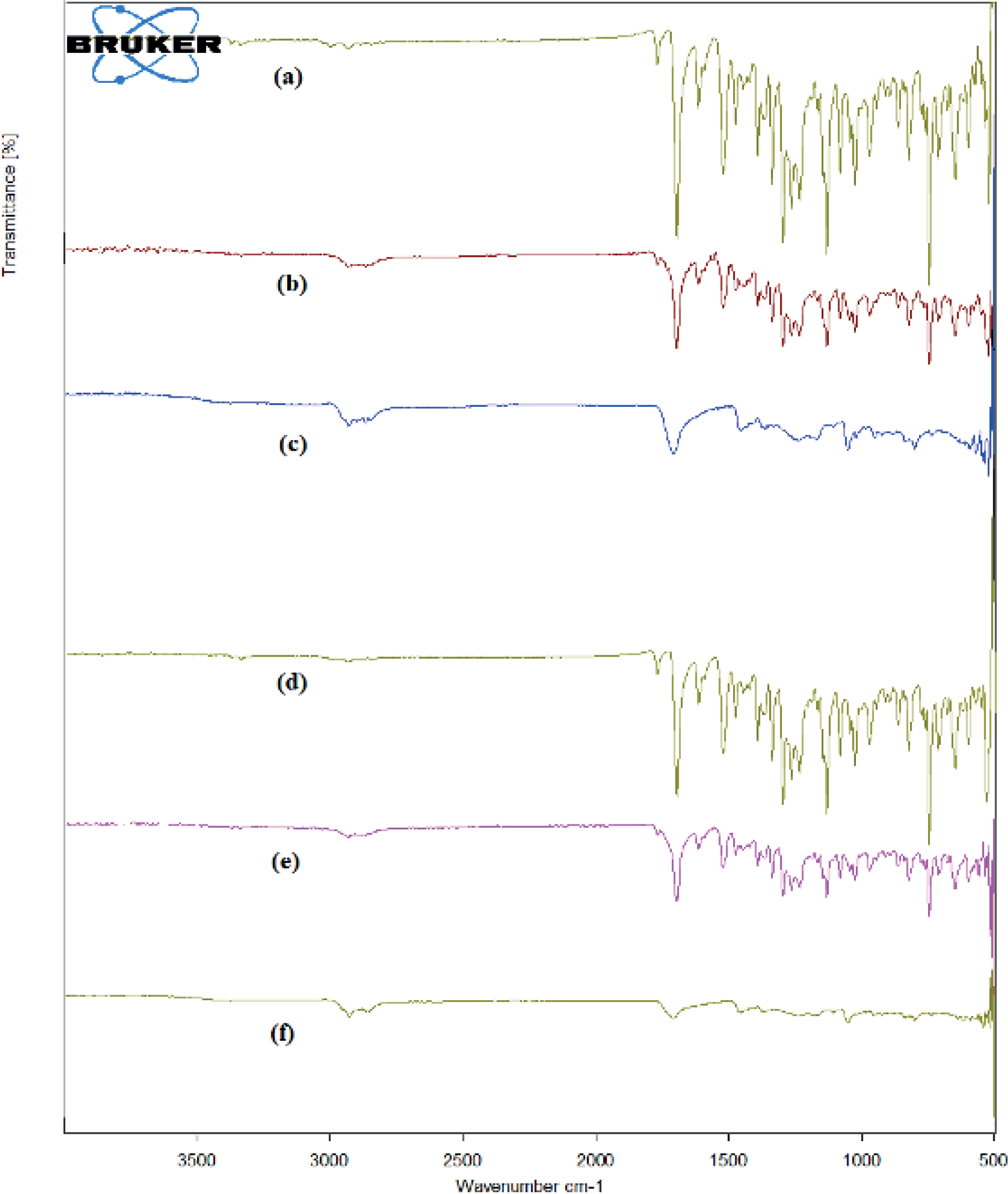
The Infrared (IR) spectra obtained by ATR technique, of APR, its mixture with Cholesterol, Poloxamer 188, and Carbomer 974P, as well as the placebo mixture containing Cholesterol, Poloxamer 188, and Carbomer 974P, are presented in Figure 1. The characteristic peaks of APR, observed at 3228 cm-1 (aromatic C-H stretch), 1504 cm-1 (aromatic C=C stretch), 1253 cm-1 (aromatic C-N stretch), and 1100 cm-1 (C-O stretch), were evident in the spectrum of APR at initial time-point, at room temperature (Figure 1a) (Rahangdale, Pandey, 2021).
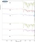
IR spectrum of APR (a); Physical mixture of APR, Cholesterol, Poloxamer 188, Carbomer 974P (b); Placebo blend (c) at initial stage. IR spectrum of APR (d); Physical mixture of APR, Cholesterol, Poloxamer 188, Carbomer 974P (e); Placebo blend (f) after exposure at 50°C for 2 weeks.
These characteristic peaks were also present in the spectrum of APR after being subjected to an elevated temperature of 50°C for two weeks (Figure 1d), indicating that APR maintains its thermal stability under these conditions. In the excipient mixture (Figure 1b), the APR peaks were observed, confirming the presence of the drug. Conversely, these peaks were absent in the placebo spectrum (Figure 1c), as expected.
Furthermore, the spectrum of the APR-excipient mixture after exposure to 50°C for two weeks (Figure 1e) showed that the APR peaks remained consistent with the initial spectrum, indicating no significant interaction or degradation of the drug. Similarly, the placebo spectrum after exposure to 50°C (Figure 1f) showed no changes, further confirming that APR is compatible with the selected excipients-Cholesterol, Poloxamer 188, and Carbomer 974P under the tested conditions.
Design of Experimentation Batches and Responses of Apremilast Niosomes
The experiment design involved the assessment of the impact of cholesterol, Poloxamer 188, and agitation speed on the entrapment efficiency, particle size, and amount of drug release of APR from niosomes after 10 hours. Table I illustrates the findings.
Entrapment Efficiency
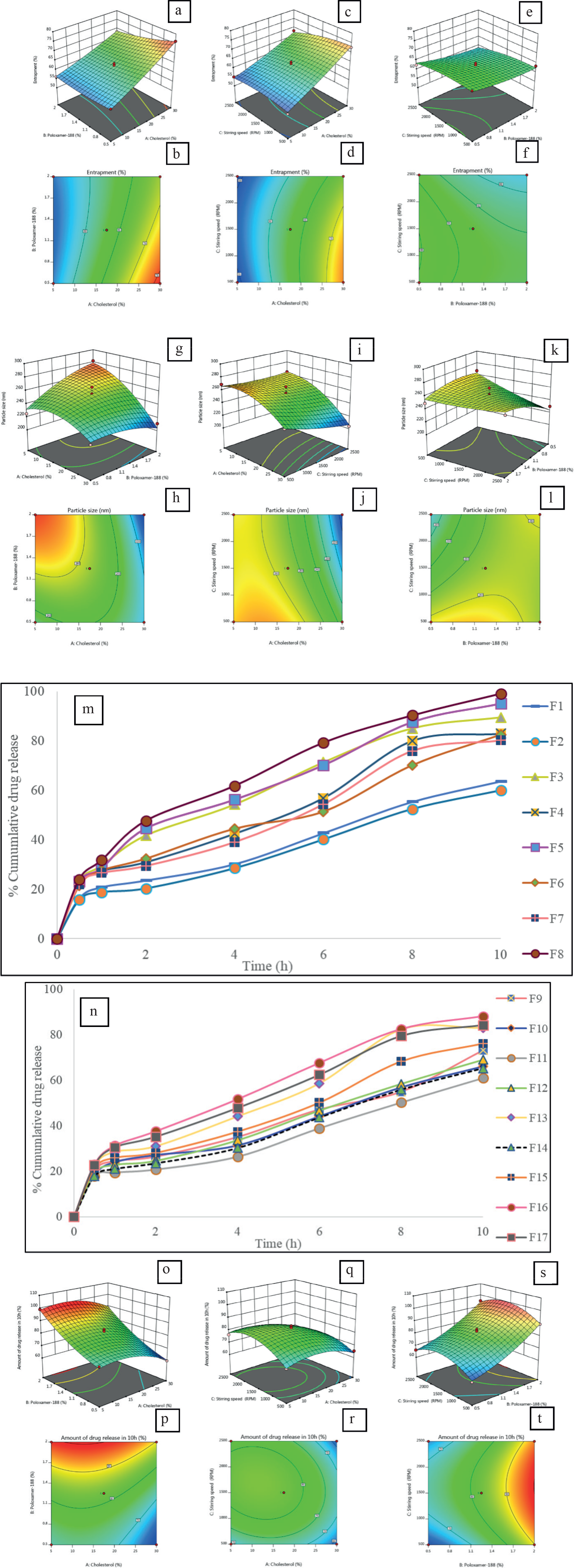
The levels of cholesterol, Poloxamer 188 and agitation speed significantly influenced the entrapment efficacy of APR in niosomes. It was observed that, formulations loaded with high amount of Cholesterol found high drug entrapment. Formulation run 2 has maximum 76.36%, run 14 and run 16 entrapped 72.71% and 68.25% respectively. Similarly, the percentage amount of Poloxamer-188 signified the entrapment efficiency as it can be seen that 0.5 mg of Poloxamer-188 in run 2 possessed highest entrapment. This could be due to emulsifying property of poloxamer-188 which entrapped lesser amount of APR. However, formulations with high amount of Cholesterol formed a hydrophobic barrier surrounding drug molecule in niosome formulation. Finally, it can be concluded that, a suitable combination of Cholesterol and Poloxamer-188 with preferable less quantity can develop a niosome with good entrapment (Goyal et al., 2015). Whereas, the rpm also contributed significantly; less processing at 500 rpm developed niosome with improved entrapment efficiency as seen in run 3 had 63.47%.
The quadratic model was significant (p < 0.0001, ANOVA) with a high F-value of 58.89. Cholesterol had the most significant effect on entrapment efficiency (F = 452.76, p < 0.0001, ANOVA), followed by Poloxamer 188 (F = 29.50, p = 0.0010) and churning speed (F = 16.41, p = 0.0049). Quadratic terms (A2 and C2) and interaction terms (AB and AC) were also significant. The residuals did not exhibit any significant lack of fit (p = 0.1287), suggesting that the model is well-fitted. The design space could be visualized in Figure 2a-2f, along with the optimal points.
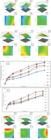
Response surface graphs showing the optimal points in the design space. 3D surface response curve (a) and 2D surface response curve (b) for the eff ect of Cholesterol and Poloxamer 188 on Entrapment Effi ciency; 3D surface response curve (c) and 2D surface response curve (d) for the eff ect of Cholesterol and Stirring speed on Entrapment Effi ciency; 3D surface response curve (e) and 2D surface response curve (f) for the eff ect of Stirring speed and Poloxamer 188 on Entrapment Effi ciency. 3D surface response curve (g) and 2D surface response curve (h) for the eff ect of Cholesterol and Poloxamer 188 on Particle size; 3D surface response curve (i) and 2D surface response curve (j) for the eff ect of Cholesterol and Stirring speed on Particle size; 3D surface response curve (k) and 2D surface response curve (l) for the eff ect of Stirring speed and Poloxamer 188 on Particle size. In vitro Drug Release from APR Niosomes for DOE runs 1 to 8 (m) and DOE runs 9 to 17 (n). 3D surface response curve (o) and 2D surface response curve (p) for the eff ect of Cholesterol and Poloxamer 188 on drug release after 10h; 3D surface response curve (q) and 2D surface response curve (r) for the eff ect of Cholesterol and Stirring speed on drug release after 10h; 3D surface response curve (s) and 2D surface response curve (t) for the eff ect of Stirring speed and Poloxamer 188 on drug release after 10h.
The polynomial equation for entrapment efficiency (Y1) is: 66.03+7.92A-2.02B-1.51C1.78AB-1.56AC-0.38BC+0.638A2+0.861B2-1.326C2.
Particle Size
The particle size of the niosomes was substantially influenced by the agitation speed, Poloxamer 188, and cholesterol. It was observed that the particle size ranged between 204 nm and 282 nm in the 17 BBD runs. The presence of high level of cholesterol usually leads to high particle size due to the formation of multilamellar vesicles (Rad et al., 2022). But it was observed that the particle size is an interplay of various factors such as cholesterol level, poloxamer level and process parameters like rpm. It was observed that the process parameter rpm had profound effect on the particle size of niosomes. A higher rpm had an inverse relation with the particle size.
The F-value of 83.06 was significant (p < 0.0001) in the ANOVA for the quadratic model. Poloxamer 188 (F = 507.32, p < 0.0001) and cholesterol (F = 66.50, p < 0.0001) had the most significant effects on particle size, while stirring speed also had a significant impact (F = 10.53, p = 0.0142, ANOVA). The quadratic terms A2 and C2 and the interaction terms AC were significant. This confirms the model's adequacy, as the residuals did not exhibit any significant lack of fit (p = 0.3092). The design space could be visualized in Figure 2g-2l, along with the optimal points.
The polynomial equation for particle size (Y2) is:255.2-21.5A+7.62B-8.12C-15.75AB-6.75AC +11.5BC-14.6A2-6.85B2+2.65C2
Drug Release from Apremilast Niosomes
The selected experimental variables had a substantial impact on the drug release after 10 hours as reflected in the drug release graphs in Figure 2(m) & 2(n). In vitro dissolution study of niosomes suggested by BBD design was evaluated. In all the formulations it was noted that, maximum 24.1% drug released in initial 30 minutes. It was found that, run 8 released maximum 99.91% of drug in 10hr. It was observed that there is direct relationship with the amount of Cholesterol and Poloxamer-188 in dissolution study. Though Cholesterol stabilizes the niosomal bilayer, the higher amount delayed drug release. Similarly, higher amount of poloxamer-188 increased the drug release. From the data available from the literature it revealed that, hydrophobic Cholesterol could develop a barrier surrounding the drug particle which can slow the process of drug release. Poloxamer-188 considered as emulsifi er while developing the formulation (Salem et al., 2021). It helps to emulsify as well as improve permeability of drug across the skin.
The quadratic model's ANOVA was signifi cant (p = 0.0014) with an F-value of 13.00. The release of the drug was signifi cantly infl uenced by cholesterol (F = 57.33, p = 0.0001), followed by Poloxamer 188 (F = 7.21, p = 0.0313) and swirling speed (F = 8.19, p = 0.0243). Quadratic terms (A2) and interaction terms (AB and BC) were statistically signifi cant. The residuals did not exhibit any signifi cant lack of fi t (p = 0.3268), suggesting that the model is well-fi tted. The design space could be visualized in Figure 2(o)-2(t), along with the optimal points.
The polynomial equation for drug release at 10th hour (Y3) is: 82.61-5.05A+13.95B+2.01C-0.62AB-2.23AC +0.10BC-5.9A2+3.47B2-8.14C2.
Evaluation of Optimized Apremilast Niosomes
The predicted optimized levels of Cholesterol, Poloxamer 188 and stirring speed were 23.82 mg, 1.61 mg/mL and 1282 rpm respectively. The predicted mean for the responses entrapment efficiency (Y1) was calculated to be 66.48%, particle size (Y2) was 240.33 nm and drug release at 10h (Y3) was 85.55%.
Entrapment Efficiency (%)
The predicted mean values were compared to the entrapment efficiency of APR niosomes. The entrapment efficiency that was observed was 68.29% ± 2.6%, which was marginally higher than the predicted mean of 66.48%. This suggests that the drug was effectively encapsulated within the niosomes during the formulation process, thereby ensuring a dependable delivery system.
Particle Size (nm) and Zeta potential (mV)
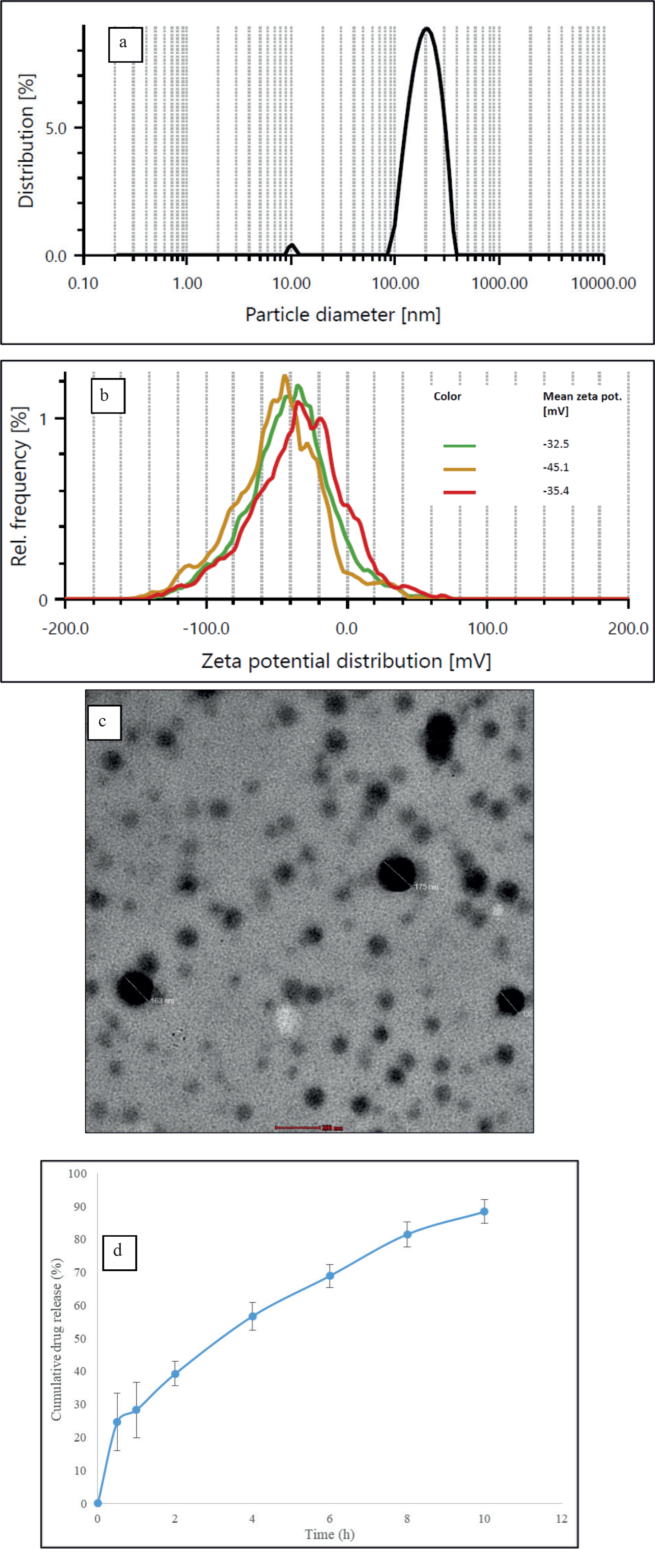
The mean particle size of 274.70 ± 42.7 nm was observed during the particle size analysis, which was higher than the predicted mean of 240.33 nm, as illustrated in Figure 3(a). The experimental conditions and minor variations in the formulation procedure may be responsible for this discrepancy. However, the particle size remained within an appropriate range for effective topical delivery, ensuring that the niosomes maintained adequate skin permeation and stability. Similarly, zeta potential was observed to be 37.6 ± 6.6 mV which suggests the existence of strong electric charges on the niosomal surface that obstruct the agglomeration, indicating an excellent physical stability (Shah et al., 2020). The zeta potential graph of optimized APR niosomes is provided in Figure 3(b).
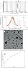
Evaluation of Optimized APR Niosomes showing Particle Size (a), Zeta Potential (b), TEM image (c) and Drug Release in 100 mL of pH 7.4 phosphate buff er containing 0.15% SLS, 40% DMSO, 20% Ethanol at 32°C.
Transmission Electron Microscopy
The morphology and size of the niosomes laden with APR were analyzed using TEM. The niosomes' uniform distribution and spherical shape were verified by the TEM images, which were consistent with the particle size analysis, as illustrated in Figure 3(c). The effectiveness of the formulation process is further validated by the structural integrity and homogeneity of the niosomes observed under TEM.
Drug Release
The observed quantity of drug released after 10 h was 88.36% ± 0.94%, which was slightly higher than the predicted mean of 85.55%, as indicated by the in vitro drug release profile, shown in Figure 3(d). This suggests that the therapeutic efficacy of APR for topical application is improved by the niosomal formulation, which provides a sustained release of the drug. The APR niosomal gel's capacity to deliver the drug over an extended period is indicated by the increased observed drug release, which reduces the necessity for frequent application.
Drug Release Kinetics of Apremilast Niosomes
The drug release data of APR from niosomes was applied to various release-kinetic models to determine the mechanism of release of drug from niosomal formulations. The best fit model for the release was evaluated by correlation using regression analysis (R2). The drug release data of APR niosomes was fitted into zero order, first order, Korsmeyer-Peppas, Hixson-Crowell and Higuchi models for all the 17 DOE runs and optimized niosomes, and the details are provided in Table III.
Regression Coefficient (R2) Values of Drug Release Kinetic Models for APR Released from Niosomes
Korsemeyer-Peppas was found to be the best fit model in all the cases with greater R2. The ‘n’ values were found to be in range of 0.48 to 0.58 which suggests the drug transport mechanism is by non-fickian (anomalous) indicating the drug release by diffusion coupled with surface erosion mechanism. These findings were in line to release kinetics for niosomes from the previous studies (Soni et al., 2024; Sambhakar et al., 2017).
Preparation of Apremilast Niosomal Gel
The gel's desired consistency and stability were achieved by incorporating a variety of excipients and APR concentrations (APR niosomes and APR as such) into each formulation as described in Table II.
Evaluation of Apremilast Niosomal Gel
The physical appearance, pH, rheology, and drug content of the gel formulations (APG1 and APG3) that were formulated, were assessed to confirm that they satisfied the criteria for topical application, as illustrated in Table IV.
Appearance and pH
The gels were uniform in appearance without phase separation and with no visible foreign particles. This suggests that the formulations were suitable for topical application and had excellent physical stability. The gel formulations' pH values, were 6.91 ± 0.12 for APG1 and 7.30 ± 0.09 for APG3, are within the permissible range for topical preparations, hereby guaranteeing skin compatibility. The pH variations between the formulations are indicative of the modifications that were implemented with triethanolamine.
Rheology
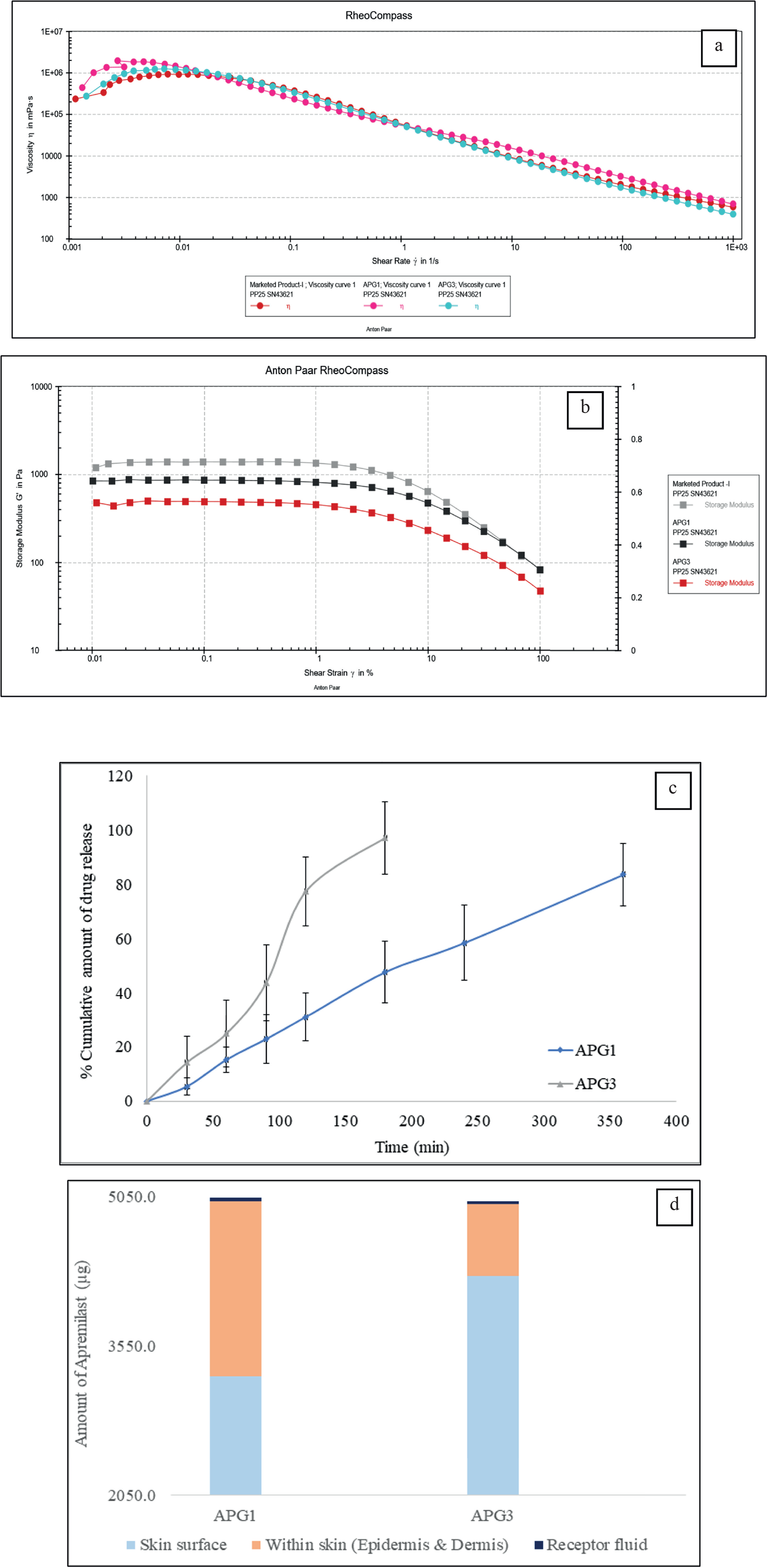
The flow curves of APG1 and APG3 were measured and compared with the marketed formulation, Omnigel®. The flow curves of the APR formulations were comparable to Omnigel® (Figure 4(a)). Similarly, the yield stress was also compared and found to be 99.24 ± 6.1 Pa for APG1, 77.32 ± 11.6 Pa for APG3 and 110.4 ± 9.6 Pa for Omnigel® (Figure 4(b)). This indicates the acceptable rheological and spread-ability properties of the APR formulations. This also indicates the acceptability of the formulation by the patients during administration of the gel on to the skin. The gel formulations' viscosities were also measured, with APG1 having a viscosity of 1498.7 ± 136.4 Pa.s and APG3 having viscosity of 1634.4 ± 124.7 Pa.s (Table IV), indicating an acceptable viscosity for administration of topical gels.
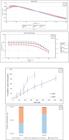
Evaluation of gels. Rheological assessment of gels - Flow curves (a) and Yield stress (b) of APG1 and APG3. In vitro release testing (c) and Skin permeation study (d) for APG1 and APG3 gels.
Quantification of Drug content Using HPLC Method
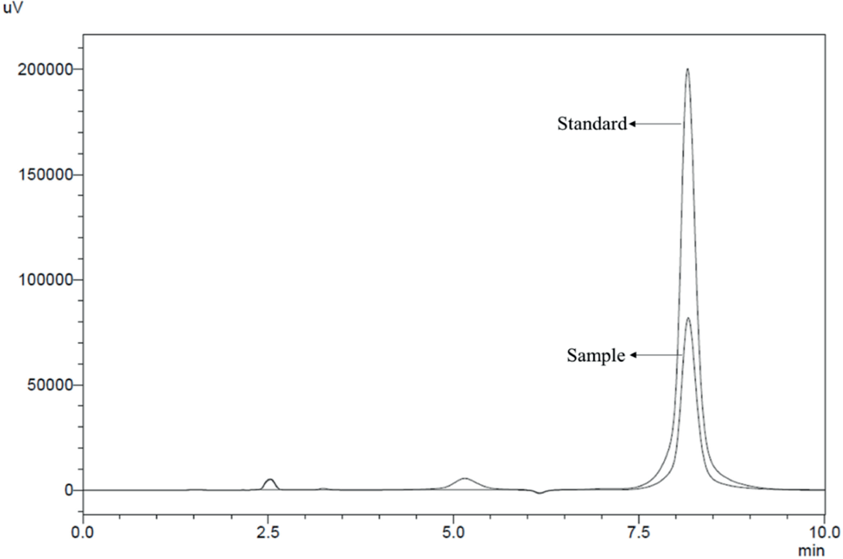
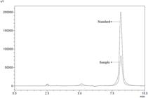
The gel formulations evaluated for drug content by HPLC were injected into the system along with the APR standard. The APR standard elution was found to ~ 7.6 min and the APR in the sample gel was also found to elute at the same time as APR standard (Figure 5). The gel formulations contained the intended quantity of the active pharmaceutical constituent, as evidenced by the drug content values for APR in APG1 was 96.5% ± 0.6% and APG3 was 98.2% ± 0.9%. This assures consistent drug loading, drug delivery and efficacy.
In Vitro Release Testing
The in vitro release profile of APR from the niosomal gel and conventional gel formulations was assessed using an immersion cell apparatus. The total quantity of drug discharged at different time intervals was quantified and is illustrated in Figure 4(c). The APG1 showed a drug release of 83.5% ± 9.4% by 6 h whereas APG3 showed a drug release of 97.1% ± 7.4% by 3 h. The niosomal formulation's efficacy in delivering sustained drug delivery is demonstrated by the release profiles, which indicate an extended and linear release pattern.
A linear and delayed release profile was observed in the niosomal gel formulations, suggesting the potential for sustained drug delivery. This sustained release is advantageous for topical applications, as it consistently delivers therapeutic levels of APR over an extended period, thereby reducing the frequency of application and improving patient compliance. Whereas for APG3, the drug release was only for 3 h duration as compared to APG1 which was for 6 h, indicating the sustained release of niosomes. The diff usion fl ux calculated as the slope of amount of drug released per unit area versus square root of time, was found to be 5.8 µg.cm-2.min-1 for APG1, which was slower to APG3 (11.3 µg.cm-2.min-1) indicating a slower drug release. Such a profi le may not only enhance therapeutic effi cacy by maintaining eff ective drug concentrations, but also reduces the frequency of application required and thereby improving patient compliance (Bhardwaj et al., 2022).
Skin Permeation Study
The ex vivo skin permeation study using excised rat dorsal skin showed notable variations in the distribution of APR across the diff erent layers of the skin and the receptor fl uid, which was infl uenced by the specifi c formulation employed. In Figure 4(d), it is evident that APG1 had the greatest concentration within the skin layers (epidermis and dermis), with 1750 ± 311.6 µg of APR detected. In contrast, the APG3 had signifi cantly lower amounts, with only 731 ± 187.8 µg. The drug remaining on the skin surface was consistent across all formulations, with APG1 measuring 3250 ± 958.8 µg. Regarding permeation into the receptor fl uid, APG1 demonstrated the highest concentration of 42.5 ± 8.9 µg, compared to 19 ± 5.3 µg of APG3, suggesting superior permeation compared to the other formulations. Based on the fi ndings, it appears that the niosomal gel formulation can improve the retention and localization of APR in the skin layers. This could potentially enhance the eff ectiveness of the topical treatment.
CONCLUSION
In conclusion, this investigation effectively developed APR niosomes and subsequently integrated them into niosomal gel formulations. The niosomal formulations were optimized exhibited using Box-Behnken design which showed sustained drug release profiles, adequate particle size for topical delivery, and high entrapment efficiency. Drug release kinetics were evaluated and found that the APR niosomal release followed Korsmeyer-Peppas model with non-fickian drug release mode. The APR niosomes were formulated into topical gel and further evaluated for acceptable pH, consistency, drug content in vitro release testing, which showed sustained drug release compared to conventional gel indicating enhanced drug effect and improving patient compliance. The ex vivo studies were performed on APR niosomal gel and APR conventional gel in which the niosomal gel formulation showed superior performance in enhancing skin permeation by retaining the drug within the targeted skin layers as observed in skin permeation studies. A concentration of 1750±311.6µg within the skin for APG1 formulation, showing more than double the concentration of APG3 provide the conclusive evidence for niosomes as drug delivery and skin permeation enhancers. These observations provide an improved and efficient way to deliver drugs and localize in the skin. In order to further establish the clinical efficacy and safety of these formulations, future research shall be focused on in vivo evaluations. Thus, the formulation of APR niosomal gel hold potential for increasing patient compliance and efficacy by enhancing skin permeation and providing sustained therapeutic effects.
ACKNOWLEDGEMENTS
The authors would like to express their gratitude to Orbicular Pharmaceutical Technologies Pvt. Ltd. for their invaluable support throughout the course of this research work. The authors would also like to thank KL College of Pharmacy for the support during the study.
DATA AVAILABILITY STATEMENT
All data is available within the article or its supplementary materials.
REFERENCE
-
Agarwal K, Das A, Das S, De A. Impact of psoriasis on quality of life. Indian J Dermatol. 2022;67(4):387-91. doi: 10.4103/ijd.ijd_572_22
» https://doi.org/10.4103/ijd.ijd_572_22 -
Alnaim AS, Shah H, Nair AB, Mewada V, Patel S, Jacob S, et al. Qbd-based approach to optimize niosomal gel of levosulpiride for transdermal drug delivery. Gels. 2023;9(3):213. doi:10.3390/gels9030213.
» https://doi.org/10.3390/gels9030213. -
Anwer MK, Mohammad M, Ezzeldin E, Fatima F, Alalaiwe A, Iqbal M. Preparation of sustained release apremilast-loaded PLGA nanoparticles: in vitro characterization and in vivo pharmacokinetic study in rats. Int J Nanomed. 2019;14:1587-95. doi:10.2147/ IJN.S195048.
» https://doi.org/10.2147/IJN.S195048 -
Bhardwaj P, Tripathi P, Gupta R, Pandey S. Niosomes: A review on niosomal research in the last decade. J Drug Deliv Technol. 2020;56:101581. doi: 10.1016/j. jddst.2020.101581.
» https://doi.org/10.1016/j.jddst.2020.101581 -
Bhardwaj P, Tripathi P, Pandey S, Gupta R, Ramchandra Patil P. Cyclosporine and Pentoxifylline laden tailored niosomes for the effective management of psoriasis: In-vitro optimization, Ex-vivo and animal study. Int J Pharm. 2022;626:122143. doi: 10.1016/j. ijpharm.2022.122143.
» https://doi.org/10.1016/j.ijpharm.2022.122143 -
El-Say KM, Abd-Allah FI, Lila AE, Hassan Ael-S, Kassem AE. Diacerein niosomal gel for topical delivery: development, in vitro and in vivo assessment. J Liposome Res. 2016;26(1):57-68. doi: 10.3109/08982104.2015.1029495.
» https://doi.org/10.3109/08982104.2015.1029495. -
Gao S, Sui Z, Jiang Q, Jiang Y. Functional evaluation of niosomes utilizing surfactants in nanomedicine applications. Int J Nanomed. 2024;19:10283-305. doi: 10.2147/IJN.S480639.
» https://doi.org/10.2147/IJN.S480639. -
Gouda R, Baishya H, Qing Z. Application of mathematical models in drug release kinetics of carbidopa and levodopa ER tablets. J Develop Drugs. 2017;6(2):1-8. doi:10.4172/2329-6631.1000171.
» https://doi.org/10.4172/2329-6631.1000171. -
Goyal G, Garg T, Malik B, Chauhan G, Rath G, Goyal AK. Development and characterization of niosomal gel for topical delivery of benzoyl peroxide. Drug Deliv. 2015 Dec;22(8):1027-42. doi: 10.3109/10717544.2013.855277.
» https://doi.org/10.3109/10717544.2013.855277. -
Gudjonsson JE, Johnston A, Sigmundsdottir H, Valdimarsson H. Immunopathogenic mechanisms in psoriasis. Clin Exp Immunol. 2004;135(1):1-8. doi: 10.1111/j.1365-2249.2004.02310.x.
» https://doi.org/10.1111/j.1365-2249.2004.02310.x. -
Keating GM. Apremilast: A review in psoriasis and psoriatic arthritis. Drugs. 2017;77(4):459-72. doi: 10.1007/s40265-017-0709-1.
» https://doi.org/10.1007/s40265-017-0709-1. -
Kim MH, Kim KT, Sohn SY, Lee JY, Lee CH, Yang H, et al. Formulation and evaluation of Nanostructured Lipid Carriers (NLCs) of 20(S)-Protopanaxadiol (PPD) by Box-Behnken Design. Int J Nanomed. 2019;14:8509-20. doi: 10.2147/IJN.S215835.
» https://doi.org/10.2147/IJN.S215835. -
Kolahdooz H, Khori V, Erfani-Moghadam V, Livani F, Mohammadi S, Memarian A. in Skin lesions of psoriatic patients: A pilot randomized controlled trial. Life. 2023;13:1076. doi: 10.3390/life13051076.
» https://doi.org/10.3390/life13051076. -
Krishnaiah YS, Xu X, Rahman Z, Yang Y, Katragadda U, Lionberger R, et al. Development of performance matrix for generic product equivalence of acyclovir topical creams. Int J Pharm. 2014;475(1-2):110-22. doi: 10.1016/j.ijpharm.2014.07.034.
» https://doi.org/10.1016/j.ijpharm.2014.07.034. -
Label of Apremilast Tablet (Otezla®). Available at: https://dailymed.nlm.nih.gov/dailymed/drugInfo.cfm?setid=f6b1f516-4972-4d82-bced-113e47b41cc5
» https://dailymed.nlm.nih.gov/dailymed/drugInfo.cfm?setid=f6b1f516-4972-4d82-bced-113e47b41cc5 -
Lebwohl M. The role of salicylic acid in the treatment of psoriasis. Int J Dermatol. 1999;38(1):16-24. doi: 10.1046/j.1365-4362.1999.00500.x.
» https://doi.org/10.1046/j.1365-4362.1999.00500.x. -
Madan JR, Khobaragade S, Dua K, Awasthi R. Formulation, optimization, and in vitro evaluation of nanostructured lipid carriers for topical delivery of Apremilast. Dermatol Ther. 2020;33(3):e13370. doi: 10.1111/dth.13370.
» https://doi.org/10.1111/dth.13370. -
Obeid MA, Khadra I, Aljabali AAA, Amawi H, Ferro VA. Characterisation of niosome nanoparticles prepared by microfluidic mixing for drug delivery. Int J Pharm X. 2022;4:100137. doi: 10.1016/j.ijpx.2022.100137.
» https://doi.org/10.1016/j.ijpx.2022.100137. -
Pandey SS, Shah KM, Maulvi FA, Desai DT, Gupta AR, Joshi SV, et al. Topical delivery of cyclosporine loaded tailored niosomal nanocarriers for improved skin penetration and deposition in psoriasis: Optimization, ex vivo and animal studies. J Drug Deliv Sci Technol. 2021;63: 102441. doi: 10.1016/j.jddst.2021.102441.
» https://doi.org/10.1016/j.jddst.2021.102441. -
Parmar PK, Bansal AK. Novel nanocrystal-based formulations of apremilast for improved topical delivery. Drug Deliv Transl Res. 2021;11:966-83. doi: 10.1007/s13346-020-00809-1.
» https://doi.org/10.1007/s13346-020-00809-1. -
Qiu F, Xi L, Chen S, Zhao Y, Wang Z, Zheng Y. Celastrol Niosome Hydrogel Has Anti-Inflammatory Effect on Skin Keratinocytes and Circulation without Systemic Drug Exposure in Psoriasis Mice. Int J Nanomed. 2021;16:6171-82. doi: 10.2147/IJN.S323208.
» https://doi.org/10.2147/IJN.S323208. -
Rad ME, Egil AC, Ince GO, Yuce M, Zarrabi A. Optimization of curcumin loaded niosomes for drug delivery applications. Coll Surfaces A. 2022;654:129921. doi: 10.1016/J.COLSURFA.2022.129921.
» https://doi.org/10.1016/J.COLSURFA.2022.129921. -
Rahangdale M, Pandey P. Development and Characterization of Apremilast Transethosomal Gel for Transdermal Delivery. Int J Pharm Sci Nanotechnol. 2021;14(3):5508-1. doi: 10.37285/ijpsn.2021.14.3.8.
» https://doi.org/10.37285/ijpsn.2021.14.3.8. -
Rapalli VK, Sharma S, Roy A, Singhvi G. Design and dermatokinetic evaluation of Apremilast loaded nanostructured lipid carriers embedded gel for topical delivery: A potential approach for improved permeation and prolong skin deposition. Colloids Surf B Biointerfaces. 2021;206:111945. doi: 10.1016/j. colsurfb.2021.111945.
» https://doi.org/10.1016/j.colsurfb.2021.111945 -
Rapalli VK, Singhvi G, Dubey SK, Gupta G, Chellappan DK, Dua K. Emerging landscape in psoriasis management: From topical application to targeting biomolecules. Biomed Pharmacother. 2018;106:707-13. doi: 10.1016/j.biopha.2018.06.136.
» https://doi.org/10.1016/j.biopha.2018.06.136. -
Riccardi D, Baldino L, Reverchon E. Liposomes, transfersomes and niosomes: production methods and their applications in the vaccinal field. J Transl Med. 2024;22:339. doi: 10.1186/s12967-024-05160-4.
» https://doi.org/10.1186/s12967-024-05160-4. -
Salem HF, Kharshoum RM, Abou-Taleb HA, Farouk HO, Zaki RM. Fabrication and appraisal of simvastatin via tailored niosomal nanovesicles for transdermal delivery enhancement: In vitro and in vivo assessment. Pharmaceutics. 2021 Jan 21;13(2):138. doi: 10.3390/ pharmaceutics13020138.
» https://doi.org/10.3390/pharmaceutics13020138 -
Sambhakar S, Paliwal S, Sharma S, Singh B. Formulation of risperidone loaded proniosomes for effective transdermal delivery: an in-vitro and in-vivo study. Bull Fac Pharm Cairo Univ. 2017;55(2):239-47. doi:10.1016/j.bfopcu.2017.09.003.
» https://doi.org/10.1016/j.bfopcu.2017.09.003. -
Sandeep G, Reddy VD, Devireddy SR. Formulation and evaluation of fluconazole pro-niosomal gel for topical Administration. J Appl Pharm Sci. 2014;4(7): 098-104. doi: 10.7324/JAPS.2014.40717.
» https://doi.org/10.7324/JAPS.2014.40717. -
Schafer P. Apremilast mechanism of action and application to psoriasis and psoriatic arthritis. Biochem Pharmacol. 2012;83(12):1583-90. doi: 10.1016/j. bcp.2012.01.001.
» https://doi.org/10.1016/j.bcp.2012.01.001 -
Shah A, Boldhane S, Pawar A, Bothiraja C. Advanced development of a non-ionic surfactant and cholesterol material based niosomal gel formulation for the topical delivery of anti-acne drugs. Mater Adv. 2020;1(6):176374. doi:10.1039/d0ma00298d.
» https://doi.org/10.1039/d0ma00298d. -
Shah P, Goodyear B, Dholaria N, Puri V, MichniakKohn B. Nanostructured Non-Ionic Surfactant CarrierBased Gel for Topical Delivery of Desoximetasone. Int J Mol Sci. 2021;22(4):1535. doi: 10.3390/ijms22041535.
» https://doi.org/10.3390/ijms22041535. -
Shetty K, Sherje AP. Nano intervention in topical delivery of corticosteroid for psoriasis and atopic dermatitis-a systematic review. J Mater Sci Mater Med. 2021;32(8):88. doi: 10.1007/s10856-021-06558-y.
» https://doi.org/10.1007/s10856-021-06558-y. -
Soni S, Baghel K, Soni ML, Kashaw SK, Soni V. Size-dependent effects of niosomes on the penetration of methotrexate in skin layers. Futur J Pharm Sci. 2024;10(48):1-18. doi: 10.1186/s43094-024-00624-2.
» https://doi.org/10.1186/s43094-024-00624-2. -
Zaid Alkilani A, Hamed R, Abdo H, Swellmeen L, Basheer HA, Wahdan W, Abu Kwiak AD. Formulation and evaluation of azithromycin-loaded niosomal gel: Optimization, in vitro studies, rheological characterization, and cytotoxicity study. ACS Omega. 2022;7(44):39782-93. doi: 10.1021/acsomega.2c03762.
» https://doi.org/10.1021/acsomega.2c03762.
Edited by
-
Associate Editor:
Marcos Bruschi