
 Screening of SIRT2 inhibitors from natural product databases using computer-aided drug design and molecular dynamics simulation
Abstract
This study is committed to searching for inhibitors of deacetylase SIRT2 within the natural product database via computer-aided drug design techniques. A comprehensive computer-aided drug design platform has been successfully established by integrating various techniques such as drug-likeness screening, pharmacokinetic prediction, molecular docking, and molecular dynamics simulation. The Traditional Chinese Medicine Systems Pharmacology Database and Analysis Platform (YaTCM) has been thoroughly explored to identify SIRT2 inhibitors, and the discovered compounds have been validated using molecular dynamics simulation. Through the computer-aided drug design method, five compounds capable of binding to SIRT2 have been successfully screened out from 47,696 natural product compounds derived from 6,220 herbs in the YaTCM database. Molecular dynamics simulation reveals that Artonin E and Paleatin B can form stable receptor-ligand complexes in the active pocket of SIRT2 inhibitors. Based on computer-aided drug design and virtual screening and verification techniques, Artonin E and Paleatin B have been identified as inhibitors of SIRT2, which is a key therapeutic target for the treatment of neuroblastoma. Applying computer aided drug design techniques to identify potential drug molecules from natural products holds profound significance for drug research.
Keywords:
SIRT2; Neuroblastoma; Computer-aided drug design; Molecular dynamics simulation; Molecular docking
INTRODUCTION
Neuroblastoma (NB) in children is one of the most prevalent solid tumors, especially in infants. It emerges from the neural crest of the sympathetic nervous system and exhibits a high degree of heterogeneity, with behaviors ranging from spontaneous regression to rapid growth and being challenging to treat (Zafar et al., 2021). Despite the progress in NB treatment, the prognosis for high-risk child patients remains extremely poor (Kameneva et al., 2021). MYCN is located on human chromosome and encodes the n-MYC protein, a transcription factor with a basic helix-loop-helix domain, which is closely associated with cell proliferation, growth, and metabolism. In approximately 20% of NB patients, the MYCN gene is amplified, which is a strong adverse prognostic factor (Bhavsar, 2023). The amplification of MYCN is closely related to the onset and prognosis of NB, thus making it an ideal therapeutic target. However, MYCN is a disordered protein, and it is difficult to intervene with traditional small-molecule drugs. Disordered proteins lack a stable tertiary structure, and their “undruggable” feature poses a significant challenge in drug design (Bartolucci et al., 2022). Although current research focuses on developing therapeutic strategies that can target MYCN, such as using small-molecule drugs to interfere with the binding of MYCN and its cooperative factors, due to the difficulties in directly targeting MYCN itself, researchers have begun to search for new therapeutic targets with “druggable” characteristics. This may include downstream signaling pathways of MYCN, interacting proteins involved in the stability and function of MYCN proteins, or regulatory factors involved in the MYCN gene amplification process (Bhardwaj, Das, Srinivasan, 2023).
It is particularly noteworthy that SIRT2 has the ability to significantly enhance the stability of n-Myc and c-Myc proteins, and simultaneously can promote the proliferation of cancer cells, which undoubtedly opens up a brand-new strategic direction for the treatment of NB (Liu et al., 2013). SIRT2 (Sirtuin 2) is essentially a deacetylase, and as a member of the Sirtuin family, plays a crucial role in many physiological and pathological processes of cells (Wu et al., 2023). Specifically, it can regulate gene expression, impact cell metabolism, participate in cell cycle process, and have a certain degree of influence on the occurrence and development of tumors (Kaya, Eren, 2024). Moreover, the function and activity of SIRT2 will also be regulated and controlled by various factors. In addition, SIRT2 plays an extremely key role in the occurrence and development processes of tumors such as prostate cancer, liver cancer, and colorectal cancer, (Fu et al., 2023; Wang et al., 2020). Through in-depth research of the Affymetrix gene array, it is found that the gene significantly inhibited by SIRT2 is the ubiquitin-protein ligase NEDD4. SIRT2 mainly combines directly with the core promoter of the NEDD4 gene and promotes the deacetylation of histone H4 lysine 16, thereby realizing the inhibition of the expression of the NEDD4 gene. And NEDD4 can directly bind to the Myc oncoprotein and treat the Myc oncoprotein as the target of ubiquitination and degradation (Liu et al., 2013).
Based on this, for small-molecule SIRT2 inhibitors, they can reactivate the expression of the NEDD4 gene, while reducing the expression of N-Myc and c-Myc proteins, thus achieving the effect of inhibiting the proliferation of NB and pancreatic cancer cells. Currently, small-molecule inhibitors targeting SIRT2 have become a research hotspot. However, the difficulty lies in that SIRT2 has a similar structure to other proteins in the Sirtuin family, which requires researchers to carefully design and vigorously develop new selective SIRT2 inhibitors to effectively reduce various side effects and adverse reactions caused by inhibiting other proteins in the Sirtuin family (Hamaidi et al., 2020; Singh et al., 2021). In this study, researchers used high-throughput virtual screening technology to actively explore acetylase SIRT2 inhibitors from the traditional Chinese medicine natural product library and strive to find selective inhibitors of SIRT2 according to the confirmed specific domain of SIRT2.
MATERIAL AND METHODS
Computer-Aided Drug Design (CADD)
Acquisition and preprocessing of the SIRT2 structure
Initially, the crystal structure of the SIRT2 target protein with PDB ID 5YQO is downloaded from the PDB database (https://www.rcsb.org/) (Yang et al., 2018). Pymol 2.6.0 is employed to visualize and remove water molecules and Zn2+ from the crystal structure, while retaining the conformation of SIRT2 and the selective inhibitor 24A. The Protein Preparation module of Schrödinger 2023-1 is utilized to preprocess the target protein, complete missing side chains and loop regions, and minimize the energy. Subsequently, the Protein Grid Generation module is used to define the active pocket of the target SIRT2 protein. The binding site coordinates of the original inhibitor 24A in 5YQO are taken as the center of the lattice box, and the space within 10 Å * 10 Å * 10 Å is designated as the inhibitor active pocket for this study.
Pre-treatment of the ligand library
The another Traditional Chinese Medicine database (YaTCM) (Li et al., 2018), a web-based free database developed by Nankai University, was utilized. A total of 47,696 Traditional Chinese Medicine (TCM) natural products were obtained from it to serve as a ligand library. The LigPrep module of Schrödinger 2023-1 was used to preprocess the molecules in the database, wich included protonation, desalination, hydrogenation, generation of tautomers, generation of stereoconformations, and energy minimization. Through preprocessing, a pre-screened compound ligand library of 58,048 was obtained.
Virtual screening
The QickProp module of Schrödinger 2023-1 was employed. Based on the drug-likeness rules “Lipinski Ro5”, “Verber Ro3” as well as ADME pharmacokinetic parameters, the first-round screening was conducted on the ligand library comprising 58,048 screened compounds. As a result, 22,227 molecules that conformed to the drug-likeness rules were selected. The ADME/Tox of the hit compounds was predicted using admetSAR (Yang et al., 2019). The hit compounds with poor pharmacokinetic properties and high toxicity were eliminated, and compounds with pharmacokinetic properties similar to the positive control 24A were chosen. The following 13 pharmacokinetic/hepatorenal toxicity indicators were referred to. (1) Molecular weight, with an ideal range of less than 650; (2) Hydrogen bond donors, having an ideal range of no more than 5; (3) Hydrogen bond acceptors, with an ideal range of no more than 10; (4) Solvent accessible surface area, having an ideal range of 300 to 1,000 Å2; (5) Compound polar surface area, with an ideal range of 7.0 to 200 Å2; (6) Oral bioavailability, with an ideal range of at least 50%; (7) LogS: The logarithm of the predicted solubility (mol/L) of the compound in a saturated aqueous solution, reflecting the compound’s water solubility. The ideal range is form -7.0 to 0.5; (8) LogHERG: The logarithm of the predicted IC50 of the compound for blocking the HERG K+ channel, with an ideal range of less than - 5; (9) Log (O/W): The predicted partition coefficient of the compound in octanol and water, reflecting oil-water partition coefficient, with an ideal range of - 2.0 to 6.5; (10) Apparent Caco-2 cell permeability: The human colorectal adenocarcinoma cell Caco-2 cell intestinal permeability model is a typical in vitro drug absorption model used to predict the absorbability of oral drugs. Less than 25 nm/s indicates very poor ability to penetrate the intestinal cell membrane, while more than 500 nm/s indicates very good ability to penetrate the intestinal cell membrane; (11) Hepatotoxicity; (12) Nephrotoxicity; (13) Acute oral toxicity: It refers to the toxic reaction of a substance to the human body in a short period of time after entering the human body through oral administration. It is divided into four levels. Generally it is considered that the acute oral toxicity (LD50) greater than 5,000 mg/kg is regarded as having no clinical exposure risk. Acute oral toxicity (LD50) between 500 and 5,000 mg/kg is considered mild toxicity (grade IV). Between 50 and 500 mg/kg is considered moderate toxicity (grade III). Theacute oral toxicity of an ideal candidate compound should be no more than grade III, which can be considered a relatively safe candidate drug.
Based on the receptor structure of the SIRT2 molecule, the second-round of screening was implemented. Initially, high-throughput virtual screening (HTVS) was conducted on the compound library screened in the previous step against the target SIRT2. According to the Glide Grid module of Schrödinger 2023-1, the top 10% compounds with the highest scores were extracted. Subsequently, standard precision screening (SP) was carried out. The screened compounds were ranked again according to the Glide Score , and the top 10% compounds with the highest scores were obtained. Finally, extra precision screening (XP) was performed. In accordance with the Glide Grid module of Schrödinger 2023-1, the top 10% compounds with the highest scores were retained. Based on flexible docking, the third-round of screening was executed. For the compounds screened in the previous step, flexible docking was conducted. The top 5 compounds with the highest scores were extracted as the final candidate compounds for this project. The binding mode of SIRT2-screened compounds was analyzed using Schrödinger 2023-1 software.
Molecular Dynamics (MD) Simulation
The computing resources employed in this simulation consisted of a Dell T3660 workstation equipped with the Ubuntu 20.04.01 operating system, an Intel Core i9 -13900k CPU, and a GeForce RTX 4070 TI SUPER GPU. The structure of SIRT2 was retrieved from the PDB database (PDB ID: 5YQO), and the heavy atoms and small molecules of the protein were repaired using SPDBV 4.10 software. The topological structure of the protein was calculated using the AMBER99SB-ILDN force field embedded in GROMACS(2023.1 single-precision version), while the topological structure of the hit compounds was computed using the AMBER force field of the online tool Acpype (http://bio2byte.be/acpype/) (Kagami et al., 2023; Sousa Da Silva et al., 2012). The complex was solved in the TIP3P water model and immersed in a cube box extending at least 1 nm on each side. By adding Na+ and Cl-ions, the system was neutralized to 0.15 M NaCl solution, bringing the system closer to the physiological state and achieving electrical neutrality. Subsequently, energy minimization and pre-equilibrium were carried out to eliminate interatomic conflicts. The steepest descent algorithm was used for 5,000 steps of energy minimization, keeping the maximum force below 1,000 kJ/mol/nm. The system was then subjected to 100 ps of the number of particles, volume, temperature (NVT) and number of particles, pressure, temperature (NPT) equilibria to ensure a well-balanced and stable system at 36.85°C (310 K). Finally, a 100 ns MD simulation of the complex was conducted using a Verlet truncation scheme with a step size of 2 fs and a Leap-frog integrator, and the trajectory data was saved every 10 ps. For each SIRT2 -hit compound system, 5 independent 100 ns MD simulations were randomly performed. Various MD indicators were utilized to evaluate the interaction between the screened compounds and SIRT2, including the Root-Mean-Square Deviation (RMSD), the Root-Mean-Square Fluctuation (RMSF) of the amino acid residues, the Radius of Gyration (Rg), molecular mechanics Poisson-Boltzmann surface area (MM/PBSA) and Free Energy Landscape (FEL).
RMSD
Use the “gmx rms” command to calculate RMSD, which initially employs the least-squares method to fit the structure to the reference structure (t2 = 0), and subsequently quantifies RMSD based on the following formula:
Where, is the the position of the atom i at frame t. In this research, unless explicitly stated otherwise, the RMSD calculations pertain solely to the protein backbone (Fatriansyah et al., 2022).
Radius of Gyration
Use the “gmx gyrate” command to calculate Rg, the Rg value is determined using the following formula:
Where, mi is the molecular weight of atom i, ri is the position of atom i relative to the center of the molecule (Ali et et al., 2023).
RMSF
Use the command “gmx rmsf”, and set relevant parameters simultaneously to ensure that the RMSF calculation is only performed on the backbone of the protein. The RMSF value is determined using the following formula:
Where, ri (t) is the position vector of the atom i at frame t and is the average atom position over all T frames (Mahdi et al., 2023).
MM/PBSA
Gmx-MM/PBSA 1.6.1 was utilized to calculate the MM/PBSA binding free energy of protein-ligand complex (Miller et al., 2012; Valdés-Tresanco et al., 2021). The MD simulation trajectories from 50 to 100 ns were exploited to calculate ∆Gbind of the complex. The representation of ∆Gbind of the protein-ligand complexes was calculated using the following formula:
ΔEMM is the gas-phase interaction energy; ΔGsolv is the solvation free energy, and TΔS represents the change in conformational entropy. Among them, ΔEMM includes Δ𝐸𝑒𝑙𝑒 and Δ𝐸𝑣𝑑W , and ΔGsolv is the interaction energy between solvent molecules and protein and ligand molecules, which can affect ∆Gbind . In this study, due to the high computational cost and low prediction accuracy of the change in conformational Entropy (TΔS), it was not considered here (Chen et al., 2018).
FEL
This study aims to evaluate the affinity and binding stability of SIRT2 and the hit compounds by means of it. The translational and rotational motions of the MD simulation trajectory were corrected using the gmx trjconv module. The FEL of the protein-ligand complex was calculated using the gmx sham module with RMSD and Rg as characteristic variables. The FEL was calculated using the following formula:
kB is Boltzmann’s constant, T is the absolute temperature, Ni is the probability density in MD data, and N max represents the maximum probability density in MD data (Chong, Im, Ham, 2019).
RESULTS
Analysis of the Results of Virtual Screening
The key breakthrough in the quest for selective SIRT2 inhibitors resides in screening inhibitors that target specific structural domains distinguishing SIRT2 from other proteins within the Sirtuins family. SIRT2 exhibits certain disparities from other Sirtuins family proteins at the interface of the Rossmann folding domain and the zinc-binding domain (near the α4 and α5 helices and loop N). The analysis of the binding mode between 24A and SIRT2 is depicted in Figure 2F. It indicates that 24A can establish hydrogen bond interactions with Arg97 (loop N) and Val233, and its aromatic ring can form Pi-Pi interactions with Phe190 and Phe235. Meanwhile, 24A is encapsulated within the hydrophobic pocket constituted by Phe119 (α4), Phe131 (α5), Leu134 (α5), Leu138 (α5), Pro140, Phr143, Phe96 (loop N), and Phe235.

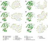
The 3D/2D combination mode of flexible molecular docking between the hit compounds and SIRT2. (A) SIRT2-Artonin E; (B) SIRT2-Paleatin B; (C) SIRT2-Neohydnocarpin; (D) SIRT2-Reticuline; (E) SIRT2-Sigmoidin B; (F) SIRT2-24A.
Referring to the binding mode of 24A with SIRT2, we found that 24A precisely binds to the distinct structural domain of SIRT2 (near the α4 and α5 helices and loop N), which distinguishes it from other proteins in the Sirtuins family. Consequently, 24A is highly likely to present a new opportunity for the design of SIRT2 selective inhibitors. Based on this, in this study, the selective inhibitor 24A of SIRT2 was selected as the reference ligand. A docking box with a 10 Å range centered around it was established for screening SIRT2 inhibitors. Subsequently, multiple rounds of virtual screening were carried out on 47,696 single compounds of TCM from 6,220 kinds of Chinese herbal medicines collected in the YaTCM database. The screening process is as shown in Figure 1.
The top 5 hit molecule structures with flexible docking scores were screened out, as shown in Figure 2. All of these compounds can bind well to the preset inhibitor active pocket of SIRT2. Specifically, as illustrated in Figure 2A, Artonin E binds to the active cavity of SIRT2. Its hydroxyl group forms hydrogen bond interactions with Pro94 (loop N), Asn168, and Asp170, while its aromatic ring forms Pi-Pi interactions with Phe96 (loop N). As shown in Figure 2B, Paleatin B binds to the active cavity of SIRT2. Its hydroxyl group forms hydrogen bond interactions with Thr101, and its aromatic ring forms Pi-Pi interactions with Phe96 (loop N) and Phe190. As shown in Figure 2C, Neohydnocarpin binds to the active cavity of SIRT2. Its hydroxyl group forms hydrogen bond interactions with Leu138 (α5) and Asp170, and its aromatic ring forms Pi-Pi interactions with Phe96 (loop N). As shown in Figure 2D, Reticuline binds to the active cavity of SIRT2. Its hydroxyl group forms hydrogen bond interactions with Pro94 (loop N), Phe96 (loop N), and Asp170, and its aromatic ring forms Pi-Pi interactions with Tyr139 (α5). As shown in Figure 2E, Sigmoidin B binds to the active cavity of SIRT2. Its hydroxyl group forms hydrogen bond interactions with Leu138 (α5) and Val233, and its aromatic ring forms Pi-Pi interactions with Phe96 (loop N). The predicted pharmacokinetic parameters of the aforementioned hit compounds and the positive control are presented in Table I and Table II. The admetSAR (Cheng et al., 2012;Yang et al., 2019) prediction shows that the positive control 24A and Sigmoidin B may have liver toxicity. In response to the above screening results, further virtual validation was conducted using MD simulation.
Analysis of Thermodynamic Properties and Binding Energies in SIRT2-Hit Compounds Complexes
In this study, 5,000 frames of trajectory files were extracted from the MD trajectories ranging from 50 to 100 ns for calculating ΔGbind of MM/PBSA and various associated energy contributions (such as ΔEvdW, ΔEelec, ΔGsolv, etc.). The aim is to evaluate and analyze the thermodynamic properties of the system (such as stability) at the energy level and explore the energy differences of the protein-ligand complexes under different states and their impacts on the binding stability. As presented in Table III, the order of ΔGbind as follows: SIRT2 - 24A > SIRT2 - Artonin E > SIRT2 - Neohydnocarpin > SIRT2 - Paleatin B > SIRT2 - Sigmoidin B > SIRT2 - Reticuline. Some studies have demonstrated that among the various energies constituting ΔGbind, the ΔEvdW is the most crucial and plays a leading role in the binding of hit compound to the target. Regarding the positive control 24A, it has a relatively strong ΔEvdW, a weaker ΔEelec, with the ΔEvdW being dominant, and a lower ΔGsolv, which is beneficial for the binding of 24A to SIRT2. In the hit compounds Artonin E, Paleatin B, Sigmoidin B, and Neohydnocarpin, energy distribution characteristics similar to those of the positive control 24A are observed, that is, the ΔEvdW is dominant and the ΔEelec is relatively weak. Sigmoidin B shows a relatively strong ΔEelec, but its ΔGsolv is relatively high, resulting in a relatively low overall binding free energy. In conclusion, among the above six protein-ligand complex systems, the binding energies are relatively strong, and the ΔGbind of these systems are all below -30 kJ/mol.
RMSD Analysis of SIRT2-Hit Compounds Complexes
In this study, we conducted rigorous simulation experiments on each protein-ligand complex system. To ensure the reliability and representativeness of the data, we repeated the simulations for each system five times. RMSD acts as a crucial indicator and plays a vital role in our research. It is employed to precisely measure the structural dynamic change conditions of the system and can vividly depict the degree of dynamic behavior change of molecules during the simulation process from a unique kinetic perspective, thereby providing a solid basis for us to judge the stability of protein-ligand binding. Through in-depth analysis of RMSD, we are able to thoroughly explore the intrinsic characteristics of the protein-ligand complex system. As shown in Figure 3 (A, C, E, G, I, K), the RMSD fluctuation situations of the protein-ligand complex throughout the simulation process are clearly presented. During detailed simulation observations, we found that in the third repeat simulation of SIRT2 -Reticuline, this system failed to converge to equilibrium successfully. This phenomenon clearly indicates that within the simulation time span of up to 100 ns, the SIRT2 -Reticuline system cannot achieve stable binding. To comprehensively evaluate the convergence of each system and ensure the high reliability of the research strategy, we carefully calculated the distribution characteristics of RMSD. The concentrated relative frequency presented by the normal distribution curve is of great significance as it can intuitively reflect whether the binding characteristics of the hit compound to SIRT2 are stable and repeatable. Similarly, Figure 3 (B, D, F, H, J, L) shows the normal distribution curves of RMSD fluctuations of the protein-ligand complex throughout the simulation process. After in-depth analysis of the five independent simulations of SIRT2 -Neohydnocarpin, we discovered that the relative frequency distribution of its RMSD values is relatively dispersed and the coincidence degree of the normal distribution curves is at a low level. This result clearly implies that the simulation repeatability of SIRT2 -Neohydnocarpin is poor. Based on the comprehensive and in-depth RMSD analysis results above, in the MD simulations that lasted for 100 ns and were repeated five times, the protein-ligand complexes SIRT2 -Artonin E, SIRT2 -Paleatin B, and SIRT2 -Sigmoidin B demonstrated remarkable characteristics. The fluctuation situations of their RMSD values and the good repeatability shown in the simulation results strongly suggest that Artonin E, Paleatin B, and Sigmoidin B can achieve stable binding with SIRT2. Thus, we have sufficient reasons to speculate that Artonin E, Paleatin B, and Sigmoidin B are highly likely to be potential inhibitors of SIRT2, providing extremely valuable clues and directions for subsequent in-depth research and application.

Display the backbone RMSD images of all complexes and the normal distribution curve conditions of their relative frequencies during the 100 ns MD simulation. (A, B) for SIRT2-Artonin E; (C, D) for SIRT2-Paleatin B; (E, F) for SIRT2-Neohydnocarpin; (G, H) for SIRT2-Reticuline; (I, J) for SIRT2-Sigmoidin B; (K, L) for SIRT2-24A.
RMSF Analysis of SIRT2-Hit Compounds Complexes
This study aims to further analyze the fluctuation status and stability of key residues in the active pocket of the complex system. To achieve this, the RMSF of the receptor-ligand complexes formed by Artonin E, Paleatin B, Sigmoidin B, and 24A with SIRT2 within the time range from 0 to 100 ns was calculated. RMSF, a crucial parameter, can reflect the positional variation situations of each amino acid residue within the protein structure during the simulation process. By monitoring the changes in RMSF values of different regions of the receptor when it binds to the ligand, the stability of key parts of the receptor, such as the amino acid residues around the binding site, can be assessed. A relatively low RMSF value indicates that the structure of the receptor in that region is relatively stable, which is beneficial for forming a stable binding with the ligand. As shown in Figure 4 and Table IV, compared with single SIRT2, the RMSF fluctuation values of the 14 key amino acid residues (Ile93, Pro94, Asp95, Phe96, Arg97, Ile118, Phe119, Phe131, Leu134, Ala135, Leu138, Tyr139, Pro140, Phe143) around the active pocket of the SIRT2 selective inhibitor in the SIRT2 Artonin E and SIRT2 -Paleatin B systems are smaller in most positions than those of the single SIRT2 protein. Moreover, their fluctuation amplitudes and characteristics are basically consistent with those of the positive control SIRT2 -24A. However, the RMSF value fluctuation situation of the SIRT2 -Sigmoidin B system in these key residues is contrary to that of the positive control SIRT2 24A system, and most of them are higher than those of the single SIRT2 protein. In conclusion, the RMSF experimental results suggest that Artonin E and Paleatin B can be encapsulated in the selective active pocket of SIRT2 to form a more stable receptor-ligand binary complex and are expected to become inhibitors of SIRT2.

Display the backbone RMSF images of all complexes during the 100 ns MD simulation. (A) SIRT2-Artonin E; (B) SIRT2-Paleatin B; (C) SIRT2-Sigmoidin B; (D) SIRT2-24A; (E) SIRT2
RMSF values of key amino acid residues within the SIRT2 active site pocket in all complexes during 100 ns MD simulation
FEL Analysis of SIRT2-Hit Compounds Complexes
In this study, to more intuitively illustrate the stability of the binding between the target protein SIRT2 and the hit compounds, FEL of the complexes formed by Artonin E, Paleatin B, and 24A with SIRT2 were plotted respectively. FEL is a graphical representation of biomolecular interactions processes. The energy well refers to the valley-like region that emerges on the energy map when molecules bind. When only one energy well exists, it indicates that the binding state between the hit compound and SIRT2 is relatively stable since the binding and dissociation of the receptor and the ligand in the body is a dynamic equilibrium process. The presence of a single energy well implies that there is only one equilibrium state during the binding and dissociation of the receptor and the ligand. By obtaining the conformation at the lowest point of the energy well, it is possible to confirm the state (bound or dissociated) of the receptor and the ligand at the lowest energy equilibrium state. As shown in Figure 5, single energy wells were formed between Artonin E, Paleatin B, and 24A with SIRT2 respectively, and the conformations at the lowest points of the energy wells were obtained. When the energy is at the lowest point, the conformations of Artonin E, Paleatin B, and 24A with SIRT2 are all in a tightly bound state. Judging from the FEL results , the binding of the SIRT2 -Artonin E and SIRT2 -Paleatin B systems is relatively stable. Consequently, Artonin E and Paleatin B are expected to become relatively good inhibitors of SIRT2.

Display the FEL images of all complexes during the 100 ns MD simulation. (A) SIRT2-Artonin E; (B) SIRT2-Paleatin B; (C) SIRT2-24A
DISCUSSION
A large number of studies have shown that the SIRT2 is a key factor in causing cancer (Chen et al., 2013; Jing et al., 2016; Lee et al., 2017; Zhou et al., 2016). The development of effective selective SIRT2 inhibitors as potential anticancer drugs is of great significance. In recent years, the development of SIRT2 inhibitors has become a hot topic in the field of anticancer drug research. Scientists in related fields at home and abroad have successively reported more than ten small molecule inhibitors of SIRT2 with diverse structures (Cui et al., 2014; Disch et al., 2013; Gertz et al., 2013; Khanfar et al., 2015; Therrien et al., 2015). It is worth noting that there are certain limitations in the above studies. SIRT2 (Yang et al., 2017) has a similar overall domain and folding structure to SIRT1, SIRT3, SIRT4, and SIRT5. This will inevitably lead to the SIRT2 inhibitor having more or less inhibitory effects on other Sirtuins family proteins. How to develop selective SIRT2 inhibitors is the current research difficulty and focus.
Finding the special structural domain of SIRT2 different from other proteins in the Sirtuins family is the breakthrough in the study of SIRT2 selective inhibitors. SIRT2 (Zhang et al., 2016) shows some differences in the catalytic site residues and α4/α5 helices, especially, SIRT2 (Yang et al., 2018) has an obvious hydrophobic pocket at the interface of the Rossmann folding domain and the zinc-binding domain (near α4 and α5 helices and loop N), which is exactly where the SIRT2 inhibitor binds. It is precisely because of the relatively special structure of SIRT2 that the SIRT2 inhibitor has a better selectivity and good inhibitory ability, and also provides a new breakthrough direction for the study of SIRT2 selective small molecule inhibitors.
Based on the previous research, this study meticulously constructed a crucial research framework centered around the position of 24A. With 24A as the reference point, a range of 10Å * 10Å * 10Å has been precisely demarcated in the junction area between the Rossmann folding domain and the zinc-binding domain (closely adjacent to the α4 and α5 helices and loop N), and it has been designated as the active pocket for SIRT2 selective inhibitors. In stark contrast to the traditional mode of drug development relying on compound design software, this study has blazed a new trail by innovatively conducting the screening of inhibitors from the database of TCM natural products.
During the implementation process of virtual screening, although it is possible to efficiently preliminarily identify hit compounds that may interact with SIRT2 from a vast number of compounds, we must clearly recognize that virtual screening has certain limitations. These hit compounds only have potential binding possibilities based on theoretical models and algorithm predictions. However, the actual biological internal environment is complex, variable, and highly dynamic. Their actual binding conditions and stabilities with SIRT2 in real biological scenarios still urgently require further in-depth verification.
Precisely because of this, MD simulation exhibits irreplaceable significant value in the field of drug screening. MD simulation can dynamically and highly realistically simulate the binding process between the hit compounds and SIRT2 at the atomic level. Through MD simulation, we can observe in detail the fine details of the interaction between the hit compounds and SIRT2 within a certain time span, covering the dynamic conformational changes between molecules, the adaptive adjustment process of the binding sites, and the dynamic change trends of various energies, and so on. For example, we can accurately monitor the changing situations of van der Waals force, electrostatic potential, etc. during the binding process, and these energy factors play a crucial role in accurately evaluating the stability of binding (Collier, Piggot, Allison, 2020). In addition, MD simulation can also simulate the binding situations under different environmental conditions, such as the influences of temperature, pressure, and solvent effects on binding, thereby providing us with more comprehensive and reliable binding stability information.
In our research, RMSD is used to measure the structural dynamic change conditions of the system, describing the degree of dynamic behavior changes of molecules during the simulation process from a kinetic perspective to judge the stability of protein-ligand binding. However, RMSD has certain limitations (Fatriansyah et al., 2022). RMSD mainly reflects the degree of dynamic structural changes during the simulation process, but relying solely on it is difficult to comprehensively and deeply analyze the inherent essential characteristics of the protein-ligand interaction system and the binding stability mechanism. For example, RMSD cannot accurately reveal the distribution and changes of energy in the system, and energy factors play a key role in evaluating the stability of protein-ligand binding. Therefore, this study adopts the method of calculating the MM/PBSA binding free energy and various energy contributions related to it. Among them, the van der Waals force reflects the strength of non-polar interactions between molecules and has a crucial impact on the tight binding between proteins and ligands; the electrostatic potential reflects the charge interactions between molecules and is of great significance in determining the specificity and directionality of binding; the solvation free energy characterizes the energy changes of the system in the solvent environment and is indispensable for understanding the stability of protein-ligand in the biological environment (Sharma et al., 2022). Through comprehensive evaluation and in-depth analysis of the thermodynamic properties of the system from the energy perspective, we can meticulously explore the energy differences of protein-ligand in different states. These energy differences are the key points for understanding the stability of protein-ligand binding. When the binding state changes, various energy contributions will also change accordingly. For example, the enhancement of the van der Waals force may promote a tighter binding between the protein and the ligand, while the change of the electrostatic potential may affect the specificity of binding. Through in-depth analysis of these energy differences and their influences on binding stability, we can construct a more accurate and detailed protein-ligand interaction model, laying a solid theoretical foundation for further research and application (Yau et al., 2020).
After a series of research approaches including virtual screening and MD simulations, Artonin E derived from the Chinese herbal medicine Licorice and the compound Paleatin B from the Chinese herbal medicine Marchantia polymorpha have stood out. Both of them are capable of forming stable receptor-ligand binary complexes in the active pocket of the SIRT2 inhibitor. Predictions indicate that Artonin E and Paleatin B have no liver and kidney toxicity and possess relatively ideal pharmacokinetic parameters. They are expected to replace the hepatotoxic 24A and become more ideal inhibitors of SIRT2.
CONCLUSION
Targeted inhibitor therapy holds an extremely crucial position in the contemporary medical field and represents a key and essential treatment approach for numerous diseases. In this study, we innovatively utilized the method of the MD simulation software GROMACS to conduct in-depth and rigorous verification of the binding stability between the hit compounds and the target protein. Through comprehensive MD simulation analysis of the five hit molecules that demonstrated relatively superior performance in the preliminary research using GROMACS, we successfully screened out the outstanding ones, namely Artonin E and Paleatin B. Specifically, from the perspective of thermodynamic indicators including detailed analysis of key parameters such as binding free energy, van der Waals force, electrostatic potential energy, and solvation free energy, it is indicated that during the interaction process with SIRT2, Artonin E and Paleatin B exhibit energy characteristics conducive to the formation of stable complexes. From the aspect of kinetic indicators such as in-depth analysis of data like RMSD, Rg, and RMSF, as well as meticulous research on conformational analysis and binding mode analysis, all consistently point to an important conclusion: Artonin E and Paleatin B can establish a more stable receptor-ligand binary complex in the active pocket of the SIRT2 inhibitor. This not only strongly substantiates their excellent performance at the MD level but also provides us with sufficient grounds to speculate that they are highly efficient inhibitors of SIRT2. Through the precise verification of the GROMACS method, we effectively identified possible false positive results in the virtual screening step of CADD, significantly enhancing the accuracy and reliability of the research results. However, we must also clearly recognize that although the MD simulation of GROMACS has irreplaceable important value, it still has certain limitations. After all, it is merely a virtual verification method and cannot be completely equivalent to the real biological in vivo environment. To further validate our research findings, the follow-up experiments of this study plan to collaborate closely with professional experimental pharmacology teams to comprehensively carry out experimental verification work and inhibition activity evaluation. We will conduct strict experimental revalidation of the results of the GROMACS virtual verification through a series of in vivo and in vitro experiments. Through these experiments, we hope to provide solid experimental evidence for Artonin E and Paleatin B to become truly effective SIRT2 inhibitors, open up new avenues for the development and application of targeted inhibitor therapy, and contribute to the cause of human health.
ACKNOWLEDGMENTS
The authors are thankful to the Key Laboratory for Early Diagnosis and Biotherapy of Malignant Tumors in Children and Women, Dalian Women and Children’s Medical Group, Dalian, Liaoning Province, China, for providing necessary facilities to carry out this research work.
REFERENCES
- Ali A, Abdi S, Ali A, Ahmad W. Hexaconazole exposure disrupt acetylcholinesterase, leading to mental illness. Toxicol Res (Camb). 2023;12(5):775-82.
- Bartolucci D, Montemurro L, Raieli S, Lampis S, Pession A, Hrelia P, et al. MYCN Impact on High-Risk Neuroblastoma: From Diagnosis and Prognosis to Targeted Treatment. Cancers (Basel). 2022;14(18).
- Bhardwaj N, Das G, Srinivasan R. Neuroblastoma-derived v-myc avian myelocytomatosis viral related oncogene or MYCN gene. J Clin Pathol. 2023;76(8):518-23.
- Bhavsar SP. Metastasis in neuroblastoma: the MYCN question. Front Oncol. 2023;13:1196861.
- Chen F, Sun H, Wang J, Zhu F, Liu H, Wang Z, et al. Assessing the performance of MM/PBSA and MM/GBSA methods. 8. Predicting binding free energies and poses of protein-RNA complexes. Rna. 2018;24(9):1183-94.
- Chen J, Chan AW, To KF, Chen W, Zhang Z, Ren J, et al. SIRT2 overexpression in hepatocellular carcinoma mediates epithelial to mesenchymal transition by protein kinase B/glycogen synthase kinase-3β/β-catenin signaling. Hepatology. 2013;57(6):2287-98.
- Cheng F, Li W, Zhou Y, Shen J, Wu Z, Liu G, et al. admetSAR: a comprehensive source and free tool for assessment of chemical ADMET properties. Chem Inf Model. 2012;52(11):3099-105.
- Chong SH, Im H, Ham S. Explicit Characterization of the Free Energy Landscape of pKID-KIX Coupled Folding and Binding. ACS Cent Sci. 2019;5(8):1342-51.
- Collier TA, Piggot TJ, Allison JR. Molecular Dynamics Simulation of Proteins. Methods Mol Biol. 2020;2073:311-27.
- Cui H, Kamal Z, Ai T, Xu Y, More SS, Wilson DJ, et al. Discovery of potent and selective sirtuin 2 (SIRT2) inhibitors using a fragment-based approach. J Med Chem. 2014;57(20):8340-57.
- Disch JS, Evindar G, Chiu CH, Blum CA, Dai H, Jin L, et al. Discovery of thieno[3,2-d]pyrimidine-6-carboxamides as potent inhibitors of SIRT1, SIRT2, and SIRT3. J Med Chem . 2013;56(9):3666-79.
- Fatriansyah JF, Rizqillah RK, Yandi MY, Fadilah, Sahlan M. Molecular docking and dynamics studies on propolis sulabiroin-A as a potential inhibitor of SARS-CoV-2. J King Saud Univ Sci. 2022;34(1):101707.
- Fu G, Li ST, Jiang Z, Mao Q, Xiong N, Li X, et al. PGAM5 deacetylation mediated by SIRT2 facilitates lipid metabolism and liver cancer proliferation. Acta Biochim Biophys Sin (Shanghai). 2023,55(9):1370-9.
- Gertz M, Fischer F, Nguyen GT, Lakshminarasimhan M, Schutkowski M, Weyand M, et al. Ex-527 inhibits Sirtuins by exploiting their unique NAD+-dependent deacetylation mechanism. Proc Natl Acad Sci U S A. 2013;110(30):E2772-81.
- Hamaidi I, Zhang L, Kim N, Wang MH, Iclozan C, Fang B, et al. Sirt2 Inhibition Enhances Metabolic Fitness and Effector Functions of Tumor-Reactive T Cells. Cell Metab. 2020;32(3):420-36.
- Jing H, Hu J, He B, Negrón Abril YL, Stupinski J, Weiser K, et al. A SIRT2-Selective Inhibitor Promotes c-Myc Oncoprotein Degradation and Exhibits Broad Anticancer Activity. Cancer Cell. 2016;29(3):297-310.
- Kagami L, Wilter A, Diaz A, Vranken W. The ACPYPE web server for small-molecule MD topology generation. Bioinformatics. 2023;39(6).
- Kameneva P, Artemov AV, Kastriti ME, Faure L, Olsen TK, Otte J, et al. Single-cell transcriptomics of human embryos identifies multiple sympathoblast lineages with potential implications for neuroblastoma origin. Nat Genet. 2021;53(5):694-706.
- Kaya SG, Eren G. Selective inhibition of SIRT2: A disputable therapeutic approach in cancer therapy. Bioorg Chem. 2024;143:107038.
- Khanfar MA, Quinti L, Wang H, Nobles J, Kazantsev AG, Silverman RB. Design and Evaluation of 3-(Benzylthio) benzamide Derivatives as Potent and Selective SIRT2 Inhibitors. ACS Med Chem Lett. 2015;6(5):607-11.
- Lee JH, Yang B, Lindahl AJ, Damaschke N, Boersma MD, Huang W, et al. Identifying Dysregulated Epigenetic Enzyme Activity in Castrate-Resistant Prostate Cancer Development. ACS Chem Biol. 2017;12(11):2804-14.
- Li B, Ma C, Zhao X, Hu Z, Du T, Xu X, et al. YaTCM: Yet another Traditional Chinese Medicine Database for Drug Discovery. Comput Struct Biotechnol J. 2018;16:600-10.
- Liu PY, Xu N, Malyukova A, Scarlett CJ, Sun YT, Zhang XD, et al. The histone deacetylase SIRT2 stabilizes Myc oncoproteins. Cell Death Differ. 2013;20(3):503-14.
- Mahdi I, Yeasmin H, Hossain I, Badhan RM, Ali MA, Kaium MA, et al. Potential antiviral peptides against the nucleoprotein of SARS-CoV-2. Chem Zvesti. 2023;77(2):813-23.
- Miller BR 3rd, Mcgee TD Jr, Swails JM, Homeyer N, Gohlke H, Roitberg AE. MMPBSA.py: An efficient program for end-state free energy calculations. J Chem Theory Comput. 2012;8(9):3314-21.
- Sharma M, Mahto JK, Dhaka P, Neetu N, Tomar S, Kumar P. MD simulation and MM/PBSA identifies phytochemicals as bifunctional inhibitors of SARS-CoV-2. J Biomol Struct Dyn. 2022;40(22):12048-61.
- Singh AP, Nigam L, Yadav Y, Shekhar S, Subbarao N, Dey S. Design and in vitro analysis of SIRT2 inhibitor targeting Parkinson’s disease. Mol Divers. 2021;25(4):2261-70.
- Sousa Da Silva AW, Vranken WF. ACPYPE -AnteChamber PYthon Parser interfacE. BMC Res Notes. 2012;5:367.
- Therrien E, Larouche G, Nguyen N, Rahil J, Lemieux AM, Li Z, et al. Discovery of bicyclic pyrazoles as class III histone deacetylase SIRT1 and SIRT2 inhibitors. Bioorg Med Chem Lett. 2015;25(12):2514-8.
- Valdés-Tresanco MS, Valdés-Tresanco ME, Valiente PA, Moreno E. gmx_MMPBSA: A New Tool to Perform End-State Free Energy Calculations with GROMACS. J Chem Theory Comput . 2021;17(10):6281-91.
- Wang B, Ye Y, Yang X, Liu B, Wang Z, Chen S, et al. SIRT2-dependent IDH1 deacetylation inhibits colorectal cancer and liver metastases EMBO Rep. 2020;21(4):e48183.
- Wu M, Zhang JB, Xiong YW, Zhao YX, Zheng MG, Huang XL, et al. Promotion of Lung Cancer Metastasis by SIRT2-Mediated Extracellular Protein Deacetylation. Adv Sci (Weinh). 2023;10(3):e2205462.
- Yang H, Lou C, Sun L, Li J, Cai Y, Wang Z, et al. admetSAR 2.0: web-service for prediction and optimization of chemical ADMET properties. Bioinformatics . 2019;35(6):1067-9.
- Yang L, Ma X, He Y, Yuan C, Chen Q, Li G, et al. Sirtuin 5: a review of structure, known inhibitors and clues for developing new inhibitors. Sci China Life Sci. 2017;60(3):249-56.
- Yang LL, Wang HL, Zhong L, Yuan C, Liu SY, Yu ZJ, et al. X-ray crystal structure guided discovery of new selective, substrate-mimicking sirtuin 2 inhibitors that exhibit activities against non-small cell lung cancer cells. Eur J Med Chem . 2018;155:806-23.
- Yau MQ, Emtage AL, Loo JSE. Benchmarking the performance of MM/PBSA in virtual screening enrichment using the GPCR-Bench dataset. J Comput Aided Mol Des. 2020;34(11):1133-45.
- Zafar A, Wang W, Liu G, Wang X, Xian W, Mckeon F, et al. Molecular targeting therapies for neuroblastoma: Progress and challenges. Med Res Rev. 2021;41(2):961-1021.
- Zhang X, Khan S, Jiang H, Antonyak MA, Chen X, Spiegelman NA, et al. Identifying the functional contribution of the defatty-acylase activity of SIRT6. Nat Chem Biol. 2016;12(8):614-20.
- Zhou W, Ni TK, Wronski A, Glass B, Skibinski A, Beck A, et al. The SIRT2 Deacetylase Stabilizes Slug to Control Malignancy of Basal-like Breast Cancer. Cell Rep. 2016;17(5):1302-17.
-
SOURCE OF FUNDING
This work was supported by Dalian Science and Technology Innovation Fund (2023JJ13SN044) to Zhong Li; Natural Science Foundation of Jiangsu Province (BK20230217) and Science and Technology Program of Suzhou (SKY2023193) to Hongli Yin; Dalian Medical Science Research Project(2022Z12007) to Huanhuan Wang.
-
AVAILABILITY OF DATA AND MATERIALS
The information about CADD and MD simulation is available on Github at https://github.com/bookasiap/li-zhong. We uploaded the representative files but the full length files are too big to upload on Github, so the trajectory files are available upon request to the corresponding author (Zhong Li).
The information about CADD and MD simulation is available on Github at https://github.com/bookasiap/li-zhong. We uploaded the representative files but the full length files are too big to upload on Github, so the trajectory files are available upon request to the corresponding author (Zhong Li).


