
 Co-administration of paclitaxel and cisplatin liposomal improves efficacy and reduces toxicity of chemotherapy agents in murine breast cancer model
Abstract
Paclitaxel (PTX) and cisplatin (CDDP) are potent cytotoxic drugs that target distinct intracellular pathways employed together for breast cancer therapy. While studies indicate enhanced treatment efficacy with drug combinations, the concomitant rise in adverse effects remains a concern. This study assessed the potential of co-administration of these drugs encapsulated into pH-sensitive liposomes (SpHL) against a murine triple-negative breast cancer model. The cytotoxicity studies revealed a concentration-dependent relationship between drug concentration and cell viability for both drugs. The combination effect of free drugs at IC50 and IC50x2 showed an additive effect, while co-treatment with SpHL-PTX:SpHL-CDDP at IC50x4 (1:3 molar ratio) displayed strong synergism (CI = 0.52). Other combinations exhibited antagonism (CI > 2.0). In vivo studies were performed at PTX:CDDP 1:3 molar ratio in two regimen protocols: single or dual dose protocol, resulting in different cumulative doses. Tumor growth was significantly decreased when two doses of free or encapsulated drugs were used compared to single-dose administration. Notably, encapsulated dual-dose treatments demonstrated enhanced antitumor efficacy, diminished systemic toxicity, and zero mortality. In conclusion, our study underscores the promising potential of co-administering encapsulated drugs into SpHL, highlighting their superior efficacy and reduced toxicity in breast cancer treatment compared to free drugs.
Keywords:
Synergism; cytotoxicity; Antitumor efficacy; Toxicity; Combined therapy
INTRODUCTION
Paclitaxel (PTX) and cisplatin (CDDP) are conventional chemotherapeutic agents widely used in clinics due to their remarkable antitumor efficacy against various solid tumors. These drugs present different physicochemical and pharmacokinetic features as well as distinct mechanisms of action and toxicity profiles (Khosravi-Shahi, Cabezón-Gutiérrez, Custodio-Cabello, 2018; Sikov, 2015). CDDP is known to bind covalently to DNA and form DNA adducts, which triggers inhibition of cell cycle progression and induces apoptosis (Dasari, Tchounwou, 2014). On the other hand, PTX is a microtubule-stabilizing agent and causes abnormal mitotic spindle assembly, chromosome segregation, and cell cycle arrest (Zhu, Chen, 2019). The efficacy of these drugs in monotherapy regimens has a response rate of 25-35%, and the median overall survival of patients is less than 12 months (Isakoff et al., 2015).
It is well-known that the combination of two or more chemotherapeutic agents is a feasible approach for improving the effectiveness of antitumor treatment, reducing the side effects, and increasing the probability of achieving a patient’s cure compared to monotherapy (Xu et al., 2015). The combination regimens can sensitize cancer response to drugs by synergetic effect and modulate different signaling pathways in cancer cells. This promising approach can result in lower doses of each drug and, consequently, reduced side effects, and offers more tolerable cancer treatments (Qin et al., 2018; Núñez et al., 2016). As CDDP and PTX exhibit distinct mechanisms of action, combined therapy with both drugs can synergistically reduce the therapeutic doses and chemoresistance to treatment. Studies have shown that the combined therapy of CDDP and PTX inhibits breast cancer growth and metastasis, hence providing a new therapeutic route to treating this disease (Wang et al., 2021; Sun et al., 2014). However, the toxicity profile is still a critical issue, and formulation development with an increased safe profile is required. Besides, a more effective combination strategy with the ability to coordinate the pharmacokinetics and biodistribution of multiple drug molecules is highly desirable to maximize combinatorial effects.
Among several strategies to reduce toxicity and increase the specificity of chemotherapeutic agents, drug delivery systems are the most promising since they act as modulators of pharmacokinetics and pharmacodynamics (Glassman, Muzykantov, 2019; van der Koog, Gandek, Nagelkerke, 2022). In the field of drug delivery, liposomes stand out with their diverse size range, permeability, fluidity, and composition, presenting a promising avenue in drug delivery for breast cancer. Their successful use in several FDA-approved medicines, with the translation of around 15 liposomal formulations into marketed products over the past three decades, underscores their potential (Bozzuto, Molinari, 2015). This system's ability to encapsulate lipophilic and hydrophilic drugs, such as PTX (aqueous solubility 5.5mg/L) and CDDP (aqueous solubility 2530mg/L), further enhances their appeal. Besides, liposomes might preferentially accumulate in tumors via the enhanced permeability and retention (EPR) effect, which results from more fenestrations in the vessels of newly vascularized tumors and defective lymphatic drainage in the tumor region (Ferreira et al., 2013; Pawar, Prabhu, 2019).
In the last years, our research group has studied the biological behavior of long-circulating and pH-sensitive liposomes carrying PTX or CDDP in different in vitro and in vivo experimental models, such as lung, breast, and pancreatic cancers. Overall, our data have shown that monotherapy regimens using CDDP or PTX liposomal were more effective in inhibiting cell proliferation and cancer progression compared to the drug in solution form (Franco et al., 2021; Monteiro et al., 2019; Carlesso et al., 2016; de Carvalho Maroni et al., 2012; Leite et al., 2012; Carvalho Júnior et al., 2007). The pH-sensitive liposomes are designed to remain stable at physiological pH and undergo structural destabilization at acidic pH, releasing the encapsulated drug at the specific tumor site. This is due to the significant pH difference between healthy tissues (pH 7.4) and the intracellular environment of tumors (pH 6.5), especially in the endosomes (pH of about 4.5–5.5). This difference in pH levels is a critical factor in the targeted drug delivery to the tumor site, allowing it to reach lethal concentrations in breast cancer cells (Ferreira et al., 2013; Franco et al., 2021; Monteiro et al., 2019).
To date, no evaluation of the combined effect of liposomal formulations has been conducted. Therefore, considering the previously reported benefits following the administration of PTX and CDDP-loaded liposomes individually, it becomes intriguing to explore the potential of co-administration of these long-circulating and pH-sensitive liposomes containing PTX and CDDP as an alternative approach for treating triple-negative breast tumors. To achieve this, liposomes were prepared and characterized, and a synergistic study was carried out to determine the best molar ratio for in vivo assays. Then, antitumor efficacy and preliminary toxicological tests were performed in a 4T1-breast tumor model to validate our hypothesis.
MATERIAL AND METHODS
Material
PTX was purchased from Quiral Quimica do Brasil S/A (Juiz de Fora, Brazil). The lipids 1,2-dioleoyl-sn-glycero-3-phosphoethanolamine (DOPE) and 1,2-distearoyl-sn-glycero-3-phosphoethanolamine- N-[methoxy(polyethylene glycol)-2000] (ammonium salt) (DSPE-PEG2000) were purchased from Lipoid GmbH (Ludwigshafen, Germany). CDDP, cholesteryl hemisuccinate (CHEMS), and Cremophor® EL were acquired from Sigma Chemical Company (St. Louis, USA). Sodium chloride (NaCl), acetonitrile high-pressure liquid chromatography (HPLC), and dimethyl sulfoxide (DMSO) were obtained from Merck (Darmstadt, Alemanha). Water was purified using a Milli-Q apparatus (Millipore, Billerica, USA). All other chemicals and reagents used in this study were of analytical grade.
Dulbecco’s modified Eagle’s medium (DMEM), Roswell Park Memorial Institute Medium (RPMI 1640), sulforhodamine B, trypsin, ethylenediaminetetraacetic acid (EDTA) solution (0.5%), and trypan blue were supplied by Sigma-Aldrich (St Louis, EUA). Fetal bovine serum (FBS) was acquired from Gibco Life Technologies (Carlsbad, USA).
Liposome preparation
PTX-loaded liposomes (SpHL-PTX) were prepared using the thin film hydration method as previously described (Monteiro et al., 2019). Briefly, liposomes composed of DOPE, CHEMS, and DSPE-PEG2000 at a molar ratio of 5.7:3.8:0.5, respectively, were prepared at a final lipid concentration of 40 mmol/L. Chloroform aliquots of the lipids and PTX (2.0 mg/mL) were transferred to a round-bottom flask, and the organic solvent was removed under reduced pressure. NaOH (0.456 mol/L) solution was added to the resulting film in equimolar ratio to CHEMS to guarantee its complete ionization. Following, the film was hydrated by NaCl 0.9% w/v (final volume equal to 10 mL). The obtained vesicles were sequentially submitted to the high-intensity probe sonication (20% amplitude) for 5 min in an ice bath using a high-intensity ultrasonic processor (R2D091109 model; Unique Instruments, Indaiatuba, Brazil). Free PTX was removed by centrifugation (HeraeusMultifage X1R Centrifuge, Germany) at 3000 rpm at 4 °C for 10 min.
The protocol described above was followed for CDDP liposomes (SpHL-CDDP) preparation except for adding the drug. In this case, the lipid film was hydrated with a CDDP (2 mg/mL) solution in NaCl 0.9% w/v (final volume 10 mL). The mixture was stirred in a vortex until liposome vesicle formation. The liposomes were then sequentially filtrated in 0.22 μm. Nonentrapped CDDP was removed by ultracentrifugation (Optima L-80XP; Beckman Coulter, Brea, California, USA) at 10 °C for 90 min. The empty liposomes (without drug, SpHL) were prepared as described for SpHL-PTX.
Preparation of free drugs
For in vitro assays, a solution of free PTX was freshly prepared by dissolving 8.5 mg of the drug in 2.0 mL of DMSO. Immediately before cell treatment, the solution was diluted at different concentrations in a cell culture medium. The highest DMSO concentration used was 0.4% (v/v). For the in vivo study, PTX was prepared in a micellar dispersion by solubilizing 6.0 mg of the drug in 1.0 mL of a mixture of Cremophor® EL: dehydrated ethanol (1:1 v/v) under vigorous stirring. Before intravenous injection, this dispersion was diluted in NaCl 0.9 % (w/v) solution at a 1.0 mg/mL concentration.
Concerning free CDDP, for in vitro e in vivo tests, 10.0 mg of the drug were dissolved in 5.0 mL of NaCl 0.9 % (w/v) at 37 °C and subsequently diluted either in cell culture medium or NaCl 0.9 % (w/v) according to the adopted protocol.
Physicochemical Characterization
SpHL-CDDP and SpHL-PTX were characterized by average diameter, zeta potential, polydispersity index (PDI), encapsulation efficiency (EE), and fixed aqueous layer thickness (FALT). The average diameter and PDI were determined by dynamic light scattering (DLS) at 25°C and a 90° angle by unimodal analysis. The zeta potential was evaluated by determining DLS associated with the electrophoretic mobility at an angle of 90° and a temperature of 25°C. All samples were diluted (30-folds) in a 0.9% NaCl (w/v) solution previously filtered in 0.45μm, and the measurements were performed in at least three batches in triplicate using a Zetasizer Nano ZS90 (Malvern Instruments Ltd, Malvern, England).
The EE of CDDP in liposomes was determined by atomic absorption spectroscopy using a Varian SpectrA–Zeeman (Varian, Freemantle, Australia) as previously described (Vieira et al., 2013). For the EE of PTX, high-performance liquid chromatography was used according to a validated method (Barbosa et al., 2015a). For both drugs, the EE was determined according to eq. (1):
FALT was evaluated by the variation of the zeta potential measured in different ionic strengths. Aliquots of liposomal formulations (SpHL, SpHL-CDDP, and SpHL-PTX) were diluted in aqueous NaCl solution at 5, 10, 20, 150, and 500 mmol/L. Zeta potential was measured, and then FALT was estimated from a plot ln (Zeta potential) versus k. The Debye Huckel parameter (k) corresponds to 3.3√C, where C represents the electrolyte concentration of the solution. The slope of the obtained plots indicates the thickness of the fixed aqueous layer in nm.
In vitro studies
Cell Culture
4T1 (murine breast cancer ATCC® CRL-2539™) cell lineage was obtained from the American Type Culture Collection (ATCC, Manassas, USA) and grown in RPMI-1640 supplemented with 10% FBS (v/v). Cells were maintained at 37°C in 5% CO2 and 95% humidity until confluency was reached. Then, they were harvested by trypsinization, centrifuged (5 min at 330 x g), and resuspended in 1.0 mL of cell medium. A viable cell count was performed in a Neubauer hemocytometer using trypan blue (1:1) dye exclusion method.
Cytotoxicity assays
The cytotoxicity was measured using sulforhodamine B (SRB) assay (Vichai, Kirtikara, 2006). Briefly, 4T1 cells were seeded (5 × 10⁴/well in 96 well-plates), and after 24 h at 37°C in a 5% CO2 humidified atmosphere, cells were exposed to free CDDP or SpHL-CDDP (0.065 to 100 µM) and free PTX or SpHL-PTX (0.01 to 10 µM) and SpHL (without drug). Subsequently, the cells were incubated for 48 h, and then 100 μL of 10% trichloroacetic acid (TCA) was added to each well to fix the cells for 1 hour. The plates were washed with Milli-Q water and stained with SRB for 30 min. Finally, the plates were washed with 1% v/v acetic acid, and 100 μL of Tris-Base solution (10 mM, pH 10.5) was added to solubilize the protein-bound dye. The cell density was determined by measuring the optical absorbance at 510 nm with a Spectra Max Plus 384 microplate spectrophotometer (Molecular Devices, Sunnyvale, USA). Cell viability was determined by the percentage of absorbance compared to the untreated cells used as a control. Half maximal inhibitory concentration (IC50) was calculated using GraphPad Prism software.
Synergism analyses
4T1 cells were treated with each drug alone or in combination at fixed ratios equivalent to the respective IC50 values (i.e., at IC50 x 0.25, x 0.5, x 1, x 2, and x 4) for 48 h. Cell viability was determined by SRB assay, as described above. A combination at a molar ratio of PTX: CDDP 1:3 was adopted from the IC50 value of the PTX liposome. This ratio was based on the doses used elsewhere (Leite et al., 2012; Monteiro et al., 2019). Considering the IC50 value equal to 0.4 μM, the PTX concentrations used were (0.1, 0.2, 0.4, 0.8, and 1.6 μM) and CDDP concentrations were (0.3, 0.6, 1.2, 2.4 and 4.8 μM). The degree of interaction between PTX and CDDP was calculated through the combination index (CI) equation, based on the median-effect principle of the mass-action law, using the software Calcusyn (Biosoft, Cambridge, U.K). According to the CI theorem, CI values lower than 0.9, between 0.9 and 1.45, and higher than 1.45 indicate synergism, additive effect, and antagonism, respectively, as described by Chou (2006).
Assessment of Antitumor Efficacy and Treatment-Related Toxicity
Female Balb/c mice (8 weeks old, 20 ± 2.0g) were obtained from the Bioterism Center of the Federal University of Minas Gerais (CEBIO-UFMG). The mice were housed in cages in a controlled environment with a temperature set at 25 ± 2 °C, a 30–70 % humidity range, a 12 h light-dark cycle, and free access to food and water. In vivo studies were conducted under the approval of the local Ethics Committee on Animal Use (CEUA/UFMG, protocol #75/2014) following the National Institutes of Health Guide for the Care and Use of Laboratory Animals.
Aliquots of 100 µL containing 2.5 × 10⁶ 4T1 cells in RPMI were injected subcutaneously into the right flank of female BALB/c mice. Tumor cells were allowed to grow for seven days; then, animals were randomly divided into five experimental groups (seven animals per group) that received: (1) SpHL; (2) free CDDP:PTX-one dose; (3) free CDDP:PTX-two doses; (4) SpHL-CDDP: SpHL-PTX-one dose or (5) SpHL-CDDP:SpHL-PTX-two doses. Each treated animal received 8.0 mg/kg and 7.5 mg/kg in one dose or 12.0 and 11.5 mg/kg split into two administrations, for CDDP and PTX, respectively. For the control group (SpHL), the corresponding volume of the SpHL-CDDP:SpHL-PTX was administered at a molar ratio of 1:3 in two doses. All injections were performed by the tail vein. The first day of treatment was considered day zero (D0) of the study. Antitumor efficacy was evaluated over six days by the determination of tumor volume.
The tumor volume (V) was evaluated each other day by the measurements of two orthogonal diameters (d1 and d2) with a slide calliper (Mitutoyo, MIP/E-103), where d1 and d2 were the smallest and the largest perpendicular diameters, respectively. It was calculated as follows: v= (d1)2 x d x 0.5.
Concerning the treatment toxicity, behavioral/clinical modifications, body weight, and mortality during treatment were observed. Six days after starting the therapeutic regimen, the mice were anaesthetized with a mixture of ketamine (80 mg/kg) and xylazine (15 mg/kg), and blood was collected by puncture of the brachial plexus in tubes with 10% w/v EDTA solution and then centrifuged at 1100 x g for 15 min to assess the plasma for biochemical tests. Urea and creatinine assays were conducted to investigate the renal function, while liver function was evaluated through measurements of alanine aminotransferase (ALT) and aspartate aminotransferase (AST) activity, employing commercially kits (Labtest, Lagoa Santa, Brazil), and performed in the Bioplus BIO-2000 semiautomatic analyzer (São Paulo, Brazil).
Statistical analysis
Statistical analyses were performed using GraphPad Prism 5.0 software. The normality and homogeneity of the variance analysis were verified by D’Agostino-Pearson’s and Bartlett’s tests, respectively. The difference among experimental groups was tested using the one-way analysis of variance (ANOVA), followed by Tukey’s test. For non-parametric parameters, Kruskal-Wallis followed by Dunn’s multiple comparison test. Differences were considered significant when P-values were lower or equal to 0.05 (p ≤ 0.05).
RESULTS AND DISCUSSION
Physicochemical Characterization
The physicochemical parameters of the liposomal formulations are summarized in Table I. Average diameter values around 200 nm and vesicles of homogeneous size with PDI less than 0.3 were detected for all formulations evaluated. The size distribution by number showed approximately 99.8, 97.9, and 94.8% of the vesicles smaller than 200 nm for SpHL-PTX, SpHL-CDDP, and SpHL, respectively. As is known, passive targeting of liposomes can occur through the EPR effect; thus, small particles can take advantage of the overexpression of fenestrations in the neovasculature to extravasate to tumor sites and consequently achieve higher tumor uptake (Sawant, Torchilin, 2012; de Barros et al., 2013).
Zeta potential values close to the neutral range were observed for all formulations. These findings were similar to those obtained previously (Barbosa et al., 2015b; Leite et al., 2012). High encapsulation efficiency was obtained for PTX formulation (~ 90%) as those obtained in previous studies (Barbosa et al., 2015b; Monteiro et al., 2018). On the other hand, higher values (~30%) compared to previous studies were detected for CDDP formulation (Leite et al., 2012). ¬In previous studies, the reverse-phase evaporation method following the extrusion process through polycarbonate membranes with pore sizes of 0.4, 0.2, and 0.1 μm (5 cycles for each) was used to prepare CDDP-loaded liposome (Leite et al., 2012). The main disadvantage of the extrusion method is the product losses as well as the relatively small working volumes (<50 mL), which represent a limit for large-scale productions (Lombardo, Kiselev, 2022). Our present study chose the thin film hydration method and the high-intensity probe sonication, which have proven more efficient, practical, and less time-consuming than the previous method. Studies have shown that the molecular organization of the bilayers is determined by interactions between the constituents, which themselves do not depend on how the bilayer is formed (Lapinski et al., 2007). Thus, the sonication or extrusion process does not affect the molecular scale organization of liposomes. Based on this information, we can suggest that the higher encapsulation efficiency found in this study is a result of minimizing material loss during the process and a more effective encapsulation.
Morphological characteristics evaluated as measures for the normal development at different time points
It is well-described that PEG-modified liposomes show a fixed aqueous layer thickness (FALT) around the vesicle due to the interaction between PEG and water, which prevents the opsonization of the vesicles. This fact can provoke a reduced uptake by the mononuclear phagocyte system and may affect biodistribution profiles. Studies have shown that FALT can be determined by zeta potential measurements in media with different ionic concentrations. This is significant as the FALT parameter decreases notably with increasing NaCl concentration, leading to alterations in the PEG conformation on the particle's surface. At least two conformations can be identified as mushrooms (isolated grafts) and brushes (extended chain conformations) (Sadzuka et al., 2002). This parameter was calculated for SpHL, SpHL-PTX, and SpHL-CDDP formulations (Table II) to compare the influence of the drugs on the FALT. Data clearly showed that with increasing NaCl concentration, the absolute value of the zeta potential of liposomes decreased. However, there was no significant difference in the average FALT values calculated. All formulations showed values near 2 nm and FALT values between 0.73 and 2.52 nm are characteristic of PEG chains with mushroom structure (Sadzuka et al., 2002). Based on the FALT values found for both formulations, we can assume that SpHL-PTX and SpHL-CDDP will display similar biological behavior, which is essential to guarantee a simultaneous delivery of the drugs to the tumor. The co-delivery of PTX and CDDP would be expected since both liposomes have the same composition and very similar physicochemical parameters. In addition, previous studies from our group showed that this type of nanosystem presents a favorable biodistribution with suitable tumor accumulation and a high tumor-to-regular tissue ratio (Carvalho Júnior et al., 2007; Monteiro et al., 2018).
Zeta potential data of PEG-functionalized liposomes measured in ionic solutions containing different NaCl concentrations and fixed aqueous layer thickness determined
Cytotoxicity and synergism evaluation
The sulforhodamine B cell viability assay was performed to investigate whether liposome encapsulation impacted the drug cytotoxicity. These cells were incubated with free drugs, SpHL-CDDP, SpHL-PTX, or SpHL, and analyzed for viability after 48 h of treatment. The data obtained, expressed as cell viability (percentage), are shown in Figure 1, and the IC50 values can be seen in Table III. Firstly, no significant cytotoxic activity was detected for the SpHL treatment (data not shown). All treatments showed a concentration-dependent relationship between drug concentration and cell viability. A similar cytotoxic effect was observed for CDDP treatment, either in free form or encapsulated into liposomes (Figure 1A).
On the other hand, SpHL-PTX showed cytotoxic effects significantly higher than those verified for the free PTX at concentrations of 2.50 and 0.04 µM (Figure 1B). There was no significant difference between the IC50 values for free and encapsulated drugs. However, our results indicated a higher cytotoxic activity from the treatment with PTX than CDDP, as evidenced by lower IC50 values. This fact suggests higher sensitivity to PTX treatment. The IC50 values for PTX and CDDP reported in the literature differ significantly among studies ranging from nM to μM. The IC50 values of PTX for the 4T1 cell line are reported to be between 50 nM to 16.52 μM, a range determined through the use of a variety of different colorimetric assay methods (WTS-1, MTT, and sulforhodamine assays) (Varan et al., 2021; Zuo et al., 2021; Gupta, Gupta, Srivastava, 2019). For CDDP, the IC50 values ranged from 10.6 to 341 μM using an MTT assay (Yerlikaya et al., 2013; Li et al., 2014; Moammeri et al., 2022). No data for these drugs loaded in liposomes were found, indicating the novelty of the study.
The next step was to evaluate the combination effect (synergy, additivity, or antagonism) since it can be affected by the associated drugs ratio (Franco, Roque, Oliveira, 2019). Further, studies have shown that drugs that may cause mitochondrial damage, like PTX, have a synergistic effect with drugs that damage the DNA as CDDP (Bidkar, Sanpui, Ghosh, 2017; Nejati-Koshki et al., 2014). Hence, the combination cytotoxicity was tested by median-effect analysis using the CalcuSyn software in order to determine the combination index values for the different ratios. Regarding the combination effect for the mixture of free drugs, IC50 and IC50x2 showed additive effects, while other combined free drugs led to antagonism (CI > 2.0). Strong synergism (CI = 0.52) was observed only for the treatment with SpHL-PTX:SpHL-CDDP at concentration 1.6:4.8 µM, 1:3 molar ratio (Figure 2). Previous studies reported in vitro data of PTX:CDDP combinations suggesting synergistic or antagonistic effects between these agents for different cell lines, besides cytotoxic effect appeared to be cell-line, time-exposure, and concentration-dependent (Uggioni et al., 2022; Feng et al., 2015; Gao et al., 2021; Cai et al., 2015). On the other hand, studies have also shown that the combined use of therapeutic agents associated with nanosystems may increase therapeutic efficacy and reduce side effects (Uggioni et al., 2022). The results of the present study are in accordance with these previous data and allow us to suggest that the combination of these drugs might result in an increased in vivo therapeutic efficacy.

Cytotoxicity activity on 4T1 cancer cells. The cell line was exposed to free (white bars) or liposome-encapsulated (black bars) cisplatin (A) and paclitaxel (B) at different concentrations for 48 hours, and cell viability was determined by sulforhodamine B assay.

Combination index values of PTX:CDDP in free or encapsulated form at five concentrations (0.25, 0.50, 1.0, 2.0, and 4.0 x IC50) combined at a molar ratio of 1:3 and adopted from the IC50 value of the PTX liposome. Range of CI: > 0.9 synergism; 0.9-1.45 additive; >1.45 antagonism.
In vivo antitumor efficacy and toxicity
In order to confirm the synergistic effect observed in vitro, free and encapsulated drugs were administered in a murine triple-negative breast cancer model (4T1). Triple-negative breast cancer is an aggressive disease and usually requires a highly potent regimen to reduce tumor growth (Wang et al., 2022). In this study, we choose to evaluate the treatment regimens in 4T1 breast cancer since it is considered a model that shares substantial molecular characteristics with human cancer with high aggressiveness and high rate of cell proliferation (Arroyo-Crespo et al., 2019). Two regimen protocols were tested: a single dose protocol (7.5 mg/kg of PTX and 8.0 mg/kg of CDDP) and a dual dose protocol resulting in a cumulative dose of 11.5 and 12.0 mg/kg for PTX and CDDP, respectively. It is essential to underscore that the molar ratio PTX: CDDP was 1:3 in both treatments. The doses of 8.0mg/kg and 7.5mg/kg were chosen from previous in vivo studies, in which these drugs individually were tested against Ehrlich and breast cancer with promising results (Leite et al., 2012; Monteiro et al., 2019). Thus, in the first step of the study, the mice were treated with these combined doses. Afterward, to improve the antitumor efficacy in the 4T1 breast cancer model, a dose 1.5-fold higher separated into two administrations was tested.
Antitumor efficacy assessed by tumor volume variation over time is shown in Figure 3A. Besides, regression analysis was performed to detect the changes in the tumor growth curves after treatments. The models that best fit and their respective determination coefficients are shown in Figure 3B. As depicted in Figure 3A, the tumor volume in the SpHL control group increased rapidly over time. Tumor volume was significantly reduced in all treated groups compared to the control group (p<0.05). In addition, tumor growth inhibition at D6 proved to be higher in mice treated with a single dose of SpHL-PTX: SpHL-CDDP than those treated with free PTX:CDDP.
On the other hand, tumor growth was significantly decreased when two doses of free or encapsulated drugs were used. Both treatments had similar tumor growth profiles with no significant change in the tumor volume throughout the experiment. These data are in agreement with in vitro studies and suggest that the synergistic effect between taxanes and platinum salts is dose-dependent.

In vivo antitumor efficacy. (A) Variation of tumor volume growth curves in mice after intravenous free or encapsulated drugs administration at ratio 1:3 (PTX: CDDP). (B) A regression analysis of the data on antitumor efficacy shows a regression model and determination coefficient (R2). Different mathematical model fits indicated significant differences between groups. The absence of the equation from the regression analysis for groups treated with two doses is justified by the determination coefficients lower than 0.5.
The toxicity of each treatment regimen was monitored using the loss of animal body weight, behavioral/clinical, and mortality. Clinical toxicity signs (prostration and intense piloerection) were more pronounced in mice that received the free drugs. A mortality rate of 57% (4/7) was observed for mice treated with free drugs at a dose of 11.5 mg/kg and 12.0 mg/kg of PTX and CDDP, respectively, while the treatment with free drugs at a dose of 7.5 mg/kg and 8.0 mg/kg of PTX and CDDP induced to death of 28% (2/7) of mice. In contrast, the absence of death (100% survival rate) was found for mice treated with the mixture of drugs encapsulated into liposomes, even at the highest dose (Figure 4A).
Signs of toxicity related to body weight loss were observed for all groups treated either with free or encapsulated drugs (Figure 4B). For the group treated with 7.5 mg/kg of PTX and 8.0 mg/kg of CDDP, the highest variation occurred at D2 for SpHL-PTX: SpHL-CDDP; however, the mice recovered the body weight from D4. By contrast, the treatment at cumulative doses equal to 11.5 mg/kg and 12.0 mg/kg of PTX and CDDP, respectively, caused pronounced systemic toxicity to mice, inducing a loss of body weight higher than 20% at D6. Because of this, the experiments were finished at this point, and the surviving mice from free or encapsulated drug treatment were euthanized.

Kaplan–Meier survival curves (A), body weight changes (B), blood urea (C), and creatinine level (D) of 4T1-bearing mice after intravenous treatment with mixtures of PTX and CDDP in free form or encapsulated into liposomes.
Biochemical indicators of nephrotoxicity and hepatotoxicity were also evaluated for survival mice (Figures 4C and D and Table IV). Figure 4C and D also show renal (urea and creatinine) parameters. Changes were not observed in serum urea concentration for mice that received the combined treatment at a dose of 7.5:8.0 mg/kg either in free or encapsulated form. On the other hand, the urea levels significantly increased in animals receiving the combinations at a dose of 11.5:12.0 mg/kg compared to the control group. This increase was around 6.6-fold for animals treated with free PTX:CDDP combination. The same dose of SpHL-PTX:SpHL-CDDP presented urea levels 2.0-fold higher than the control group and significantly decreased the urea level compared to free PTX:CDDP. Concerning the creatinine levels, significant enhancement was detected only in mice receiving free PTX:CDDP combination at a dose of 11.5:12.0 mg/kg. Other groups showed creatinine levels comparable to the saline group. It is worth noting that urea and creatinine levels after SpHL-PTX:SpHL-CDDP treatment at the highest dose were significantly reduced compared to free PTX:CDDP.
Regarding the clinical chemistry parameters indicators of hepatic toxicity (Table IV), an increase in serum enzymes (AST and ALT) was detected in mice treated with the highest combination dose in free or encapsulated form compared to the control group. No significant clinical alteration was observed after treatment with a combination at the lowest dose in free or encapsulated liposome form. Although mice submitted to free PTX:CDDP and SpHL-PTX:SpHL-CDDP at a dose of 11.5:12.0 mg/kg showed increased levels of AST and ALT, the AST/ALT ratio remained constant in all groups treated compared to the control group. Previous studies reported that the combined chemotherapy with PTX and CDDP induced a transient elevation of ALT and/or AST (Wang, Su, Yin, 2017).
In vivo, the results showed improved antitumor efficacy, a significant survival benefit, and reduced systemic toxicity when the combined encapsulated drugs were used, even in the highest dose. Clinical trials have provided evidence that progression-free survival was better and adverse effects were more common after combining CDDP and PTX compared to CDDP and gemcitabine (Li et al., 2019; Wang et al., 2022). In this study, we were able to demonstrate that regimen protocols with drugs encapsulated into SpHL significantly decrease toxicity and exhibit high potency for the treatment of triple-negative breast cancer compared to association with free drugs.
AST and ALT levels of 4T1-bearing mice treated with the PTX:CDDP combination in the free or encapsulated form
CONCLUSION
In conclusion, the combined therapy with PTX and CDDP encapsulated into pH-sensitive liposomes achieved greater efficacy with manageable toxicity, and hence, it can be an option as a first-line treatment of breast cancer.
ACKNOWLEDGMENTS
The authors would like to thank Conselho Nacional de Desenvolvimento Científico e Tecnológico (CNPq, Brazil), Fundação de Amparo à Pesquisa do Estado de Minas Gerais (FAPEMIG, Brazil), and Coordenação de Aperfeiçoamento de Pessoal de Nível Superior (CAPES, Brazil) for their financial support and fellowships.
DATA AVAILABILITY STATEMENT
Data available from the corresponding author upon reasonable request.
REFERENCES
- Arroyo-Crespo JJ, Armiñán A, Charbonnier D, Deladriere C, Palomino-Schätzlein M, Lamas-Domingo R, et al. Characterization of triple-negative breast cancer preclinical models provides functional evidence of metastatic progression. Int J Cancer. 2019;145(8):2267-2281.
- Barbosa MV, Monteiro LOF, Carneiro G, Malagutti AR, Vilela JMC, Andrade MS, et al. Experimental design of a liposomal lipid system: A potential strategy for paclitaxel-based breast cancer treatment. Colloids Surf B Biointerfaces. 2015b;136:553-561.
- Barbosa MV, Monteiro LOF, Malagutti AR, Oliveira MC, Carvalho Junior AD, Leite EA. Comparative Study of First-Derivative Spectrophotometry and High Performance Liquid Chromatography Methods for Quantification of Paclitaxel in Liposomal Formulation, J Braz Chem Soc. 2015a;26.
- Bidkar AP, Sanpui P, Ghosh SS. Efficient induction of apoptosis in cancer cells by paclitaxel-loaded selenium nanoparticles. Nanomedicine (Lond). 2017;12(21):2641-2651.
- Bozzuto G, Molinari A. Liposomes as nanomedical devices. Int J Nanomed. 2015:10:975-999.
- Cai L, Xu G, Shi C, Guo D, Wang X, Luo J. Telodendrimer nanocarrier for co-delivery of paclitaxel and cisplatin: A synergistic combination nanotherapy for ovarian cancer treatment. Biomaterials 2015;37:456-468.
- Carlesso FN, Araújo RS, Fuscaldi LL, Mendes Miranda SE, Rubello D, Teixeira CS, et al. Preliminary data of the antipancreatic tumor efficacy and toxicity of long-circulating and pH-sensitive liposomes containing cisplatin. Nucl Med Commun. 2016;37(7):727-734.
- Carvalho Júnior AD, Vieira FP, Melo VJ, Lopes MT, Silveira JN, Ramaldes GA, et al. Preparation and cytotoxicity of cisplatin-containing liposomes. Braz J Med Biol Res. 2007;40(8):1149-1157.
- Chou TC. Theoretical basis, experimental design, and computerized simulation of synergism and antagonism in drug combination studies. Pharmacol Rev. 2006;58(3):621-681.
- Dasari S, Tchounwou PB. Cisplatin in cancer therapy: molecular mechanisms of action. Eur J Pharmacol. 2014;740:364-78.
- de Barros AL, Mota LD, Soares DC, Souza CM, Cassali GD, Oliveira MC, et al. Long-circulating, pH-sensitive liposomes versus long-circulating, non-pH-sensitive liposomes as a delivery system for tumor identification. J Biomed Nanotechnol. 2013;9(9):1636-1643.
- de Carvalho Maroni L, de Oliveira Silveira AC, Leite EA, Melo MM, de Carvalho Ribeiro AF, Cassali GD et al. Antitumor effectiveness and toxicity of cisplatin-loaded long-circulating and pH-sensitive liposomes against Ehrlich ascitic tumor. Exp Biol Med (Maywood). 2012;237(8):973-984.
- Feng L, E LL, Soloveiv MM, Wang DS, Zhang BO, Dong YW, et al. Synergistic cytotoxicity of cisplatin and Taxol in overcoming Taxol resistance through the inhibition of LDHA in oral squamous cell carcinoma. Oncol Lett. 2015;9(4):1827-1832.
- Ferreira DDS, Lopes SCDA, Franco MS, Oliveira MC. pH-sensitive liposomes for drug delivery in cancer treatment. Ther Deliv. 2013;4:1099-1123.
- Franco MS, Roque MC, Oliveira MC. Short and Long-Term Effects of the Exposure of Breast Cancer Cell Lines to Different Ratios of Free or Co-Encapsulated Liposomal Paclitaxel and Doxorubicin. Pharmaceutics. 2019;11(4):30979090.
- Franco MS, Silva CA, Leite EA, Silveira JN, Teixeira CS, Cardoso VN, et al. Investigation of the antitumor activity and toxicity of cisplatin loaded pH-sensitive-pegylated liposomes in a triple negative breast cancer animal model. J Drug Deliv Sci Technol. 2021;62:102400.
- Gao J, Wang Z, Fu J, A J, Ohno Y, Xu C. Combination treatment with cisplatin, paclitaxel and olaparib has synergistic and dose reduction potential in ovarian cancer cells. Exp Ther Med. 2021;22(3):935.
- Glassman PM, Muzykantov VR. Pharmacokinetic and Pharmacodynamic Properties of Drug Delivery Systems. J Pharmacol Exp Ther. 2019;370(3):570-580.
- Gupta N, Gupta P, Srivastava SK. Penfluridol overcomes paclitaxel resistance in metastatic breast cancer. Sci Rep. 2019;9(1):5066.
- Isakoff SJ, Mayer EL, He L, Traina TA, Carey LA, Krag KJ, et al. TBCRC009: A Multicenter Phase II Clinical Trial of Platinum Monotherapy With Biomarker Assessment in Metastatic Triple-Negative Breast Cancer. J Clin Oncol. 2015;33(17):1902-1909.
- Khosravi-Shahi P, Cabezón-Gutiérrez L, Custodio-Cabello S. Metastatic triple negative breast cancer: Optimizing treatment options, new and emerging targeted therapies, Asia Pac J Clin Oncol. 2018;14(1):32-39.
- Lapinski MM, Castro-Forero A, Greiner AJ, Ofoli RY, Blanchard GJ. Comparison of liposomes formed by sonication and extrusion: rotational and translational diffusion of an embedded chromophore. Langmuir. 2007;6,23(23):11677-83.
- Leite EA, Souza CM, Carvalho-Júnior AD, Coelho LG, Lana AM, Cassali GD, et al. Encapsulation of cisplatin in long-circulating and pH-sensitive liposomes improves its antitumor effect and reduces acute toxicity. Int J Nanomed. 2012;7:5259-5269.
- Li Q, Tian Y, Li D, Sun J, Shi D, Fang L, et al. The effect of lipocisplatin on cisplatin efficacy and nephrotoxicity in malignant breast cancer treatment. Biomaterials. 2014;35(24):6462-72.
- Li Y, Zhao Y, Gong C, Xie Y, Hu X, Zhang J, et al. Cisplatin shows greater efficacy than gemcitabine when combined with nab-paclitaxel in metastatic triple-negative breast cancer. Sci Rep. 2019;9(1):3563.
- Lombardo D, Kiselev MA. Methods of Liposomes Preparation: Formation and Control Factors of Versatile Nanocarriers for Biomedical and Nanomedicine Application. Pharmaceutics. 2022;28,14(3):543.
- Moammeri A, Abbaspour K, Zafarian A, Jamshidifar E, Motasadizadeh H, Dabbagh Moghaddam F, et al. pH-Responsive, Adorned Nanoniosomes for Codelivery of Cisplatin and Epirubicin: Synergistic Treatment of Breast Cancer. ACS Appl Bio Mater. 2022;5(2):675-690.
- Monteiro LOF, Fernandes RS, Castro L, Reis D, Cassali GD, Evangelista F, et al. Paclitaxel-Loaded Folate-Coated pH-Sensitive Liposomes Enhance Cellular Uptake and Antitumor Activity. Mol Pharm. 2019;16(8):3477-3488.
- Monteiro LOF, Fernandes RS, Oda CMR, Lopes SC, Townsend DM, Cardoso VN, et al. Paclitaxel-loaded folate-coated long circulating and pH-sensitive liposomes as a potential drug delivery system: A biodistribution study. Biomed Pharmacother. 2018;97:489-495.
- Nejati-Koshki K, Mesgari M, Ebrahimi E,Abbasalizadeh F, Fekri Aval S, Khandaghi AA, et al. Synthesis and in vitro study of cisplatin-loaded Fe3O4 nanoparticles modified with PLGA-PEG6000 copolymers in treatment of lung cancer. J Microencapsul. 2014;31(8):815-823.
- Núñez C, Capelo JL, Igrejas G, Alfonso A, Botana LM, Lodeiro C. An overview of the effective combination therapies for the treatment of breast cancer. Biomaterials. 2016;97:34-50.
- Pawar A, Prabhu P. Nanosoldiers: a promising strategy to combat triple negative breast cancer. Biomed Pharmacother. 2019;110:319-341.
- Qin SY, Cheng YJ, Lei Q, Zhang AQ, Zhang XZ. Combinational strategy for high-performance cancer chemotherapy. Biomaterials. 2018;171:178-197.
- Sadzuka Y, Nakade A, Hirama R, Miyagishima A, Nozawa Y, Hirota S, et al. Effects of mixed polyethyleneglycol modification on fixed aqueous layer thickness and antitumor activity of doxorubicin containing liposome. Int J Pharm. 2002;238(1-2):171-180.
- Sawant RR, Torchilin VP. Challenges in development of targeted liposomal therapeutics. AAPS J. 2012;14(2):303-315.
- Sikov WM. Assessing the role of platinum agents in aggressive breast cancers. Curr Oncol Rep. 2015;17:3.
- Sun S, Tang L, Zhang J, Lv F, Wang Z, Wang L, et al. Cisplatin improves antitumor activity of weekly nab-paclitaxel in patients with metastatic breast cancer. Int J Nanomed. 2014;19(9):1443-1452.
- Uggioni MLR, Feuser PE, Possato JC, Melo ME, De Pieri E, Cercena R, et al. Synergic effect of paclitaxel and cisplatin associated with gold nanoparticles on HeLa cervical cells. Gold Bulletin. 2022;55:65-75.
- van der Koog L, Gandek TB, Nagelkerke A. Liposomes and Extracellular Vesicles as Drug Delivery Systems: A Comparison of Composition, Pharmacokinetics, and Functionalization. Adv Healthc Mater. 2022;11(5):e2100639.
- Varan G, Varan C, Öztürk SC, Benito JM, Esenda?l? G, Bilensoy E. Therapeutic Efficacy and Biodistribution of Paclitaxel-Bound Amphiphilic Cyclodextrin Nanoparticles: Analyses in 3D Tumor Culture and Tumor-Bearing Animals In Vivo. Nanomaterials (Basel). 2021;11(2):515.
- Vichai V, Kirtikara K. Sulforhodamine B colorimetric assay for cytotoxicity screening. Nat Protoc. 2006;1:1112-1116.
- Vieira FP, Mesquita TL, Lara PC, Ramaldes GA, Beinner MA, Silva JB, et al. ET AAS evaluation of the stability and pH-sensitivity of, pH-sensitive stealth liposomes containing cisplatin in mouse plasma. J Pharm Biomed Anal. 2013;84:135-139.
- Wang B, Sun T, Zhao Y, Wang S, Zhang J, Wang Z, et al. A randomized phase 3 trial of Gemcitabine or Nab-paclitaxel combined with cisPlatin as first-line treatment in patients with metastatic triple-negative breast cancer. Nat Commun. 2022;13(1):4025.
- Wang G, Su C, Yin T, Paclitaxel and platinum-based chemotherapy results in transient dyslipidemia in cancer patients. Mol Clin Oncol. 2017;6(2):261-265.
- Wang H, Guo S, Kim SJ, Shao F, Ho JWK, Wong KU, et al. Cisplatin prevents breast cancer metastasis through blocking early EMT and retards cancer growth together with paclitaxel. Theranostics. 2021; 11(5):2442-2459.
- Yerlikaya A, Alt?kat S, Irmak R, Cavga FZ, Kocacan SA, Boyaci I. Effect of bortezomib in combination with cisplatin and 5-fluorouracil on 4T1 breast cancer cells. Mol Med Rep. 2013;8(1):277-81.
- Xu X, Ho W, Zhang X, Bertrand N, Farokhzad O. Cancer nanomedicine: from targeted delivery to combination therapy. Trends Mol Med. 2015;21(4):223-232.
- Zhu L, Chen L. Progress in research on paclitaxel and tumor immunotherapy. Cell Mol Biol Lett. 2019;24:40.
- Zuo S, Wang Z, An X, Wang J, Zheng X, Shao D, Zhang Y. Self-Assembly Engineering Nanodrugs Composed of Paclitaxel and Curcumin for the Combined Treatment of Triple Negative Breast Cancer. Front Bioeng Biotechnol. 2021;9:747637.
Edited by
-
Associated Editor:
Michele Carvalho

 Note: Data are expressed as mean ± error of three independent experiments (n=3). * p<0.05, *** p<0.001, Tukey’s test.
 Note: To improve the figure's readability, the values on the Y-axis were restricted to a maximum of 3.0.
 Note: Data are presented as the mean ± error standard. *p < 0.05, ANOVA followed by Tukey’s test.NA means not adjustable. Abbreviations – SpHL: pH-sensitive liposome; PTX: paclitaxel; CDDP: cisplatin.
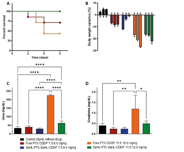 Note: Data are displayed as mean ± error standard. *, ** and **** P-values less than 0.05, 0.01 and 0.0001, respectively were set as the significance level by the ANOVA, followed by Tukey’ test.