Abstracts
Bovine karyotyping has become an important diagnostic tool in animal breeding. In the prenatal period it can diagnose several chromosomal abnormalities such as Robertsonian translocations, testicle feminization syndrome, gonadal dysgenesis and Klinefelter’s syndrome. An important cell source for karyotype analysis is the amniotic fluid. It has been extensively used in humans but in bovine, however, this is not the case despite its diagnostic value. Since a small percentage of cells is viable, cells and their growth conditions as well as the handling of the material should be optimal to insure a successful analysis. For this, we have compared the growth efficiency for bovine amniocytes in two media, employing cells from 10 to 14 weeks of gestation. Amniocytes were cultured in the Amniomax (Gibco-BRL/ Life Technologies, Rockville, MD USA) medium during eleven days and in the RPMI 1640 (Gibco-BRL) medium during sixteen days at 37ºC and 5% CO2, then fixed and GTG banded. All the cultures with RPMI showed a poor cell growth, regardless the gestational age. Out of the samples cultured in Amniomax one presented 100% of cellular confluence at day 11 (10 weeks of gestation) and the others resulted in an increased proliferation compared with those that were cultured in RPMI. To ensure a successful karyotyping, amniotic fluid from cows with gestational ages of 10-12 weeks should be used and care should be taken for critical steps in preparation of spread metaphases - hypotonic and trypsin treatments.
Karyotypes; Cell culture; Amnion; Cattle
A cariotipagem em bovinos é uma importante ferramenta diagnóstica. Pode ser utilizada no período prenatal para diagnóstico de várias anormalidades cromossômicas, tais como translocações Robertsonianas, síndrome da feminilização testicular, disgenesia gonadal e síndrome de Klinefelter. O fluido amniótico é uma importante fonte de células para cariotipagem e tem sido extensivamente utilizado para humanos mas não para bovinos, apesar de seu valor diagnóstico. Uma vez que pequena porcentagem dessas células é viável, suas condições de crescimento, assim como o processamento do material, devem ser otimizadas para se assegurar uma análise bem sucedida. Para tanto, comparamos a eficiência de crescimento de amniócitos bovinos em dois meios de cultura, usando células de 10 a 14 semanas de gestação. Os amniócitos foram cultivados no meio Amniomax (Gibco-BRL/ Life Technologies, Rockville, MD USA) durante onze dias e no meio RPMI (Gibco-BRL) durante dezesseis dias a 37ºC e 5% CO2, fixados e corados de acordo com a técnica GTG de bandeamento. Todas as culturas no meio RPMI apresentaram baixo crescimento celular, independente da idade gestacional. Das amostras cultivadas em Amniomax, uma apresentou 100% de confluência celular no 11ºdia de cultivo (10 semanas de gestação) e as outras apresentaram proliferação maior em relação àquelas cultivadas em RPMI. O líquido amniótico proveniente de gestações entre 10 e 12 semanas deve ser utilizado para se assegurar uma boa qualidade de material para cariotipagem. Além disso, deve-se atentar para os passos durante o processamento para melhor visualização das metáfases - choque hipotônico e tempo de tripsina.
Cariótipos; Cultura de células; Âmnio; Bovinos
Amniotic cell culture during different ages of gestation for karyotype analysis in bovine
Cultivo de células amnióticas durante diferentes idades gestacionais para análise do cariótipo em bovinos
Paola Ribeiro Seabra EIRAS11 Departamento de Medicina Veterinária da Universidade Federal de Lavras, Lavras MG ; João Bosco BARRETO FILHO11 Departamento de Medicina Veterinária da Universidade Federal de Lavras, Lavras MG ; Romain Rolland GOLGHER21 Departamento de Medicina Veterinária da Universidade Federal de Lavras, Lavras MG ; Sandra Regina Quintino dos SANTOS31 Departamento de Medicina Veterinária da Universidade Federal de Lavras, Lavras MG
CORRESPONDENCE TO:
Paola Ribeiro Seabra Eiras
Departamento de Medicina Veterinária da Universidade Federal de Lavras
Caixa Postal 37 Campus Universitário
37200-000 Lavras MG
e-mail: paolarse@uol.com.br
SUMMARY
Bovine karyotyping has become an important diagnostic tool in animal breeding. In the prenatal period it can diagnose several chromosomal abnormalities such as Robertsonian translocations, testicle feminization syndrome, gonadal dysgenesis and Klinefelters syndrome. An important cell source for karyotype analysis is the amniotic fluid. It has been extensively used in humans but in bovine, however, this is not the case despite its diagnostic value. Since a small percentage of cells is viable, cells and their growth conditions as well as the handling of the material should be optimal to insure a successful analysis. For this, we have compared the growth efficiency for bovine amniocytes in two media, employing cells from 10 to 14 weeks of gestation. Amniocytes were cultured in the Amniomax (Gibco-BRL/ Life Technologies, Rockville, MD USA) medium during eleven days and in the RPMI 1640 (Gibco-BRL) medium during sixteen days at 37ºC and 5% CO2, then fixed and GTG banded. All the cultures with RPMI showed a poor cell growth, regardless the gestational age. Out of the samples cultured in Amniomax one presented 100% of cellular confluence at day 11 (10 weeks of gestation) and the others resulted in an increased proliferation compared with those that were cultured in RPMI. To ensure a successful karyotyping, amniotic fluid from cows with gestational ages of 10-12 weeks should be used and care should be taken for critical steps in preparation of spread metaphases - hypotonic and trypsin treatments.
UNITERMS: Karyotypes; Cell culture; Amnion; Cattle.
INTRODUCTION
Bovine karyotyping has become an important diagnostic tool in animal breeding. Cattle karyotype consists of 58 acrocentric autosomes and submetacentric X and Y sex chromosomes6. In the domestic species, the X chromosome is substantially longer than the Y chromosome and this dimorphism forms the basis of sex determination by cytological methods8.
Karyotyping can indicate males affected by several chromosomal abnormalities, helping breeders to choose bulls, semen and embryos11. Some abnormalities can be attributed to errors that have arisen during cell divisions leading to gamete production (meiosis) and during fertilization. Others are a result of errors in mitotic divisions. Although the mechanisms of production of these two groups of abnormalities are different, the final appearance is often indistinguishable16. Robertsonian translocations are the most common congenital chromosomal abnormalities related to reproductive disorders in cattle5,10. Slapnika; Havrankova18 verified a high incidence of this translocation in beef and dairy cattle and its relationship with increased open days, embryo loss and inherited infertility from carrier females. Among other reproductive disorders, in which cytogenetic analysis is indicated, it can be quoted: testicle feminization syndrome12, gonadal dysgenesis and Klinefelters syndrome17. Karyotyping is also an important tool for sexing embryos in multiple ovulations in embryo transfer programs and for the identification of transgenic fetuses in utero in genetic engineering9.
Bovine karyotyping can be performed using a variety of cell sources. The choice of the cell source depends on several factors such as: (i) the type of patient (postnatal or prenatal); (ii) the purpose of the diagnosis (constitutional or acquired) and (iii) the clinical indication. For neonatal and adult patients, the most common tissue used for constitutional chromosome diagnosis is the peripheral blood. For acquired chromosome abnormalities, frequently associated with neoplasms, the tissue involved is used for cytogenetic evaluation19. In prenatal cases, the fetal cells are employed to diagnose constitutional chromosome complement and the common cells used for this purpose are the amniocytes from the amniotic fluid, cells from the chorionic villi3 and fetal blood lymphocytes, in the order of preference.
Amniocytes are collected by amniocentesis and this technique has been applied for cytogenetic analysis in prenatal medicine of human pregnancies in a variety of clinical situations. In bovines, it can be performed between the 8th and 22nd weeks of gestation, without major risks of fetal wastage9. However, despite its diagnostic value, this technique has not been widely used.
To ensure that an adequate number of quality metaphase chromosomal spreads will be available for karyotype study, a minimum proliferation of amniocytes is required. This is a relevant factor for diagnostic purposes, specially when samples with a low number of viable amniocytes are examined. It is known, for example, that only 20% of amniocytes are viable in women from 14 to 18 weeks of gestation14. To accomplish a minimum proliferation, amniotic fluid cells need to be cultivated in a nutrient-enriched culture medium and the resultant colonies are subsequently analyzed. Tissue culture media, however, vary significantly in their contents. They consist of a basal salt solution, glucose, vitamins, nucleotides and amino acids. Additional substances such as growth factors and hormones are now added to promote rapid cell proliferation18.
Therefore, it is most important to compare the efficiency of various culture media for the growth of amniocytes. For human cells, Biddle et al.2 have shown that when amniocytes were cultivated in Amniomax, Chang and Minimum Essential (MEM) media, the cells grew more vigorously in the first medium than in the other two. Although several culture media have been used to culture bovine cells such as RPMI 1640, Hams F-10, TCM 199, Eagles MEM, Medium-1992, no study was undertaken to define an improved medium for the multiplication of bovine amniocytes.
Another important factor to ensure successful karyotyping is the gestational age for amniocentesis. Human amniotic fluid contains a heterogeneous population of cells according to the stage of gestation. The number of fetal cells present in the amniotic fluid increases according to the gestational age, but as pregnancy progresses an increasing number of these cells become not viable. Thus, a sample taken at 12 weeks of pregnancy may have a reduced cell pellet after centrifugation, but the majority of these cells are likely to be viable; on the other hand, a sample taken at 20 weeks may have a very large pellet, but with many dead cells16. These authors, working with samples of human amniotic fluid ranging from 13 to 20 weeks of gestation, observed that the pregnancies from 13 to 14 weeks and from 15 to 20 weeks did not differ significantly regarding the number of days in vitro after which the culture could be examined. In cattle, Leibo and Rall9 compared the median age of amniotic cell culture when harvested as a function of the age of the fetuses when samples of amniotic cells were collected. On average, amniotic samples from older fetuses reached harvest age faster than those from younger fetuses; delaying amniocentesis from 8 to 12 weeks of fetal age shortened the culture time by 3.5 days.
Since karyotyping by amniocentesis in bovine has many indications and the current karyotyping techniques could be improved, we have studied several variables to refine the technique, adapting from the protocols used for human amniocytes. We have concluded that the Amniomax medium is superior to RPMI 1640 medium; the 10-week gestational age is appropriate to collect the amniocytes. We have also observed that the harvest of the cultures can be reduced to 6 days.
MATERIAL AND METHOD
The protocol used to culture bovine amniotic cells was a modification of those commonly used for human cells, described by Verma; Babu19.
Cultures with Amniomax medium (GIBCO-BRL / Life Technologies, Rockville, MD USA) - Five amniotic fluid samples were aseptically drawn in slaughterhouse from the amniotic sac of pregnant cows, by needle puncture, at 10 (n = 2) and 12 (n = 2) weeks of gestation and kept at room temperature until they get to the laboratory. The fetus age was determined according to Richardson15. The fluids were cultured in duplicate at 37ºC and 5% CO2 in polystyrene dishes (60 x 15 mm) in the proportion of 4 ml of amniotic fluid in 2 ml of culture media, supplemented with 20% of fetal calf serum, and gentamicin at a concentration of 50 ml/ml. The dishes were observed every day and cultures were harvested when several macroscopic colonies were visible (day 11); 2 ml of fresh medium were added containing Colcemid (0.05 mg/ml; Sigma, Saint Louis, USA) and the cells were reincubated for three hours. The material was then processed by hypotonic treatment for 20 minutes with 2 ml 0.075 M KCl solution. The solution was drained and fixative (methanol/acetic acid (3:1) solution) was added and changed five times. Chromosome staining was performed by Giemsa banding (GTG-banding)19. The dishes were rinsed with phosphate buffer saline (PBS 0.01 M), pH = 7.4 and immersed in 1:1 solution consisting of 0.12% trypsin solution (Gibco-BRL) /PBS 1X, for 7 seconds. Then each dish was rinsed again in PBS, stained with Giemsa for 3 minutes and examined and photographed under light microscopy.
Cultures with RPMI 1640 medium (Gibco-BRL) - Other six amniotic fluid samples were obtained at 10 (n = 1), 12 (n = 2) and 14 (n = 3) weeks of gestation. They were cultured in triplicate in the proportion of 4 ml of amniotic fluid and 2 ml of culture medium at 37ºC and 5% CO2. The medium was supplemented with 20% of fetal calf serum and 50 mg/ml of Gentamicin. The other steps were the same as described above.
RESULTS AND DISCUSSION
Behavior of amniotic cells, which were cultured in the two media, was different. Amniotic cells cultured in RPMI medium during sixteen days were daily observed. At day 10, the dishes had few attached cells without colony formation and fresh medium was added. In spite of low growth, at day 16, cells were harvested. We could add fresh medium waiting for more proliferation but growth factors could be diluted1.The cells cultured in RPMI did not yield sufficient material for karyotype analysis in the studied period.
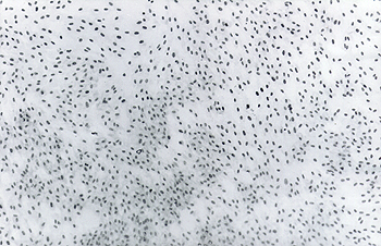
All samples cultured in Amniomax medium presented outstanding cell growth rates (Fig. 1) at day eleven, except for one sample. This medium was developed specifically for in vitro diagnostic testing of human amniotic fluid specimens. According to Biddle et al.2, it was designed to help minimize diagnostic turn around time by maximizing colony growth2. Indeed, these authors demonstrated that when the Amniomax, Chang and MEM media were used for in vitro cultures of human amniocytes, cultivation in Amniomax resulted in an increased proliferation rate and a corresponding reduction in cellular doubling time when compared to MEM. In addition, Amniomax medium cultivation increased the resulting number of amniocytes when compared with Chang medium. As we have observed (Fig. 1), the Amniomax is also superior to the RPMI 1640 medium for bovine amniocytes growth.
Amniocytes cultured in Amniomax medium at day 11 cells were collected for culture at 10 weeks of gestation. Magnification 85 X.
Another important factor for a successful karyotyping is the age of cultures in vitro. With Amniomax medium, a 10-week gestation sample can reach a 100% cellular confluence in less than 11 days (Fig. 1). A full confluence, however, precludes cytogenetic analysis, as metaphases can not be visualized on the borders of the colonies, presumably, because of contact inhibition. Therefore, cultures should be examined when colony borders are still visible (Fig. 2). Our results are similar to those obtained by Leibo and Rall9 in cattle, when ten days or less were necessary to reach a cell concentration sufficient for analysis in gestations of 10 to 15 weeks.
Colony border of an amniocyte culture grown in Amniomax medium. Several metaphases can be seen. Magnification 428 X.
The gestational age when amniotic fluid is drawn influences the length of time that is necessary for a sufficient growth for karyotyping. Out of the samples that were cultured in Amniomax, one presented 100% of cellular confluence at day 11 (10 weeks of gestation) (Fig. 1) and the others showed an efficient proliferation of 11.11 ± 8.98 colonies/dish. Cells from 10-week gestation samples grew at a higher rate if compared with those from advanced pregnancy stages (12 weeks of gestation). Faller et al.4 working with samples of amniotic fluid ranging from 13 to 20 weeks of gestation from 115 women observed that the younger pregnancies (13 to 14 weeks of gestation) did not delay the harvesting in relation to the advanced gestational ages (15 to 20 weeks of gestation) with a rate of 12.62 ± 2.62 and 12.74 ± 2.93 days of culture, respectively.
Cells from the 30 week-gestation stage did not proliferate in the Amniomax medium. Human amniotic fluid contains a heterogeneous population of cells according to the stage of gestation16 and only approximately 20% of cells are viable in amniotic fluids from women with 14 to 18 weeks of gestation14. In bovine, Kamimura et al.7 observed that the sediment of centrifuged amniotic fluid, throughout gestation, increases considerably presenting more cellular materials. It is possible that increased cellular sediment means less cellular viability as our results show that the 10-week gestational age culture is better than 12 weeks. These results are at variance with those from Leibo and Rall 9, in which older pregnancies (15 weeks of gestation) presented a higher number of colonies than the earlier (7 weeks) gestations for harvesting. However, our experiments show that results can be easily obtained in a 10-week gestational age. There is no need to postpone karyotyping for further 12 weeks. Another point that could explain why some samples of amniotic cells did not grow is the fact that the fetuses could be dead or dying at the time of amniocentesis9.
It is well known that small differences in the methods used to arrest mitotic cells and to prepare the material may affect the morphological appearance of chromosomes13. Besides the factors already pointed out, our experience showed that, to obtain a better chromosomal image, the length of the hypotonic treatment should be no less than 20 minutes and the trypsinizing time should be less than 10 seconds (Fig. 3A and 3B).
Thus, our results indicate that amniotic fluid should be collected early in pregnancy, up to 10-12 weeks, and the culture of amniotic cells in Amniomax medium can be examined in less than ten days. In addition, caution should be taken for critical steps in preparation of spread metaphases.
RESUMO
A cariotipagem em bovinos é uma importante ferramenta diagnóstica. Pode ser utilizada no período prenatal para diagnóstico de várias anormalidades cromossômicas, tais como translocações Robertsonianas, síndrome da feminilização testicular, disgenesia gonadal e síndrome de Klinefelter. O fluido amniótico é uma importante fonte de células para cariotipagem e tem sido extensivamente utilizado para humanos mas não para bovinos, apesar de seu valor diagnóstico. Uma vez que pequena porcentagem dessas células é viável, suas condições de crescimento, assim como o processamento do material, devem ser otimizadas para se assegurar uma análise bem sucedida. Para tanto, comparamos a eficiência de crescimento de amniócitos bovinos em dois meios de cultura, usando células de 10 a 14 semanas de gestação. Os amniócitos foram cultivados no meio Amniomax (Gibco-BRL/ Life Technologies, Rockville, MD USA) durante onze dias e no meio RPMI (Gibco-BRL) durante dezesseis dias a 37ºC e 5% CO2, fixados e corados de acordo com a técnica GTG de bandeamento. Todas as culturas no meio RPMI apresentaram baixo crescimento celular, independente da idade gestacional. Das amostras cultivadas em Amniomax, uma apresentou 100% de confluência celular no 11ºdia de cultivo (10 semanas de gestação) e as outras apresentaram proliferação maior em relação àquelas cultivadas em RPMI. O líquido amniótico proveniente de gestações entre 10 e 12 semanas deve ser utilizado para se assegurar uma boa qualidade de material para cariotipagem. Além disso, deve-se atentar para os passos durante o processamento para melhor visualização das metáfases choque hipotônico e tempo de tripsina.
UNITERMOS: Cariótipos; Cultura de células; Âmnio; Bovinos.
Received: 11/11/1998
Accepted: 26/04/2000
2 Centro de Pesquisas René Rachou da FIOCRUZ MS, Belo Horizonte MG
3 Laboratório Origen, Belo Horizonte MG
-
1- ALBERTS, B.; BRAY, D.; LEWIS, J.; RAFF, M.; ROBERTS, K.; WATSON, J.D. Molecular biology of the cell 3.ed. New York : Garland, 1994. 1362p.
-
2- BIDDLE, W.C.; KULIGWSKI, S.; LOCKWOOD, D.H.; FILBY, J.; HAGEN, T.C. Amniomax - C 100: a new specialized cell culture medium for the propagation of human amniocytes. Focus, v.14, n.3, p.80-5, 1992.
-
3- COSTA, H.B.M.; CARVALHO, T.B.; PINHEIRO, L.E.L. Preparo de cromossomos bovinos utilizando cultura rápida de células coriônicas. Revista Brasileira de Reprodução Animal, v.11, n.3, p.127-31, 1987.
-
4- FALLER, M.S.; MALUF, W.S.; ALMEIDA, I.; PASSOS, P. Amniocentese precoce e diagnóstico pré-natal citogenético. In: CONGRESSO NACIONAL DE GENÉTICA, 44., 1998, Águas de Lindóia, SP. Anais... Águas de Lindóia : Sociedade Brasileira de Genética, 1998. p.286.
-
5- FIGUEIREDO, T.R.; IANNUZZI, L. A cattle breed close to 58 diploid number due to high frequency of rob (1; 29). Hereditas, v.115, n.1, p.73-8, 1991.
-
6- GALLAGHER JR., D.S.; WOMACK, J.E. Chromosome conservation in the bovidae. Journal of Heredity, v.83, n.4, p.287-98, 1992.
-
7- KAMIMURA, S.; NISHIYAMA, N.; OOKUTSU, S.; GOTO, K.; HAMANA, K. Determination of bovine fetal Sex by PCR using fetal fluid aspirated by transvaginal ultrasound-guided amniocentesis. Theriogenology, v.47, n.8, p.1563-9, 1997.
-
8- KING, W.A. Sexing embryos by cytological methods. Theriogenology, v.21, n.1, p.7-17, 1984.
-
9- LEIBO, S.P.; RALL, W.F. Prenatal diagnosis of sex in bovine fetuses by amniocentesis. Theriogenology, v.33, n.2, p.531-53, 1990.
-
10- MURAKAMI, R.K.; MIYAKE, Y.I.; KANEDA, Y. Cases of XY female, single-birth freemartin and trisomy (61, XX, + 20) observed in cytogenetical studies on 18 sterile heifers. Japanese Journal of Veterinary Science, v.51, n.5, p.941-5, 1989.
-
11- PINHEIRO, L.E.L. Perfil genético da raça holandesa. Revista Gado Holandês, ano 56, n.181, p.15-9, 1990.
-
12- PIRES, R.M.L.; REICHERT, R.H.; ARCARO, J.R.P.; MACRUZ, R. Disgenesia gonadal XY em uma novilha holandesa. Boletim de Indústria Animal, v.46, n.2, p.235-9, 1989.
-
13- PRIEST, J.H., 1997 apud LEIBO, S.P.; RALL, W.F., 1990. p.535
-
14- REID, R.; SEPULVEDA, W.; KYLE, P.M.; DAVIES, G. Amniotic fluid failure: clinical significance and association with aneuploidy. Obstetrics and Gynecology, v.16, n.4, p.588-92, 1996.
-
15- RICHARDSON, C. Personal communication. In: ARTHUR, G.H.; NOAKES, D.E.; PEARSON, H. Veterinary reproduction and obstetrics 6.ed. London: Baillière Tindall, 1989. p.48-59.
-
16- ROONEY, D.E.; CZEPULKOWSKI, B.H. Human cytogenetics; a practical approach - constitutional analysis. 1.ed. New York : Oxford University Press, 1992. 274p.
-
17- SCHMUTZ, S.M.; BARTH, A.D.; MOKER, J.S. A Klinefelter bull with a 1;29 translocation born to a fertile 61, XXX cow. Canadian Veterinary Journal, v.35, n.2, p.182-4, 1994.
-
18- SLAPNICKA, J.; HAVRANKOVA, J. Chromosome aberrations as cause of reproductive disorders in cattle. Nas Chov, v.51, n.4, p.154-6, 1991.
-
19- VERMA, R.S.; BABU, A. Human chromosomes; principles and techniques. 2.ed. New York : McGraw-Hill, 1994. p.6-63.
Publication Dates
-
Publication in this collection
11 May 2001 -
Date of issue
2000
History
-
Received
11 Nov 1998 -
Accepted
26 Apr 2000