Abstracts
The goal of this study was to evaluate skeletal, dental and chronological development in an HIV-positive group of children, as compared with a control group, during a four-year period. Panoramic radiographs and hand and wrist radiographs of 60 children were taken. The children, of both sexes, aged 5 years and 2 months to 15 years and 5 months, were selected as follows: 30 HIV-positive volunteers who had acquired the disease vertically, and 30 volunteers who did not present the HIV infection or any other systemic disease. All radiographs were technically standardized and analyzed according to criteria established by Nolla (dental age), Greulich and Pyle (bone age), and Eklöf and Ringertz (bone age). The results were submitted to Student's t-test at a 5% level of significance. Based on the comparison between the chronological age and the dental or the skeletal age, significant differences were observed between HIV-positive and HIV-negative children, both in 1999 and in 2003 (p < 0.05). Considering the results obtained with the methodology used, it was concluded that HIV-positive children of both sexes presented delayed bone development despite the administration of antiretroviral drugs, and that HIV-positive female children presented younger dental ages compared with their chronological ages in 1999 and in 2003; and HIV-positive males, in 1999.
Age determination by skeleton; Age determination by teeth; HIV; Acquired immunodeficiency syndrome
Este trabalho teve como objetivo avaliar, em um intervalo de quatro anos, o desenvolvimento ósseo, dentário e cronológico de um grupo de crianças portadoras do HIV, comparando-o com um grupo controle. Foram realizadas radiografias panorâmica e de mão e punho, de uma amostra de 60 crianças, com idades variando entre 5 anos e 2 meses e 15 anos e 5 meses, dos sexos feminino e masculino, sendo: 30 crianças, voluntárias, portadoras de infecção pelo HIV, adquirida verticalmente, e 30 crianças voluntárias, que não apresentavam infecção pelo HIV ou qualquer outra doença sistêmica. Todas as radiografias foram padronizadas tecnicamente e analisadas segundo os critérios dos métodos de Nolla (idade dentária), Greulich e Pyle (idade óssea) e Eklöf e Ringertz (idade óssea). Os resultados foram submetidos ao teste estatístico t-Student em um nível de significância de 5%. Verificou-se, através da comparação da idade cronológica com as idades dentária e óssea, que houve diferença entre as crianças HIV+ e as não portadoras do vírus, tanto em 1999 como em 2003 (p < 0,05). Concluiu-se que as crianças HIV+ deste estudo sofreram um retardo no desenvolvimento ósseo. A idade dentária foi inferior à idade cronológica no grupo feminino HIV+, em 1999 e 2003, e masculino HIV+ em 1999.
Determinação da idade pelo esqueleto; Determinação da idade pelos dentes; HIV; Síndrome de imunodeficiência adquirida
ORIGINAL ARTICLES
RADIOLOGY
Comparison among dental, skeletal and chronological development in HIV-positive children: a radiographic study
Comparação entre o desenvolvimento dentário, ósseo e cronológico em crianças HIV+: estudo radiográfico
Rejane Maria HolderbaumI; Elaine Bauer VeeckII; Helena Willhelm OliveiraIII; Carmem Lúcia da SilvaIV; Ângela FernandesV
IPhD in Clinical Stomatology
IIChair Professor
IIIAdjunct Professor School of Dentistry, Pontifical Catholic University of Rio Grande do Sul
IVCoordinator, Pediatric AIDS Service, Clinical Hospital of Porto Alegre
VPhD in Clinical Stomatology, Professor, School of Dentistry, Federal University of Paraná
ABSTRACT
The goal of this study was to evaluate skeletal, dental and chronological development in an HIV-positive group of children, as compared with a control group, during a four-year period. Panoramic radiographs and hand and wrist radiographs of 60 children were taken. The children, of both sexes, aged 5 years and 2 months to 15 years and 5 months, were selected as follows: 30 HIV-positive volunteers who had acquired the disease vertically, and 30 volunteers who did not present the HIV infection or any other systemic disease. All radiographs were technically standardized and analyzed according to criteria established by Nolla (dental age), Greulich and Pyle (bone age), and Eklöf and Ringertz (bone age). The results were submitted to Student's t-test at a 5% level of significance. Based on the comparison between the chronological age and the dental or the skeletal age, significant differences were observed between HIV-positive and HIV-negative children, both in 1999 and in 2003 (p < 0.05). Considering the results obtained with the methodology used, it was concluded that HIV-positive children of both sexes presented delayed bone development despite the administration of antiretroviral drugs, and that HIV-positive female children presented younger dental ages compared with their chronological ages in 1999 and in 2003; and HIV-positive males, in 1999.
Descriptors: Age determination by skeleton; Age determination by teeth; HIV; Acquired immunodeficiency syndrome.
RESUMO
Este trabalho teve como objetivo avaliar, em um intervalo de quatro anos, o desenvolvimento ósseo, dentário e cronológico de um grupo de crianças portadoras do HIV, comparando-o com um grupo controle. Foram realizadas radiografias panorâmica e de mão e punho, de uma amostra de 60 crianças, com idades variando entre 5 anos e 2 meses e 15 anos e 5 meses, dos sexos feminino e masculino, sendo: 30 crianças, voluntárias, portadoras de infecção pelo HIV, adquirida verticalmente, e 30 crianças voluntárias, que não apresentavam infecção pelo HIV ou qualquer outra doença sistêmica. Todas as radiografias foram padronizadas tecnicamente e analisadas segundo os critérios dos métodos de Nolla (idade dentária), Greulich e Pyle (idade óssea) e Eklöf e Ringertz (idade óssea). Os resultados foram submetidos ao teste estatístico t-Student em um nível de significância de 5%. Verificou-se, através da comparação da idade cronológica com as idades dentária e óssea, que houve diferença entre as crianças HIV+ e as não portadoras do vírus, tanto em 1999 como em 2003 (p < 0,05). Concluiu-se que as crianças HIV+ deste estudo sofreram um retardo no desenvolvimento ósseo. A idade dentária foi inferior à idade cronológica no grupo feminino HIV+, em 1999 e 2003, e masculino HIV+ em 1999.
Descritores: Determinação da idade pelo esqueleto; Determinação da idade pelos dentes; HIV; Síndrome de imunodeficiência adquirida.
INTRODUCTION
Before 1983, it was believed that AIDS (acquired immunodeficiency syndrome) was restricted to a specific group of people. In that year, however, the first cases were reported of the syndrome affecting children14. Among children, most cases of infection by HIV are transmitted vertically, i.e., from mother to child. Newborn children present an immature immune system, which may cause the rapid and severe progression of the disease1.
Children who are vertically infected present considerable weight loss and delayed development2,5,9,12,15. They may also present the following changes: delayed dental eruption, fewer permanent teeth, prolonged retention of primary teeth16 and a slightly larger number of dental anomalies6. The establishment of antiretroviral protocols has reduced the occurrence of oral changes7,10. However, some authors have reported delayed development even with the use of antiretroviral drugs17.
In this study, the assessment of dental and skeletal growth and development was performed by means of significant parameters of skeletal maturation and dental development. The stages of dental calcification were assessed according to the method proposed by Nolla13 (1960). Skeletal development was assessed according to the methods proposed by Greulich, Pyle8 (1959) and Eklöf, Ringertz4 (1967).
Recently, the digitalization of radiographic images has been performed to obtain greater accuracy with these methods. Linear measurements have also been made using a specific computer program, to demonstrate the level of bone development3,11.
The aim of this study was to carry out a longitudinal analysis of skeletal and dental development in HIV+ children and in control children during a period of 4 years.
MATERIAL AND METHODS
A total of 60 children, aged 5 years and 2 months to 11 years and 8 months, in the first phase of the research (1999), and 8 years and 5 months to 15 years and 5 months, in the second phase of the research (2003), participated in this study. Among the children, 30 were HIV-positive volunteers, 17 male and 13 female, who had been vertically infected and were under outpatient treatment at the Pediatric service of the Clinical Hospital, Federal University of Rio Grande do Sul, Porto Alegre; and 30 were HIV-negative volunteers, 17 male and 13 female, who did not present any other systemic condition, and were seen at the School of Dentistry, Pontifical Catholic University of Rio Grande do Sul (PUCRS), Porto Alegre. The HIV-negative children presented chronological ages similar to those of the HIV-positive children in both phases of the study, and subjects were paired according to the following criteria: age, socioeconomic level, gender and race. All HIV-positive children were under antiretroviral therapy.
The children were examined and radiographed from January 1999 to October 1999, in phase 1, and from December 2002 to September 2003, in phase 2. They were submitted to routine clinical examination at the School of Dentistry, PUCRS, and panoramic radiographs, as well as hand and wrist radiographs, were taken. All radiographs were technically standardized using the same equipment (Orthophos Siemens, Munich, Germany) and processed using automatic processor AT2000 (Air Techniques, Hicksville, New York, USA). During the radiographic take, patients used a long lead apron for protection. Analysis of the radiographs was performed by a single radiologist at 3 different times with a 10-day interval between readings.
The study was approved by the Research Ethics Committees of the Clinical Hospital of Porto Alegre (HCPA) and of the Pontifical Catholic University of Rio Grande do Sul (PUCRS).
Dental age assessment Nolla method
Dental age assessment was performed by analyzing the dental calcification stages observed in the panoramic radiographs according to the method established by Nolla13 (1960). The teeth analyzed were the mandibular left permanent teeth. Each tooth was classified according to its calcification stage and the values were added up, resulting in one single value per child. When teeth were found to be between 2 stages, an average value was assigned.
Bone age assessment Greulich and Pyle method
Each hand and wrist radiograph was compared with the atlas pattern of similar age and sex. Older and younger patterns were also observed8. Bones and their epiphyses were then analyzed following a standardized sequence: distal surfaces of the radius and ulna bones, carpal bones (capitate, hamate, triquetral, lunate, scaphoid, trapezium, trapezoid and pisiform bones), metacarpal bones and distal phalanges. In cases where there was a sesamoid bone, its presence was also recorded, since it indicates the beginning of puberty.
Bone age assessment Eklöf and Ringertz method
This method measures the distances that correspond to the width and length of the hand and wrist bones, according to 10 parameters established by Eklöf, Ringertz4 (1967). To perform this analysis, the radiographs were scanned using a flatbed scanner with transparency module (SnapScan TPO, AGFA, Shanghai, People's Republic of China), and assessed using the Image Tool program for digitalized images (UTHSCSA, Texas, USA). The data obtained were tabulated and statistically analyzed by means of Student's t-test for paired samples at a 5% significance level (p < 0.05).
RESULTS
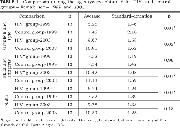
Female sex
Based on the Greulich and Pyle, and the Nolla methods, the values observed for the HIV-positive group were significantly lower than those observed for the control group in 1999 (p < 0.01). In 2003, significant differences were also observed between the two groups according to the methods established by Greulich and Pyle (p < 0.02) and by Eklöf and Ringertz (p < 0.01); moreover, the values obtained for the HIV-positive group were lower than those obtained for the control group (Table 1).
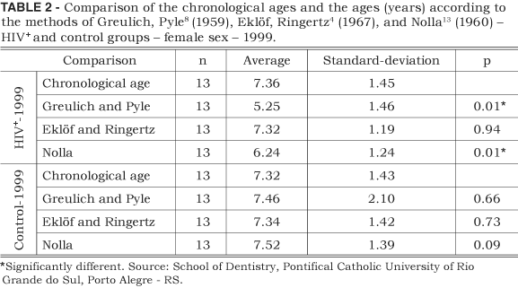
According to the Greulich and Pyle, and the Nolla methods, HIV-positive females, in 1999, presented significantly older chronological age than the standard ages described by these authors (p < 0.01) (Table 2).
In 2003, HIV-positive female children presented older chronological ages than those obtained by Greulich, Pyle8 (1959) and Nolla13 (1960) (p < 0.01) and by Eklöf, Ringertz4 (1967) (p < 0.02). In the control group, the chronological age was significantly older than that obtained according to the Nolla method (p < 0.01) (Table 3).
Male sex
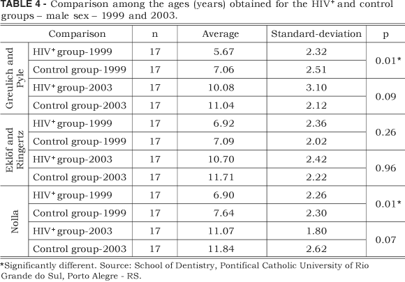
Among males of the HIV-positive and control groups, in 1999, a significant age difference was observed based on the Greulich and Pyle, and the Nolla methods. Values obtained for the HIV-positive group were lower than those obtained for the control group. No significant difference between the two groups was observed according to the Eklöf, Ringertz4 (1967) method. In 2003, no statistically significant differences were observed between the two groups (p > 0.01) (Table 4).
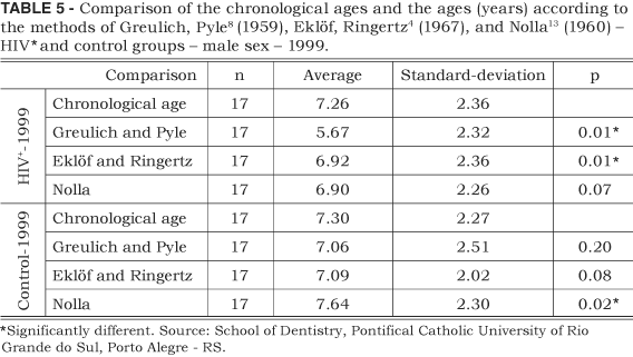
In 1999, HIV-positive patients presented significantly older chronological ages than those observed according to the Greulich, Pyle8 (1959) and the Eklöf, Ringertz4 (1967) methods (p < 0.01). According to the Nolla method, no significant difference between chronological and dental ages was observed. The control group presented a significantly older dental age compared with chronological age in 1999 (p < 0.02) (Table 5).
In 2003, HIV-positive patients presented significantly older chronological ages than those observed according to the Greulich and Pyle, and the Eklöf and Ringertz methods (p < 0.01). The control group also presented significantly older chronological ages than those observed according to the methods established by Greulich and Pyle (p < 0.03) and by Eklöf and Ringertz (p < 0.01) (Table 6).
DISCUSSION
The first reports of HIV infection in children date back to 1983, when Oleske et al.14 reported the different characteristics observed in children when compared with adults, including the transmission pattern. HIV infection may cause deficient bone maturation9,17 and delayed dental development6,16. In the present study, when HIV-positive and HIV-negative children of both sexes were compared using the methods proposed by Nolla13 (1960), by Greulich, Pyle8 (1959) and by Eklöf, Ringertz4 (1967), delayed skeletal and dental development was also observed. The present study revealed that, both in 1999 and in 2003, HIV-positive females presented younger skeletal and dental ages when compared with their chronological ages. However, the difference between control and HIV-positive patients was greater in 1999. This indicates that the medication given to HIV-positive patients was a favorable factor1,2.
In 1999, HIV-positive males presented significantly younger skeletal and dental ages than those of the control group. These results are consistent with the findings of Fine et al.7 (2003).
According to the Nolla method, the HIV-positive female group presented a deficiency in dental development in 1999, as did both the HIV-positive and control groups in 2003. The same happened to the HIV-positive male group in 1999, whereas the control group presented older dental age compared with their chronological age.
Sierra18 (1987) and Vallejo-Bolaños et al.19 (1999) also observed significant differences between chronological and dental ages, although using different methods. Fine et al.7 (2003) showed that HIV-positive children present prolonged retention of primary teeth and delayed tooth eruption when compared with non-infected children. These findings, similar to the results of our study, may indicate impairment to normal development. Ramos-Gomez et al.16 (2000) reported a smaller number of erupted teeth in HIV-positive children. However, Fernandes et al.6 (2002) found no significant differences between HIV-positive and HIV-negative children. Laskaris10 (2000) suggested that antiretroviral drugs may have caused a decrease in the number of oral alterations. This could explain why some HIV-positive children presented dental development similar to that of non-infected children.
According to the Greulich and Pyle method, HIV-positive female patients presented older chronological ages compared with their bone ages in 1999 and in 2003. According to the Eklöf and Ringertz method, HIV-positive female patients presented older chronological ages compared with their bone ages only in 2003. This indicates a deficiency in bone growth. The same was observed for both the male HIV-positive and the control groups.
Moye et al.12 (1996) and Abuzaitoun, Hanson1 (2000) reported that an immature immune system such as that of children may cause deficient organic growth and development, especially in HIV-positive children. This explains why, in this study, most HIV-positive children presented younger bone ages when compared with their chronological ages. However, in this study, little difference was observed between results for HIV-positive and HIV-negative children. This may be explained by the fact that the former were undergoing antiretroviral therapy, as demonstrated by Arpadi2 (2000); Abuzaitoun, Hanson1 (2000). Our results are corroborated by Oliveira15 (2002), who observed the influence of the HIV virus on skeletal development in female children. Even though the European Collaborative Study5 (1995) and Moye et al.12 (1996) reported a strong correlation between vertically-acquired infection and growth deficiency, the results of the present study do not completely corroborate this fact.
Adequate antiretroviral therapy may be an important factor to reestablish normal development in HIV-positive children. Further studies must be carried out to evaluate children whose diagnosis and therapy occurred early at birth and children whose diagnosis was confirmed only after the appearance of the first signs and symptoms of HIV-infection.
CONCLUSIONS
Considering the results obtained with the methodology used, it was concluded that HIV-positive children of both sexes present delayed bone development despite the administration of antiretroviral drugs, and that HIV-positive female children present younger dental ages compared with their chronological ages in 1999 and in 2003; and HIV-positive males, in 1999.
Received for publication on Mar 21, 2005
Sent for alterations on May 24, 2005
Accepted for publication on Jun 27, 2005
- 1. Abuzaitoun OR, Hanson IC. Organ-specific manifestations of HIV disease in children. Pediatr Clin North Am 2000;47(1):109-25.
- 2. Arpadi SM. Growth failure in children with HIV infection. J Acquir Immune Defic Syndr 2000;25(I Suppl):37-42.
- 3. Cao F, Huang HK, Pietka E, Gilsanz V. Digital hand atlas and web-based bone assessment: system design and implementation. Comput Med Imaging Graph 2000;24(5):297-307.
- 4. Eklöf O, Ringertz H. A method for assessment of skeletal maturity. Ann Radiol 1967;10(3):330-6.
- 5. European Collaborative Study. Weight, height and human immunodeficiency virus infection in young children of infected mothers. Pediatr Infect Dis J 1995;14(8):685-90.
- 6. Fernandes Â, Cherubini K, Veeck EB, Grando LJ, Birman EG, Silva CLO. Avaliação radiográfica das anomalias dentárias de número, forma, tamanho, posição e estrutura em crianças infectadas pelo HIV. Rev ABO Nac 2002;10(2):93-7.
- 7. Fine DH, Tofsky N, Nelson EM, Schoen D, Barasch A. Clinical implications of the oral manifestations of HIV infection in children. Dent Clin North Am 2003;47(1):159-74.
- 8. Greulich WW, Pyle SI. Radiographic atlas of skeletal development of the hand and wrist. 2nd ed. Stanford: Stanford University Press; 1959.
- 9. Kaufman FR, Gomperts ED. Growth failure in boys with hemophilia and HIV infection. Am J Pediatr Hematol Oncol 1989;11(3):292-4.
- 10. Laskaris G. Oral manifestations of HIV disease. Clin Dermatol 2000;18(4):447-55.
- 11. Mahmoodi S, Sharif BS, Chester G, Owen JP, Lee R. Skeletal growth estimation using radiographic image processing and analysis. IEEE Trans Inf Technol Biomed 2000;4(4):292-7.
- 12. Moye J Jr, Rich KC, Kalish LA, Sheon AR, Diaz C, Cooper L, et al. Natural history of somatic growth in infants born to women infected by human immunodeficiency virus. Women and Infants Transmission Study Group. J Pediatr 1996;128(1):58-69.
- 13. Nolla CM. The development of the permanent teeth. ASDC J Dent Child 1960;27(14):254-66.
- 14. Oleske J, Minnefor A, Cooper R, Thomas K, Dela Cruz A, Ahdieh H, et al. Immune deficiency syndrome in children. JAMA 1983;249(17):2345-9.
- 15. Oliveira HW. Avaliação do desenvolvimento ósseo em crianças infectadas pelo HIV por contaminação vertical [Tese de Doutorado em Odontologia]. Porto Alegre: Faculdade de Odontologia da Pontifícia Universidade Católica do Rio Grande do Sul; 2002.
- 16. Ramos-Gomez FJ, Petru A, Hilton JF, Canchola AJ, Wara D, Greenspan JS. Oral manifestations and dental status in pediatric HIV infection. Int J Paediatr Dent 2000;10(1):3-11.
- 17. Saavedra JM, Henderson RA, Perman JA, Hutton N, Livingston R, Yolken RH. Longitudinal assessment of growth in children born to mothers with human immunodeficiency virus infection. Arch Pediatr Adolesc Med 1995;149(5):497-502.
- 18. Sierra AM. Assessment of dental and skeletal maturity a new approach. Angle Orthod 1987;57(3):194-207.
- 19. Vallejo-Bolaños E, España-López AJ, Muñoz-Hoyos A, Fernandez-Garcia JM. The relationship between bone age, chronological age and dental age in children with isolated growth hormone deficiency. Int J Paediatr Dent 1999;9(3):201-6.
Publication Dates
-
Publication in this collection
21 Nov 2005 -
Date of issue
Sept 2005
History
-
Accepted
27 June 2005 -
Received
21 Mar 2005 -
Reviewed
24 May 2005