Abstract
Obstructive urolithiasis is common in farmed sheep and has a multifactorial etiology, but inadequate nutritional management is considered the most relevant condition for its occurrence. The objectives of this study were to verify the influence of two diets with different concentrations of calcium (Ca) and phosphorus (P) on the development of obstructive urolithiasis, and to describe the clinical and anatomopathological findings of the urinary system in sheep. Thirty male crossbred Santa Inês and Ile de France lambs were randomly distributed into two groups: Group 1 (G1, n = 15) - Ca: 1.9:1 P and 0.42% P; Group 2 (G2, n = 15) - Ca: P 1.5:1 and 0.65% P. The diets consisting of Coast-cross hay, soybean meal, wheat, and corn were provided for 90 consecutive days with water ad libitum. After the diagnosis of the disease, the lambs were subjected to clinical and surgical treatment, when necessary. Urolithiasis was detected in 36.7% (11/30) of lambs, 26.7% were asymptomatic and 10% (3/30) had urethral obstruction. A lamb was unobstructed after amputation of the urethral process and urethral catheterization, one died of bladder and uroperitoneum rupture, and another was sacrificed after the failure of perineal urethrostomy and cystostomy. The most frequent renal histopathological changes were vascular congestion, dilation, and tubular degeneration. Proteins in the tubular lumen were more pronounced in G2. The diets were rich in concentrate and had adequate Ca:P ratios, but caused calculogenesis, showing that excess minerals and a small amount of roughage can cause disease in the herd.
Keywords:
feedlot; phosphorus; sheep; urethral obstruction; urolith
Resumo
A urolitíase obstrutiva é frequente na ovinocultura e possui etiologia multifatorial, porém o manejo nutricional inadequado é considerado o mais relevante para sua ocorrência. Os objetivos deste estudo foram verificar a influência de duas dietas com diferentes proporções e concentrações de cálcio (Ca) e fósforo (P) no desenvolvimento da urolitíase obstrutiva, e descrever os achados clínicos e anatomopatológicos do sistema urinário de ovinos. Utilizaram-se 30 cordeiros, machos, mestiços das raças Santa Inês e Ile de France, que foram aleatoriamente distribuídos em dois grupos: Grupo 1 (G1, n=15) - Ca:P de 1,9:1 e 0,42% de P; Grupo 2 (G2, n=15) - Ca:P de 1,5:1 e 0,65% de P. As dietas foram fornecidas por 90 dias consecutivos com feno de Coast-cross, farelo de soja, trigo e milho, e água ad libitum. Após o diagnóstico da doença, os cordeiros foram submetidos ao tratamento clínico e cirúrgico, quando necessário. A urolitíase foi detectada em 36,7% (11/30) dos cordeiros, sendo 26,7% assintomáticos e 10% (3/30) apresentaram obstrução uretral. Um cordeiro foi desobstruído após amputação do processo uretral e sondagem uretral; outro foi a óbito por ruptura vesical e uroperitôneo; outro foi sacrificado após uretrostomia perineal e cistostomia sem sucesso. Em ambos os grupos, as alterações histopatológicas renais mais frequentes foram congestão vascular, dilatação e degeneração tubular. A presença de proteínas na luz tubular foi mais pronunciada no G2. As dietas fornecidas, ricas em concentrado, embora com relação Ca:P adequadas, provocaram a calculogênese, o que comprovou que o excesso de minerais e pouca quantidade de volumoso podem causar a enfermidade no rebanho.
Palavras-chave:
cálculo; confinamento; fósforo; obstrução uretral; ovinos
Introduction
A goal for sheep farmers is to obtain greater weight gain in their herd earlier, and the intensification of sheep rearing has led to a higher prevalence of metabolic and nutritional diseases, especially urolithiasis(11 Guimarães JA, Mendonça CL, Sousa Guaraná EL, Dantas AC, Azevêdo CN, Câmara ACL, Farias CC, Afonso JAB. Estudo retrospectivo de 66 casos de urolitíase obstrutiva em ovinos. Pesquisa Veterinária Brasileira. 2012; 32(9), 824-830. doi: 10.1590/S0100-736X2012000900002
https://doi.org/10.1590/S0100-736X201200...
,22 Maciel TA, Ramos IA, Silva RJ, Soares PC, Carvalho CCD, Maior Júnior RJS, Amoroso L, Artoni SMB, Afonso JAB, Oliveira D. Clinical and Biochemical Profile of Obstructive Urolithiasis in Sheep. Acta Scientiae Veterinariae. 2017; 45, 1-15. Available from: https://www.redalyc.org/articulo.oa?id=289053641089.
https://www.redalyc.org/articulo.oa?id=2...
). The etiology of this disease is complex and multifactorial. It particularly affects males due to the shape of the urethra, which is short and narrow(33 Riet-Côrrea F, Simões SVD, Vasconcelos JS. Urolitíase em caprinos e ovinos. Pesquisa Veterinária Brasileira. 2008; 28(6), 319-322. ), and causes economic losses from treatment expenses, inadequate reproduction, animal demise, and genetic material losses(44 Antonelli AC, Barrêto Júnior RA, Mori CS, Sucupira MCA, Marcello ACS, Ortolani EL. Efeito de diferentes fontes energéticas na predisposição para urolitíase em cabritos. Ciência Animal Brasileira. 2012; 13, 487-493. doi: 10.5216/cab.v13i4.15617
https://doi.org/10.5216/cab.v13i4.15617...
,55 Ferreira DOL, Santarosa BP, Amorim RM, Chiacchio SB, Gonçalves RC. (2015). Urolitíase obstrutiva em ovinos. Veterinária e Zootecnia. 2015; 22(2), 183-197.).
Productive and reproductive integrity is more effectively maintained with disease prevention prior to the formation of calculi(66 Ewoldt JM, Jones ML, Miesner MD. Surgery of obstructive urolithiasis in ruminants. Veterinary Clinics of North America: Food Animal Practice. 2008; 24, 455-465. doi: 10.1016/j.cvfa.2008.06.003
https://doi.org/10.1016/j.cvfa.2008.06.0...
,77 Kinsley MA, Semevolos S, Parker JE, Duesterdieck-Zellmer K, Huber M. Use of plain radiography in the diagnosis, surgical management, and postoperative treatment of obstructive urolithiasis in 25 goats and 2 sheep. Veterinary Surgery. 2013; 42, 663-668. doi: 10.1111/j.1532-950X.2013.12021.
https://doi.org/10.1111/j.1532-950X.2013...
,88 Van Metre D, Fecteau G, House JK. Obstructive urolithiasis in ruminants: surgical management and prevention. Compendium on Continuing Education for the Practicing Veterinarian. 1996; 19: 275-289.), because after the appearance of clinical signs, the prognosis is generally poor(99 Dória RGS, Canola PA, Dias DPM, Pereira RN, Valadão CAA. Surgical Techniques for obstructive urolithiasis in small ruminants: case report. Arquivo Brasileiro de Medicina Veterinária e Zootecnia. 2007; 59(6), 1425-1432. doi:10.1590/S0102-09352007000600012
https://doi.org/10.1590/S0102-0935200700...
). When calculi occur, it is necessary to determine the mineral profiles and chemical composition of the stones to rectify the possible factors associated with their formation(1010 Freeman SR, Poorea MH, Young GA, Anderson KL. Influence of calcium (0.6 or 1.2%) and phosphorus (0.3 or 0.6%) content and ratio on the formation of urolithogenic compounds in the urine of Boer-cross goats fed high-concentrate diets. Small Ruminant Research. 2010; 93, 94-102. doi: 10.1016/j.smallrumres.2010.05.007
https://doi.org/10.1016/j.smallrumres.20...
,1111 Streeter RN, Washburn KE, Mccauley CT. Percutaneous tube cystostomy and vesicular irrigation for treatment of obstructive urolithiasis in a goat. Jornal of the American Veterinary Medical Association. 2002; 221, 546-549. doi: 10.2460/javma.2002.221.546
https://doi.org/10.2460/javma.2002.221.5...
,1212 Sun WD, Zhang KC, Wang JY, Wang XL. The chemical composition and ultrastructure of uroliths in Boer goats. The Veterinary Journal. 2010; 86, 70-75. doi: 10.1016/j.tvjl.2009.07.009
https://doi.org/10.1016/j.tvjl.2009.07.0...
).
The highest occurrence of the disease is related to intensive management systems with considerable amounts of grains and their by-products, because of the high concentrations of phosphorus and magnesium, and low levels of calcium, which results in an imbalance in the ideal Ca:P ratio of 1:1 to 2:1(33 Riet-Côrrea F, Simões SVD, Vasconcelos JS. Urolitíase em caprinos e ovinos. Pesquisa Veterinária Brasileira. 2008; 28(6), 319-322. ). This Ca:P imbalance results in exacerbated phosphorus excretion in the urine, which at the alkaline urinary pH found in ruminants, favors its precipitation and consequent formation of calculi(1010 Freeman SR, Poorea MH, Young GA, Anderson KL. Influence of calcium (0.6 or 1.2%) and phosphorus (0.3 or 0.6%) content and ratio on the formation of urolithogenic compounds in the urine of Boer-cross goats fed high-concentrate diets. Small Ruminant Research. 2010; 93, 94-102. doi: 10.1016/j.smallrumres.2010.05.007
https://doi.org/10.1016/j.smallrumres.20...
,1313 Aquino Neto HM, Facury Filho EJ, Carvalho AU, Souza FA, Jordão LR. Urolitíase obstrutiva em ovinos: revisão de literatura. Veterinária em Foco. 2007; 4(2), 191-202.,1414 Ferreira DOL, Santarosa BP, Sacco SR, Dias A, Amorim RM, Chiacchio SB, Lis bôa JAN, Gonçalves RC. Efeito da suplemen tação de cloreto de amônio sobre o equilíbrio ácido-básico e o pH urinário de ovinos confinados. Pesquisa Veterinária Brasileira. 2014a; 34(8), 797-804. doi: 10.1590/S0100-736X2014000800016
https://doi.org/10.1590/S0100-736X201400...
,1515 Maciel TA, Júnior NL, Araújo VV, Silva Filho AB, Gomes DLS, Barbosa AMS, Farias CC, Magalhães ALR, Lima MJM, Melo SAX, Oliveira D. Avaliação dos perfis minerais séricos, urinários e sedimentares de ovinos recebendo dieta calculogênica. Arquivo Brasileiro de Medicina Veterinária e Zootecnia. 2016; 68(4), 967-976. doi: 10.1590/1678-4162-8363
https://doi.org/10.1590/1678-4162-8363...
).
In response to the calculogenic potential of a diet rich in concentrate with a high mineral content, the objectives of this study were to verify the nutritional influence on the occurrence of obstructive urolithiasis in feedlot lambs fed two diets with different levels of phosphorus, and to analyze the clinical findings in affected animals and the anatomopathological characteristics of the urinary system in the development of the disease, as well as to describe the treatments applied to the obstructed animals.
Material and methods
This work was submitted and approved by the Animal Use Ethics Committee of the Faculty of Veterinary Medicine and Animal Science (FMVZ), Universidade Estadual Paulista (UNESP), Campus de Botucatu, under protocol 156/2012.
For this study, we used 30 male, uncastrated, crossbred Ile de France and Santa Inês lambs, approximately 120 days old, newly weaned, with an average live weight (BW) of 18.7 ± 1.2 kg, from commercial breeding. These animals were separated into two groups of 15 animals, identified, and randomly distributed [Group 1 (G1) and Group 2 (G2)]. The lambs were initially dewormed with 1% injectable moxidectin (200 µg/kg) (Cidectin® Zoetis Indústria de Produtos Veterinários Ltda, Campinas-SP, Brazil) and vaccinated against Clostridioses (Sintoxan polyvalent T®, Boehringer Ingelheim, Campinas-SP, Brazil).
After an adaptation period of 15 d to the environment and to the diets, the animals remained confined for another 75 d, totaling 90 d of confinement. The sheep were housed in four 12 m2 masonry collective pens, with seven or eight animals each (average of 1.5 m2/head), arranged in the same location, inside the Veterinary Hospital of FMVZ/UNESP-Botucatu, under equal temperature conditions, air humidity, and luminosity. Two pens were designated for G1, one with seven and one with eight animals, and another two were designated for G2, in which the animals were under the same nutritional conditions, in an ad libitum manner. The animals received water ad libitum in automatic drinkers.
There was a visual assessment of the leftovers, but it was not quantified in order to guarantee the daily adjustment of 10% of leftovers. The hay was crushed and mixed with the concentrate, which provided a complete homogeneous diet, and was offered in feeders with sufficient area available for all sheep to feed without dispute. The experiment covered the usual growth and finishing time of lambs for slaughter, weaning at 60 to 70 days of age at an average weight of 18 kg, confining for 90 days, and reaching a final average weight of 40 kg at 150 to 160 days old. During this period, the sheep presented an average daily consumption of 3% of the dry matter (DM) PV, following the recommendations of the NRC(1616 National Research Council - NRC. Nutrient Requirements of Small Ruminants: sheep, goats, cervids and New World camelids. Natl Acad. Press, Washington, DC. 2007, 384p.). Thus, the animals had an average daily intake of DM of 1 to 1.3 kg and an average daily weight gain of 244 g.
The groups received diets differing in Ca and P ratios, but within the recommendations indicated by the NRC (2007) for raising sheep, using ratios of 1.9:1 for G1 and 1.5:1 for G2, as follows: G1-Ca 0.8% (8 g/kg DM), P 0.43% (4.3 g/kg DM); and G2-Ca 0.97% (9.7 g/kg DM), P 0.65% (6.5 g/kg DM). Based on the average consumption of 1 kg/day of DM, lambs from G1 ingested 8 g of Ca and 4.3 g of P per day, and animals in G2 consumed 9.7 g of Ca and 6.5 g of P per day. These values were above the absolute requirement for this animal category of 183 mg of Ca and 103 mg of P per kg of BW/day(1616 National Research Council - NRC. Nutrient Requirements of Small Ruminants: sheep, goats, cervids and New World camelids. Natl Acad. Press, Washington, DC. 2007, 384p.). Based on this, at the end of the experiment lambs with 40 kg BW would require 7.32 g of Ca and 4.12 g of P daily.
The chemical-bromatological composition and percentage of ingredients in the diets are listed in Table 1. In order to ensure experimental safety, the diet of 10% crushed Coast-cross hay and 90% concentrate (soy bran, wheat, and crushed corn), was subjected to chemical-bromatological analysis by the Department of Animal Improvement and Nutrition of FMVZ/UNESP, Botucatu. The DM, Crude Protein (CP), Etheric Extract (EE), and Mineral Matter (MM) were analyzed by the Weende method, and the neutral detergent fiber (NDF) and acid detergent fiber (ADF) by the Van Soest procedure. Mineral analysis was performed at the Campineiro Institute for Soil and Fertilizer Analysis (ICASA) to determine the percentage of phosphorus, calcium, magnesium, potassium, sodium, and sulfur.
Chemical-bromatological composition and percentage of ingredients in the diets of feedlot lambs subjected to diets with different levels of phosphorus, 0,43% (G1) and 0,65% (G2)
The effect of feeding on the occurrence of urolithiasis in animals was evaluated by confirming the diagnosis based on clinical signs and necropsies in animals from both groups. The animals were inspected twice daily (at 6:00 a.m. and 6:00 p.m.) in terms of the behaviors of food and water consumption, rumination, and urination, along with an evaluation of the body condition score (BCS)(1717 Russel A. Body condition scoring of sheep. In: E. Boden (Ed.) Sheep and Goat Practice. Bailliere Tindall, Philadelphia. 1991. p3.). Ultrasound evaluation (My LabTM30 Gold VET Esaote® Ultrasound Device, Esaote Healthcare do Brasil, São Paulo-SP, Brazil) of the entire length of the penile urethra, bladder, and kidneys was performed only in animals that presented signs suggestive of obstructive urolithiasis, using a convex transducer with a frequency of 3.5 to 5.0 MHz. For ultrasonography, the animals were manually restrained in left lateral decubitus for access to the right kidney (RK). For better image quality, the animals were clipped, and a trichotomy was performed in the region of contact with the transducer. For renal visualization, the transducer was positioned on the caudal margin of the costal cage, in the dorsal region of the paralumbar fossa, ventral to the lumbar transverse processes. The RK was visualized juxtaposed to the tail pole of the liver(1818 Scott PR, Sargison ND. Ultrasonography as an adjunct to clinical examination in sheep. Small Ruminant Research. 2010;92, 108-119. doi: 10.1016/j.smallrumres.2010.04.011
https://doi.org/10.1016/j.smallrumres.20...
,1919 Scott P. Transabdominal Ultrasonographic Examination of 26 Sheep with Suspected Urinary Tract Disease (2010- 2012 Journal of Veterinary Science & Medical Diagnosis. 2013; 2, 1-5. doi: 10.4172/2325-9590.1000107
https://doi.org/10.4172/2325-9590.100010...
). In the left kidney (LK) the transducer was directed cranially below the last rib, due to the presence of the rumen. After identifying the kidneys in two-dimensional mode, the measurement of the renal length in the sagittal section was performed and the architecture of the organ was observed(2020 Ferreira DOL, Santarosa BP, Belotta AF, Mamprim MJ, Silva AA, Dias A, Chiacchio SB, Gonçalves RC. Alterações ultrassonográficas renais e vesicais de ovinos confinados e suplementados com cloreto de amônio. Pesquisa Veterinária Brasileira. 2014b; 34(supl1), 99-106. doi:10.1590/S0100-736X2014001300018
https://doi.org/10.1590/S0100-736X201400...
). For the use of pulsed Doppler and measurement of the resistivity index (RI), only the RK was examined, since the visualization of the LK vascularization was complicated by the rumen. Color Doppler was activated to visualize the renal and interlobar arteries. The pulsed Doppler sample volume was positioned over the renal artery and then over the interlobar artery at an insonation angle of less than 60º, obtaining at least three waves of arterial spectral tracing. The RI was automatically calculated by the equipment after manual delimitation of the peak systolic velocity and the end-diastolic velocity. This procedure was performed according to the literature(2121 Santarosa BP, Ferreira DOL, Rodrigues MMP, Dantas GN, Sacco SR, Lopes RS, Dias A, Gonçalves RC. Clinical, laboratory and anatomopathological evaluation of the urinary system of feedlot sheep with or without ammonium chloride supplementation. Pesquisa Veterinária Brasileira. 2016; 36(1), 1-12. doi: 10.1590/S0100-736X2016000100001
https://doi.org/10.1590/S0100-736X201600...
), and the measurements were used as a reference.
When the diagnosis of urolithiasis was confirmed, the sheep underwent amputation of the vermiform appendix for retrograde urethral hydropropulsion and, when necessary, surgical treatment of urethrostomy or cystostomy with placement of a Foley catheter for urine drainage(99 Dória RGS, Canola PA, Dias DPM, Pereira RN, Valadão CAA. Surgical Techniques for obstructive urolithiasis in small ruminants: case report. Arquivo Brasileiro de Medicina Veterinária e Zootecnia. 2007; 59(6), 1425-1432. doi:10.1590/S0102-09352007000600012
https://doi.org/10.1590/S0102-0935200700...
,2222 Oman RE, Reppert EJ, Jones SM. Outcome and complications in goats treated by perineal urethrostomy for obstructive urolithiasis: 25 cases (2010-2017). Journal of Veterinary Internal Medicine. 2019; 33, 292-296. doi: 10.1111/jvim.15360
https://doi.org/10.1111/jvim.15360...
). The anesthetic protocol used for amputation of the vermiform appendix was intramuscular sedation with acepromazine (0.05 mg/kg). For other surgical procedures, in addition to acepromazine, general anesthesia was used with intravenous propofol (10 mg/kg), and maintenance with isoflurane, via inhalation.
At the end of the experimental period, the sheep, an average weight of 40 kg, were euthanized under the protocol of 0.2 mg/kg of intravenous xylazine, for sedation. After entering decubitus, 10 mg/kg of thiopental (5% solution) was administered intravenously, followed by 1 mL/kg of potassium chloride (19.1% KCl ampoule, 2.5 mEq/mL). This method was approved by the Federal Council of Veterinary Medicine in Resolution no. 714, June 20, 2002 and included in acceptable and accepted methods under restriction as per annex 1 of CFMV Resolution no. 1000/2012 for ruminants. We then performed macroscopic analysis of the urinary system and microscopic analysis of the kidneys. For the histopathological examination, fragments of the kidneys were fixed in a 10% buffered formalin solution. After 48 h, the samples were transferred to vials with 70% ethyl alcohol, embedded in paraffin, cut in a microtome (RM2255-Leica Biosystems, Leica do Brasil Importação e Comércio Ltda, São Paulo-SP, Brazil) at a thickness of 5 µm, and stained with hematoxylin and eosin (HE) at the Department of Veterinary Pathology at FMVZ/UNESP, Botucatu. All slides were analyzed under an optical microscope and histopathological changes in the kidneys were classified as absent, mild, moderate, or severe(2323 Guedes Jr FS, Cruz DS, Rodrigues MMP, Silva LM, Amorim RL, Vianna PTG, Castiglia YM M. Renal histology and immunohistochemistry after acute hemorrhage in rats under sevoflurane and ketoprofen effect. Acta Cirurgica Brasileira. 2012; 27(1), 37-42. doi: 10.1590/S0102-86502012000100007
https://doi.org/10.1590/S0102-8650201200...
).
Clinical and macroscopic findings were only described qualitatively. Microscopy data were analyzed using the Statistical Analysis System software (SAS - SAS Institute, Cary, NC, USA). To analyze the effects of G1 and G2 treatments on the degree of impairment and type of microscopic change, non-parametric tests (two-tailed) were performed, as the data were not normally distributed and the Proc Freq was used. Results that presented p ≤ 0.05 by the Chi-square test were considered significant.
Results
At the beginning of the experiment, all lambs had an adequate ECC (3.89 ± 0.64), of between 3.25 and 4.75(1717 Russel A. Body condition scoring of sheep. In: E. Boden (Ed.) Sheep and Goat Practice. Bailliere Tindall, Philadelphia. 1991. p3.), were alert, and active. The onset of the disease occurred on the 28th day of the experiment, in an animal from G2, which presented, on physical examination, acute prostration and anuria, cyanotic glans penis, and blackened urethral process. For the initial treatment, intravenous dipyrone (25 mg/kg) and hyoscine were administered, along with intramuscular acepromazine (0.05 mg/kg), followed by amputation of the urethral process and urethral probe and retrograde hydropropulsion with saline solution, which was unsuccessful, and the animal continued to complete urethral obstruction. On abdominal ultrasound, the bladder was filled with hyperechoic dots and 0.72 cm calculi producing an acoustic shadow (Fig. 1), and a left renal artery resistivity index (RI) of 0.60 (Fig. 2). Although there was pelvic dilation in both kidneys (0.46 cm and 0.52 cm), the length of the kidneys at the sagittal cut was normal at 5.73 cm (left kidney) and 5.89 cm (right kidney), and there was no hydronephrosis. However, it was not possible to visualize the site of obstruction of the urethra, as it was proximal to the bladder (pelvic region), and therefore, without access for the transducer.

Bladder of Lamb 17 (G2) with diffuse hyperechogenic punctiform structures forming a discrete reverberation artifact and producing a slight acoustic shadow posterior to the dependent bladder wall, suggesting the presence of bladder stones. 5 MHz frequency.

Normal resistivity index (RI) (0.60) and spectral tracing of the renal artery of the right kidney of Lamb 17 (G2).
Later, laparotomy and cystostomy were performed for placement of the Foley catheter (Fig. 3). There was a small rupture point in the bladder, measuring 2 cm, which was not seen on the ultrasound performed hours before; therefore, cystorrhaphy was performed at the rupture site and a Foley catheter was introduced for urinary excretion, followed by perineal urethrostomy(99 Dória RGS, Canola PA, Dias DPM, Pereira RN, Valadão CAA. Surgical Techniques for obstructive urolithiasis in small ruminants: case report. Arquivo Brasileiro de Medicina Veterinária e Zootecnia. 2007; 59(6), 1425-1432. doi:10.1590/S0102-09352007000600012
https://doi.org/10.1590/S0102-0935200700...
, 1111 Streeter RN, Washburn KE, Mccauley CT. Percutaneous tube cystostomy and vesicular irrigation for treatment of obstructive urolithiasis in a goat. Jornal of the American Veterinary Medical Association. 2002; 221, 546-549. doi: 10.2460/javma.2002.221.546
https://doi.org/10.2460/javma.2002.221.5...
,2424 Van Metre DC, Fubini SL. Ovine and caprine urolithiasis: Another piece of the puzzle veterinary surgery. Veterinary Surgery. 2006; 35, 413-416. doi: 10.1111/j.1532-950X.2006.00168.
https://doi.org/10.1111/j.1532-950X.2006...
).
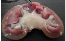
The animal was kept in a masonry pen, without bedding, to check urination, in addition to supportive therapy with flunixin meglumine (2.2 mg/kg, intramuscularly, every 24 h) and enrofloxacin (7.5 mg/kg, subcutaneously, every 24 h). However, the lamb was euthanized four days after the onset of clinical signs due to new obstructions of both the Foley catheter of the cystostomy and the perineal urethrostomy. At necropsy, the penile and urethral mucosa were found to be necrotic (Fig. 4A, 4B), with stone deposition from the bladder trigone to the sigmoid flexure. Although calculi were not observed in the kidneys, clumps of organic or mineral material were revealed which acted in their formation (Fig.5).

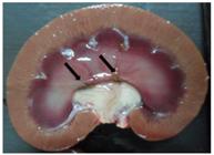
Ovine kidney cut surface (G2), marrow with greenish-brown organic matter deposition (arrows) in the pelvis and renal medulla region.
At 42 days of the experiment, a second animal, from G1, presented abdominal distension. Physical examination revealed the presence of fluid, and after laparocentesis, bladder rupture was confirmed. Due to the animal’s uremic condition from the uroperitoneum, and although preoperative supportive therapy was administered, the lamb died at the time of anesthetic induction. At necropsy, there were accumulations of serosanguineous fluid in the abdominal cavity and stones in the urethra proximal to the bladder with blood clots at the rupture site, and the urinary bladder wall was thin and friable with the mucosa showing diffuse hemorrhage (Fig. 6A/B). The kidneys and penile mucosa were normal.
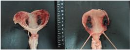
Lamb bladder 15 (G1), with rupture. A) Bladder mucosa with bilateral necrohemorrhagic cystitis with multifocal hemorrhages. B) Bladder serosa with bilateral necrosis and hemorrhage.
On day 54, a third animal, from G1 presented clinical symptoms of obstructive urolithiasis, with dysuria, pollakiuria, and anorexia. After application of hyoscine with dipyrone and acepromazine, the animal was positioned and sedated for amputation of the urethral process because of a stone discovered at the base of the glans (Fig.7A). A urethral probe (Fig.7B) and retrograde hydropropulsion were performed, with success. Urinary flow was reestablished and the animal received the same treatment as described above with an anti-inflammatory and antibiotics, in addition to remaining in a stall without a bed to monitor the characteristics of urination. The animal presented no subsequent obstruction recurrence and urinated normally until the end of the experiment. After the experimental period, a necropsy of this third animal was performed, and calculi were found in the renal medullary region. The other structures of the urinary system were normal.
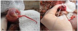
Lamb 02 (G1) with urethral obstruction. A) calculus at the base of the urethral process, with edema and hyperemia. B) urethral probe after amputation of the urethral process.
At the end of 90 days of the experiment, the necropsy of 28 animals was performed (14 lambs from G1 and 14 from G2) and the kidneys were harvested for macro- and microscopic analysis. Stones were detected in six (6/15) sheep from G1 and five (5/15) from G2, including two animals that died due to obstructive urolithiasis during the experiment, one from each group.
The macroscopic changes and location of urinary stones are shown in Table 2. In G1, urinary stones were located in the urethral process and renal medulla, urethra, and bladder, whereas in G2 they were found in the renal medulla and pelvis, urethra, bladder, urethral probe, and Foley catheter (cystostomy). The majority of stones were in the medullary and renal pelvis regions, while bladder stones were found in only two animals that did not present kidney stones. Kidney stones formed clumps ranging from 0.5 to 10 mm in diameter, mostly grayish-white or yellowish in color, with irregular edges (Fig. 8). Renal pelvis dilatation and mild hydronephrosis were noted in some animals with kidney stones, although the renal size was within the normal parameters for the species (Fig. 9) of up to 7.5 cm(2020 Ferreira DOL, Santarosa BP, Belotta AF, Mamprim MJ, Silva AA, Dias A, Chiacchio SB, Gonçalves RC. Alterações ultrassonográficas renais e vesicais de ovinos confinados e suplementados com cloreto de amônio. Pesquisa Veterinária Brasileira. 2014b; 34(supl1), 99-106. doi:10.1590/S0100-736X2014001300018
https://doi.org/10.1590/S0100-736X201400...
,2525 Santarosa BP, Ferreira DOL, Belotta AF, Dias A, Mamprim MJ, Gonçalves RC. B-Mode and pulsed Doppler sonography of kidney in healthy sheep according to age. Pesquisa Veterinária Brasileira. 2016; 36(6), 545-550. doi: 10.1590/S0100-736X2016000600014
https://doi.org/10.1590/S0100-736X201600...
).
Location of calculi and macroscopic changes in the urinary system of the two experimental groups (G1 and G2)

Kidney of Lamb 18 (G2), cut surface, pyelonephritis, whitish color in the medullary region showing dilation of the renal pelvis, presence of multiple calculi in the pelvis, and renal medullary region forming clusters, alteration in the medullary cortex relationship.
Lamb 18 (G2), cut surface of the kidney. There is deposition of multiple whitish stones in the pelvis region, moderate pelvic dilatation, hydronephrosis, and mild atrophy of the renal parenchyma with alteration in the cortical medulla relationship.
Alterations in the kidneys were observed in the histopathological examination of the urinary system (Figs 10, 11, and 12), as described in Table 3. It was observed that there was no significant difference in the frequency of the types of alterations among the sheep of groups G1 and G2, which were vascular congestion (p=0.57), tubular dilation (p=0.75), tubular degeneration (p=0.93), glomerular synechia (p=0.91), tubular necrosis (p= 0.75), inflammatory infiltrate of mononuclear cells (p=0.96), tubular lumen protein (p=0.904), and fibrosis (p=0.82).
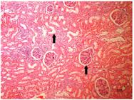
Kidney. Sheep. Nephrosis. Glomerular synechia and marked acute tubular necrosis (arrows) in dilated proximal tubules and degeneration (presence of intracytoplasmic vacuoles in tubular cells) marked in renal parenchyma, moderate vascular congestion. HE. Obj.10x.
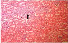
Kidney. Sheep. Nephritis. Moderate interstitial mononuclear inflammatory infiltrate (lymphocytic, arrow), moderate vascular congestion. HE. Obj.10x.

Kidney. Sheep. Proliferative glomerulonephritis. Glomerular basement membrane detachment (arrow), severe vascular congestion, moderate presence of tubular lumen protein (arrow). HE. Obj. 20x.
Microscopic changes in the kidneys of feedlot lambs from the two experimental groups (G1 and G2)
Regarding the frequency of involvement of microscopic changes, there was no difference between groups G1 and G2 in any of the lesions, which were absent (p=0.129), mild (p=0.165), moderate (p=0.752), or severe (p =0.472). In both groups, the mild degree of histopathological alterations was more frequent, with 42% in G1 and 52% in G2, while the accentuated degree was less common, with 4% of the animals from G1 and 2% from G2.
Comparing the two experimental groups, there was a difference in the presence of protein in the tubular lumen in mild (p=0.02) and moderate (p=0.03) degrees. Of the animals in G1 57.14% (8/14) had mild proteinuria, while G2 showed 92.86% (13/14). However, four animals from G1 had moderate levels of protein in the tubular lumen, whereas from G2 no lambs showed moderate protein content. The other changes were similar between groups, so it was not possible to relate them to the level of phosphorus ingested in the diets.
Although the groups received diets with different Ca:P ratios (G1: 1.9:1 and G2: 1.5:1), the results of both groups were similar. Of the 30 animals, 36.67% (11/30) showed urolithiasis, 26.67% (8/30) were asymptomatic and 10% (3/30) had obstructive urolithiasis. Of these three animals, one recovered after clinical treatment and the other two died, one after a bladder rupture and the other due to recurrence after surgical procedures of perineal urethrostomy and cystostomy to reestablish the urinary flow.
Discussion
In the present study, the first signs of obstructive urolithiasis occurred after 28 days, when one animal presented complete urethral obstruction. Therefore, it can be inferred that the diet, based on 90% concentrate and 10% crushed Coast-cross hay, with 17% NDF, could cause early clinical manifestation of the disease. In contrast, other authors(2626 Unanian MM, Rosa JSR, Silva EDF. Urolitíase Experimental Em Caprinos: Possíveis Causas E Profilaxia. Pesquisa Agropecuária brasileira. 1985; 20(4), 467-474.)described symptoms of urethral obstruction later in goats, after 92 days of confinement, with animals fed elephant grass ad libitum, which did not prevent the onset of the disease, similar to experiments with corn grain (59.5 g/day) and cotton cake (40 g/day).
The lambs were fed diets with Ca:P ratios of 1.9:1 (G1) and 1.5:1 (G2), and despite these ratios being within the values proposed in the literature(33 Riet-Côrrea F, Simões SVD, Vasconcelos JS. Urolitíase em caprinos e ovinos. Pesquisa Veterinária Brasileira. 2008; 28(6), 319-322. ,2727 Samal L, Pattanaik AK, Mishra C, Maharana BR, Sarangi LN, Baithalu RK. Nutritional strategies to prevent urolithiasis in animals. Veterinary World. 2011; 4(3), 142-144. ), of 2:1 and 1:1 respectively, they were not effective in preventing urolithiasis in this experiment. However, the total amount of Ca and P ingested by the animals in both groups was high (G1: 8 g of Ca and 4.3 g of P/day; G2: 9.7 g of Ca and 6.5 g of P/day) and above those recommended for growing lambs(1616 National Research Council - NRC. Nutrient Requirements of Small Ruminants: sheep, goats, cervids and New World camelids. Natl Acad. Press, Washington, DC. 2007, 384p.). Along with the high total consumption of minerals, the animals were fed 10% of ground roughage, which, despite not having caused digestive alterations due to previous adaptation to the diet, provided less salivation and, consequently, less P excretion by the rumination process. Thus, the excretion of excess P from the body occurred largely through the kidneys instead of via feces, which favored calculogenesis(1010 Freeman SR, Poorea MH, Young GA, Anderson KL. Influence of calcium (0.6 or 1.2%) and phosphorus (0.3 or 0.6%) content and ratio on the formation of urolithogenic compounds in the urine of Boer-cross goats fed high-concentrate diets. Small Ruminant Research. 2010; 93, 94-102. doi: 10.1016/j.smallrumres.2010.05.007
https://doi.org/10.1016/j.smallrumres.20...
,1313 Aquino Neto HM, Facury Filho EJ, Carvalho AU, Souza FA, Jordão LR. Urolitíase obstrutiva em ovinos: revisão de literatura. Veterinária em Foco. 2007; 4(2), 191-202.,1515 Maciel TA, Júnior NL, Araújo VV, Silva Filho AB, Gomes DLS, Barbosa AMS, Farias CC, Magalhães ALR, Lima MJM, Melo SAX, Oliveira D. Avaliação dos perfis minerais séricos, urinários e sedimentares de ovinos recebendo dieta calculogênica. Arquivo Brasileiro de Medicina Veterinária e Zootecnia. 2016; 68(4), 967-976. doi: 10.1590/1678-4162-8363
https://doi.org/10.1590/1678-4162-8363...
).
Another relevant factor was mortality, since only one of the three animals with obstructive urolithiasis remained alive until the end of the experiment. The eventual success of urethral flow clearance is related to the obstruction site, number of stones, and early diagnosis, since after the onset of the disease, the chance of returning to a productive life is low(99 Dória RGS, Canola PA, Dias DPM, Pereira RN, Valadão CAA. Surgical Techniques for obstructive urolithiasis in small ruminants: case report. Arquivo Brasileiro de Medicina Veterinária e Zootecnia. 2007; 59(6), 1425-1432. doi:10.1590/S0102-09352007000600012
https://doi.org/10.1590/S0102-0935200700...
). Therefore, the best outcomes occur through disease prevention(55 Ferreira DOL, Santarosa BP, Amorim RM, Chiacchio SB, Gonçalves RC. (2015). Urolitíase obstrutiva em ovinos. Veterinária e Zootecnia. 2015; 22(2), 183-197.,77 Kinsley MA, Semevolos S, Parker JE, Duesterdieck-Zellmer K, Huber M. Use of plain radiography in the diagnosis, surgical management, and postoperative treatment of obstructive urolithiasis in 25 goats and 2 sheep. Veterinary Surgery. 2013; 42, 663-668. doi: 10.1111/j.1532-950X.2013.12021.
https://doi.org/10.1111/j.1532-950X.2013...
).
The ultrasonographic findings regarding the RI of the renal artery of the lamb that presented complete urethral obstruction (Lamb 17), were normal for age and species(2121 Santarosa BP, Ferreira DOL, Rodrigues MMP, Dantas GN, Sacco SR, Lopes RS, Dias A, Gonçalves RC. Clinical, laboratory and anatomopathological evaluation of the urinary system of feedlot sheep with or without ammonium chloride supplementation. Pesquisa Veterinária Brasileira. 2016; 36(1), 1-12. doi: 10.1590/S0100-736X2016000100001
https://doi.org/10.1590/S0100-736X201600...
). These authors described an RI of 0.73 in an obstructed sheep, which demonstrated that, although the sheep in this study presented a large number of stones in the bladder, at the time of the examination, there was no hemodynamic change in the kidneys. Furthermore, despite the pelvis dilation meeting the reference measurements(2020 Ferreira DOL, Santarosa BP, Belotta AF, Mamprim MJ, Silva AA, Dias A, Chiacchio SB, Gonçalves RC. Alterações ultrassonográficas renais e vesicais de ovinos confinados e suplementados com cloreto de amônio. Pesquisa Veterinária Brasileira. 2014b; 34(supl1), 99-106. doi:10.1590/S0100-736X2014001300018
https://doi.org/10.1590/S0100-736X201400...
), the ureters were not completely occluded and the urine reached the bladder; however, it was not being eliminated due to the presence of stones in the proximal urethra. Hyperchoic spots were visible in the urinary vesicle of this lamb (Fig. 1), which revealed the presence of calculi(2020 Ferreira DOL, Santarosa BP, Belotta AF, Mamprim MJ, Silva AA, Dias A, Chiacchio SB, Gonçalves RC. Alterações ultrassonográficas renais e vesicais de ovinos confinados e suplementados com cloreto de amônio. Pesquisa Veterinária Brasileira. 2014b; 34(supl1), 99-106. doi:10.1590/S0100-736X2014001300018
https://doi.org/10.1590/S0100-736X201400...
,2828 Riedi AK, Knubben-Schweizer G, Meylan M (2018) Clinical findings and diagnostic procedures in 270 small ruminants with obstructive urolithiasis. Journal of Veterinary Internal Medicine. 2018; 32: 1274-1282. doi: 10.1111/jvim.15128
https://doi.org/10.1111/jvim.15128...
). However, in Lamb 15, which presented bladder rupture, it was not possible to readily detect free fluid in the abdominal cavity, as reported by other authors(2828 Riedi AK, Knubben-Schweizer G, Meylan M (2018) Clinical findings and diagnostic procedures in 270 small ruminants with obstructive urolithiasis. Journal of Veterinary Internal Medicine. 2018; 32: 1274-1282. doi: 10.1111/jvim.15128
https://doi.org/10.1111/jvim.15128...
) in 33/243 of the small ruminants examined.
Amputation of the urethral process, associated with urethral probing and retrograde hydropropulsion were performed in two animals with urethral obstruction, and urinary flow was reestablished in one lamb, as calculi were found at the base of the vermiform appendix, which is a common location for small ruminants(2929 Halland S, Phelps M, House JK. New methods to treat and prevent obstructive urolithiasis in small ruminants and pot-bellied pigs. In: Proceedings of the 18o American College of Veterinary Medicine Forum; Lafayette, Colorado. Lafayette: American College of Veterinary Medicine Forum. 2000; 268-270.). Amputation of this urethral extension is considered an effective technique in close to 50% of the cases of urolithiasis in small ruminants, providing immediate relief(66 Ewoldt JM, Jones ML, Miesner MD. Surgery of obstructive urolithiasis in ruminants. Veterinary Clinics of North America: Food Animal Practice. 2008; 24, 455-465. doi: 10.1016/j.cvfa.2008.06.003
https://doi.org/10.1016/j.cvfa.2008.06.0...
), in addition to being necessary for urethral probing. Immediate restoration of urethral flow was reported in 50%(3030 Fortier LA, Gregg AJ, Fubini SL. Caprine obstructive urolithiasis Requirement for 2nd surgical intervention and mortality after percutaneous tube cystotomy, surgical tube cystotomy, or urinary bladder marsupialization. Veterinary Surgery. 2004; 33(6), 661-667. doi: 10.1111/j.1532-950X.2004.04089
https://doi.org/10.1111/j.1532-950X.2004...
) to 66%(3131 Haven ML, Bowman KF, Englebert TA, Blikslager AT. Surgical management of urolithiasis in small ruminants. Cornell Veterinary. 1993; 83(1), 47-55.) of animals after amputation of this structure. However, the recurrence rate is high and, in approximately 80 to 90% of cases, reobstruction occurs(3030 Fortier LA, Gregg AJ, Fubini SL. Caprine obstructive urolithiasis Requirement for 2nd surgical intervention and mortality after percutaneous tube cystotomy, surgical tube cystotomy, or urinary bladder marsupialization. Veterinary Surgery. 2004; 33(6), 661-667. doi: 10.1111/j.1532-950X.2004.04089
https://doi.org/10.1111/j.1532-950X.2004...
). In the animal that reestablished urinary flow, no recurrence of the obstruction was observed for the 36 days to the end of the experiment, but stones were found in the renal medulla at necropsy, since the diet with high concentration of minerals was maintained.
In another animal from G2, perineal urethrostomy surgery was performed due to the failure of the urethral probe, which is a surgical procedure indicated in case of complete obstruction or urethral rupture(99 Dória RGS, Canola PA, Dias DPM, Pereira RN, Valadão CAA. Surgical Techniques for obstructive urolithiasis in small ruminants: case report. Arquivo Brasileiro de Medicina Veterinária e Zootecnia. 2007; 59(6), 1425-1432. doi:10.1590/S0102-09352007000600012
https://doi.org/10.1590/S0102-0935200700...
,2222 Oman RE, Reppert EJ, Jones SM. Outcome and complications in goats treated by perineal urethrostomy for obstructive urolithiasis: 25 cases (2010-2017). Journal of Veterinary Internal Medicine. 2019; 33, 292-296. doi: 10.1111/jvim.15360
https://doi.org/10.1111/jvim.15360...
). This technique is not a cure, as stenosis occurs, being a contraindicated procedure for breeders because it makes the animal unsuitable for reproduction(88 Van Metre D, Fecteau G, House JK. Obstructive urolithiasis in ruminants: surgical management and prevention. Compendium on Continuing Education for the Practicing Veterinarian. 1996; 19: 275-289.,2222 Oman RE, Reppert EJ, Jones SM. Outcome and complications in goats treated by perineal urethrostomy for obstructive urolithiasis: 25 cases (2010-2017). Journal of Veterinary Internal Medicine. 2019; 33, 292-296. doi: 10.1111/jvim.15360
https://doi.org/10.1111/jvim.15360...
,3232 Janke JJ, Osterstock JB, Washburn KE, Bissett WT, Roussel Jr AJ, Hooper RN. Use of Walpole’s solution for treatment of goats with urolithiasis: 25 cases (2001-2006). Journal of the American Veterinary Medical Association. 2009; 53, 234-249. doi: 10.2460/javma.234.2.249
https://doi.org/10.2460/javma.234.2.249...
). However, even after the procedure, the animal was unable to urinate due to total obstruction of the proximal portion of the urethra. After retrograde hydropropulsion with saline solution, reestablishment of urinary flow did not occur. Therefore, this lamb underwent a cystostomy procedure for placement of a Foley catheter(99 Dória RGS, Canola PA, Dias DPM, Pereira RN, Valadão CAA. Surgical Techniques for obstructive urolithiasis in small ruminants: case report. Arquivo Brasileiro de Medicina Veterinária e Zootecnia. 2007; 59(6), 1425-1432. doi:10.1590/S0102-09352007000600012
https://doi.org/10.1590/S0102-0935200700...
,3333 Riedi AK, Christina N, Gabriela KS, Karl N, Mireille M. Variables of initial examination and clinical management associated with survival in small ruminants with obstructive urolithiasis Journal of Veterinary Internal Medicine. 2018; 32, 2105-2114. doi: 10.1111/jvim.15336
https://doi.org/10.1111/jvim.15336...
). The animal was followed-up with post-operative care and after four days was euthanized and sent for necropsy due to recurrence of the obstruction.
The use of cystostomy with urethral hydropropulsion is not generally adopted, especially in the field, due to the prolonged surgical time, resulting in an increased risk of urethral rupture at the site of obstruction(3030 Fortier LA, Gregg AJ, Fubini SL. Caprine obstructive urolithiasis Requirement for 2nd surgical intervention and mortality after percutaneous tube cystotomy, surgical tube cystotomy, or urinary bladder marsupialization. Veterinary Surgery. 2004; 33(6), 661-667. doi: 10.1111/j.1532-950X.2004.04089
https://doi.org/10.1111/j.1532-950X.2004...
). However, the procedure with application of a probe (Foley catheter) allows for longer survival time and a return to normal reproductive function(1111 Streeter RN, Washburn KE, Mccauley CT. Percutaneous tube cystostomy and vesicular irrigation for treatment of obstructive urolithiasis in a goat. Jornal of the American Veterinary Medical Association. 2002; 221, 546-549. doi: 10.2460/javma.2002.221.546
https://doi.org/10.2460/javma.2002.221.5...
,3434 Fazili MR, Malik HU, Bhattacharyya HK, Buchoo BA, Moulvi BA, Makhdoomi DM. Minimally invasive surgical tube cystotomy for treating obstructive urolithiasis in small ruminants with an intact urinary bladder. Veterinary Record. 2010; 166, 528-532. doi:10.1136/vr.b4831
https://doi.org/10.1136/vr.b4831...
,3535 Pearce SG, Dearo AC, Howard BE, Brisson BA. Management of obstructive urolithiasis and concurrent urethral rupture in a goat. Australian Veterinary Journal. 2003; 81, 268-270. doi: 10.1111/j.1751-0813.2003.tb12568
https://doi.org/10.1111/j.1751-0813.2003...
) with an average of 80% success in small ruminants. However, surgical time, costs, and the need for extensive veterinary follow-up in the postoperative period restrict its use(99 Dória RGS, Canola PA, Dias DPM, Pereira RN, Valadão CAA. Surgical Techniques for obstructive urolithiasis in small ruminants: case report. Arquivo Brasileiro de Medicina Veterinária e Zootecnia. 2007; 59(6), 1425-1432. doi:10.1590/S0102-09352007000600012
https://doi.org/10.1590/S0102-0935200700...
,3636 Chigerwe M, Heller MC, Balcomb CC, Angelos JA. Use of a percutaneous transabdominal catheter for management of obstructive urolithiasis in goats, sheep, and potbellied pigs: 69 cases (2000-2014). Jornal of the American Veterinary Medical Association. 2016; 248(11), 1287-1290. doi: 10.2460/javma.248.11.1287
https://doi.org/10.2460/javma.248.11.128...
). The macroscopic changes observed at necropsy occurred according to the type of urolithiasis, with findings related to bladder rupture, foci of hemorrhagic necrosis, and bladder mucosa congested with ecchymosis. Such alterations are similar to the descriptions by other authors(3737 Davoodi F, Zakian A, Raisi A, Rocky A, FarjaniKish G. Report of urinary bladder rupture following obstructive urolithiasis in rams of Romanov sheep flock. Comparative Clinical Pathology. 2020; 29, 1277-1282. doi: 10.1007/s00580-020-03165-1.
https://doi.org/10.1007/s00580-020-03165...
), who reported a bladder rupture in a Romanov ram.
Although mild hydronephrosis and dilation of the renal pelvis were observed in two animals, in addition to a large abundance of stones, the kidneys were of normal size. Other authors(11 Guimarães JA, Mendonça CL, Sousa Guaraná EL, Dantas AC, Azevêdo CN, Câmara ACL, Farias CC, Afonso JAB. Estudo retrospectivo de 66 casos de urolitíase obstrutiva em ovinos. Pesquisa Veterinária Brasileira. 2012; 32(9), 824-830. doi: 10.1590/S0100-736X2012000900002
https://doi.org/10.1590/S0100-736X201200...
,2121 Santarosa BP, Ferreira DOL, Rodrigues MMP, Dantas GN, Sacco SR, Lopes RS, Dias A, Gonçalves RC. Clinical, laboratory and anatomopathological evaluation of the urinary system of feedlot sheep with or without ammonium chloride supplementation. Pesquisa Veterinária Brasileira. 2016; 36(1), 1-12. doi: 10.1590/S0100-736X2016000100001
https://doi.org/10.1590/S0100-736X201600...
,3838 Alimi OA, Bello M, Baraya YS, Raji A, Bashir S, Bello AA, Shoyinka SVO. Obstructive Urolithiasis In Ouda-Yankasa Ram: Case Report. Animal Research International. 2018; 15(2), 2989-2993. doi:10.4314/as.v17i1.7
https://doi.org/10.4314/as.v17i1.7...
) found alterations in sheep such as pyelonephritis and hydroureter, which were not observed here.
In the present study, the presence of mild and moderate protein levels in the tubular lumen of 85.71% (12/14) of sheep in G1 and 100% (14/14) of G2, can be related to crude protein (CP) of the diets, which was 19.22% and 18.39%, respectively. The protein in the renal tubule generally precedes a lesion, as the majority comes from an alteration in glomerular filtration or glomerulopathy(3939 Frelier PF, Armstrong DL, Pritchard, J. Ovine mesangiocapillary glomerulonephritis type I and crescent formation. Veterinary Pathology. 1990; 27, 26-34. doi: 10.1177/030098589002700104
https://doi.org/10.1177/0300985890027001...
). Other authors(2525 Santarosa BP, Ferreira DOL, Belotta AF, Dias A, Mamprim MJ, Gonçalves RC. B-Mode and pulsed Doppler sonography of kidney in healthy sheep according to age. Pesquisa Veterinária Brasileira. 2016; 36(6), 545-550. doi: 10.1590/S0100-736X2016000600014
https://doi.org/10.1590/S0100-736X201600...
), who also studied microscopic findings of confined sheep, found 37.5% of animals that received ammonium chloride and 25% of the control group had mild tubular lumen protein, and the diet contained 20.34% of CP.
Tubular necrosis, ranging from mild to severe, was found in 78.57% (11/14) of lambs of both groups (22/28), which is a relevant finding for this study, corroborating other authors(2525 Santarosa BP, Ferreira DOL, Belotta AF, Dias A, Mamprim MJ, Gonçalves RC. B-Mode and pulsed Doppler sonography of kidney in healthy sheep according to age. Pesquisa Veterinária Brasileira. 2016; 36(6), 545-550. doi: 10.1590/S0100-736X2016000600014
https://doi.org/10.1590/S0100-736X201600...
), who reported this change as the most frequent, with 40% in the group that received urinary acidifiers and 70% in the control group. In a case report, this alteration in the histopathological analysis of an Ouda-Yankasa ram was also described along with glomerular atrophy(3838 Alimi OA, Bello M, Baraya YS, Raji A, Bashir S, Bello AA, Shoyinka SVO. Obstructive Urolithiasis In Ouda-Yankasa Ram: Case Report. Animal Research International. 2018; 15(2), 2989-2993. doi:10.4314/as.v17i1.7
https://doi.org/10.4314/as.v17i1.7...
), which was not a common finding in the present study. Another study(3737 Davoodi F, Zakian A, Raisi A, Rocky A, FarjaniKish G. Report of urinary bladder rupture following obstructive urolithiasis in rams of Romanov sheep flock. Comparative Clinical Pathology. 2020; 29, 1277-1282. doi: 10.1007/s00580-020-03165-1.
https://doi.org/10.1007/s00580-020-03165...
) reported, in addition to tubular and glomerular degeneration, fibrosis of the renal tissue in a Romanov sheep, which was not found in the lambs in this study.
Conclusion
The diets used, even with different levels of phosphorus, induced urolithiasis in a similar way between groups. The Ca:P ratio of the diets in the proportion 1.9:1 or 1.5:1 did not prevent the formation of calculi, due to the high concentration of minerals. The concentration of crude protein in the diet resulted in the presence of protein in the tubular lumen. Treatment success depends on early diagnosis, site of obstruction, and number of stones that obstruct the urinary flow, as well as diet adequacy.
Therefore, it was concluded that urolithiasis is a multifactorial disease, and nutritional management is particularly relevant for calculogenesis. The presence of the disease in one animal in the herd is an indication that others may be affected. Therefore, prevention is recommended, since when established, the chance of economic losses is high, due to the costs of treatment and loss of animals of high genetic value.
Acknowledgments
The authors thank FMVZ/UNESP, Campus Botucatu-SP, for the physical structure and equipment used to carry out the experiment, and FAPESP (São Paulo State Research Support Foundation) for the Regular Research Support granted (Process 2012/22620-8).
References
-
1Guimarães JA, Mendonça CL, Sousa Guaraná EL, Dantas AC, Azevêdo CN, Câmara ACL, Farias CC, Afonso JAB. Estudo retrospectivo de 66 casos de urolitíase obstrutiva em ovinos. Pesquisa Veterinária Brasileira. 2012; 32(9), 824-830. doi: 10.1590/S0100-736X2012000900002
» https://doi.org/10.1590/S0100-736X2012000900002 -
2Maciel TA, Ramos IA, Silva RJ, Soares PC, Carvalho CCD, Maior Júnior RJS, Amoroso L, Artoni SMB, Afonso JAB, Oliveira D. Clinical and Biochemical Profile of Obstructive Urolithiasis in Sheep. Acta Scientiae Veterinariae. 2017; 45, 1-15. Available from: https://www.redalyc.org/articulo.oa?id=289053641089
» https://www.redalyc.org/articulo.oa?id=289053641089 -
3Riet-Côrrea F, Simões SVD, Vasconcelos JS. Urolitíase em caprinos e ovinos. Pesquisa Veterinária Brasileira. 2008; 28(6), 319-322.
-
4Antonelli AC, Barrêto Júnior RA, Mori CS, Sucupira MCA, Marcello ACS, Ortolani EL. Efeito de diferentes fontes energéticas na predisposição para urolitíase em cabritos. Ciência Animal Brasileira. 2012; 13, 487-493. doi: 10.5216/cab.v13i4.15617
» https://doi.org/10.5216/cab.v13i4.15617 -
5Ferreira DOL, Santarosa BP, Amorim RM, Chiacchio SB, Gonçalves RC. (2015). Urolitíase obstrutiva em ovinos. Veterinária e Zootecnia. 2015; 22(2), 183-197.
-
6Ewoldt JM, Jones ML, Miesner MD. Surgery of obstructive urolithiasis in ruminants. Veterinary Clinics of North America: Food Animal Practice. 2008; 24, 455-465. doi: 10.1016/j.cvfa.2008.06.003
» https://doi.org/10.1016/j.cvfa.2008.06.003 -
7Kinsley MA, Semevolos S, Parker JE, Duesterdieck-Zellmer K, Huber M. Use of plain radiography in the diagnosis, surgical management, and postoperative treatment of obstructive urolithiasis in 25 goats and 2 sheep. Veterinary Surgery. 2013; 42, 663-668. doi: 10.1111/j.1532-950X.2013.12021.
» https://doi.org/10.1111/j.1532-950X.2013.12021. -
8Van Metre D, Fecteau G, House JK. Obstructive urolithiasis in ruminants: surgical management and prevention. Compendium on Continuing Education for the Practicing Veterinarian. 1996; 19: 275-289.
-
9Dória RGS, Canola PA, Dias DPM, Pereira RN, Valadão CAA. Surgical Techniques for obstructive urolithiasis in small ruminants: case report. Arquivo Brasileiro de Medicina Veterinária e Zootecnia. 2007; 59(6), 1425-1432. doi:10.1590/S0102-09352007000600012
» https://doi.org/10.1590/S0102-09352007000600012 -
10Freeman SR, Poorea MH, Young GA, Anderson KL. Influence of calcium (0.6 or 1.2%) and phosphorus (0.3 or 0.6%) content and ratio on the formation of urolithogenic compounds in the urine of Boer-cross goats fed high-concentrate diets. Small Ruminant Research. 2010; 93, 94-102. doi: 10.1016/j.smallrumres.2010.05.007
» https://doi.org/10.1016/j.smallrumres.2010.05.007 -
11Streeter RN, Washburn KE, Mccauley CT. Percutaneous tube cystostomy and vesicular irrigation for treatment of obstructive urolithiasis in a goat. Jornal of the American Veterinary Medical Association. 2002; 221, 546-549. doi: 10.2460/javma.2002.221.546
» https://doi.org/10.2460/javma.2002.221.546 -
12Sun WD, Zhang KC, Wang JY, Wang XL. The chemical composition and ultrastructure of uroliths in Boer goats. The Veterinary Journal. 2010; 86, 70-75. doi: 10.1016/j.tvjl.2009.07.009
» https://doi.org/10.1016/j.tvjl.2009.07.009 -
13Aquino Neto HM, Facury Filho EJ, Carvalho AU, Souza FA, Jordão LR. Urolitíase obstrutiva em ovinos: revisão de literatura. Veterinária em Foco. 2007; 4(2), 191-202.
-
14Ferreira DOL, Santarosa BP, Sacco SR, Dias A, Amorim RM, Chiacchio SB, Lis bôa JAN, Gonçalves RC. Efeito da suplemen tação de cloreto de amônio sobre o equilíbrio ácido-básico e o pH urinário de ovinos confinados. Pesquisa Veterinária Brasileira. 2014a; 34(8), 797-804. doi: 10.1590/S0100-736X2014000800016
» https://doi.org/10.1590/S0100-736X2014000800016 -
15Maciel TA, Júnior NL, Araújo VV, Silva Filho AB, Gomes DLS, Barbosa AMS, Farias CC, Magalhães ALR, Lima MJM, Melo SAX, Oliveira D. Avaliação dos perfis minerais séricos, urinários e sedimentares de ovinos recebendo dieta calculogênica. Arquivo Brasileiro de Medicina Veterinária e Zootecnia. 2016; 68(4), 967-976. doi: 10.1590/1678-4162-8363
» https://doi.org/10.1590/1678-4162-8363 -
16National Research Council - NRC. Nutrient Requirements of Small Ruminants: sheep, goats, cervids and New World camelids. Natl Acad. Press, Washington, DC. 2007, 384p.
-
17Russel A. Body condition scoring of sheep. In: E. Boden (Ed.) Sheep and Goat Practice. Bailliere Tindall, Philadelphia. 1991. p3.
-
18Scott PR, Sargison ND. Ultrasonography as an adjunct to clinical examination in sheep. Small Ruminant Research. 2010;92, 108-119. doi: 10.1016/j.smallrumres.2010.04.011
» https://doi.org/10.1016/j.smallrumres.2010.04.011 -
19Scott P. Transabdominal Ultrasonographic Examination of 26 Sheep with Suspected Urinary Tract Disease (2010- 2012 Journal of Veterinary Science & Medical Diagnosis. 2013; 2, 1-5. doi: 10.4172/2325-9590.1000107
» https://doi.org/10.4172/2325-9590.1000107 -
20Ferreira DOL, Santarosa BP, Belotta AF, Mamprim MJ, Silva AA, Dias A, Chiacchio SB, Gonçalves RC. Alterações ultrassonográficas renais e vesicais de ovinos confinados e suplementados com cloreto de amônio. Pesquisa Veterinária Brasileira. 2014b; 34(supl1), 99-106. doi:10.1590/S0100-736X2014001300018
» https://doi.org/10.1590/S0100-736X2014001300018 -
21Santarosa BP, Ferreira DOL, Rodrigues MMP, Dantas GN, Sacco SR, Lopes RS, Dias A, Gonçalves RC. Clinical, laboratory and anatomopathological evaluation of the urinary system of feedlot sheep with or without ammonium chloride supplementation. Pesquisa Veterinária Brasileira. 2016; 36(1), 1-12. doi: 10.1590/S0100-736X2016000100001
» https://doi.org/10.1590/S0100-736X2016000100001 -
22Oman RE, Reppert EJ, Jones SM. Outcome and complications in goats treated by perineal urethrostomy for obstructive urolithiasis: 25 cases (2010-2017). Journal of Veterinary Internal Medicine. 2019; 33, 292-296. doi: 10.1111/jvim.15360
» https://doi.org/10.1111/jvim.15360 -
23Guedes Jr FS, Cruz DS, Rodrigues MMP, Silva LM, Amorim RL, Vianna PTG, Castiglia YM M. Renal histology and immunohistochemistry after acute hemorrhage in rats under sevoflurane and ketoprofen effect. Acta Cirurgica Brasileira. 2012; 27(1), 37-42. doi: 10.1590/S0102-86502012000100007
» https://doi.org/10.1590/S0102-86502012000100007 -
24Van Metre DC, Fubini SL. Ovine and caprine urolithiasis: Another piece of the puzzle veterinary surgery. Veterinary Surgery. 2006; 35, 413-416. doi: 10.1111/j.1532-950X.2006.00168.
» https://doi.org/10.1111/j.1532-950X.2006.00168 -
25Santarosa BP, Ferreira DOL, Belotta AF, Dias A, Mamprim MJ, Gonçalves RC. B-Mode and pulsed Doppler sonography of kidney in healthy sheep according to age. Pesquisa Veterinária Brasileira. 2016; 36(6), 545-550. doi: 10.1590/S0100-736X2016000600014
» https://doi.org/10.1590/S0100-736X2016000600014 -
26Unanian MM, Rosa JSR, Silva EDF. Urolitíase Experimental Em Caprinos: Possíveis Causas E Profilaxia. Pesquisa Agropecuária brasileira. 1985; 20(4), 467-474.
-
27Samal L, Pattanaik AK, Mishra C, Maharana BR, Sarangi LN, Baithalu RK. Nutritional strategies to prevent urolithiasis in animals. Veterinary World. 2011; 4(3), 142-144.
-
28Riedi AK, Knubben-Schweizer G, Meylan M (2018) Clinical findings and diagnostic procedures in 270 small ruminants with obstructive urolithiasis. Journal of Veterinary Internal Medicine. 2018; 32: 1274-1282. doi: 10.1111/jvim.15128
» https://doi.org/10.1111/jvim.15128 -
29Halland S, Phelps M, House JK. New methods to treat and prevent obstructive urolithiasis in small ruminants and pot-bellied pigs. In: Proceedings of the 18o American College of Veterinary Medicine Forum; Lafayette, Colorado. Lafayette: American College of Veterinary Medicine Forum. 2000; 268-270.
-
30Fortier LA, Gregg AJ, Fubini SL. Caprine obstructive urolithiasis Requirement for 2nd surgical intervention and mortality after percutaneous tube cystotomy, surgical tube cystotomy, or urinary bladder marsupialization. Veterinary Surgery. 2004; 33(6), 661-667. doi: 10.1111/j.1532-950X.2004.04089
» https://doi.org/10.1111/j.1532-950X.2004.04089 -
31Haven ML, Bowman KF, Englebert TA, Blikslager AT. Surgical management of urolithiasis in small ruminants. Cornell Veterinary. 1993; 83(1), 47-55.
-
32Janke JJ, Osterstock JB, Washburn KE, Bissett WT, Roussel Jr AJ, Hooper RN. Use of Walpole’s solution for treatment of goats with urolithiasis: 25 cases (2001-2006). Journal of the American Veterinary Medical Association. 2009; 53, 234-249. doi: 10.2460/javma.234.2.249
» https://doi.org/10.2460/javma.234.2.249 -
33Riedi AK, Christina N, Gabriela KS, Karl N, Mireille M. Variables of initial examination and clinical management associated with survival in small ruminants with obstructive urolithiasis Journal of Veterinary Internal Medicine. 2018; 32, 2105-2114. doi: 10.1111/jvim.15336
» https://doi.org/10.1111/jvim.15336 -
34Fazili MR, Malik HU, Bhattacharyya HK, Buchoo BA, Moulvi BA, Makhdoomi DM. Minimally invasive surgical tube cystotomy for treating obstructive urolithiasis in small ruminants with an intact urinary bladder. Veterinary Record. 2010; 166, 528-532. doi:10.1136/vr.b4831
» https://doi.org/10.1136/vr.b4831 -
35Pearce SG, Dearo AC, Howard BE, Brisson BA. Management of obstructive urolithiasis and concurrent urethral rupture in a goat. Australian Veterinary Journal. 2003; 81, 268-270. doi: 10.1111/j.1751-0813.2003.tb12568
» https://doi.org/10.1111/j.1751-0813.2003.tb12568 -
36Chigerwe M, Heller MC, Balcomb CC, Angelos JA. Use of a percutaneous transabdominal catheter for management of obstructive urolithiasis in goats, sheep, and potbellied pigs: 69 cases (2000-2014). Jornal of the American Veterinary Medical Association. 2016; 248(11), 1287-1290. doi: 10.2460/javma.248.11.1287
» https://doi.org/10.2460/javma.248.11.1287 -
37Davoodi F, Zakian A, Raisi A, Rocky A, FarjaniKish G. Report of urinary bladder rupture following obstructive urolithiasis in rams of Romanov sheep flock. Comparative Clinical Pathology. 2020; 29, 1277-1282. doi: 10.1007/s00580-020-03165-1.
» https://doi.org/10.1007/s00580-020-03165-1 -
38Alimi OA, Bello M, Baraya YS, Raji A, Bashir S, Bello AA, Shoyinka SVO. Obstructive Urolithiasis In Ouda-Yankasa Ram: Case Report. Animal Research International. 2018; 15(2), 2989-2993. doi:10.4314/as.v17i1.7
» https://doi.org/10.4314/as.v17i1.7 -
39Frelier PF, Armstrong DL, Pritchard, J. Ovine mesangiocapillary glomerulonephritis type I and crescent formation. Veterinary Pathology. 1990; 27, 26-34. doi: 10.1177/030098589002700104
» https://doi.org/10.1177/030098589002700104
Publication Dates
-
Publication in this collection
13 Aug 2021 -
Date of issue
2021
History
-
Received
23 Feb 2021 -
Accepted
09 June 2021 -
Published
14 July 2021


