 Comparative analysis of MS and PC-L2 media on growth, photosynthetic performance, elemental composition, and anatomical traits of Selenicereus undatus (Haw.) D.R. Hunt (Cactaceae) in vitro culture
ABSTRACT
The composition of the culture medium is a critical factor in the development of plants in vitro, influencing the growth and morphophysiological characteristics of micropropagated plants. This study evaluated the suitability of MS (Murashige & Skoog) and PC-L2 (Phillips & Collins) culture media for the in vitro development of Selenicereus undatus (Haw.) D.R. Hunt based on growth characteristics, photosynthetic responses, mineral composition, and anatomy. Cladodes were cultured for 75 days under a 16-h photoperiod and a photosynthetic photon flux density of 90 μmol m-² s-¹. Shoot and root growth, fresh and dry biomass, photosynthetic pigments, chlorophyll a fluorescence parameters, elemental composition, and anatomical characteristics were evaluated. Plants grown in PC-L2 medium exhibited greater shoot elongation, fresh biomass accumulation, root area, and root diameter, whereas MS medium promoted a greater cladode cross-sectional area and a higher number of vascular bundles. Chlorophyll b concentration was higher in PC-L2 medium, whereas chlorophyll a fluorescence parameters did not differ between the media, indicating the maintenance of photosynthetic performance. Mineral analysis revealed greater Mg and P detection in PC-L2 medium, whereas K predominated in both treatments. Overall, both media promoted the development of S. undatus; however, PC-L2 showed greater potential for promoting in vitro growth under the conditions evaluated.
Index terms:
Micropropagation; mineral composition; modulated fluorescence; pitahaya; vascular anatomy
RESUMO
A composição do meio de cultura é um fator crítico para o desenvolvimento de plantas in vitro, influenciando o crescimento e as características morfofisiológicas de plantas micropropagadas. Este estudo avaliou a adequação dos meios MS (Murashige & Skoog) e PC-L2 (Phillips & Collins) para o desenvolvimento in vitro de Selenicereus undatus (Haw.) D.R. Hunt, com base em características de crescimento, respostas fotossintéticas, composição mineral e anatomia. Os cladódios foram cultivados por 75 dias sob fotoperíodo de 16 h e densidade de fluxo de fótons fotossintéticos de 90 μmol m-² s-¹. Foram avaliados o crescimento da parte aérea e das raízes, biomassa fresca e seca, pigmentos fotossintéticos, parâmetros de fluorescência da clorofila a, composição elementar e características anatômicas. As plantas cultivadas em meio PC-L2 apresentaram maior alongamento da parte aérea, acúmulo de biomassa fresca, área e diâmetro radiculares, enquanto o meio MS promoveu maior área transversal dos cladódios e maior número de feixes vasculares. A concentração de clorofila b foi maior no meio PC-L2, enquanto os parâmetros de fluorescência da clorofila a não diferiram entre os meios, indicando manutenção do desempenho fotossintético. A análise mineral revelou maior detecção de Mg e P no meio PC-L2, enquanto K apresentou predominância em ambos os tratamentos. De modo geral, ambos os meios favoreceram o desenvolvimento de S. undatus, porém o PC-L2 mostrou-se mais promissor para promover o crescimento in vitro nas condições avaliadas.
Termos para indexação:
Micropropagação; composição mineral; fluorescência modulada; pitahaya; anatomia vascular
Introduction
Plant tissue culture is widely recognized as one of the most efficient techniques for clonal propagation, enabling the large-scale production of genetically uniform plants (Nimavat & Parikh, 2024). Based on the regenerative capacity of plant cells, this technique has broad applications in basic research, micropropagation, and plant breeding, including the development and selection of transgenic organisms (Long et al., 2022).
The success of in vitro regeneration depends on several factors, particularly plant growth regulators (Gerdakaneh et al., 2020), explant type (Minutolo et al., 2020), and culture medium composition (Chimdessa, 2020). Culture media provide essential nutrients required for plant growth and morphogenesis under controlled conditions (Wawrosch & Zotchev, 2021). Therefore, an appropriate medium formulation containing mineral nutrients in adequate proportions is essential to support cell division, differentiation, and the development of regenerative tissues (Rupert & Collins, 1985).
Among the available formulations, Murashige and Skoog (MS) medium is one of the most widely used for the propagation of medicinal and aromatic plants (Duta-Cornescu et al., 2023; Pasternak & Steinmacher, 2024; Xiyan et al., 2024; Gidas et al., 2025). However, alternative formulations have also shown promising results, including the Phillips and Collins (PC) medium (Phillips & Collins, 1979), which has demonstrated efficiency in tissue proliferation and rooting in several plant species (Phillips & Garda, 2019).
Despite advances in plant tissue culture, the large-scale production of some species remains limited by environmental constraints under conventional cultivation systems, restricting their commercial expansion (Anuruddi, Nakandalage & Fonseka, 2023). Among these species, Selenicereus undatus (Haw.) D.R. Hunt, commonly known as red dragon fruit or pitahaya, has gained global importance due to its adaptability, nutritional value, and economic potential. Native to Mexico and Central America, this species is currently cultivated in tropical and subtropical regions worldwide.
Belonging to the family Cactaceae, S. undatus is valued for its nutritional properties (Yadav et al., 2025; Clairvil et al., 2025) and increasing international market acceptance (Safira et al., 2021). It is extensively cultivated in Southeast Asia, particularly in southern China, where its exotic appearance, delicate texture, and functional properties have contributed to its economic importance (Guo et al., 2014; Balendres & Bengoa, 2019). Commercial production is also established in countries such as China, Vietnam, Thailand, Malaysia, Mexico, Colombia, Ecuador, Nicaragua, Indonesia, Australia, and the United States (Arivalagan et al., 2021). In addition, bioactive compounds present in its pulp have been associated with potential health-promoting effects, including the modulation of blood glucose levels (Yang et al., 2024).
Although pitahaya can be efficiently propagated by stem cuttings, in vitro culture provides important advantages over conventional propagation methods. It enables the rapid multiplication of genetically uniform plants under controlled conditions, facilitates germplasm conservation, and provides standardized plant material for physiological, anatomical, and biotechnological studies. These advantages are particularly relevant for optimizing propagation protocols and improving the quality of planting material.
Previous studies on indirect organogenesis have demonstrated the feasibility of using both MS (Murashige & Skoog) and PC-L2 (Phillips & Collins) media for S. undatus regeneration, including evaluations by scanning electron microscopy and flow cytometry (Rodrigues et al., 2022). However, comparative information regarding the effects of these media on plant growth, photosynthetic performance, mineral composition, and anatomical traits remains limited.
Therefore, this study aimed to evaluate the suitability of MS (Murashige & Skoog) and PC-L2 (Phillips & Collins) culture media for the in vitro development of Selenicereus undatus (Haw.) D.R. Hunt by assessing plant growth, photosynthetic performance, mineral composition, and anatomical traits.
Material and Methods
Study location
The experiment was conducted at the Plant Tissue Culture Laboratory, Department of Agriculture, Federal University of Lavras (UFLA), in Lavras, Minas Gerais, Brazil. Biometric analyses and the assessment of photosynthetic pigment accumulation were also carried out at this location.
Chemical analyses, using energy-dispersive X-ray spectroscopy (EDX), were performed at the Seed Microscopy Laboratory. Analyses of chlorophyll a fluorescence and anatomical evaluations of cladodes and roots were conducted at the Plant Anatomy Laboratory, Department of Biology, UFLA.
Plant material and in vitro cultivation
Plants (2.5 ± 0.5 cm) of Selenicereus undatus (Haw.) D.R. Hunt, previously established in vitro from seed germination on MS medium (Murashige & Skoog, 1962) without plant growth regulators, were used as the source of explants. Cladode segments (1.5 cm in length) excised from these stock plants were cultured in 250 mL flasks containing 50 mL of either MS (Murashige & Skoog, 1962) or PC-L2 (Level 2, revision R11.1) (Phillips & Collins, 1979) culture medium (Table 1).
The pH of the media was adjusted to 6.0 ± 0.2, and sterilization was carried out in an autoclave at 121 °C, under 1.2 atm, for 20 minutes.
Five cladodes per flask were inoculated in a laminar flow hood, with 20 flasks per treatment (100 explants). The flasks were placed in a growth room at 25 ± 2 °C for 75 days, under a 16-hour photoperiod, using Empalux® FT8 HO LED lamps (36 W/6400 K), with an average illuminance of approximately 5000-6000 lux, corresponding to 90 µmol m⁻² s⁻¹ of photosynthetically active radiation (PAR). The culture flask was considered the experimental unit because all cladodes within a flask shared the same culture medium and growth conditions. When analyses required the use of a subset of plants, one plant was randomly selected from each sampled flask, and the number of experimental units corresponded to the number of flasks evaluated, thereby avoiding pseudoreplication.
Biometric characteristics
Biometric characteristics, including shoot length and shoot and root fresh and dry masses, were evaluated after 75 days of in vitro cultivation. One plant was randomly selected from each of ten culture flasks per treatment (n = 10 experimental units), with the culture flask considered the experimental unit.
Shoot length was measured using a millimeter ruler. Shoot and root fresh masses were determined using a precision balance (OHAUS, model PR224BR; 0.0001 g readability). For dry mass determination, plant material was dried in a forced-air circulation oven at 65 °C for 72 h. Dry masses were recorded after thermal stabilization in a Styrofoam box to minimize variations caused by condensation or moisture absorption.
Photosynthetic pigments
Photosynthetic pigment analysis was performed after 75 days of in vitro cultivation using cladodes (approximately 0.050 g) collected from one randomly selected plant from each of five culture flasks per treatment (n = 5 experimental units), with the culture flask considered the experimental unit. Cladode fragments were transferred to test tubes containing 5 mL of 80% acetone for chlorophyll and carotenoid extraction. The tubes were wrapped in aluminum foil to prevent pigment degradation and stored at 4 ± 2 °C for 24 h before analysis.
Absorbance was measured at room temperature (25 ± 2 °C) using an Elisa Multiskan GO spectrophotometer (Thermo Fisher Scientific) at wavelengths of 470 nm (Abs470), 647 nm (Abs647), and 663 nm (Abs663) (Scopel, Barbosa & Vieira, 2011). Each biological sample was analyzed in triplicate (technical replicates), and the average value was used for statistical analyses.
Chlorophyll a, chlorophyll b, total chlorophyll, and carotenoid contents were calculated according to the equations of Lichtenthaler and Wellburn (1983) and Lichtenthaler and Babani (2021). Data were processed using SkanIt 5.0 for Microplate Readers software (version 5.0.0.42).
Modulated chlorophyll a fluorescence
Modulated chlorophyll a fluorescence was measured in ten biological replicates (plants) randomly selected from independent culture flasks for each treatment after 75 days of in vitro cultivation. Although each flask initially contained five cladodes, the individual plant was considered the experimental unit for this analysis, and only one plant was sampled from each selected flask to avoid pseudoreplication. Measurements were performed between 8:00 and 10:00 a.m. using a portable fluorometer MINI-PAM-I (Heinz Walz GmbH, Effeltrich, Germany), following the protocol described by Kramer et al. (2004). Plants were maintained inside the culture flasks and dark-adapted for at least 30 min before measurements.
Fluorescence was recorded in the median region of the cladodes using the MINI-PAM-I sensor attached to a leaf clip (DLC-8, Walz GmbH), which was covered with aluminum foil to prevent external light interference during light-adapted measurements. Dark-adapted measurements were used to determine the initial fluorescence (F₀), maximum fluorescence (Fₘ), variable fluorescence (Fᵥ = Fₘ − F₀), and the maximum quantum yield of photosystem II [Fᵥ/Fₘ = (Fₘ − F₀)/Fₘ]. Light-adapted measurements were used to determine the maximum fluorescence of the illuminated sample (Fₘ′), steady-state fluorescence (Fₛ), effective quantum yield of photosystem II [ΦPSII or Y(II) = (Fₘ′ − Fₛ)/Fₘ′], electron transport rate (ETR = ΦPSII × 0.50 × PPFDₐ, where PPFDₐ is the photosynthetic photon flux density absorbed by the sample), and non-photochemical quenching [NPQ = (Fₘ − Fₘ′)/Fₘ′]. Each measurement required approximately 30 s. The fluorescence parameters and their calculations followed the recommendations of Goltsev et al. (2016), Martins et al. (2020), and Clairvil et al. (2022).
Energy-Dispersive X-ray Spectroscopy (EDX)
Energy-dispersive X-ray spectroscopy (EDX) mapping analyses were performed using a scanning electron microscope (TESCAN-CLARA, Czech Republic) equipped with an energy-dispersive X-ray detector (Bruker Quantax EDX, XFlash Detector 6|60). Due to logistical constraints associated with sample preparation and instrument availability, three biological replicates (plants) per treatment were randomly selected from independent culture flasks, and their cladodes were used for EDX analyses. The EDX analysis was intended to provide semi-quantitative information on the relative elemental composition and distribution in the cladode tissues rather than absolute quantification of mineral concentrations.
Fresh cladode samples from S. undatus plants cultivated in vitro in MS and PC-L2 media were mounted on aluminum stubs using double-sided carbon tape (HOGOTEK, model 7321, 8 mm width; Jiangsu HOGO Technology Co., Ltd.). Samples were dried in a forced-air oven at 70 °C for 12 h and subsequently transferred to a desiccator containing silica gel for at least 24 h before analysis.
EDX evaluations were carried out at an accelerating voltage of 20 kV and magnifications of 200× and 400×. X-ray microanalysis was used to obtain the relative atomic percentages of K, Ca, S, Mg, P, Cl, Fe, Mn, Na, and Zn in the cladodes of S. undatus, particularly in the central ribs and rib margins (Clairvil et al., 2026).
Anatomical analyses
Anatomical analyses were performed on cladodes and roots randomly collected from five biological replicates (plants) per treatment after 75 days of in vitro cultivation. The samples were fixed in FAA solution (formalin, acetic acid, and 50% ethanol, in the proportion 0.05:0.05:0.90, v/v/v) for 72 h. Dehydration was then carried out using an ethanol series of increasing concentrations (70, 80, 90, and 100%), with 2 h at each concentration (Johansen, 1940). The samples were subsequently immersed for 72 h in a 1:1 mixture of 50% ethanol and pure resin and then embedded in activated Historesin® (glycol methacrylate resin; Leica Biosystems, Heidelberg, Germany) according to the manufacturer’s instructions.
Five cross-sections per plant were obtained from the median region of the cladodes and roots. Sections (9 μm thick) were prepared using a semi-automatic rotary microtome (MRS 3500). The sections were stained with 0.05% toluidine blue and permanently mounted using Entellan according to standard histological procedures.
The anatomical analyses included the number of vascular bundles, cladode and root cross-sectional area (mm²), and root diameter (μm). To ensure statistical representativeness, at least five sections were evaluated for each biological replicate.
Observations were performed using a light microscope (Nikon Eclipse E100) coupled to an Infinity digital camera for image acquisition. The obtained photomicrographs were analyzed using UTHSCSA ImageTool® software after prior calibration. The cladode cross-sectional area was measured using a 4× objective, whereas vascular bundle counting was performed using a 10× objective.
Because the cladode cross-sections exceeded the camera field of view, each section was photographed in two overlapping images (upper and lower portions), allowing accurate determination of the total cross-sectional area. The evaluated anatomical parameters were selected based on their functional relevance, including cladode cross-sectional area (associated with growth and storage capacity), vascular bundle number (related to vascular development and the transport of water and nutrients), and root diameter (an indicator of root structural development and absorption capacity), in order to characterize the anatomical responses of in vitro cultivated plants grown in different culture media.
Statistical analysis
The experiment was conducted in a completely randomized design using S. undatus (Haw.) D.R. Hunt plants cultivated in vitro in two culture media (MS and PC-L2). Statistical analyses were performed using RStudio software (version 4.2.2).
Prior to hypothesis testing, data were assessed for normality using the Shapiro-Wilk test and for homogeneity of variances using Levene’s test. As Welch’s t-test is robust to unequal variances, comparisons between the two culture media were performed using Welch’s two-sample t-test, adopting a significance level of α = 0.05.
Results are presented as boxplots showing the median, interquartile range, and data distribution. Differences between treatments were considered statistically significant when p ≤ 0.05 and are indicated in the corresponding figures and tables by different letters.
Results and Discussion
Biometric characteristics
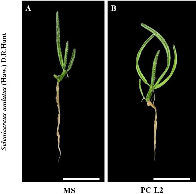
The initial growth of Selenicereus undatus (Haw.) D.R. Hunt was influenced by the culture media (MS and PC-L2) after 75 days of in vitro cultivation (Figure 1).

Specimens of Selenicereus undatus (Haw.) D.R. Hunt after 75 days of in vitro culture in two culture media (MS and PC-L2). Images represent plants randomly selected from the experimental units. Scale bar = 2 cm.
During this period, differences were observed in biometric characteristics, including cladode length and fresh mass, between plants grown in the two media (Figures 2 and 3).

Cladode length (cm) of pitahaya (S. undatus) plants after 75 days of in vitro cultivation in two types of culture media (MS and PC-L2). Boxplots represent the median, interquartile range, and data distribution of 10 biological replicates (n = 10) per treatment. Different letters indicate significant differences according to Welch’s t-test (p ≤ 0.05).

Fresh mass of the shoot (FMAP, g plant-¹) and root (FMR, g plant-¹), and dry mass of the shoot (DMAP, g plant-¹) and root (DMR, g plant-¹) of pitahaya (S. undatus) plants after 75 days of in vitro cultivation in two types of culture media (MS and PC-L2). Boxplots represent the median, interquartile range, and data distribution of 10 biological replicates (n = 10) per treatment. Different letters indicate significant differences according to Welch’s t-test (p ≤ 0.05).
The mean shoot length of plants grown in the PC-L2 medium (5.90 ± 1.00 cm) was greater than that of plants cultivated in the MS medium (4.5 ± 0.17 cm) (Figure 2).
The greater shoot length observed in plants cultured on PC-L2 medium, compared with MS medium, indicates that this formulation provided more favorable conditions for cladode elongation under the conditions evaluated. This response may be related to differences in the mineral and vitamin composition of the culture medium, which are known to influence in vitro plant growth and morphogenesis (Phillips & Collins, 1979; Phillips & Garda, 2019; Rodrigues et al., 2022). However, the specific physiological or biochemical mechanisms underlying this response were not investigated in the present study.
Previous studies have shown that modifications in culture medium composition can affect the growth of S. undatus during in vitro cultivation (Rodrigues et al., 2022; Clairvil et al., 2025). Likewise, physical properties of the medium, including the gelling agent concentration, may also influence shoot development by altering the culture environment (Sarwar et al., 2024). Therefore, the superior shoot elongation observed in PC-L2 medium suggests that its formulation is more suitable for promoting cladode growth, although further studies are required to clarify the physiological mechanisms involved.
The fresh mass of the shoot was higher in plants grown in the PC-L2 medium (1.69 ± 0.19 g) compared to those cultivated in the MS medium (1.44 ± 0.20 g). For root fresh mass, values of 0.68 ± 0.13 g were observed in plants grown in the PC-L2 medium and 1.50 ± 0.02 g in the MS medium. On the other hand, shoot and root dry mass did not differ between the culture media (Figure 3).
The differences in shoot fresh mass observed between the MS and PC-L2 media indicate that the culture medium influenced cladode development, despite the absence of significant differences in dry mass accumulation (Pinheiro et al., 2022). The higher fresh mass obtained in PC-L2 suggests greater tissue hydration and expansion during in vitro growth, although the underlying physiological mechanisms were not investigated in this study. Similar responses have been reported in studies demonstrating that culture medium composition can influence the growth of pitahaya plantlets (Sá et al., 2018; Cristina et al., 2023).
Therefore, the combined increase in shoot length and fresh mass indicates that PC-L2 provided more favorable conditions for cladode development under the experimental conditions evaluated.
Photosynthetic pigments
The chlorophyll b content was significantly higher in pitahaya plants grown in the PC-L2 medium (0.23 ± 0.03 μg g⁻¹) compared to the MS medium (0.18 ± 0.03 μg g⁻¹). On the other hand, the contents of chlorophyll a, total chlorophylls, and carotenoids did not differ between the MS and PC-L2 culture media (Figure 4).

Photosynthetic pigment contents, including chlorophyll a (Chl a), chlorophyll b (Chl b), total chlorophyll (Chl a+b), and carotenoids (Car), in pitahaya (S. undatus) cladodes after 75 days of in vitro cultivation in two types of culture media (MS and PC-L2). Data are expressed as μg g-¹ fresh mass. Boxplots represent the median, interquartile range, and data distribution of five biological replicates (n = 5) per treatment. Each biological sample was analyzed in triplicate as technical replicates. Different letters indicate significant differences according to Welch’s t-test (p ≤ 0.05).
The photosynthetic pigment profile of Selenicereus undatus plants cultivated in vitro showed a differential response to the MS and PC-L2 media, with PC-L2 promoting higher chlorophyll b content. This result suggests that the composition of this medium provided more favorable conditions for chlorophyll biosynthesis and pigment accumulation during cladode development. Since chlorophyll formation is influenced by mineral nutrition, particularly the availability of nitrogen and magnesium, differences in the nutritional composition of the media may have contributed to the observed variation (Hauer-Jákli & Tränkner, 2019; Chaudhry et al., 2021). Similar variation in chlorophyll-related responses has been reported in dragon fruit under different growing conditions (Karunakaran et al., 2025). However, the specific mechanisms underlying the higher chlorophyll b content observed in PC-L2 were not directly evaluated in the present study.
The absence of significant differences in carotenoid content indicates that both culture media maintained similar levels of accessory pigments involved in light harvesting and photoprotection. Previous studies have shown that modifications in culture medium composition can affect pigment accumulation and photosynthetic performance in in vitro-grown plants (Sarwar et al., 2024). Therefore, the higher chlorophyll b concentration observed in PC-L2 suggests a potential advantage of this medium in promoting photosynthetic pigment development, which may contribute to improved physiological performance during subsequent acclimation, although further studies are required to confirm this response.
Modulated chlorophyll a fluorescence
The modulated chlorophyll a fluorescence parameters (F₀ and Fₘ), the maximum quantum efficiency of photosystem II (Fᵥ/Fₘ), the effective quantum yield [Y(II)], the electron transport rate (ETR), and non-photochemical quenching (NPQ) did not differ between plants grown in the MS and PC-L2 media (Table 2).
Chlorophyll a fluorescence analysis revealed similar photosynthetic performance of Selenicereus undatus plants cultivated in MS and PC-L2 media, as indicated by the absence of significant differences in Fᵥ/Fₘ, Y(II), ETR, and NPQ parameters. This approach provides useful information on the functioning of the photosynthetic apparatus and can be used to characterize physiological responses in plants (Tatagiba & Pezzopane, 2007). The Fᵥ/Fₘ values remained within the range generally considered indicative of functional PSII in plants, suggesting that both culture media supported adequate photochemical efficiency and did not induce apparent photoinhibition under the evaluated conditions (Campostrini, 2001; Azevedo Neto et al., 2011).
The similar values of F₀ and Fₘ between treatments indicate comparable excitation energy capture and PSII photochemical capacity, resulting in equivalent maximum quantum efficiency. Likewise, the maintenance of Y(II) and ETR values in both treatments suggests that the culture media provided sufficient conditions for sustaining photosynthetic electron transport during in vitro growth. The similar ETR values between treatments further indicate that electron transport through photosystem II was maintained under both culture conditions, although ETR should be interpreted cautiously as an indicator of photosynthetic performance when gas exchange measurements are not available (Perera-Castro & Flexas, 2023). The absence of differences in NPQ indicates that both formulations maintained similar capacities for regulating excess excitation energy dissipation.
Although PC-L2 showed slight numerical increases in some fluorescence parameters, these differences were not statistically significant, indicating that both MS and PC-L2 media maintained functional photosynthetic machinery in S. undatus. Therefore, the choice between these formulations may depend on other growth, anatomical, or biochemical responses rather than differences in PSII performance alone.
Energy-dispersive X-ray spectroscopy (EDX)
The elemental composition of S. undatus cladodes showed significant variations between the central ribs (CR) and the rib margins (RM) (Table 3 and Figure 5).
Percentage distribution (atomic percentage, %) of chemical elements (CE) in Selenicereus undatus (Haw.) D.R. Hunt cladodes. Values are expressed as mean ± standard error (SE) of three biological replicates (n = 3). CR = central ribs; RM = rib margins.

Photomicrographs of cross-sections of Selenicereus undatus cladodes after 75 days of in vitro cultivation in two types of culture media (MS and PC-L2). Images were obtained from five biological replicates (n = 5) per treatment. Labels: cr = central ribs; rm = rib margins; ab = axillary bud (areole); vb = vascular bundle; w = wing. Scale bar = 400 µm.
Potassium (K) was the predominant element in Selenicereus undatus cladodes, regardless of the culture medium. Both in the rib margins and central ribs, higher concentrations were observed in plants grown in MS medium compared to PC-L2 (Table 3).
The mineral profile of S. undatus cladodes cultivated in vitro followed the general order K > Ca > N > Mg ≈ P > S, followed by micronutrients, including Zn, Mn, Fe, and Cl. This distribution is consistent with the predominance of macronutrients commonly reported in pitahaya tissues, reflecting the high demand for elements involved in growth, osmotic regulation, and metabolic processes (Lima et al., 2019; Lima et al., 2021).
Potassium was the most abundant element in both treatments, with higher concentrations detected in plants cultured on MS medium. The greater K accumulation may be related to the higher availability of potassium-containing salts in this formulation, such as KNO₃ and KH₂PO₄. Given the role of K in stomatal regulation, enzyme activation, and osmotic balance, its accumulation may contribute to the maintenance of tissue development under in vitro conditions (Lima et al., 2019).
Calcium was the second most abundant element, highlighting its importance in structural development and cellular functions. Calcium contributes to cell wall stabilization and participates in signaling processes associated with plant growth and responses to environmental conditions (Viñas & Jiménez, 2016). Differences in sulfur accumulation between treatments may reflect variations in nutrient uptake and assimilation associated with the composition of each culture medium.
Magnesium and phosphorus were more evident in plants grown on PC-L2 medium, suggesting that this formulation favored the accumulation of these nutrients in cladode tissues. Both elements are essential for photosynthetic metabolism, energy transfer, and biosynthetic processes, which may contribute to the maintenance of plant physiological activity during in vitro cultivation (Moreira et al., 2016). However, further studies evaluating nutrient uptake dynamics and biochemical responses are required to clarify the mechanisms underlying these differences.
Micronutrients showed lower accumulation compared with macronutrients, which is consistent with their requirement in smaller quantities for plant metabolism. Elements such as Fe, Mn, Zn, and Cl participate in several physiological processes, including electron transport, enzymatic activity, and photosynthetic functions (Shah et al., 2023; Meier, Mariani & Peiter, 2025; Roychoudhury, Mukherjee & Ghosh, 2025). Chloride also plays important roles in plant ion homeostasis and physiological regulation (Silva-Herrera et al., 2025).
Overall, MS and PC-L2 media resulted in distinct mineral accumulation patterns in S. undatus cladodes. While MS favored higher accumulation of K and Ca, PC-L2 promoted greater accumulation of Mg and P, indicating that modifications in culture medium composition can influence the nutritional status of micropropagated plants.
Anatomical analyses
MS medium produced plants with a higher number of vascular bundles (NVB) and a larger cladode cross-sectional area (CSAC), with values of 4.60 ± 0.25 and 0.65 ± 0.02 mm², respectively, compared to plants grown in PC-L2 medium (Figure 5 A-B). On the other hand, root area (RA) and root diameter (RD) were greater in plants cultivated in PC-L2 medium (0.21 ± 0.01 mm² and 532.66 ± 10.50 µm, respectively) compared to MS medium (Figures 6 and 7).

A. Number of vascular bundles (NVB); B. Cladode cross-sectional area (CSAC); C. Root diameter (RD); and D. Root area (RA) of pitahaya (S. undatus) plants after 75 days of in vitro cultivation in two types of culture media (MS and PC-L2). Boxplots represent the median, interquartile range, and data distribution of five biological replicates (n = 5) per treatment. Different letters indicate significant differences according to Welch’s t-test (p ≤ 0.05).

(A-B) Cross-sections of Selenicereus undatus cladodes and (C-D) roots after 75 days of in vitro cultivation in two types of culture media (MS and PC-L2). Images were obtained from five biological replicates (n = 5) per treatment. Labels: ep = epidermis; pr = storage parenchyma; pm = medullary parenchyma; cx = cortex; w = wing; ab = axillary bud (areole); vb = vascular bundle; cv = vascular cylinder; ex = exodermis; rh = root hairs. Scale bar = 100 µm.
Plants cultured in MS medium exhibited a greater number of vascular bundles and a larger cladode cross-sectional area, indicating enhanced vascular development and tissue expansion. These responses may be related to differences in the nutritional composition of the culture media, which influence plant growth and anatomical differentiation under in vitro conditions (Phillips & Garda, 2019; Pasternak & Steinmacher, 2024). A more developed vascular system may contribute to improved transport capacity within cladodes, supporting tissue development and plant growth. In contrast, PC-L2 medium promoted greater root area (RA) and root diameter (RD), suggesting a distinct pattern of anatomical development that may be advantageous during subsequent acclimation stages (Clairvil et al., 2025).
The anatomical responses observed were consistent with the differences found in biometric traits, pigment accumulation, and mineral composition, indicating that the two media promoted distinct growth patterns in S. undatus. The greater cladode development in MS medium was associated with increased vascular traits, whereas PC-L2 favored root anatomical characteristics related to belowground development. These differences highlight the influence of culture medium composition on the morpho-anatomical characteristics of micropropagated plants.
Overall, the results suggest that MS and PC-L2 media provide complementary advantages for S. undatus micropropagation. While MS favors shoot structural development, PC-L2 promotes root system development, which may contribute to better plant establishment during acclimation. Further studies evaluating physiological responses during the transition to ex vitro conditions are required to confirm the functional significance of these anatomical traits.
Conclusions
The culture medium significantly influenced the development of Selenicereus undatus (Haw.) D.R. Hunt during in vitro cultivation. PC-L2 medium promoted greater shoot elongation, fresh biomass accumulation, and root development, whereas MS medium favored cladode anatomical development. Both media maintained functional photosynthetic performance, with differences observed in mineral accumulation patterns. These results demonstrate that MS and PC-L2 provide distinct advantages for the micropropagation of S. undatus, with PC-L2 showing greater potential for promoting overall plant development under the evaluated conditions.
Acknowledgments
The authors of this work thank the Coordenação de Aperfeiçoamento de Pessoal de Nível Superior - Brasil (CAPES), the Conselho Nacional de Desenvolvimento Científico e Tecnológico (CNPq), and the Fundação de Amparo à Pesquisa do Estado de Minas Gerais (FAPEMIG) for financial support for the experiments and for funding scholarships and research productivity grants.
Data Availability Statement
Data available upon request to authors.
References
- Anuruddi, H. I. G. K., Nakandalage, N., & Fonseka, D. K. (2023). New insights for the production of medicinal plant materials: Ex vitro and in vitro propagation. In N. Kumar., & R. S. Singh. (eds). Biosynthesis of bioactive compounds in medicinal and aromatic plants Food Bioactive Ingredients. Springer, Cham, (pp.181-212).
- Arivalagan, M. et al. (2021). Biochemical and nutritional characterization of dragon fruit (Hylocereus species). Food Chemistry, 353:129426.
- Azevedo Neto, A. D. D. et al. (2011). Fluorescência da clorofila como uma ferramenta possível para seleção de tolerância à salinidade em girassol.Revista Ciência Agronômica,42(4):893-897.
- Balendres, M. A. et al. (2019). Diseases of dragon fruit (Hylocereus species): Etiology and current management options. Crop Protection, 126:104920.
-
Campostrini, E. (2001). Fluorescência da Clorofila a: considerações teóricas e aplicações práticas UFNF: Rio de Janeiro, Brasil. 31p. Available in: https://uenf.br/UENF_ARQUIVOS/Downloads/CENTRO_CCTA_1629_1112121492.pdf
» https://uenf.br/UENF_ARQUIVOS/Downloads/CENTRO_CCTA_1629_1112121492.pdf - Chaudhry, A. H. et al. (2021). Current understandings on magnesium deficiency and future outlooks for sustainable agriculture.International journal of molecular sciences,22(4):1819.
- Chimdessa, E. (2020). Composition and preparation of plant tissue culture medium. Journal of Tissue Culture and Bioengineering, 3:281-286.
- Clairvil, E. et al. (2025). Agar concentration interferes with the biometry, photosynthetic pigment content, and anatomy of Selenicereus undatus in vitroCiência e Agrotecnologia,49:e021624.
- Clairvil, E. et al. (2026). Light quality and zinc concentrations interfere with the in-vitro cultivation of Selenicereus undatusBragantia, 85:e20250157.
- Clairvil, E. et al. (2022). Modulation of physiological responses and tolerance of Alternanthera tenella Colla (Amaranthaceae) to cadmium during in vitro cultivation. Israel Journal of Plant Sciences, 69:181-192.
- Cristina, M. A. M. et al. (2023). Evaluation of the effect of different culture systems on photomixotrophic capacity during in vitro multiplication of pitahaya (Hylocereus undatus).South African Journal of Botany, 159:396-404.
- Duta-Cornescu, G. et al. (2023). Somaclonal variation-Advantage or disadvantage in micropropagation of the medicinal plants.International Journal of Molecular Sciences,24(1):838.
- Gerdakaneh, M. et al. (2020). Effect of different media and growth regulators on micropropagation of GF677.Plant Productions,43(2):241-254.
- Gidas, J. D. et al. (2025). LED lights and plant growth regulators enhance the in vitro mass propagation of rosemary (Rosmarinus officinalis L.). European Journal of Horticultural Science, 90(1): 0008.
- Goltsev, V. N. et al. (2016). Variable chlorophyll fluorescence and its use for assessing physiological condition of plant photosynthetic apparatus. Russian Journal of Plant Physiology, 63:869-893.
- Guo, L. W. et al. (2014). First report of dragon fruit (Hylocereus undatus) anthracnose caused by Colletotrichum truncatum in China. Journal of Phytopathology, 162:272-275.
- Hauer-Jákli, M., & Tränkner, M. (2019). Critical leaf magnesium thresholds and the impact of magnesium on plant growth and photo-oxidative defense: A systematic review and meta-analysis from 70 years of research. Frontiers in Plant Science, 10:766.
- Johansen, D. A. (1940). Plant microtechnique (2nd ed.). McGraw-Hill Book, New York, 523p.
- Karunakaran, G. et al. (2025). Sunburn mitigation in dragon fruit (Hylocereusspp.): unravelling genotype-specific physiological and biochemical responses.Frontiers in Plant Science,16:1661147.
- Kramer, D. M. et al. (2004). New fluorescence parameters for the determination of QA, redox state and excitation energy fluxes.Photosynthesis Research,79:209-218.
- Lichtenthaler, H. K., & Babani, F. (2021). Contents of photosynthetic pigments and ratios of chlorophyll a/b and chlorophylls to carotenoids (a+ b)/(x+ c) in C4 plants as compared to C3 plants.Photosynthetica,60(1):3-9.
- Lichtenthaler, H. K., & Wellburn, A. R. (1983). Determinations of total carotenoids and chlorophyll a and b of leaf extracts in different solvents. Biochemical Society Transactions, 11:591-592.
- Lima, D. D. C. et al. (2019). Growth and nutrient accumulation in the aerial part of red Pitaya (Hylocereus sp.).Revista Brasileira de Fruticultura,41(5):e-030.
- Lima, D. D. C. et al. (2021). Initial growth and nutrient accumulation in pitaya plants at different phenological stages.Revista Caatinga,34(3):720-727.
- Long, Y. et al. (2022). New insights into tissue culture plant-regeneration mechanisms.Frontiers in Plant Science,13:926752.
- Martins, J. P. R. et al. (2020). Morphophysiological responses, bioaccumulation and tolerance of Alternanthera tenella Colla (Amaranthaceae) to excess copper under in vitro conditions.Plant Cell, Tissue and Organ Culture (PCTOC),143:303-318.
- Meier, B., Mariani, O., & Peiter, E. (2025). Manganese handling in plants: Advances in the mechanistic and functional understanding of transport pathways.Quantitative Plant Biology, 6:e16.
- Minutolo, M. et al. (2020). Accumulation of ascorbic acid in tomato cell culture: Influence of the genotype, source explant and time of in vitro cultivation.Antioxidants, 9(3):222.
- Moreira, R. A. et al. (2016). Nutrient accumulation at the initial growth of pitaya plants according to phosphorus fertilization.Pesquisa Agropecuária Tropical,46(3):230-237.
- Murashige, T., & Skoog, F. (1962). A revised medium for rapid growth and bio assays with tobacco tissue cultures.Physiologia Plantarum, 15(3):473-497.
- Nimavat, N., & Parikh, P. (2024). Innovations in date palm (Phoenix dactylifera L.) micropropagation: Detailed review of in vitro culture methods and plant growth regulator applications.Plant Cell, Tissue and Organ Culture (PCTOC),159:6.
- Pasternak, T. P., & Steinmacher, D. (2024). Plant growth regulation in cell and tissue culture in vitroPlants,13(2):327.
- Perera-Castro, A. V., & Flexas, J. (2023). The ratio of electron transport to assimilation (ETR/AN): underutilized but essential for assessing both equipment’s proper performance and plant status.Planta,257:29.
- Phillips, G. C., & Collins, G. B. (1979). In vitro tissue culture of selected legumes and plant regeneration from callus cultures of red clover.Crop Science,19(1):59-64.
- Phillips, G. C., & Garda, M. (2019). Plant tissue culture media and practices: An overview.In Vitro Cellular & Developmental Biology-Plant,55:242-257.
- Pinheiro, M. V. M. et al. (2022) Led enhance the growth characteristics and affects the ex vitro acclimatization of sugarcane.Plant Cell Culture & Micropropagation, 18:e184.
- Rodrigues, M. A. et al. (2022). Regeneration of pitaya by indirect organogenesis evaluated by scanning electron microscopy and flow cytometry.Pesquisa Agropecuária Brasileira,57:e02312.
- Roychoudhury, A., Mukherjee, S., & Ghosh, A. (2025). Molecular and physiological studies of the role of micronutrients in sustainable growth and development of crop plants. In A. Husen.Agricultural crop improvement CRC Press, (pp.135-147).
- Rupert, E. A., & Collins, G. B. (1985). Chapter 16, tissue culture. In N. L. Taylor (Ed.). Clover science and technology American Society of Agronomy, Inc., Crop Science Society of America, Inc., Soi1 Science Society of Arnerica, Inc., Madison, Wisconsin, USA, (pp.405-416).
- Sá, J. F. D. et al. (2018). Meios de cultura para a multiplicação de espécies silvestres de Manihot.Ciência e Agrotecnologia,42(6):598-607.
- Saad, A. I. M., & Elshahed, A. M. (2012). Plant tissue culture media. In A. Leva., & L. M. R. Rinaldi. Recent advances in plant in vitro culture IntechOpen, (pp.29-40).
- Safira, A. et al. (2021). Review on the pharmacological and health aspects of Hylocereus or pitaya: An update.Journal of Drug Delivery and Therapeutics,11(6):297-303.
- Sarwar, G. et al. (2024). Optimizing germination: comparative assessment of various growth media on dragon fruit germination and early growth.BMC Plant Biology,24:533.
- Scopel, W., Barbosa, J. Z., & Vieira, M. L. (2011). Extração de pigmentos foliares em plantas de canola.Unoesc & Ciência-ACET, 2(1):87-94.
- Shah, K. et al. (2023). Pitaya nutrition, biology, and biotechnology: A review.International Journal of Molecular Sciences,24(8):13986.
- Silva-Herrera, H. et al. (2025). Chloride transport and homeostasis in plants.Quantitative Plant Biology, 6:e20.
- Tatagiba, S. D., & Pezzopane, J. E. M. (2007). Cinética de emissão de fluorescência das clorofilas em dois clones de Eucalyptus.Revista Científica Eletrônica de Engenharia Florestal,10:1-14.
- Viñas, M., & Jiménez, V. M. (2016). Occurrence and characterisation of calcium oxalate crystals in stems and fruits of Hylocereus costaricensis and Selenicereus megalanthus (Cactaceae: Hylocereeae).Micron,89:21-27.
- Wawrosch, C., & Zotchev, S. B. (2021). Production of bioactive plant secondary metabolites through in vitro technologies-status and outlook. Applied Microbiology and Biotechnology,105:6649-6668.
- Xiyan, L. et al. (2024). Establishment of regeneration system for tissue culture of Euphorbia milii European Journal of Horticultural Science, 89(3):013.
- Yadav, A. et al. (2025). A review on genetic resources, breeding status and strategies of dragon fruit.Genetic Resources and Crop Evolution,72:2511-2531.
- Yang, Z. Y. et al. (2024). Selenicereus undatus (Dragon Fruit) phytochemicals for managing three human pathogenic bacteria: An In Vitro and in silico Approach . Metabolites,14(11):577.