Abstracts
BACKGROUND: Release of the neuronal protein S-100B into the circulation has been suggested as a specific indication of neuronal damage. The hypothesis that S-100B is a useful and cost-effective screening tool for the management of minor head injuries was tested. METHODS: Fifty consecutive patients sustaining isolated minor head injury were prospectively evaluated in the emergency room of a Brazilian hospital by routine cranial computed tomography scan. Venous blood samples (processed to serum) were assssayed for S-100B using a newly developed immunoassay test kit. Twenty-one normal healthy individuals served as negative controls. Data are presented as median and 25 to 75 percentiles. RESULTS: Patients reached the emergency room an average of 45 minutes (range: 30-62 minutes) after minor head injury. Six of 50 patients (12%) showed relevant posttraumatic lesions in the initial cranial computed tomography scan and were counted as positive. The median systemic concentration of S-100B in those patients was 0.75 µg/L (range: 0.66-6.5 µg/L), which was significantly different (U-test, P < .05) from the median concentration of 0.26 µg/L (range: 0.12-0.65 µg/L), of patients without posttraumatic lesions as counted by the cranial computed tomography. A sensitivity of 100%, a specificity of 20%, a positive predictive value of 15%, and a negative predictive value of 100% was calculated for the detection of patients suffering from intracranial lesions. CONCLUSIONS: Protein S-100B had a very high sensitivity and negative predictive value and could have an important role in ruling out the need for cranial computed tomography scan after minor head injury. This appears to be of substantial clinical relevance, particularly in countries where trauma incidence is high and medical resources are limited, such as in Brazil.
Cranial computed tomography; Glasgow coma scale; Minor head injury; Neuronal protein S-100B
INTRODUÇÃO: A liberação da proteína neuronal S-100B na circulação tem sido sugerida como indicadora de dano neuronal. Foi testada a hipótese de que a S-100B é um marcador útil e custo efetivo para a triagem de pacientes com trauma craniano leve. MÉTODO: Cinqüenta pacientes consecutivos com trauma craniano isolado foram prospectivamente avaliados na sala de emergência de um Centro de Trauma brasileiro pela tomografia computadorizada de crânio e por amostras de sangue venoso, para a medida no soro da proteína S-100B utilizando um teste recentemente desenvolvido; 21 pessoas normais foram utilizadas como controles negativos. Os resultados são apresentados como mediana e percentis 25-75. RESULTADOS: Os pacientes chegaram ao Centro de Trauma em média 45 min (30-62) após o trauma craniano leve. Seis dos 50 pacientes tiveram lesões pós-traumáticas relevantes segundo a tomografia computadorizada de crânio inicial (12%) e foram considerados como positivos. A concentração mediana de S-100B nestes pacientes foi de 0,75µg/L (0,66-6,5), significativamente maior (U-teste, p<0,05) do que a concentração mediana de 0,26µg/L (0.12-0.65) dos pacientes sem lesões pós-traumáticas, segundo a tomografia computadorizada de crânioCCT-. O cálculo para a detecção dos pacientes com lesões intra-cranianas revelou sensibilidade de 100%, especificidade de 20%, valor preditivo positivo de 15% and valor preditivo negativo de 100%. CONCLUSÃO: A proteína S-100B tem altas taxas de sensibilidade e valor preditivo negativo, podendo ter um importante papel para descartar a necessidade de tomografia de crânio após trauma craniano leve. Acreditamos que este achado é de relevância clínica, principalmente em países onde o trauma é muito frequente e os recursos médicos limitados.
Tomografia de crânio; Trauma de crânio; Escala de Coma de Glasgow; Proteína S100B
ORIGINAL RESEARCH
Measurement of S-100B for risk classification of victims sustaining minor head injury - first pilot study in Brazil
Medida da proteína S-100B sérica para classificação de risco no trauma craniano leve estudo piloto no Brasil
Luiz F Poli-de-FigueiredoI, II; Peter BiberthalerIII; Charles Simao FilhoIV; Christopher HauserIII; Wolf MutschlerIII; Marianne JochumIII
IDepartment of Surgery, Federal University of São Paulo São Paulo/SP, Brazil. Email: lpoli@uol.com.br
IIHeart Institute, São Paulo University Medical School São Paulo/SP, Brazil
IIIDepartment of Surgery and Traumatology, Ludwig-Maximilians-University - Munich, Germany
IVEmergency Department, João XXIII Hospital - Belo Horizonte/MG, Brazil
ABSTRACT
BACKGROUND: Release of the neuronal protein S-100B into the circulation has been suggested as a specific indication of neuronal damage. The hypothesis that S-100B is a useful and cost-effective screening tool for the management of minor head injuries was tested.
METHODS: Fifty consecutive patients sustaining isolated minor head injury were prospectively evaluated in the emergency room of a Brazilian hospital by routine cranial computed tomography scan. Venous blood samples (processed to serum) were assssayed for S-100B using a newly developed immunoassay test kit. Twenty-one normal healthy individuals served as negative controls. Data are presented as median and 25 to 75 percentiles.
RESULTS: Patients reached the emergency room an average of 45 minutes (range: 3062 minutes) after minor head injury. Six of 50 patients (12%) showed relevant posttraumatic lesions in the initial cranial computed tomography scan and were counted as positive. The median systemic concentration of S-100B in those patients was 0.75 µg/L (range: 0.666.5 µg/L), which was significantly different (U-test, P < .05) from the median concentration of 0.26 µg/L (range: 0.120.65 µg/L), of patients without posttraumatic lesions as counted by the cranial computed tomography. A sensitivity of 100%, a specificity of 20%, a positive predictive value of 15%, and a negative predictive value of 100% was calculated for the detection of patients suffering from intracranial lesions.
CONCLUSIONS: Protein S-100B had a very high sensitivity and negative predictive value and could have an important role in ruling out the need for cranial computed tomography scan after minor head injury. This appears to be of substantial clinical relevance, particularly in countries where trauma incidence is high and medical resources are limited, such as in Brazil.
Keywords: Cranial computed tomography. Glasgow coma scale. Minor head injury. Neuronal protein S-100B.
RESUMO
INTRODUÇÃO: A liberação da proteína neuronal S-100B na circulação tem sido sugerida como indicadora de dano neuronal. Foi testada a hipótese de que a S-100B é um marcador útil e custo efetivo para a triagem de pacientes com trauma craniano leve.
MÉTODO: Cinqüenta pacientes consecutivos com trauma craniano isolado foram prospectivamente avaliados na sala de emergência de um Centro de Trauma brasileiro pela tomografia computadorizada de crânio e por amostras de sangue venoso, para a medida no soro da proteína S-100B utilizando um teste recentemente desenvolvido; 21 pessoas normais foram utilizadas como controles negativos. Os resultados são apresentados como mediana e percentis 25-75.
RESULTADOS: Os pacientes chegaram ao Centro de Trauma em média 45 min (30-62) após o trauma craniano leve. Seis dos 50 pacientes tiveram lesões pós-traumáticas relevantes segundo a tomografia computadorizada de crânio inicial (12%) e foram considerados como positivos. A concentração mediana de S-100B nestes pacientes foi de 0,75µg/L (0,66-6,5), significativamente maior (U-teste, p<0,05) do que a concentração mediana de 0,26µg/L (0.12-0.65) dos pacientes sem lesões pós-traumáticas, segundo a tomografia computadorizada de crânioCCT-. O cálculo para a detecção dos pacientes com lesões intra-cranianas revelou sensibilidade de 100%, especificidade de 20%, valor preditivo positivo de 15% and valor preditivo negativo de 100%.
CONCLUSÃO: A proteína S-100B tem altas taxas de sensibilidade e valor preditivo negativo, podendo ter um importante papel para descartar a necessidade de tomografia de crânio após trauma craniano leve. Acreditamos que este achado é de relevância clínica, principalmente em países onde o trauma é muito frequente e os recursos médicos limitados.
Unitermos: Tomografia de crânio. Trauma de crânio. Escala de Coma de Glasgow. Proteína S100B.
INTRODUCTION
Risk estimation for clinically relevant intracranial lesions after minor head injury (MHI) (Glasgow Coma Scale Score of 1315 points) remains a major diagnostic challenge. The gold standard diagnostic tool to rule out posttraumatic lesions is cranial computed tomography (CCT). The need for an immediate CCT scan after moderate or severe head injury is undisputed.1 However, substantial controversy exists regarding the cost effectiveness of routine CCT scan after a minor head injury.2,3 Although the reported frequency of intracranial injuries, varying from 5.9% to 21.6%,4,5 is much lower than after moderate or severe head injury, this proportion represents a high risk group, since prognosis is also dependent on rapid detection of potentially life-threatening complications, eg, skull fracture, hemorrhage, or brain edema. Some reports recommend routine CCT, because clinical evaluation may not always provide sufficient information regarding the decision for further diagnosis and special treatment, preferably in patients in whom the level of consciousness and the quality of neurological examination may have been altered by alcohol, drugs, or other associated injuries.6,7
Patients at risk are characterized by relevant pathological findings in the CCT scan. As shown in a study of 2,766 patients, none required further treatment in the absence of pathological findings in the initial CCT scan.7,8 However, due to the large number of patients sustaining minor head injury, routine CCT scanning and the need for repetitive neurological supervision, requiring admittance to a ward, may generate serious financial and logistic burdens, especially in areas where trauma incidence is high and medical resources are limited, such as in many parts of Brazil. If the victim suffers from a posttraumatic intracranial lesion, additional neurological symptoms are likely to occur, ie, focal neurological symptoms, increasing dizziness, or disturbances of the pupils. Although a CCT scan is usually performed in this situation, the development of symptomatic intracerebral lesions, such as epidural or subdural hemorrhages, may have just started and thus might be overlooked at this point. This delay may cause further damage or worsen the initial prognosis. Therefore, a rapid and sensitive screening test for the presence of intracranial lesions in patients suffering from minor MHI is very much needed.
In recent years, measurement of a specific neuronal protein S-100B released into the circulation has been evaluated for identification of risk groups among victims sustaining head trauma.9-12 Protein S-100B is predominantly found in glial and Schwann cells as well as in neurons; it is normally released in small amounts due to metabolic cell processes, but in larger quantities as a consequence of cell damage. It is known to modulate glial fibrillary acidic protein (GFAP) and intermediate filament assembly in neuronal and glial cells as well as proliferation of neuronal cells.13 In physiological conditions, it has been detected in serum only in very low amounts, requiring a highly sensitive immunoassay.14 The half-life is about 80-120 minutes. Therefore, measurement is valid only if the blood sample is obtained within 2 hours from trauma. However, an early peak of S-100B serum levels after severe head injury or ischemic brain conditions reflects cellular damage and elevated permeability of the blood brain barrier.14,15 Additional studies also indicate S-100B as a promising quantitative, objective tool for identifying high-risk patients with MHI, including those under the influence of alcohol.9,10,12,16,17 There is no influence of gender on S-100B measurement. In infants, the normal concentration is higher, and there is a current study addressing year-adjusted concentration ranges. The majority of studies have included only patients 18 years old or older.
The aim of this pilot study was to further investigate the diagnostic value of S-100B serum measurements in patients suffering from MHI. Therefore the hypothesis that S-100B may be a rapid and useful screening tool for the management MHI in Brazilian patients, enabling the reduction of often unnecessary and costly CCT scans was tested.
PATIENTS AND METHODS
This study was approved by the Ethics Committee of João XXIII Hospital. João XXIII Hospital is a large emergency center located in downtown Belo Horizonte (2.5 million inhabitants), in central Brazil. In 2003 during September and October, we prospectively studied 50 consecutive patients sustaining isolated MHI, defined by a Glasgow Coma Scale Score (GCS) of 13 to 15 and presenting at least one of the following symptoms: amnesia, loss of consciousness, nausea, vomiting, vertigo, or severe headache on admission. Patients with focal neurological deficits were excluded. In addition, a negative control group of healthy volunteers (n = 21) was studied. Informed consent was given by volunteers, patients or relatives. Venous blood samples were drawn on admission and processed to serum. The deep-frozen serum samples were transported to the Ludwig-Maximilians-University Hospital in Munich, Germany, and levels of S-100B were measured using a newly developed heterogeneous immunoassay (Elecsys 2010®) according to the manufacturer's instructions. In our earlier studies,12,17 we established a cut-off point at a concentration of 0.1 µg/L of S-100B, which was the highest level measured in healthy volunteers without any sign of intracranial injury. Thus, patients presenting a S-100B level below 0.1 µg/L were defined as "negative," while those with concentration above 0.1 µg/L were defined as "positive."
Cranial computed tomography (CCT) was performed within 6 hours of emergency room admission, and radiological findings were defined as pathological (CCT+) if intracranial hemorrhage, skull fracture, and/or diffuse brain swelling (edema) were detected. Sensitivity, specificity, positive, and negative predictive values of S-100B serum levels were calculated for signs of intracranial injury at the initial CCT scan.
Statistical analysis was performed using the Mann-Whitney rank sum test and Dunn's method (SigmaStat 2.0, Jandel Corporation); significance was accepted at P < .05. Data are shown as median and 25%75% quartile.
RESULTS
Fifty consecutive patients (28 males, 22 females) sustaining isolated minor head injury (MHI) were included. There were 37 patients with a GCS of 15, 11 with a GCS of 14, and 2 with a GCS of 13. Out of these, 6 patients had trauma-relevant intracranial lesions according to the radiological criteria given in the Patients and Methods section and were thereby counted as CCT+. In contrast, 44 patients had none of these lesions and were counted as CCT.
The median time interval from trauma to blood sampling for the S-100B assay was 82 minutes (25%-75% quartiles: 60-110 min). The median serum concentration of S-100B was 0.29 µg/L (25%-75% quartiles: 0.140.76 µg/L) for the entire MHI group, which was significantly elevated (P < .001) compared to that of the negative control group of 21 healthy volunteers (median, 0.04 µg/L; 25%-75% quartiles: 0.030.05 µg/L). As shown in Figure 1, MHI patients (n = 44) without radiological findings exhibited a median S-100B serum value of 0.26 µg/L (25%-75% quartiles: 0.120.65 µg/L), which was significantly lower (P < .05) than the levels of CCT+ patients (n = 6); median: 0.75 µg/L; 25%-75% quartiles: 0.626.5 µg/L).
The contingency table (Table 1) based on the cut-off at 0.1 µg/L demonstrates that all 6 individuals presenting trauma-relevant intracranial lesions in the initial CCT scan exhibited a serum concentration above the cut-off level and were thereby detected correctly as CCT+/S-100B+. Nine individuals were classified as S-100B negative and had no lesions in CCT scan. From those 9 patients, 8 presented a GCS of 15 and only one presented GCS of 14.
In 35 patients, the S-100B serum concentrations were above the cut-off at 0.1 µg/L and were counted as S-100B+ but as negative for intracranial lesions from the CCT scan (CCT-). From these data, for the ruling-out test of S-100B measurement, a sensitivity of 100%, a negative predictive value of 100%, a specificity of 20%, and a positive predictive value of 15% were calculated.
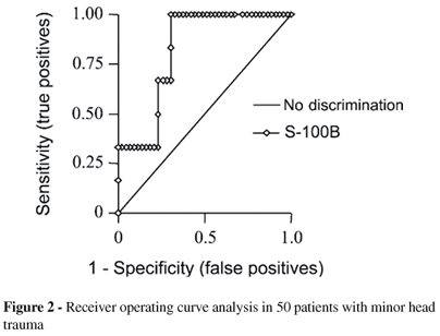
The receiver operating curve (ROC) analysis (Figure 2) revealed an area under the curve (AUC) of 0.82 (95% CI 0.690.96) for discrimination of patients according to their S-100B levels into the groups CCT+ or CCT-, which was highly significant (P < .0001).
DISCUSSION
Although the studied patient population was limited, our results demonstrate that measurement of serum S-100B levels in victims sustaining isolated minor head injury (MHI) is a highly sensitive procedure for the detection of intracranial injury. Due to its excellent negative predictive value, a normal level of protein S-100B may rule out the need for a CCT scan. The speed (determination within 17 minutes) and lower cost of this diagnostic screening tool (about 10%20% of a CCT scan), suggest a significant positive impact for S-100B measurements on the management of MHI patients in countries such as Brazil. The cost of S100B would probably vary between 10 and 20 US dollars, and needle-to-result time would be about 20 minutes. However, if a patient presents classical neurological symptoms, such as a wide pupil or loss of conciousness, a CT scan should be performed even is the S-100B is below the cut-off value.
The benefit of a triage test for this patient population is obvious. The management of patients suffering from MHI is associated with related problems concerning diagnosis and treatment.18 The incidence of MHI in Brazil is not known, but we believe it may be as high or even higher than that observed in the USA, causing 400,000 to 450,000 admissions per year.19 Due to the large number of MHI victims, routine CCT scanning of these patients would generate financial and logistical burdens for health systems, especially in developing countries where medical resources are limited and CT availability may be limited to some larger cities. Based on the 100% negative predictive value of protein S-100B measurement, a much more selective approach to CCT scanning for those patients can be achieved. In addition, the lack of an influence of alcohol use on the S-100B determination is a highly desired characteristic of this test.12,15,17 The first promising results regarding this diagnostic test showed a strong correlation between serum levels of S-100B and the outcomes of severe head injury victims as well as of patients suffering from a cerebral hypoxia during circulatory arrest.20,21-23 A study on the neurobehavioral outcomes revealed that MHI patients with a serum concentration above 0.14 µg/L have a 6.9 likelihood ratio for developing posttraumatic neuropsychological deficits.24 Yet, the authors did not mention the frequencies of pathological radiological results in comparison to the S-100 B data. In an earlier study, Rothoerl et al compared levels of S-100B serum concentrations for 11 patients with MHI versus 30 patients with severe head injury using a radioimmunoassay method for S-100B measurement.15 The MHI group revealed a mean serum concentration of 0.35 µg/L versus 2.6 µg/L for the severe head injury group. From these concentrations, the authors defined a cut-off level of 0.5 µg/L to indicate relevant brain injury.15 Yet, this study suffered from the limitation of a great variation of the interval between trauma and blood sampling, which was up to 330 minutes, and had no negative control group. Hence, the diagnostic value of measurement of S-100B in plasma of MHI patients for identifying high-risk groups still remained unclear; importantly, a reliable cut-off level for a safe differentiation between uncomplicated and problematic MHI had not been determined.
Therefore, Biberthaler et al conducted 2 studies on MHI patients establishing a reliable cut-off value of 0.1 µg/L.12,16 The review of Klauber et al analyzing data of the USA National Traumatic Coma Data Bank clearly showed that mortality of patients suffering from MHI and an initial GCS of 13-15 points with an occult intracerebral lesion is considerably higher than that estimated for the risk of death.25 These lesions, eg intracranial hematomas or diffuse brain swelling, express themselves clinically within a time frame of hours to days after first neurological examination.26 Although not all of these symptoms have to be treated by immediate surgery, close clinical observation is essential, because any relevant posttraumatic intracerebral lesion may result in severe disability or even early death.8 It is well known that screening methods that are intended to identify high-risk groups have to be very sensitive. In this respect, routine testing of newborn infants for phenylketonuria or neonatal hypothyroidism, which are 2 exemplary diseases of similar deleterious consequences for the affected patient if they are missed within the first weeks of life,27 presents a comparable problem. Because measurement of serum S-100B is proposed as a screening method of a similar potentially life-threatening complication in patients suffering from MHI, a low cut-off point has to be established providing high sensitivity for the test. Therefore, the aim of our present study was to analyze the diagnostic value of measurements of S-100B levels in serum of patients with MHI and to re-evaluate the cut-off level for practical use. With a cut-off point of 0.1 µg/L, sensitivity and negative predictive value were 100%, but specificity and positive predictive value were only 20% and 15% at an overall prevalence of 12% of intracerebral injury signs on our present study.
In our study, 60% of the patients defined as "positive" by the S-100B assay did not show relevant intracranial lesions in the CCT scan. Nevertheless, our data indicated that a reduction of 40% of routine CCT scans on MHI patients might be possible by using this simple and practical test.
Last but not least, this is a first report on serum S-100B levels in a Brazilian population, either healthy or traumatized. They were shown to be similar to those of European individuals. This is especially interesting in view of the fact that in addition to neuronal cells, melanocytes may also be able to release S-100B into the circulation. Thus, the origin of elevated levels of S-100B in Brazilian MHI patients appears to be due to release from neuronal cells rather than from melanocytes.
In conclusion, in spite of the low specificity for the detection of intracranial lesions, using a cut-off value of 0.1 µg/L of serum S-100B as determined by this heterogeneous immunoassay system, we find that it has a high sensitivity and, most importantly, a 100% negative predictive value, ruling out the need for CCT scan in MHI patients with a serum S-100B concentration below 0.1µg/L. These results appear to be of substantial clinical and financial relevance for countries such as Brazil where trauma incidences are very high and medical resources are limited.
ACKNOWLEDGEMENT
The study was supported by the program "CAPES-BAVARIA" which is a project of the Bavarian ministry of science, research and art (Staatsministerium für Wissenschaft, Forschung und Kunst) and the "Coordenação de Aperfeiçoamento de Pessoal de Nível Superior (CAPES)" administration in Brazil, grant number Z4-L0142B2-8/30321, to increase scientific exchange between both countries. The test systems were provided by ROCHE Diagnostics, Mannheim, Germany.
The paper is dedicated to Professor Claudio A. Sampaio ( 17.01.2005), former head of the Department of Biochemistry, Federal University of São Paulo, Brazil.
Received for publication on August 09, 2005.
Accepted for publication on October 04, 2005.
- 1. Maas AI, Dearden M, Teasdale GM Braakman R, Cohadon F, Iannotti F, et al. EBIC-guidelines for management of severe head injury in adults. European Brain Injury Consortium. Acta Neurochir (Wien ). 1997;139:286-94.
- 2. Mohanty SK, Thompson W, Rakower S. Are CT scans for head injury patients always necessary? J Trauma. 1991;31:801-4.
- 3. Miller EC, Holmes JF, Derlet RW. Utilizing clinical factors to reduce head CT scan ordering for minor head trauma patients. J Emerg Med. 1997;15:453-7.
- 4. Stein SC, Ross SE . The value of computed tomographic scans in patients with low-risk head injuries. Neurosurgery. 1990;26:638-40.
- 5. Borczuk P. Predictors of intracranial injury in patients with mild head trauma. Ann Emerg Med. 1995;25:731-6.
- 6. Harad FT, Kerstein MD. Inadequacy of bedside clinical indicators in identifying significant intracranial injury in trauma patients. J Trauma. 1992;32:359-61.
- 7. Kelly DF . Alcohol and head injury: an issue revisited. J Neurotrauma. 1995;12:883-90.
- 8. Shackford SR, Wald SL, Ross SE, Cogbill TH, Hoyt DB, Morris JA, et al. The clinical utility of computed tomographic scanning and neurologic examination in the management of patients with minor head injuries. J Trauma. 1992;33:385-94.
- 9. Ingebrigtsen T, Romner B, Kongstad P, Langbakk B. Increased serum concentrations of protein S-100 after minor head injury: a biochemical serum marker with prognostic value? J Neurol Neurosurg Psychiatry. 1995;59:103-4.
- 10. Ingebrigtsen T, Romner B. Serial S-100 protein serum measurements related to early magnetic resonance imaging after minor head injury. Case report. J Neurosurg. 1996;85:945-8.
- 11. Biberthaler P, Mussack T, Wiedemann E, Gilg T, Soyka M, Koller G, et al. Elevated serum levels of S-100B reflect extent of brain injury in alcohol intoxicated patients after mild head trauma (MHT). Shock. 2001;16:97-101.
- 12. Biberthaler P, Mussack T, Wiedemann E, Kanz KG, Koelsch M, Gippner-Steppert C, et al. Evaluation of S-100b as a specific marker for neuronal damage due to minor head trauma. World J Surg. 2001;25:93-7.
- 13. Bianchi R, Garbuglia M, Verzini M, Giambanco I, Spreca A, Donato R. S-100 protein and annexin II2-p11(2) (calpactin I) act in concert to regulate the state of assembly of GFAP intermediate filaments. Biochem Biophys Res Commun. 1995;208:910-8.
- 14. Rosen H, Rosengren L, Herlitz J, Blomstrand C. Increased serum levels of the S-100 protein are associated with hypoxic brain damage after cardiac arrest. Stroke. 1998;29:473-7.
- 15. Rothoerl RD, Woertgen C, Holzschuh M, Metz C, Brawanski A. S-100 serum levels after minor and major head injury. J Trauma. 1998;45:765-7.
- 16. Biberthaler P, Mussack T, Wiedemann E, Kanz KG, Mutschler W, Linsenmaier U, et al. Rapid identification of high-risk patients after minor head trauma (MHT) by assessment of S-100B: ascertainment of a cut-off level. Eur J Med Res. 2002;7:164-170.
- 17. Biberthaler P, Mussack T, Wiedemann E, Gilg T, Soyka M, Koller G, et al. Elevated serum levels of S-100B reflect the extent of brain injury in alcohol intoxicated patients after mild head trauma. Shock. 2001;16:97-101.
- 18. Rocha-e-Silva M, Poli de Figueiredo LF. Small volume hypertonic resuscitation of circulatory shock. Clinics. 2005;60:159-72.
- 19. Kraus JF. Epidemiology of head injury. In: Cooper PR, editor. Head injury. Baltimore: Wiliams &Wilkins; 1987:1-19.
- 20. Mussack T, Biberthaler P, Gippner-Steppert C, Kanz KG, Wiedemann E, Mutschler W, et al. Early cellular brain damage and systemic inflammatory response after cardiopulmonary resuscitation or isolated severe head trauma: a comparative pilot study on common pathomechanisms. Resuscitation. 2001;49:193-9.
- 21. Pelinka LE, Kroepfl A, Schmidhammer R, Krenn M, Buchinger W, Redl H, et al. Glial fibrillary acidic protein in serum after traumatic brain injury and multiple trauma. J Trauma. 2004;57:1006-12.
- 22. Pelinka LE, Kroepfl A, Leixnering M, Buchinger W, Raabe A, Redl H. GFAP versus S100B in serum after traumatic brain injury: relationship to brain damage and outcome. J Neurotrauma. 2004;21:1553-61.
- 23. Mussack T, Biberthaler P, Kanz KG, Wiedemann E, Gippner-Steppert C, Mutschler W, et al. Serum S-100B and interleukin-8 as predictive markers for comparative neurologic outcome analysis of patients after cardiac arrest and severe traumatic brain injury. Crit Care Med. 2002;30:2669-74.
- 24. Herrmann M, Jost S, Wunderlich MT. Neuron specific enolase and protein S-100B as early neurobiochemical predictors of the short and long term neurobehavioral outcome after TBI. Zentralbl Neurochir. 1998;59:208.
- 25. Klauber MR. Determinants of head injury mortality: importance of the low risk patient. Neurosurgery. 1989;24:31-36.
- 26. Miller JD, Murray LS, Teasdale GM. Development of a traumatic intracranial hematoma after a "minor" head injury. Neurosurgery. 1990;27:669-73.
- 27. Sackett DL, Tugwell P, Haynes R. Clinical epidemiology. New York: 2005:59-77.
Publication Dates
-
Publication in this collection
06 Mar 2006 -
Date of issue
Feb 2006
History
-
Accepted
04 Oct 2005 -
Received
09 Aug 2005