 LncRNA GATA3-AS1-mediated miR-2116-5p interaction modulates TCF12 and contributes to podocyte dysfunction in childhood nephrotic syndrome
Abstract
Objectives Childhood Nephrotic Syndrome (NS) is a common kidney disease. This study investigates the role of LncRNA GATA3-AS1 and miR-2116-5p in childhood NS and their impact on podocyte function.
Methods This study employed qRT-PCR to detect the levels of GATA3-AS1 and miR-2116-5p in serum and evaluated their diagnostic value. An in vitro injury model was established by treating MPC5 podocytes with TGF-β1. Following the overexpression of GATA3-AS1, miR-2116-5p, or TCF12, cell proliferation, inflammatory responses, and oxidative stress were assessed. RNA interactions were examined using dual-luciferase reporter and RIP assays, and podocyte-specific proteins were analyzed by Western Blot.
Results In childhood NS patients, serum GATA3-AS1 is downregulated and miR-2116-5p upregulated, with a negative correlation. ROC curves show they have good diagnostic value. In vitro, GATA3-AS1 overexpression restored TGF-β1 -inhibited cell proliferation, reduced inflammatory factors, lowered MDA, and raised SOD activity, with miR-2116-5p mimic reversing this effect. Bioinformatics analysis revealed TCF12 as a high-confidence target gene of miR-2116-5p, and luciferase assays confirmed the binding between miR-2116-5p and TCF12. Childhood NS patients have lower serum TCF12 than controls, and TCF12 overexpression can repair miR-2116-5p -induced podocyte damage. TCF12 overexpression can restore the expression of podocyte marker proteins such as Synaptopodin and Podocin, and inhibit Cleaved Caspase-3-mediated apoptosis.
Conclusions LncRNA GATA3-AS1 is downregulated in pediatric NS and mediates the expression of miR-2116-5p, thereby influencing TCF12, a mechanism that may potentially have a protective effect on the kidneys in children.
Keywords
Childhood nephrotic syndrome; LncRNA GATA3-AS1; miR-2116--5p; TCF12; Podocyte
Introduction
Childhood Nephrotic Syndrome (NS) is a common kidney disease characterized by massive proteinuria, hypoalbuminemia, hyperlipidemia, and edema, with high morbidity and mortality rates. NS is typically diagnosed in children aged 1- to 6-years, initially presenting as facial swelling that may gradually spread throughout the body.1 NS can be classified into primary and secondary types, among which primary NS is mostly caused by glomerular diseases, while secondary NS may be triggered by various systemic diseases or infections.2 It is currently believed that the pathogenesis of NS is associated with various factors such as immune response, alterations in renal hemodynamics, and endocrine disorders.3 The pathological process is complex and can lead to a gradual deterioration of renal function, potentially progressing to chronic renal failure.4 Currently, the diagnosis of NS relies on clinical manifestations, laboratory tests, and renal biopsy, but traditional markers cannot reflect its damage at an early stage.5 Although glucocorticoids and immunosuppressants are commonly used in treatment, their efficacy is limited with numerous side effects, and they are not effective for refractory NS.6 Therefore, in-depth research into the pathogenesis of childhood NS and the identification of novel early diagnostic markers and therapeutic targets are crucial for improving the clinical outcomes of childhood NS.
The critical roles of non-coding RNAs (such as lncRNA and miRNA) in various diseases have gradually garnered attention.7 miRNA influences key processes such as cell proliferation, differentiation, apoptosis, and immune responses by regulating multiple disease-related genes.8 lncRNA, as a competitive endogenous RNA (ceRNA), sponges miRNAs to interfere with their repression of target genes, thereby becoming a crucial node in the gene regulatory network.9 GATA3-AS1 is an antisense transcript located at the GATA3 locus, extensively involved in various physiological and pathological processes.10 In breast cancer, GATA3 gene mutations can impair its DNA-binding activity and protein stability, alter gene regulatory networks, and promote tumor invasion and growth.11 In preeclampsia, LncRNA GATA3-AS1 promotes trophoblast cell apoptosis and inhibits their proliferation, migration, and invasion capabilities by acting as a molecular sponge for miR-488-3p and regulating ROCK1 expression.12 Research has found that GATA3 is a key transcription factor in embryonic kidney development. GATA3 is specifically expressed in the glomerulus, playing a crucial role in maintaining its function and preventing glomerular diseases,13 offering protective effects on the glomerulus.14 In addition, relevant reports have indicated that the downregulation of GATA3-AS1 is associated with the aggressive phenotype of renal cell tumors.15 However, its expression pattern and functional significance in childhood NS have not yet been explored. Therefore, this study hypothesizes that GATA3-AS1 may play a potential role in NS.
In the initial phase of this investigation, the lncRNASNP2 database was employed to screen for miRNAs with potential interactions against GATA3-AS1. This predictive analysis identified miR-2116-5p as a principal candidate, a designation based on its high binding affinity score derived from the computational model. Further literature review revealed that research on this molecule in the field of pediatric NS and even kidney diseases remains a blank, which presents an opportunity for exploring novel regulatory mechanisms. Although not reported in NS, recent evidence suggests its association with microvascular complications. miR-2116-5p is upregulated in the serum of patients with Diabetic Retinopathy (DR), a disease also characterized by microvascular damage and capable of promoting retinal endothelial cell dysfunction.16,17 Given the common pathological mechanisms of endothelial/podocyte injury in diabetic microvascular complications and the similarities between retinal and glomerular vascular lesions.18 Based on this, we propose the hypothesis that GATA3-AS1 may influence the development of childhood NS by specifically adsorbing miR-2116-5p, thereby affecting podocyte function. Preliminary studies suggest that GATA3-AS1 may play a role in renal pathology.19 However, its specific function and molecular mechanisms in children with NS remain unclear. An important paradigm of LncRNA function is acting as a competitive endogenous RNA, which specifically sequesters microRNAs through sequence complementarity, thereby relieving miRNA-mediated repression of their target genes.20 Therefore, by integrating computational predictions, the established ceRNA paradigm, and the potential renoprotective role of GATA3-AS1, this study hypothesizes that in children with NS, GATA3-AS1 may participate in the disease pathology by acting as a ceRNA to adsorb miR-2116-5p, thereby regulating the expression of downstream target genes.
Therefore, based on the aforementioned reports, this study aims to delve into the mechanism of lncRNA GATA3-AS1 in childhood NS, particularly its molecular mechanism in regulating podocyte function through the miR-2116-5p-mRNA axis. Aimed at discovering new early diagnostic markers and therapeutic targets, it is expected to provide stronger support for the clinical management of childhood NS.
Materials and methods
Research subjects
This study enrolled 128 healthy individuals and 105 childhood NS patients. All participants were recruited from Dazhou Central Hospital between January 2022 and December 2023. The study subjects are children under 10-years of age. Children in the NS group must meet the following diagnostic criteria: 24-hour urinary protein excretion > 50 mg/kg, plasma albumin < 30 g/L, elevated blood lipids (cholesterol > 5.7 mmoL/L or triglycerides > 1.7 mmoL/L), edema (in the lower limbs or throughout the body). The healthy control group must have no history of kidney disease, no proteinuria or hematuria, and normal renal function. This study was performed in line with the principles of the Declaration of Helsinki. Approval was granted by the Ethics Committee of Dazhou Central Hospital (approval number: 2021-05-14). The legal guardians of all study participants are required to sign the informed consent form. Exclusion criteria included: concurrent systemic diseases, use of immunosuppressants or glucocorticoids within the past three months that may affect renal function, presence of hereditary or congenital kidney diseases, being in the acute phase of infection, or having secondary childhood NS.
Quantitative real-time PCR analysis
Additive-free serum tubes were used to collect blood samples, which were left to stand at room temperature for 30‒60 min to allow blood coagulation. The serum was then separated by centrifugation at 1900 g and 4 °C for 15-minutes. The upper serum was then gently aspirated and transferred into sterile cryovials, and stored in a −80 °C freezer for subsequent RNA extraction. The relative expression levels of miR-2116-5p and LncRNA GATA3-AS1 in serum and cells were detected using qRT-PCR. Total RNA was extracted using the miRNeasy Serum/Plasma Kit (Qiagen), then reverse-transcribed into cDNA using the PrimeScript RT Kit (TaKaRa), followed by qRT-PCR performed with SYBR Premix Ex Taq (TaKaRa). GAPDH was used for the normalization of LncRNA GATA3-AS1, and U6 was used for the normalization of miR-2116-5p, with relative expression levels calculated using the 2-ΔΔCt method.
Cell culture and induction
The mouse renal podocyte cell line MPC5 cells (Purchased from ATCC, USA) were grown in high-glucose DMEM with 10% FBS and 1% penicillin-streptomycin, incubated at 37 °C with 5% CO2. Thaw the cryopreserved MPC5 cells rapidly in a water bath, transfer them to a centrifuge tube with complete medium, and collect the cells by centrifugation at 1000 rpm for 5 min. After resuspension in fresh complete medium, inoculate into culture flasks. When the cells reach 80% confluence, digest them with 0.25% trypsin-EDTA (Gibco) and continue culturing at a 1:3 split ratio. For TGF-β1 induction, MPC5 cells were seeded in 6-well plates. After culturing until 80% confluence, cells in the TGF-β1 group were exposed to a 10 ng/mL TGF-β1 solution. The control group was supplemented with an equal volume of serum-free medium and continued to be cultured for 24-hours.
Cell transfection
Perform cell transfection on the TGF-β1-induced MPC5 cells mentioned above. When transfecting the GATA3-AS1 overexpression plasmid, follow the instructions provided with Lipofectamine 3000 (Invitrogen). 2 μg of plasmid and 10 μL of Lipofectamine 3000 were diluted separately in serum-free medium, incubated at room temperature for 5 min, mixed, and incubated for an additional 15 min. By gently shaking the mixture to evenly distribute the plasmid within the cells, subsequent experiments were conducted 48-hours after transfection. For the group co-transfected with miR-2116-5p mimic, the transfection procedure was the same but included 50 nM miR-2116-5p mimic alongside the overexpression plasmid. For the rescue experiment, we transfected a plasmid designed for overexpressing full-length human TCF12 (designated oe-TCF12) into cells, and the observed restoration of TCF12 levels is attributable to the transfection of this overexpression construct.
Cell proliferation
Cell proliferation was assessed via CCK-8 method. MPC5 cells were seeded in a 96-well plate at 100 μL/well (≈5 × 10³ cells). Set up three replicates to ensure data reliability. At 0, 24, 48, and 72 h, 10 μL CCK-8 solution (Dojindo Molecular Technologies, Inc.) was added per well, followed by 4-hour incubation at 37 °C with 5% CO2. OD values at 450 nm were measured using a microplate reader.
Inflammatory factor detection
Collect the cell supernatant and perform the operation according to the instructions of the ELISA kit (R&D Systems). Add the standard and samples to the wells pre-coated with specific antibodies, incubate and wash, then add enzyme-labeled antibodies, incubate again, and finally add the substrate solution for color development. Finally, the absorbance is measured at a specific wavelength using a microplate reader, and the concentrations of various inflammatory factors in the samples are calculated through the standard curve.
Detection of oxidative stress markers
The intracellular MDA content and SOD activity were detected using the colorimetric method. The MDA content was determined by the thiobarbituric acid reaction method using the MDA assay kit (Cayman Chemical), and the absorbance was measured at a wavelength of 530 nm. The SOD activity was determined by the xanthine oxidase method using the SOD assay kit (Cayman Chemical), and the absorbance was measured at a wavelength of 450 nm. The SOD activity was calculated according to the formula provided in the kit.
Western blot analysis
After extracting total cellular proteins, the concentration was determined using the BCA method. Equal amounts of proteins were separated by SDS-PAGE electrophoresis, transferred to PVDF membranes, and blocked with 5% skim milk. Then incubate with the primary antibody at 4 °C overnight. Anti-Synaptopodin antibody, anti-Podocin antibody, anti-Cleaved Caspase-3 antibody, and anti-GAPDH antibody were all purchased from Cell Signaling Technology company, with a dilution of 1:1000. After washing with TBST, incubate with the corresponding secondary antibody at room temperature for 1 hour. Develop using ECL chemiluminescence reagent, and analyze the band gray values with ImageJ software. The expression of target proteins was normalized to GAPDH as an internal reference.
Dual-luciferase assay validation
First, the LncRNA GATA3-AS1 fragment containing the miR-2116-5p binding site was cloned into the psiCHECK-2 vector to construct the WT plasmid, and point mutations were introduced at the binding site to construct the MUT plasmid. Then, cells were seeded in 24 ‒ well plates at 5 × 10⁴ cells per well. When they reached 70% confluence, WT or MUT plasmids (0.5 μg/well) were co-transfected with miR-2116-5p mimic or mimic NC into cells using Lipofectamine 3000, and three replicates were set for each experimental group. Forty-eight hours post-transfection, luciferase activity was measured to determine the direct binding between miR ‒ 2116 ‒ 5p and TCF12.
RNA immunoprecipitation (RIP)
EZ-Magna RIP™ RNA-Binding Protein Immunoprecipitation Kit (Millipore) was applied for RIP. Immunoprecipitation was performed on TGF-β1-treated MPC5 cell lysates using anti-Ago2 antibody (or isotype control IgG), followed by extraction of co-precipitated RNA. The enrichment levels of GATA3-AS1 and miR-2116-5p were respectively detected by qRT-PCR.
Bioinformatics
Predict the potential target genes of miR-2116-5p using three databases: miRmap (https://mirmap.ezlab.org/), miRWalk (http://mirwalk.umm.uni-heidelberg.de/), and TargetScan (https://www.targetscan.org/vert_80/). Through Venn analysis, identify the commonly predicted target genes to enhance the reliability of the prediction. Subsequently, GO enrichment (https://www.geneontology.org) and KEGG pathway (https://www.genome.jp/kegg/) analyses were performed on the predicted common target genes. Finally, the miRNA-target gene network was constructed using the STRING (https://cn.string-db.org/) database.
Data analysis
Data were analyzed with SPSS 26.0 and GraphPad Prism. They are shown as mean ± SD. Group differences were assessed via independent ‒ samples t-test or ANOVA. Correlations were determined using Pearson's test. All in vitro experiments in this study were conducted with three independent biological replicates. Within each biological replicate, we performed three technical replicate measurements to ensure data accuracy. A p-value < 0.05 was considered statistically significant.
Results
Basic characteristics
The baseline comparison between childhood NS patients and the healthy group (Table 1) revealed that the levels of WBC, PLT, BUN, serum creatinine, and PRO in the NS group were significantly higher than those in the healthy control group (all p-values < 0.001). However, there were no significant differences between the two groups in terms of age, BMI, and gender ratio (all p-values > 0.05).
The expression and correlation of LncRNA GATA3-AS1 and miR-2116-5p in childhood NS
In the childhood NS group, the relative level of GATA3-AS1 (0.82 ± 0.08) was significantly lower than that in the healthy control group (0.99 ± 0.11, p < 0.0001, Fig. 1A), while the relative level of miR-2116-5p (1.21 ± 0.12) was significantly higher than that in the healthy control group (1.02 ± 0.12, p < 0.0001, Fig. 1B). ROC revealed that the AUC of GATA3-AS1 was 0.892 (95% CI 0.852‒0.933, Fig. 1C), with a sensitivity of 0.867, specificity of 0.781, and a cutoff value of 0.904. The AUC of miR-2116-5p is 0.867 (95% CI 0.823‒0.912, Fig. 1D), with a sensitivity of 0.762, a specificity of 0.813, and a cutoff value of 1.124, both of which possess certain diagnostic value. Correlation analysis showed that GATA3 ‒ AS1 expression was negatively correlated with miR-2116-5p (Fig. 1E), WBC (Fig. 1F), PLT (Fig. 1G), BUN (Fig. 1H), serum creatinine (Fig. 1I), and PRO (Fig. 1J), all p < 0.0001. In contrast, the expression of miR-2116-5p showed a significant positive correlation with WBC (Fig. 1K), PLT (Fig. 1L), BUN (Fig. 1M), Serum creatinine (Fig. 1N), and PRO (Fig. 1O), all p < 0.0001.

Expression and correlation analysis of LncRNA GATA3-AS1 and miR-2116-5p in childhood NS. Relative levels of GATA3-AS1 (A) and miR-2116-5p (B) in NS patients versus healthy controls; ROC curves of GATA3-AS1 (C) and miR-2116-5p (D); Correlation of GATA3-AS1 with miR-2116-5p (E) and clinical indicators WBC (F), PLT (G), BUN (H), serum creatinine (I), PRO (J); Correlation of miR-2116-5p with WBC (K), PLT (L), BUN (M), serum creatinine (N), PRO (O). Continuous variables were compared by independent samples t-test, categorical variables by Chi-square test. ****p < 0.0001.
The impact of LncRNA GATA3-AS1 on podocyte function and its mechanism
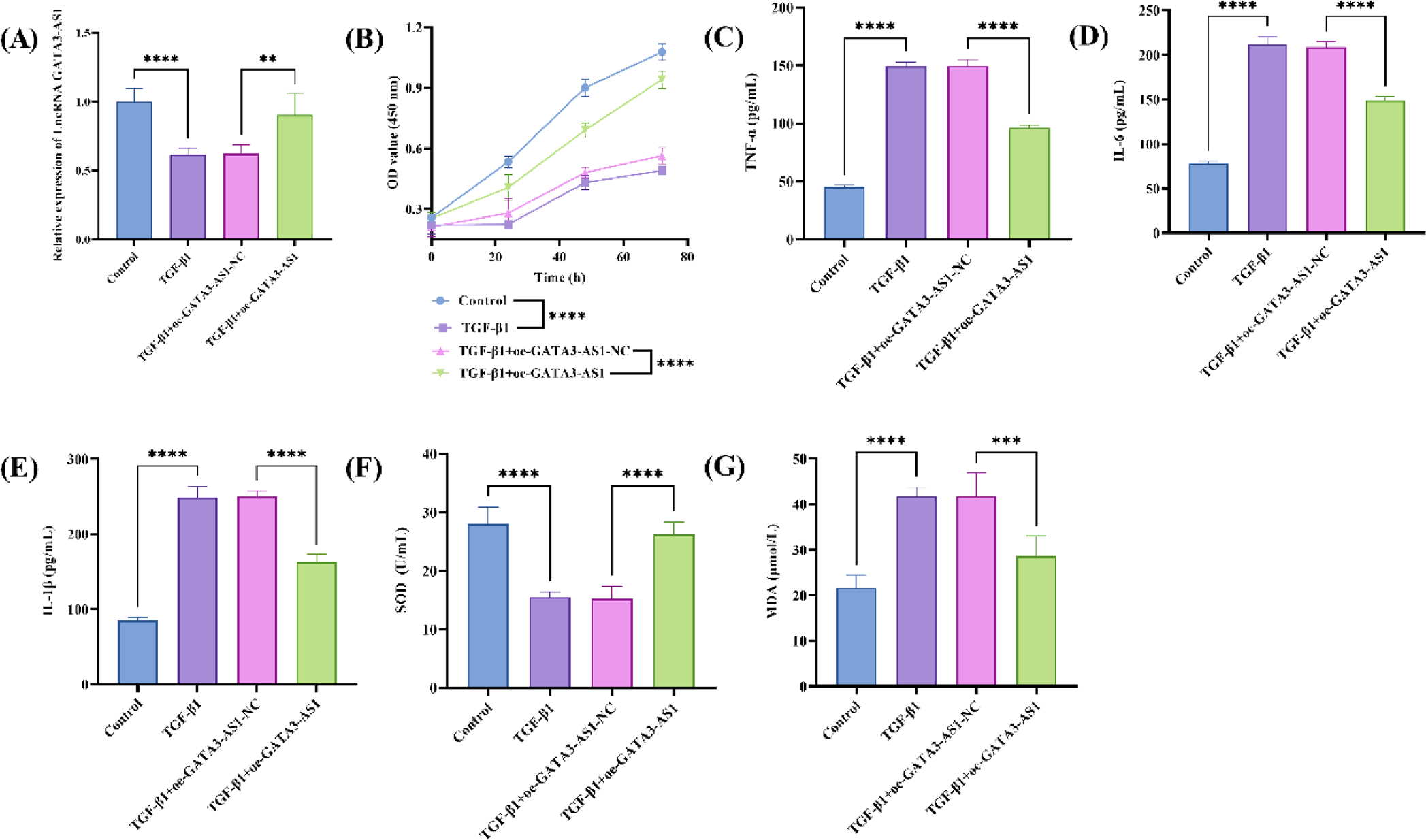
Further analysis of the results revealed that, compared to the Control, the relative level of LncRNA GATA3-AS1 was significantly reduced in TGF-β1-treated samples, and TGF-β1 could markedly inhibit the expression of GATA3-AS1 (Fig. 2A, p < 0.0001). The CCK-8 assay results demonstrated that TGF-β1 treatment significantly inhibited the proliferative capacity of MPC5 cells, while overexpression of GATA3-AS1 markedly restored cell proliferation (Fig. 2B, p < 0.0001). The ELISA results showed that TGF-β1 treatment significantly increased the secretion levels of TNF-α (Fig. 2C), IL-6 (Fig. 2D), and IL-1β (Fig. 2E), while overexpression of GATA3-AS1 markedly reduced the secretion of these inflammatory factors. In addition, TGF-β1 treatment reduced SOD activity and increased MDA content, while overexpression of GATA3-AS1 significantly enhanced SOD activity (Fig. 2F, p < 0.0001) and decreased MDA content (Fig. 2G, p < 0.001).

Impact of LncRNA GATA3-AS1 on TGF-β1-induced MPC5 cell function and mechanism analysis. The relative expression of GATA3-AS1 in TGF-β1-induced MPC5 cells (A); The effects of LncRNA GATA3-AS1 overexpression on TGF-β1-induced MPC5 cell proliferation capacity (B), TNF-α (C), IL-6 (D), IL-1β (E) secretion, SOD activity (F), and MDA content (G); n = three biological replicates; Data were analyzed using one-way ANOVA for intergroup comparisons, followed by Tukey's post hoc test; ****p < 0.0001, ***p < 0.001, **p < 0.01.
The relationship between LncRNA GATA3-AS1 and miR-2116-5p
According to the analysis, in the WT sequence, GATA3-AS1 and miR-2116-5p have complementary binding sites, whereas in the MUT sequence, this binding site is disrupted (Fig. 3A). The dual-luciferase reporter assay showed that, compared with NC-mimic, miR-2116-5p mimic significantly reduced the luciferase activity in the WT-GATA3-AS1 group (p < 0.0001), while it had no significant effect on the MUT-GATA3-AS1 group (Fig. 3B). Similarly, the miR-2116-5p inhibitor significantly increased the luciferase activity in the WT-GATA3-AS1 group (p < 0.0001), while the NC-inhibitor had no significant effect. The RIP experimental results demonstrated that both GATA3-AS1 (p < 0.01) and miR-2116-5p (p < 0.0001) were significantly enriched in the Ago2 immunoprecipitation complex compared to the IgG control group (Fig. 3C). Fig. 3D shows that TGF-β1 treatment significantly upregulates the expression of miR-2116-5p, and overexpression of GATA3-AS1 reverses this effect, while miR-2116-5p mimic counteracts the regulatory role of GATA3-AS1 (p < 0.0001). In addition, TGF-β1 inhibited cell proliferation, overexpression of GATA3-AS1 promoted proliferation, but the miR-2116-5p mimic significantly attenuated the proliferative effect of GATA3-AS1 (Fig. 3E, p < 0.0001). Overexpression of GATA3-AS1 reduced the secretion of TNF-α (Fig. 3F), IL-6 (Fig. 3G), and IL-1β (Fig. 3H), while miR-2116-5p mimic reversed this inhibitory effect. Further analysis revealed that overexpression of GATA3-AS1 enhanced SOD (Fig. 3I) and reduced MDA (Fig. 3J), while the miR-2116-5p mimic attenuated this protective effect.

The interaction between GATA3-AS1 and miR-2116-5p on the functional impact of TGF-β1-induced MPC5 cells. The binding sites sequence of GATA3-AS1 with miR-2116-5p (A); Dual-luciferase reporter assay to validate the binding of GATA3-AS1 with miR-2116-5p (B); RIP assay detects the enrichment of GATA3-AS1 and miR-2116-5p (C); Relative expression levels of miR-2116-5p under different treatment condition (D); The cell proliferation capacity under different treatment conditions (E), TNF-α (F), IL-6 (G), IL-1β (H) levels, SOD activity (I), and MDA content (J); n = three biological replicates; Data were analyzed using one-way ANOVA for intergroup comparisons, followed by Tukey's post hoc test; ****p < 0.0001,***p < 0.001, **p < 0.01.
Prediction and functional analysis of miR-2116-5p target genes
Using the miRmap, miRWalk, and TargetScan databases to predict potential target genes of miR-2116-5p, the Venn diagram shows 433 co-predicted genes, which may be high-confidence target genes of miR-2116-5p (Fig. 4A).

Prediction and Functional Analysis of miR-2116-5p Target Genes. Venn diagram of miR-2116-5p target gene prediction results (A), KEGG pathway enrichment analysis (B), GO functional enrichment analysis (C), and miRNA-target gene network analysis (D).
KEGG analysis showed these target genes were significantly enriched in signaling pathways closely related to inflammatory response, cell proliferation, and apoptosis (ErbB signaling pathway, Transcriptional misregulation in cancer, Cholinergic synapse, Phosphatidylinositol signaling system). miR-2116-5p may participate in the pathological process of childhood NS by regulating these pathways (Fig. 4B). GO enrichment analysis revealed (Fig. 4C) that in terms of biological processes, the target genes were significantly enriched in the regulation of cell morphogenesis, positive regulation of fatty acid oxidation, ERBB signaling pathway, and cellular response to transforming growth factor beta. These processes are closely related to the normal physiological functions of cells and the changes under pathological conditions, suggesting that miR-2116-5p may influence the progression of childhood NS by regulating these biological processes. The analysis of cellular components revealed that the target genes are primarily associated with cellular structures such as the phosphatidylinositol 3-kinase complex, the endoplasmic reticulum protein-containing complex, and the postsynaptic membrane. This indicates that the target genes of miR-2116-5p may play a significant role in cell signal transduction, protein synthesis and transport, as well as synaptic function. In terms of molecular functions, the target genes are significantly enriched in activities such as phosphatidylinositol bisphosphate kinase activity and nucleocytoplasmic transport signal receptor activity. These molecular functions are crucial for maintaining intracellular signal transduction, substance metabolism, and the stability of the cytoskeleton. In addition, the constructed miRNA-target gene network visually demonstrates the regulatory relationship between miR-2116-5p and its predicted target genes (Fig. 4D). Network analysis revealed the pivotal position of miR-2116-5p in the regulatory network and identified the top 10 core genes with the highest scores, including TP53, TCF12, TAL1, SRC, SEMA3A, PRKDC, PLXNA4, NRG4, NHEJ1, and JUN. The scores of both TP53 and TCF12 are 0.999, indicating their significant functional roles, and they are used for further research.
The regulation of TCF12 by miR-2116-5p and its functional impact
In childhood NS, the expression of TP53 was significantly upregulated (Fig. 5A, p < 0.0001), while the relative level of TCF12 was significantly lower than that in the healthy control group (Fig. 5B, p < 0.0001). The levels of TP53 and miR-2116-5p were consistent, indicating that there was no targeted relationship between them. The dual-luciferase confirmed the binding of miR-2116-5p to TCF12 (Fig. 5C). TGF-β1 induction significantly reduced TCF12 expression levels (p < 0.0001). Overexpression of GATA3-AS1 significantly restored TCF12 levels (p < 0.0001), while the miR-2116-5p mimic further suppressed TCF12 expression (p < 0.0001). Transfection with oe-TCF12 reversed the TCF12 inhibition mediated by the miR-2116-5p mimic (p < 0.001), further validating TCF12 as a direct functional target of miR-2116-5p (Fig. 5D). Meanwhile, oe-TCF12 rescued the miR-2116-5p mimic-induced suppression of proliferation (Fig. 5E), and significantly regulated intracellular inflammatory factors (p < 0.0001), including TNF-α (Fig. 5F), IL-6 (Fig. 5G), and IL-1β (Fig. 5H), as well as oxidative stress markers (p < 0.01), MDA (Fig. 5I) and SOD (Fig. 5J). Western Blot analysis showed that TGF-β1 treatment significantly reduced the expression of Synaptopodin and Podocin while upregulating Cleaved Caspase-3, whereas overexpression of GATA3-AS1 effectively reversed this damage. However, co-transfection with the miR-2116-5p mimic partially counteracted the protective effect of GATA3-AS1. Under these conditions, further overexpression of TCF12 was able to reverse the damage induced by the miR-2116-5p mimic, as reflected in the expression levels of Synaptopodin (Fig. S1A), Cleaved Caspase-3 (Fig. S1B), and Podocin (Fig. S1C).
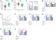
Functional analysis of the GATA3-AS1/miR-2116-5p/TCF12 axis in childhood NS. The relative expression of TP53 (A) and TCF12 (B) in childhood NS patients and healthy controls; Luciferase activity validation of TCF12 and miR-2116-5p (C); Relative expression of TCF12 under different treatment conditions (D); Detection of cell proliferation capacity under different treatment conditions (E), TNF-α (F), IL-6 (G), IL-1β (H) levels, MDA content (I), and SOD activity (J); n = three biological replicates; Data were analyzed using one-way ANOVA for intergroup comparisons, followed by Tukey's post hoc test; ****p < 0.0001,***p < 0.001,**p < 0.01.
Knockdown of TCF12 reverses the protective effect of GATA3-AS1 on podocytes
To determine whether TCF12 is essential in the GATA3-AS1-mediated protective pathway, we knocked down TCF12 while overexpressing GATA3-AS1. The results showed that, compared with the control, knockdown of TCF12 significantly reversed the protective effects of GATA3-AS1 on podocytes. This reversal was manifested as decreased cell proliferation (Fig. S2A), increased secretion of inflammatory factors including TNF-α (Fig. S2B), IL-6 (Fig. S2C), and IL-1β (Fig. S2D), and aggravated oxidative stress reflected by changes in SOD (Fig. S2E) and MDA (Fig. S2F) levels. At the protein level, knockdown of TCF12 also downregulated the expression of the podocyte cytoskeletal protein Synaptopodin (Fig. S2G) and the slit diaphragm protein Podocin (Fig. S2I), while reactivating the apoptosis marker protein Cleaved Caspase-3 (Fig. S2H).
Discussion
The complexity of childhood NS stems from the interaction of various molecular and cellular mechanisms, as well as genetic and environmental factors, and its pathogenesis has not yet been fully elucidated. Previous studies have found that GATA3 expression is downregulated in renal cell carcinoma, and its overexpression can reduce tumor metastasis by inhibiting the STAT3 signaling pathway,19 suggesting that the dysregulation of GATA3-AS1 is associated with renal pathology. We extended this research direction to pediatric NS, focusing on circulating molecules as noninvasive probes for disease pathology. Although renal biopsy is considered the gold standard, it is rarely performed in children with NS, especially in new-onset cases, due to ethical and clinical constraints. Therefore, this study utilized serum samples for analysis. Available evidence indicates that the levels of specific lncRNAs and miRNAs in serum have been shown to correlate with their expression in renal tissue and with the severity of kidney disease.21-23 This study is the first to identify a significant downregulation of LncRNA GATA3-AS1 in the serum of pediatric NS patients, and the degree of this downregulation shows a negative correlation with key clinical indicators such as WBC, PLT, BUN, serum creatinine, and PRO.24-26 In summary, the systematic association of GATA3-AS1 with key clinical indicators provides clues for its potential role as a circulating biomarker involved in the disease progression of childhood NS, and its clinical application value warrants further elucidation in future studies. In our cohort, LncRNA GATA3-AS1 showed preliminary diagnostic efficacy for childhood NS (AUC = 0.892), which supports its potential for auxiliary diagnosis. Future studies with larger, longitudinal cohorts are warranted to validate the potential of serum GATA3-AS1 and miR-2116-5p as a combined non-invasive biomarker panel for aiding diagnosis, monitoring disease activity, or assessing treatment response in childhood NS. Therefore, further investigation into the regulatory mechanisms of GATA3-AS1 may provide new insights into the pathogenesis and diagnosis of childhood NS.
Podocytes are crucial for preserving the glomerular filtration barrier's integrity, their damage can lead to proteinuria and glomerulosclerosis.27 TGF-β1 plays a critical role in various kidney-related diseases and can exacerbate podocyte injury. For example, TGF-β1 can induce an increase in miR-155 expression in mouse podocytes, while simultaneously elevating the levels of pro-inflammatory cytokines.28 In addition, TGF-β1 can increase the expression of mitochondrial Nox4 by activating the mTORC1 pathway, leading to the generation of ROS and mitochondrial dysfunction, ultimately resulting in podocyte apoptosis.29 This study found that TGF-β1 treatment inhibited cell proliferation, upregulated inflammatory levels, and enhanced oxidative stress, while overexpression of GATA3-AS1 significantly reversed these alterations. lncRNA can influence the processing, maturation, or stability of miRNA by directly binding to its precursor or mature form, thereby regulating miRNA function. This interaction can also be involved in the mechanisms of disease development and progression.30 The study found that lncRNA GATA3-AS1 is upregulated in breast cancer tissues, and by binding to miR-495-3p, it indirectly regulates the expression of CENPU, thereby affecting the prognosis of breast cancer patients.31 The downregulation of GATA3-AS1 can inhibit the proliferation of childhood ALL cells and induce cell apoptosis by targeting the upregulation of miR-515-5p expression.32 Based on this, we utilized the lncRNASNP2 database to predict the downstream miRNA of GATA3-AS1 and selected miR-2116-5p with the highest binding score for further analysis. The luciferase activity assay confirmed the interaction between GATA3-AS1 and miR-2116-5p Additionally, the Ago2 RIP experiment confirmed that GATA3-AS1 binds with miR-2116-5p in the RISC complex, providing certain biochemical evidence for the ceRNA mechanism. Based on their genomic localization, it was found that GATA3-AS1 (chromosome 10, https://www.ncbi.nlm.nih.gov/gene) and MIR2116 (chromosome 15, http://www.mirbase.org/) are located on different chromosomes. This analysis ruled out the possibility of both being regulated by a common upstream cis-regulatory element, further supporting the sponging mechanism. Further analysis revealed that miR-2116-5p is upregulated in childhood NS and significantly negatively correlated with GATA3-AS1, and possesses good diagnostic efficacy for childhood NS. In addition, miR-2116-5p can reverse the inhibitory effects of GATA3-AS1 on inflammation and oxidative stress in TGF-β1-induced podocyte injury. It is speculated that miR-2116-5p may play a role in childhood NS by mediating GATA3-AS1.
Through the construction of a miRNA-mRNA regulatory network and screening of miR-2116-5p target genes, TCF12 was identified as the research focus. Although previous studies have shown that TCF12 acts as a transcriptional repressor of E-cadherin and promotes tumor metastasis in colorectal cancer,33 its role in kidney diseases remains unknown. This study found that TCF12 expression is reduced in childhood NS. Overexpression of TCF12 rescued miR-2116-5p-induced podocyte injury, while knockdown of TCF12 reversed the protective effect of GATA3-AS1, demonstrating that TCF12 is an indispensable downstream factor in this axis. Relevant studies have also reported that E-cadherin is a core molecule in EMT, and its downregulation is closely associated with pathologies such as renal fibrosis.34 Additional experiments further indicate that the GATA3-AS1/miR-2116-5p/TCF12 axis significantly upregulates the expression of key podocyte cytoskeletal and slit‑diaphragm proteins, Synaptopodin and Podocin, and suppresses the activation of the apoptosis marker Cleaved Caspase-3. Therefore, this finding reveals a new function of GATA3-AS1/miR-2116-5p/TCF12 axis in podocytes and provides a novel perspective for its investigation in kidney diseases.
This study offers fresh insights into the pathogenesis of childhood nephrotic syndrome and the screening of diagnostic markers, yet it is subject to several limitations. First, this study has a limited sample size, and all molecular tests were performed based on serum samples. Although changes in serum biomarkers are significantly correlated with disease activity, their expression and functional status in renal tissues, particularly in podocytes, still require direct confirmation. Future work will expand the cohort samples and utilize archived renal biopsy tissues or animal models to concurrently analyze the expression and correlation of this molecular axis in both serum and tissues. Second, the mechanism study relies entirely on a TGF-β1-induced mouse podocyte model, which, although capable of simulating key injury characteristics, cannot fully replicate the complex in vivo microenvironment and systemic disease processes. Future studies will validate the universality and functionality of this regulatory axis in models that more closely resemble human pathology, such as puromycin-induced nephropathy animal models. Third, the elucidation of the downstream transcriptional regulatory network of TCF12 remains incomplete, with E-cadherin being only one of its potential targets. Future studies plan to systematically screen the gene sets regulated by TCF12 through RNA-seq and validate its direct binding to key target gene promoters using ChIP-qPCR, thereby clarifying the specific mechanisms through which TCF12 mediates podocyte protective effects via downstream molecules.
Conclusion
This study found that GATA3-AS1 is downregulated in childhood NS patients and negatively correlated with the expression of miR-2116-5p Overexpression of GATA3-AS1 can restore TGF-β1-induced damage in MPC5 cells, while miR-2116-5p can reverse this protective effect. The study also revealed the direct binding relationship between miR-2116-5p and TCF12, indicating that GATA3-AS1 influences podocyte function by regulating the miR-2116-5p-TCF12 axis. These results provide a certain research background for subsequent studies on the roles of LncRNA GATA3-AS1 and miR-2116-5p in childhood NS.
Data availability
All data generated or analyzed during this study are included in this article. Further enquiries can be directed to the corresponding author.
-
Funding
No funds, grants, or other support was received.
Acknowledgement
Not applicable.
Supplementary materials
Supplementary material associated with this article can be found, in the online version, at doi:10.1016/j.clinsp.2026.101053.
Supplementary PDF
References
- 1 Wang L., Wang J., Wang Z., Zhou J., Zhang Y. Higher urine exosomal miR-193a is associated with a higher probability of primary focal segmental glomerulosclerosis and an increased risk of poor prognosis among children with nephrotic syndrome. Front Cell Dev Biol. 2021;9:727370.
- 2 Kolb A., Gallacher P.J., Campbell J., O'Neill M., Smith J.R., Bell S., et al. A National Registry Study of patient and renal survival in Adult Nephrotic Syndrome. Kidney Int Rep. 2021;6(2):449-59.
- 3 Wang Y.N., Hu H.H., Zhang D.D., Wu X.Q., Liu J.L., Guo Y., et al. The dysregulation of Eicosanoids and Bile acids correlates with impaired kidney function and renal fibrosis in Chronic Renal failure. Metabolites. 2021;11(2):89.
- 4 Gao S., Chao Y., Li N., Li H., Zhao H., Liu X., et al. An integrated Proteomics and Metabolomics Strategy for the mechanism of calcium oxalate crystal-induced kidney injury. Front Med (Lausanne). 2022;9:805356.
- 5 Jorge S., Capelo J.L., LaFramboise W., Satturwar S., Korentzelos D., Bastacky S., et al. Absolute quantitative proteomics using the total protein approach to identify novel clinical immunohistochemical markers in renal neoplasms. BMC Med. 2021;19(1):196.
- 6 Wu L., Chen M., Mao H., Wang N., Zhang B., Zhao X., et al. Albumin-based nanoparticles as methylprednisolone carriers for targeted delivery towards the neonatal Fc receptor in glomerular podocytes. Int J Mol Med. 2017;39(4):851-60.
- 7 Yang F., Shen Y., Zhang W., Jin J., Huang D., Fang H., et al. An androgen receptor negatively induced long non-coding RNA ARNILA binding to miR-204 promotes the invasion and metastasis of triple-negative breast cancer. Cell Death Differ. 2018;25(12):2209-20.
- 8 Benderska N., Dittrich A.L., Knaup S., Rau T.T., Neufert C., Wach S., et al. miRNA-26b overexpression in ulcerative colitis-associated carcinogenesis. Inflamm Bowel Dis. 2015;21(9):2039-51.
- 9 Ren J., Xu N., Zhou R., Huang F., Zhang H., Li W. Long non-coding RNA PCED1B antisense RNA 1 promotes gastric cancer progression via modulating microRNA-215-3p /C-X-C motif chemokine receptor 1 axis. Bioengineered. 2021;12(1):6083-95.
- 10 Figueiredo J.C., Hsu L., Hutter C.M., Lin Y., Campbell P.T., Baron J.A., et al. Genome-wide diet-gene interaction analyses for risk of colorectal cancer. PLoS Genet. 2014;10(4):e1004228.
- 11 Takaku M., Grimm S.A., Roberts J.D., Chrysovergis K., Bennett B.D., Myers P., et al. GATA3 zinc finger 2 mutations reprogram the breast cancer transcriptional network. Nat Commun. 2018;9(1):1059.
- 12 Zhang Y., Liu H., Shu X., Sun Y., Chen X. Overexpressed IncRNA GATA3-AS1 in Preeclampsia and its effects on trophoblast proliferation and migration by the miR-488-3p/ROCK1 Axis. Crit Rev Eukaryot Gene Expr. 2022;32(5):33-45.
- 13 Moriguchi T., Yu L., Otsuki A., Ainoya K., Lim K.C., Yamamoto M., et al. Gata3 Hypomorphic Mutant Mice rescued with a yeast artificial chromosome transgene suffer a Glomerular Mesangial cell defect. Mol Cell Biol. 2016;36(17):2272-81.
- 14 Guo F., Zhang W., Su J., Xu H., Yang H. Prediction of drug positioning for Quan-Du-Zhong capsules against Hypertensive Nephropathy based on the robustness of disease network. Front Pharmacol. 2019;10:49.
- 15 Bohosova J., Kasik M., Kubickova A., Trachtova K., Stanik M., Poprach A., et al. LncRNA PVT1 is increased in renal cell carcinoma and affects viability and migration in vitro. J Clin Lab Anal. 2022;36(6):e24442.
- 16 Ji H., Yi Q., Chen L., Wong L., Liu Y., Xu G., et al. Circulating miR-3197 and miR-2116-5p as novel biomarkers for diabetic retinopathy. Clin Chim Acta. 2020;501:147-53.
- 17 Xue L., Hu M., Li J., Li Y., Zhu Q., Zhou G., et al. Ginsenoside Rg1 inhibits high glucose-induced Proliferation, migration, and angiogenesis in retinal endothelial cells by regulating the lncRNA SNHG7/miR-2116-5p/SIRT3 Axis. J Oncol. 2022;2022:6184631.
- 18 Wong C.W., Wong T.Y., Cheng C.Y., Sabanayagam C. Kidney and eye diseases: common risk factors, etiological mechanisms, and pathways. Kidney Int. 2014;85(6):1290-302.
- 19 Shi Q., Xu R., Song G., Lu H., Xue D., He X., et al. GATA3 suppresses human fibroblasts-induced metastasis of clear cell renal cell carcinoma via an anti-IL6/STAT3 mechanism. Cancer Gene Ther. 2020;27(9):726-38.
- 20 Wu C., Wang Z., Tian X., Wang J., Zhang Y., Wu B. Long non-coding RNA DDX11-AS1 promotes esophageal carcinoma cell proliferation and migration through regulating the miR-514b-3p/RBX1 axis. Bioengineered. 2021;12(1):3772-86.
- 21 Zhou L.J., Yang D.W., Ou L.N., Guo X.R., Wu B.L. Circulating expression level of LncRNA Malat1 in diabetic kidney disease patients and its clinical significance. J Diabetes Res. 2020;2020:4729019.
- 22 Qi P., Zhou X.Y., Du X. Circulating long non-coding RNAs in cancer: current status and future perspectives. Mol Cancer. 2016;15(1):39.
- 23 Masum M.A., Ichii O., Elewa Y.H.A., Kon Y. Podocyte injury through interaction between Tlr8 and its endogenous ligand miR-21 in obstructed and its collateral kidney. Front Immunol. 2020;11:606488.
- 24 Xu-Yang Z., Pei-Yu B., Chuan-Tao Y., Wei Y., Hong-Wei M., Kang T., et al. Interferon-Induced transmembrane protein 3 Inhibits Hantaan virus infection, and its single Nucleotide Polymorphism rs12252 Influences the Severity of Hemorrhagic Fever with Renal Syndrome. Front Immunol. 2016;7:535.
- 25 Ishida M., Ueki M., Morishita J., Ueno M., Shiozawa S., Maekawa N. T-5224, a selective inhibitor of c-Fos/activator protein-1, improves survival by inhibiting serum high mobility group box-1 in lethal lipopolysaccharide-induced acute kidney injury model. J Intensive Care. 2015;3:49.
- 26 Torres-González L., Cienfuegos-Pecina E., Perales-Quintana M.M., Alarcon-Galvan G., Muñoz-Espinosa L.E., Pérez-Rodríguez E., et al. Nephroprotective effect of Sonchus oleraceus extract against kidney injury induced by Ischemia-Reperfusion in Wistar rats. Oxid Med Cell Longev. 2018;2018:9572803.
- 27 Chen J., Chen J.K., Harris R.C. EGF receptor deletion in podocytes attenuates diabetic nephropathy. J Am Soc Nephrol. 2015;26(5):1115-25.
- 28 Zheng X., Zhong Q., Lin X., Gu X., Ling X., Liang Z., et al. Transforming growth factor-β1-induced podocyte injury is associated with increased microRNA-155 expression, enhanced inflammatory responses and MAPK pathway activation. Exp Ther Med. 2021;21(6):620.
- 29 Das R., Xu S., Nguyen T.T., Quan X., Choi S.K., Kim S.J., et al. Transforming Growth Factor β1-induced Apoptosis in Podocytes via the Extracellular Signal-regulated Kinase-Mammalian Target of Rapamycin Complex 1-NADPH Oxidase 4 Axis. J Biol Chem. 2015;290(52):30830-42.
- 30 Liu S., Lai W., Shi Y., Liu N., Ouyang L., Zhang Z., et al. Annotation and cluster analysis of long noncoding RNA linked to male sex and estrogen in cancers. NPJ Precis Oncol. 2020;4:5.
- 31 Lin S., Zhao M., Lv Y., Mao G., Ding S., Peng F. The lncRNA GATA3-AS1/miR-495-3p/CENPU axis predicts poor prognosis of breast cancer via the PLK1 signaling pathway. Aging (Albany NY). 2021;13(10):13663-79.
- 32 Qin Y., Yun X.X., Zheng Z.M., Xu Q., Zuo L.M. [Effect of LncRNA GATA3-AS1 targeting MiR-515-5p on cell Proliferation and Apoptosis in Childhood Acute Lymphoblastic Leukemia Cells]. Zhongguo Shi Yan Xue Ye Xue Za Zhi. 2023;31(4):1032-7.
- 33 Lee C.C., Chen W.S., Chen C.C., Chen L.L., Lin Y.S., Fan C.S., et al. TCF12 protein functions as transcriptional repressor of E-cadherin, and its overexpression is correlated with metastasis of colorectal cancer. J Biol Chem. 2012;287(4):2798-809.
- 34 Barriere G., Fici P., Gallerani G., Fabbri F., Rigaud M. Epithelial Mesenchymal Transition: a double-edged sword. Clin Transl Med. 2015;4:14.
Edited by
-
Edited by:
José Maria Soares Junior