Abstract
BACKGROUND: There are no reports on the long-term follow-up of patients with swine-origin influenza A virus infection that progressed to acute respiratory distress syndrome. METHODS: Four patients were prospectively followed up with pulmonary function tests and high-resolution computed tomography for six months after admission to an intensive care unit. RESULTS: Pulmonary function test results assessed two months after admission to the intensive care unit showed reduced forced vital capacity in all patients and low diffusion capacity for carbon monoxide in two patients. At six months, pulmonary function test results were available for three patients. Two patients continued to have a restrictive pattern, and none of the patients presented with abnormal diffusion capacity for carbon monoxide. All of them had a diffuse ground-glass pattern on high-resolution computed tomography that improved after six months. CONCLUSIONS: Despite the marked severity of lung disease at admission, patients with acute respiratory distress syndrome caused by swine-origin influenza A virus infection presented a late but substantial recovery over six months of follow-up.
Swine-origin influenza A virus; Intensive care unit; Acute respiratory failure; Recovery; Pulmonary function test
CLINICAL SCIENCE
IPulmonary Division - InCor - Faculdade de Medicina da Universidade de São Paulo, SP, Brazil
IIInfectious and Parasitic Diseases Division - Faculdade de Medicina da Universidade de São Paulo, SP, Brazil
IIIPhysiotherapy Service - Instituto Central do Hospital das Clínicas da Faculdade de Medicina da Universidade de São Paulo, SP, Brazil
ABSTRACT
BACKGROUND: There are no reports on the long-term follow-up of patients with swine-origin influenza A virus infection that progressed to acute respiratory distress syndrome.
METHODS: Four patients were prospectively followed up with pulmonary function tests and high-resolution computed tomography for six months after admission to an intensive care unit.
RESULTS: Pulmonary function test results assessed two months after admission to the intensive care unit showed reduced forced vital capacity in all patients and low diffusion capacity for carbon monoxide in two patients. At six months, pulmonary function test results were available for three patients. Two patients continued to have a restrictive pattern, and none of the patients presented with abnormal diffusion capacity for carbon monoxide. All of them had a diffuse ground-glass pattern on high-resolution computed tomography that improved after six months.
CONCLUSIONS: Despite the marked severity of lung disease at admission, patients with acute respiratory distress syndrome caused by swine-origin influenza A virus infection presented a late but substantial recovery over six months of follow-up.
Keywords: Swine-origin influenza A virus; Intensive care unit; Acute respiratory failure; Recovery; Pulmonary function test.
INTRODUCTION
In 2009, the novel swine-origin influenza A (H1N1) virus (S-OIV) was identified as the cause of a severe influenza pandemic.1 The clinical spectrum of the pandemic H1N1 virus was broad, from mild upper respiratory tract illness to severe complications, such as acute respiratory distress syndrome, multiorgan failure, and death.1
Approximately 10% to 30% of hospitalized patients required intensive care unit (ICU) admission, and 60% to 88% of those patients who were admitted to the ICU needed mechanical ventilation (MV).3,4 Risk factors for death included high acute physiology and chronic health evaluation II (APACHE II) scores, a low ratio of arterial partial pressure of oxygen to fraction of inspired oxygen (PaO /FIO ratio), use of inotropic drugs, hemodialysis, lymphocytopenia, and high levels of lactic dehydrogenase (LDH) and C-reactive protein (CRP).3,5
The lungs were extensively involved, and bilateral areas of consolidation and/or ground-glass opacities on high-resolution computed tomography (HRCT) were already present 4-9 days after hospital admission caused by H1N1 virus infection.6 The main pathological changes associated with H1N1 virus infection were diffuse alveolar damage, necrotizing bronchiolitis, and extensive hemorrhage.7 The mortality rate in Brazil was 70 deaths per 100,000 people.1 General management of lung involvement in our Institution has been extensively covered.8-11
Survivors of acute respiratory distress syndrome (ARDS) might present reduced carbon monoxide diffusion capacity (DLCO), impaired lung function, and diminished physical capacity six months after hospital discharge.12 At 3-month follow-up, patients who have suffered from H1N1 pneumonia can still present with ground-glass opacities and reduced carbon monoxide diffusion capacity.13 There are no reports regarding the follow-up of patients admitted to the ICU as a result of ARDS during the influenza pandemic. Our goal in this case series was to describe the long-term recovery of patients diagnosed with influenza A (H1N1) virus who developed ARDS.
MATERIALS AND METHODS
From July 9 to August 31, 2009, patients over 18 years old at a tertiary hospital located in Sao Paulo, who were admitted to the ICU with H1N1 virus infection confirmed with nasopharyngeal swab specimens (by using the real-time reverse transcriptase polymerase chain reaction [rRT-PCR] test) and by the fulfillment of ARDS criteria, were prospectively included. A written informed consent, approved by the hospital's ethical committee, was obtained from the patients' next-of-kin. Patients with ages below 18 years old, more than three organ system failures, persistent hemodynamic instability, severe cardiac disease, immunosuppression by chemotherapy or radiation therapy, or acute brain injury, as well as pregnant women, were excluded.
Patients were managed with a lung-protective ventilation strategy (tidal volume 6 mL/kg of predicted body weight and driving pressure [plateau pressure - PEEP] up to maximum 15 cm H2O). Using bedside electrical impedance tomography, a recruitment strategy with a decremental PEEP titration was performed to obtain the optimal PEEP value.14
Electrical impedance tomography is a new imaging tool that is noninvasive and radiation-free and that drives harmless electrical currents across the thorax, using 16 electrodes placed at the transverse plane and crossing the fifth intercostal space at the midclavicular line. The electrodes generate a potential gradient at the surface, which is then transformed into a two-dimensional image of the electric impedance distribution within the thorax.15 When comparing regional ventilation across different thoracic regions, the quantitative information provided by electrical impedance tomography was closely proporcional to changes in air content, as calculated by dynamic computed tomography scanning.15
All patients received a corticosteroid (hydrocortisone, 200 mg/day) and oseltamivir (150 mg/day). Sepsis was defined as infection plus systemic manifestations of infection, and treatment for sepsis was based on the Surviving Sepsis Campaign guidelines.16
Patients were followed during their stays in the hospital and at regular intervals (1-2 months and 6 months after admission) with pulmonary function tests (lung volume assessed by body plethysmography), inspiratory/expiratory high-resolution computed tomography, and a 6-min walk test (6-MWT) if the patient was able to walk. All measurements were performed in extubated patients.
RESULTS
A total of 54 patients, 18 of whom required invasive mechanical ventilation, were admitted to the ICU because of H1N1 virus infection. Of the 18 intubated patients, 14 were excluded (2 with more than 48 hours of ARDS, 2 with acute brain injury, 3 with severe cardiac disease, 3 with immunosuppression due to chemotherapy or radiation therapy, 3 with more than 3 organ system failures, and 1 because of pregnancy). Four patients were included.
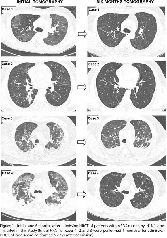
Admission and demographic data assessed during the ICU stay are summarized in Table 1. The PFTs after 2 and 6 months are summarized in Table 2, as well as the 6-MWD performed after 6 months. High-resolution computed tomography, performed initially and after 6 months, is shown in Figure 1. Six months after ICU admission, none of the patients presented with air trapping in expiration on high-resolution computed tomography.
Case 1
ARDS caused by H1N1 virus was diagnosed in a 28-year-old man after one week of fever and dyspnea. He was admitted to the ICU and was started on mechanical ventilation. Patient data during his ICU stay are shown in Table 1. Thirty-four days after admission, he was discharged without supplemental oxygen but with a significant limitation in his physical capacity. He resumed work after four months, although reassigned to a different, less physically demanding, position. The PFTs during follow-up demonstrated improvement in the DLCO, with the forced vital capacity and total lung capacity still reduced after six months (Table 2). The 6-minute walk distance was also reduced 6 months after admission, and the high-resolution computed tomography scan showed an improvement in the initial ground-glass pattern (Figure 1).
Case 2
A 25-year-old man, in whom ARDS caused H1N1 virus was diagnosed after four days with fever and progressive dyspnea, was admitted to the ICU with respiratory failure requiring mechanical ventilation (Table 1). He stayed in the hospital for 29 days and returned to his usual activities 2 months after he was discharged. After 6 months of follow-up, a pulmonary function test demonstrated an improvement in forced vital capacity, total lung capacity, and DLCO (Table 2), as well an improvement on his high-resolution computed tomography scan (Figure 1). He still had physical limitations, as measured with the 6-minute walk test, 6 months after ICU discharge (Table 1).
Case 3
A 54-year-old man with a past medical history of diabetes and arterial hypertension was admitted to the ICU caused by respiratory and acute renal failure associated with the H1N1 virus (Table 1). He was discharged from the hospital 38 days later but was readmitted twice because of pulmonary edema. His pulmonary function test after 2 months showed a severe restrictive pattern and reduced DLCO (Table 2). The high-resolution computed tomography scan showed an interstitial infiltration (Figure 1). Six months after the initial admission, the patient was hospitalized because of pulmonary congestion, and he was therefore unable to perform the pulmonary function test or the 6-minute walk test. At this time, high-resolution computed tomography was performed and was compatible with pulmonary edema (Figure 1).
Case 4
The patient was a previously healthy 43-year-old man who presented to the hospital with severe acute respiratory failure caused by the H1N1 virus, which required immediate intubation and ICU admission (Table 1). He remained in the hospital for 23 days and resumed his normal activities after 2 months. Two months after admission, his pulmonary function test showed a mild restrictive pattern with a normal DLCO. At the six-month follow-up, his pulmonary function test improved (Table 2). In comparison to the high-resolution computed tomography scan at admission, his computed tomography at six months was markedly improved, but there remained rare peripheral lung infiltrates (Figure 1). Six months after admission, his physical capacity was normal (Table 2).
DISCUSSION
In this follow-up of four patients with ARDS caused by H1N1 virus infection, we found a restrictive pattern in the pulmonary function test in three patients and a reduced DLCO in two patients two months after admission. These results are consistent with data from ARDS survivors published previously,12,17 in which a reduced forced vital capacity and DLCO could be observed three months after ARDS.
Conversely, the marked improvement observed at six-month follow-up, especially the changes observed in DLCO and on high-resolution computed tomography, are in disagreement with the expected long-term functional outcome in ARDS patients. In previous studies,12 pulmonary function abnormalities were common in ARDS survivors after 6 months, with a persistently low DLCO and with residual lung parenchyma changes suggestive of fibrosis on the high-resolution computed tomography scan.18 Considering the severity of the respiratory failure that our patients with ARDS caused by H1N1 virus infection underwent (all patients had PaO2/FIO2 ratios <100 mm Hg at admission), we had expected a worse outcome at six months.
The reduction of physical capacity observed at six months of follow-up in three of our cases was probably multi-factorial and may be partially explained by corticosteroid-induced and/or critical-illness-associated myopathy,12 in agreement with previous studies.
Patients in this case series were younger and presented an APACHE II score lower than ARDS survivors in previous studies12 and than critically ill influenza A (H1N1) patients,3,4 favoring a good outcome. However, they presented laboratory test abnormalities (elevated LDH and C-reactive protein levels and reduced lymphocyte count) and a reduced PaO2/FIO2 ratio that were previously associated with poor outcomes of H1N1 virus infection.3,5
Another point to consider when interpreting the results is the mechanical ventilation strategy applied. Patients with H1N1 virus infection who require mechanical ventilation can be very difficult to oxygenate, and the use of adjuvant methods to improve hypoxemia, such as recruitment maneuvers,3 prone positioning,3 and extracorporeal membrane oxygenation, was frequent in previous studies.4,19,20 We used a ventilatory strategy with low tidal volumes and recruitment maneuvers and with a careful selection of the PEEP set to minimize the lung injury caused by cyclic alveolar recruitment and collapse,14 according to the bedside PEEP titration strategy guided by electrical impedance tomography. It is possible that this ventilatory strategy was responsible for the good functional recovery of our patients, although we cannot exclude that the functional improvement was merely a characteristic of infection with the H1N1 virus.
The main limitations of this case series are as follows: (1) the small number of patients with complete follow-up, (2) the exclusion of patients with more severe symptoms, and (3) the absence of control patients. In particular, the latter limitation prevents us from drawing definitive conclusions about the aforementioned associations between treatment and outcome, which should be a matter for future studies.
CONCLUSION
In summary, selected patients diagnosed with the H1N1 virus and managed with a bedside recruitment strategy using electrical impedance tomography had functional limitations two months after ICU admission, which recovered partially or fully after six months. Additional studies should be conducted to assess the impact of new protective strategies during mechanical ventilation on the long-term recovery of patients with ARDS.
Received for publication on January 3, 2011; First review completed on January 20, 2011; Accepted for publication on February 20, 2011
E-mail: toufenjr@ig.com.br Tel.: 55 11 3069-5000
- 1. World Health Organization. Transmission dynamics and impact of pandemic influenza A (H1N1) 2009 virus. Wkly Epidemiol Rec. 2009;46:481-4.
- 2. Hui DS, Lee N, Chan PKS. Clinical management of pandemic (H1N1) infection. Chest. 2010;137:916-25, doi: 10.1378/chest.09-2344.
- 3. Estenssoro E, Rios FG, Apezteguia C, Reina R, Neira J, Ceraso DH, et al. Pandemic 2009 influenza A(H1N1) inArgentina: Astudyof337patients on mechanical ventilation. Am J Respir Crit Care Med. 2010;182:41-8, doi:10.1164/201001-0037OC.
- 4. Miller RR, Markewitz BA, Rolfs RT, Brown SM, Dascomb KK, Grissom CK, et al. Clinical findings and demographic factors associated with ICU admission in Utah due to novel 2009 influenza A (H1N1) infection. Chest. 2010;137:752-8, doi: 10.1378/chest.09-2517.
- 5. Cui W, Zhao H, Lu H, Wen Y, Zhou Y, Deng B, et al. Factors associated with dead in hospitalized pneumonia patients with 2009 H1N1 influenza in Shenyang, China. BMC Infect Dis. 2010;10:1-9, doi: 10.1186/1471-2334-10-145.
- 6. Marchiori E, Zanetti G, Hochhegger B, Rodrigues RS, Fontes CA, Nobre LF, et al. High-resolution computed tomography findings from adult patients with Influenza A (H1N1) virus-associated pneumonia. Eur J Radiol. 2010;74:93-8, doi: 10.1016/j.ejrad.2009.11.005.
- 7. Mauad T, Hajjar LA, Callegari GD, da Silva LF, Schout D, Galas FR, et al. Lung Pathology in fatal novel human influenza A (H1N1) infection. AmJRespirCritCareMed. 2010;181:72-9,doi: 10.1164/rccm.200909-1420OC.
- 8. Capelozzi VL, Parra ER, Ximenes M, Bammann RH, Barbas CS, Duarte MI. Pathological and ultrastructural analysis of surgical lung biopsies in patients with swine-origin influenza type A/H1N1 and acute respiratory failure. Clinics. 2010;65:1229-37, doi: 10.1590/S1807-59322010001200003.
- 9. Biatto JF, Costa EL, Pastore L, Kallás EG, Deheinzelin D, Schettino G. Prone position ventilation, recruitment maneuver and intravenous zanamivir in severe refractory hypoxemia caused by influenza A (H1N1). Clinics. 2010;65:1211-3, doi: 10.1590/S1807-59322010001100026.
- 10. Schout D, Hajjar LA, Galas FR, Uip DE, Levin AS, Caiaffa Filho HH, et al. Epidemiology of human infection with the novel virus influenza A (H1N1) in the Hospital das Clínicas, São Paulo, Brazil-June-September 2009. Clinics. 2009;64:1025-30, doi: 10.1590/S1807-59322009001000014.
- 11. Hajjar LA, Schout D,GalasFR,Uip DE, LevinAS,Caiaffa FilhoHH,et al. Guidelines on management of human infection with the novel virus influenza A (H1N1) -a report from the Hospital das Clínicas of the University of São Paulo. Clinics. 2009;64:1015-24, doi: 10.1590/S1807-59322009001000013.
- 12. Herridge MS, Cheung AM,Tansey CM, Matte-Matyn A, Diaz-Granados N, Al-Saidi F, et al. One-Year outcomes in survivors of the acute respiratory distress syndrome. N Engl J Med. 2003;348: 683-93, doi: 10.1056/ NEJMoa022450.
- 13. Bai L, Gu L, Cao B, Zhai XL, Lu M, Lu Y, et al. Clinical features of pneumonia caused by influenza A (H1N1) virus inBeijing, China. Chest. 2010; Published online Sep 23 (ahead of print).
- 14. Costa EL, Borges JB, Melo A, Suarez-Sipmann F, Toufen C Jr, Bohm SH, et al. Bedside estimation of recruitable alveolar collapse and hyperdis-tension by electrical impedance tomography. Intensive Care Med. 2009;35:1132-7, doi: 10.1007/s00134-009-1447-y.
- 15. Victorino JA, Borges JB, Okamoto VN, Matos GFJ, Tucci M, Caramez MPR, et al. Imbalances in regional lung ventilation. A validation study on electrical impedance tomography. Am J Respir Crit Care Med. 2004;169:791-800, doi: 10.1164/rccm.200301-133OC.
- 16. Dellinger RP, Levy MM, Carlet JM, Bion J, Parker MM, Jaeschke R, et al. Surviving sepsis campaign: International guidelines for management of severe sepsis and septic shock. Crit Care Med. 2008;36:296-327, doi: 10. 1097/01.CCM.0000298158.12101.41.
- 17. Schelling G, Stoll C, Vogelmeier C, Hummel T, Behr J, Kapfhammer HP, et al. Pulmonary function and health-related quality of life in a sample of long-term survivors of the acute respiratory distress syndrome. Intensive Care Med. 2000;26: 1304-11, doi: 10.1007/s001340051342.
- 18. Lindeín VB, Lidegran MK, Frisén G, Dahlgren P, Frenckner BP, Larsen F. ECMO in ARDS: a long-term follow-up study regarding pulmonary morphology and function and health-related quality of life. Acta Anaesthesiol Scand. 2009;53:489-95, doi: 10.1111/j.1399-6576.2008. 01808.x.
- 19. Davies A, Jones D, Bailey M, Beca J, Bellomo R, Blackwell N et al. Australia and New Zealand extracorporeal membrane oxygenation (ANZ ECMO) influenza investigators. Extracorporeal membrane oxyge-nation for 2009 influenza A (H1N1) acute respiratory distress syndrome. JAMA. 2009;302:1888-95, doi: 10.1001/jama.2009.1535.
- 20. Hubmayr RD, Farmer JC. Should we ''rescue'' patients with 2009 influenza A (H1N1) and lung injury from conventional mechanical ventilation? Chest. 2010;137:745-7, doi: 10.1378/chest.09-2915.
Follow-up after acute respiratory distress syndrome caused by influenza a (H1N1) virus infection
Publication Dates
-
Publication in this collection
21 July 2011 -
Date of issue
2011
History
-
Received
03 Jan 2011 -
Accepted
20 Feb 2011