TECHNICAL NOTE
IDepartamento de Mecânica, Universidade Estadual Paulista, Guaratinguetá, SP, Brasil
IIDepartamento de Morfologia e Fisiologia, Faculdade de Medicina do ABC, Santo Andre, SP, Brasil
IIIDepartamento de Medicina, Disciplina de Cardiologia, Universidade Federal de Saio Paulo (UNIFESP), Saio Paulo, SP, Brasil
IVFaculdade de Tecnologia de São Paulo (FATEC), São Paulo, SP, Brasil
VDepartamento de Educação Física e Motricidade Humana, Universidade Federal de Saio Carlos, Saio Carlos, SP, Brasil.Email: vitor.valenti@gmail.com Tel.: 55 12 31232850
INTRODUCTION
Accurate assessment of the performance of the human muscular system has been the object of scientists and practitioners of physical medicine and rehabilitation for many years.1-3 Evaluation of muscle strength is a common practice carried out by professionals and researchers. Few medical professionals have accurate measuring instruments, and almost in its entirety, the muscle strength tests are carried out manually by physicians, physiotherapists, physical educators, among others. The concern about the lack of quantitative data based on tests carried out by hand strength resulted in the development of instrumental forms of muscle testing, such as Jamar®, Kratos® and computerized isokinetic dynamometer Biodex®. These instruments have earned wide acceptance in clinical research of muscular strength evaluation.4
Rabin and Post5 performed a comparison between the evaluation by manual methods and the instrumental evaluation of the flexor moment and external rotator of the shoulder before and after surgery. It was found that applying the manual method time was rated higher, however, this increase was not evident when using the instrumental evaluation.
Hsu et al6 indicated that increased muscle strength improves the functionality of the patient and suggested that the methods of muscle strength measurement should be accurate and reliable. Therefore, it is important equipments that assist in the accurate assessment of muscle strength and an experimental analysis of engineering possibilities. This analysis refers to applications where measurement provided by an instrument is designed to be used as a post-measurement for the determination of some parameters, models and / or validation.7
Measurement devices extend the possibilities of physical examination, particularly in the case of complex musculos-keletal problems during rehabilitation of patients with muscular weakness or restricted range of motion.8 The grip strength refers to any process that seeks to measure the forces, thus, the dynamometer is a type of equipment that measures the behaviour of the load and strain deformation of structures.9 The use of dynamometers to measure muscle strength has increased in recent decades and various types of instruments have been improved and made available for researchers.10
In a review conducted by Jaric11 on strength tests most studies involving muscle strength evaluation are performed using the lower limbs, suggesting new studies involving the upper limbs. There is little information available regarding the functional connections of the shoulder and wrist and the factors involved in its strength.12
Given the necessity of obtaining quantitative data in Biomechanics, it is necessary the design and development of equipments to measure the forces that interact with the locomotor system. Thus, the aim of this study was to evaluate a new device from our laboratory capable of measuring the strength generated by the muscles of the upper limbs and to compare the muscle strength of shoulder between dominant and non-dominant upper limbs in healthy subjects.
METHODS
Subjects and Procedures
This study received prior ethical approval from the Institutional Ethics Board Committee. Twenty two healthy subjects, aged 18 and 19 years old, body mass between 57.7 and 93 kg (71.8 + 9.45 kg) and height between 1.67 and 1.90 m (1.75 + 0.06 m), healthy and without a history of orthopaedic disease or any type of musculoskeletal injury were studied. All subjects were soldiers from the Air Force Infantry Battalion, physically active and regular military activity in the School of the Specialists Air - Brazilian Air Force / Guaratingueta-SP. According to a previous study,6 each individual was given standardized instructions with an objective to reduce the margin of error during testing. Three maximum voluntary contractions (MVC) for 10 seconds were request, with an interval of 30 seconds between each MVC. The position of the upper limb for this test was extract from the guidelines of Bohannon13 where the shoulder joint remained in neutral position, the elbow flexed approximately 90° and the forearm in a neutral position. The subject was instructed to perform a MVC for the attainment of the movement of internal and external rotation of the shoulder joint (horizontal force) and flexion of the wrist and elbow joints (vertical force).
Tests of muscle strength were applied first in the right upper limb (RUL) and then in the left upper limb (LUL), always in this sequence. For each type of effort three maximal voluntary contractions (MVC) for 10 seconds were requested with an interval of 30 seconds between each MVC. We analyzed three types of efforts and the three tests were: (1) Flexion of the wrist and elbow joints, (2) Internal rotation of the shoulder joints and (3) External rotation of the shoulder joints.
Equipment
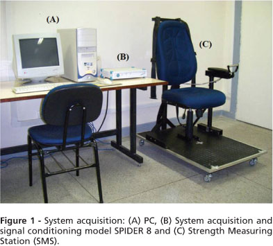
For the tests of muscle strength we idealized a Strength Measuring Station (SMS) from isometric muscle contractions in the vertical (flexion) and horizontal (rotation) directions with transducers, signal conditioning, data acquisition board and a computer. The definition of the Strength Measuring Station (SMS) model was based on the desired positioning for the testing of strength from isometric contraction of the muscles of the upper limbs (both sides). The position value should allow assessment of muscle strength with the subject in a sitting position with the limb near the trunk, with feet flat on the Strength Measuring Device (SMD) and the trunk stabilized in the vertical backrest of the chair.
The SMS is composed of a commercial type swivel chair, injected foam, fabric covering and no arm-rests. The SMS provided a structure which housed the sensing element and at the same time served as a support for the forearm during the strength tests. This structure was called SMD and allowed connections to two devices for placement of all wrist / hand and sensors. The basic dimensions of SMS such as size, height and width of the seat size of the pegs have been identified through anthropometric analysis considering a young adult male. To support the SMS a base was built.
The SMD is formed by a steel tube instrumented with 8 (eight) strain gauges, Kiowa KFG-3-120-c1-11 model, 2 (two) at the top and 2 (two) at the bottom of the tube to measure the efforts made in the vertical direction (flexion of the elbow and wrist). To measure the efforts made in the horizontal direction (internal and external rotation of the shoulder) two strain gages in each lateral portion of the steel tube were attached. The strain gages were attached at 200 mm from the point of application of force by the subject, according to the calculations performed earlier.
The SMD was dimensioned to support 500 N as a maximum load with (AE/V) max = 0, 002. The steel tube dimensions are as follows: external diameter d2 = 21,34 mm, internal diameter dj = 16, 11 mm and thickness t = 2, 74 mm.
Figure 1 shows the acquisition made by the device constructed, the system of acquisition, the signal conditioning and a computer.
Calibration Device
The electrical signals from the Wheatstone bridge circuit formed by the strain gauges were transmitted to a signal conditioning system, Spider 8 model (HBM, Darmstadt, Germany) and processed by software - Catman (version 3.1, release 3, 1997 - 2000).
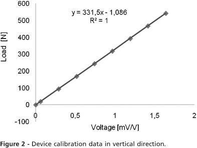
The calibration was performed statically through predefined loads applied to the SMD catcher (vertical and horizontal). The calibration values were entered into the Catman so that the test results were automatically converted into force (N). We applied the mass of 1.98 [kg], 9.59 [kg], 17.22 [kg], 24.85 [kg], 32.45 [kg], 40.07 [kg], 47.68 [kg] and 55.31 [kg] in the free end of the steel tube of SMD and they were measured on an accurate scale, corresponding to the forces of 19.42 [N], 94.08 [N], 168.93 [N], 243.78 [N], 318. 38 [N], 393.09 [N], 467.74 [N], 542.59 [N].
Figure 2 illustrates the result of efforts to vertical calibration, the calibration equation and R-squared value. It is observed good linearity between the electrical signal and applied loads.
Figure 3 shows the result of the horizontal forces' calibration, the equation of calibration and R-squared value. It is observed excellent linearity between the electrical signal and applied loads. Negative values indicate change in direction of effort (internal rotation and external rotation of the shoulder).
Statistical Analysis
We applied the Normality Test of Anderson-Darling in order to evaluate the distribution. In order to compare the means of the variables we initially used the term quartile and the Student's t- test. Differences were considered significant when the probability of a Type I error was less than 5% (p < 0.05).
RESULTS
We compared the maximum values obtained in muscle strength tests between the right upper limb (RUL) and the left upper limb (LUL). We used quartile as a measure of asymmetry in the first attempt to compare the values obtained from tests at the right upper limb with the data obtained from the muscle strain in the left upper limb.
Table 1 shows the data from the strength tests performed in the right and left upper limbs, the values of the first (Q1) and third quartiles (Q3).
Table 2 shows the average values of force (N) regarding flexion of the right upper limb (F RUL), flexion the left upper limb (F LUL), internal rotation of the right upper limb (IR RUL), internal rotation of the left upper limb (IR LUL), external rotation of the right upper limb (ER RUL), external rotation of the left upper limb (ER LUL) and the comparisons between RUL and LUL strength tests. It is noted that the average flexion strength in RUL was similar to the average flexion strength in the LUL. The average strength of internal rotation of the RUL was not different from the average strength of internal rotation of the LUL. In relation to the external rotation test, we did not observe significant differences between the external rotation strength of the RUL and the external rotation of the LUL.
DISCUSSION
In this study we aimed to evaluate and implement a new device from our laboratory which is able to measure the strength of upper limbs muscles and check the efficiency and adaptability of the device. As a main finding, we validated the device, since all subjects evaluated performed the test without difficulty and the device tested several possibilities for biomechanical analysis and we reported similarity between the forces generated by the right and left upper limbs. Examination of the force produced by the musculoskeletal system by using instrument is widely used by researchers from commercially available equipment such as computerized isokinetic dynamometers,14-16 hand dynamometers13,17 and dispositive manufactured by individual laboratories.18,19-22 However, it lacks information in the literature related to the functional connections of the shoulder and wrist.12
All the subjects in this study reported right dominance, i.e., they were right-handed and used preferentially the right upper limb (RUL) in daily activities. It is worth to note that the relationship between dominance and non-dominance is not well understood. Comparative studies involving muscle strength as well as considerations about the relationship of dominance and non-dominance has been studied by many researchers.23-25 Shklar and Dvir26 support our findings, since they found no difference between the dominant and the non-dominant sides regarding strength in flexion and internal rotation of shoulder.
The population was chosen in an attempt to homogenize the sample with regards to demographics, age, frequency and intensity of physical activity, diet and rest periods. Bohannon13 sought to establish reference values for muscle strength in upper limbs in an adult population aged between 20 and 79 years old from both genders. To measure the strength, the researcher used a manual dynamometer and obtained values of strength in elbow flexion of 285 [N] for the dominant side and 278.5 [N] for the non-dominant side. These strength values are from men aged 20 to 29 years old. When they examined the external rotation of the shoulder, the same author found values of 206.8 [N] for the dominant and 205 [N] for the non-dominant side. However, Bohannon13 did not evaluate internal rotator muscles strength.
Values of bending strength measured in our investigation are close to the values presented by MacKinnon24, which built a device for analysis of muscle strength in the upper limbs in different positions, using a load cell (Load Cell Service, Pretoria, South Africa). He evaluated eight subjects (5 men and 3 women) aged between 20 to 43 years old. Information such as occupational therapy and physical activity as well as the general state of health of the subjects were not informed. The average force (N) values found by him varied according to the position of the subject during testing. For instance, on the sitting position the averages for efforts in the sagittal plane (flexion) ranged between 75 and 204 [N]. When the subjects were tested on a standing position, the averages ranged from 99 to 241 [N]. There was no statistical difference between the dominant and the non-dominant limb with respect to contraction strength. It is also worth to note that the author made no mention of the bending articulation and considered the movement of the upper limb as a whole.
Other studies also investigated differences between dominant and non-dominat limbs. In the Poulis et al27 study statistical similarity between the dominant and non-dominant handles were observed when analyzing the peak flexor and extensor torque generated in that joint. Ertem et al25 investigated the relationship of dominance, body mass and age as factors in the process of functional evaluation of the hands. Regarding the evaluation of strength between the dominant and non-dominant limbs, the authors suggested that the dominant limb created a greater force than the non-dominant limb. Thus, according to those and our studies, we believe that there is no consensus among researchers about the relationship of dominance and that further studies are essential to better understand the phenomena involved in the generation of muscular strength.
Feasibility of muscle strength data acquisition during maximum voluntary contractions test was demonstrated by us in healthy human subjects. Our results are relevant, since it is necessary to obtain quantitative data in Biomechanics in the academic-scientific and clinical field. It is clear that there is a need for projects and developments of instruments which measure forces that interact with the locomotion system, as well as to develop low cost equipments availability for biomechanical analysis.
In conclusion, we have validated a new device that evaluates upper limbs muscle strength which was effective and we noted no differences between dominant and non-dominant upper limbs regarding force generation. This device is a tool that will help in the complex study of muscle strength and its intervening factors. Therefore, its availability is necessary to aids researchers and clinical professionals interested in this type of biomechanical analysis.
- 1. Souza PM, Jacob Filho W, Santarém JM, Silva AR, Li HY, Burattini MN. Progressive resistance training in elderly HIV-positive patients: does it work? Clinics. 2008;63:619-24, doi: 10.1590/S1807-59322008000500009.
- 2. de Amorim Aquino M, Leme LE. Isokinetic dynamometry in elderly women undergoing total knee arthroplasty: a comparative study. Clinics. 2006;61:215-22.
- 3. Carvalho NA, Bittar ST, Pinto FR, Ferreira M, Sitta RR. Manual for guided home exercises for osteoarthritis of the knee. Clinics. 2010;65:775- 80
- 4. Demura S, Miyaguchi K, Aoki H. The difference in output properties between dominant and nondominant limbs as measured by various muscle function tests. J Strength Cond Res. 2010; 24: 2816-20, doi: 10. 1519/JSC.0b013e3181e38293.
- 5. Rabin SI, Post M. A comparative study of clinical muscle testing and Cybex evaluation after shoulder operations. Clin Orthop Rel Res. 1990 258 147-156.
- 6. Hsu A, Tang P, Jan M. Test-retest reability of isokinetic muscle strength of the lower extremities in patients with stroke and traumatic brain injury. Arch Phys Med Rehabil. 2000;83:483-90.
- 7. Doeblin EO. Measurement Systems. Ed. McGraw-Hill, Kogakusha, 1975, USA, 307.
- 8. Standaert CJ, Herring SA. Expert opinion and controversies in musculoskeletal and sports medicine: stingers. Arch Phys Med Rehabil. 2009;90:402-6, doi: 10.1016/j.apmr.2008.09.569.
- 9. Nicholas JJ, Robinson LR, Logan A, Robertson R. Isokinetic testing in young nonathletic able-bodied subjects. Arch Phys Med Rehabil. 1989;70:210-3, doi: 10.1016/0003-9993(89)90033-6.
- 10. van Wilgen CP, Akkerman L, Wieringa J, Dijkstra PU. Muscle strength in patients with chronic pain. Clin Rehabil. 2003;17: 885-9, doi: 10.1191/ 0269215503cr693oa.
- 11. Jaric S. Muscle Strength Testing - Use of Normalization for Body Size. Sports Med. 2002;32:615-31, doi: 10.2165/00007256-200232100-00002.
- 12. Kaelin DL, Oh TH, Lim PA, Brander VA, Biundo JJ Jr. Rehabilitation of orthopedic and rheumatologic disorders. 4. Musculoskeletal disorders. Arch Phys Med Rehabil. 2000;81:S73-7.
- 13. Bohannon RW. Reference Values for Extremity Muscle Strength Obtained by Hand-Held dynamometry From Adults Aged 20 to 79 Years. Arch Phys Med Rehabil. 1997;78:26-32, doi: 10.1016/S0003-9993(97)90005-8.
- 14. Wilkin LD, Haddock BL. Isokinetic strength of collegiate baseball pitchers during a season. Journ Streng Condit Res. 2006; 20: 829-32.
- 15. Hughes RE, Johnson ME, O'Driscoll SW, An KN. Age-Related Changes in Normal Isometric Shoulder Strength. Am J Sports Med. 1999;27: 651-7.
- 16. Kasprisin JE, Grabiner MD. Joint angle-dependence of elbow flexor activation levels during isometric and isokinetic maximum voluntary contractions. Clin Biomec. 2000;15:743-9, doi: 10.1016/S0268-0033(00)00036-X.
- 17. Memberg WD, Murray WM, Ringleb SI, Kilgore KL, Snyder SA. A transducer to measure isometric elbow moments. Clin Biomech. 2001;16:918-20, doi: 10.1016/S0268-0033(01)00071-7.
- 18. Bohannon RW. Manual muscle testing: does it meet the standards of an adequate screening test? Clin Rehabil. 2005;19: 662-7, doi: 10.1191/ 0269215505cr873oa.
- 19. Ericson K, Werner H, Styf J, Hansson T. Unintentional forces developed during isometric test of the shoulder. Clinic Biomech. 2002;17:383-9, doi: 10.1016/S0268-0033(02)00027-X.
- 20. Madeleine P, Nie H, Arendt-Nielsen L. Dynamic shoulder dynamome-try: a way to develop delay onset muscle soreness in shoulder muscles. Journ Biomech. 2006;39:184-8, doi: 10.1016/j.jbiomech.2004.10.027.
- 21. de Groot JH, Rozendaal LA, Meskers CG, Arwert HJ. Isometric shoulder muscle activation patterns for 3-D planar forces: A methodology for musculo-skeletal model validation. Clinic Biomech. 2004;19:790-800, doi: 10.1016/j.clinbiomech.2004.05.013.
- 22. Garner BA, Shi J. Isometric shoulder girdle strength of healthy young adults. Clinic Biomech. 2008;23:30-7, doi: 10.1016/j.clinbiomech.2007.07. 018.
- 23. McGarvey SR, Morrey BF, Askew LJ, An KN. Reliability of isometric strength testing - temporal factors and strength variations. Clinic Orthop Rel Res. 1984;185:301-5.
- 24. MacKinnon SN. Isometric pull forces in the sagittal plane. Appl Ergon.1998;29:319-24, doi: 10.1016/S0003-6870(98)00003-9.
- 25. Ertem K. An investigation of hand dominance, average versus maximum grip strength, body mass index and ages as determinants for hand evaluation. Isokin Exerc Sc. 2005;13:223-7.
- 26. Shklar A, Dvir Z. lsokinetic strength relationships in shoulder muscles. Clinic Biomech. 1995;10:369-73, doi: 10.1016/0268-0033(95)00007-8.
- 27. Poulis S. Force-velocity relationship of the wrist flexors and extensors: The influence of small and large handgrips. Isokin Exerc Scien. 2003;11:101-8.
A new device to measure isometric strength in upper limbs: comparison between dominant and non-dominant limbs*
Publication Dates
-
Publication in this collection
31 Mar 2011 -
Date of issue
2011