Abstract
INTRODUCTION: Dysphagia is the important symptom in achalasia, and surgery is the most common treatment. The Heller-Pinotti technique is the method preferred by Brazilian surgeons. For many years, this technique was performed by laparotomy, and now the laparoscopic method has been introduced. The objective was to evaluate the immediate and long-term results of patients submitted to surgery by either laparotomy or laparoscopy. MATERIALS AND METHODS: A total of 67 patients submitted to surgery between 1994 and 2001 with at least 5 years of follow-up were evaluated retrospectively and divided into two groups: laparotomy (41 patients) and laparoscopy (26 patients). Chagas was the etiology in 76.12% of cases. Dysphagia was evaluated according to the classification defined by Saeed et al. RESULTS: There were no cases of conversion to open surgery. The mean duration of hospitalization was 3.32 days for laparotomy and 2.54 days for laparoscopy (p<0.05). An improvement in dysphagia occurred with both groups reporting good or excellent results (laparotomy: 73.17% and laparoscopy: 73.08%). Mean duration of follow-up was 8 years. CONCLUSIONS: There was no difference between the two groups with respect to relief from dysphagia, thereby confirming the safety and effectiveness of the Heller-Pinotti technique, which can be performed by laparotomy or laparoscopy, depending on the surgeon's experience.
Achalasia; Laparoscopy; Laparotomy; Cardiomyotomy; Dysphagia
CLINICAL SCIENCE
Results of the surgical treatment of non-advanced megaesophagus using Heller-Pinotti's surgery: Laparotomy vs. Laparoscopy
Luiz Roberto Lopes; Nathália da Silva Braga; Gustavo Carvalho de Oliveira; João de Souza Coelho Neto; Marcelo Amade Camargo; Nelson Adami Andreollo
Department of Surgery, University of Campinas, Campinas, São Paulo, Brazil. E-mail: lopeslr@fcm.unicamp.br Tel.: 55 19 35219450
ABSTRACT
INTRODUCTION: Dysphagia is the important symptom in achalasia, and surgery is the most common treatment. The Heller-Pinotti technique is the method preferred by Brazilian surgeons. For many years, this technique was performed by laparotomy, and now the laparoscopic method has been introduced. The objective was to evaluate the immediate and long-term results of patients submitted to surgery by either laparotomy or laparoscopy.
MATERIALS AND METHODS: A total of 67 patients submitted to surgery between 1994 and 2001 with at least 5 years of follow-up were evaluated retrospectively and divided into two groups: laparotomy (41 patients) and laparoscopy (26 patients). Chagas was the etiology in 76.12% of cases. Dysphagia was evaluated according to the classification defined by Saeed et al.
RESULTS: There were no cases of conversion to open surgery. The mean duration of hospitalization was 3.32 days for laparotomy and 2.54 days for laparoscopy (p<0.05). An improvement in dysphagia occurred with both groups reporting good or excellent results (laparotomy: 73.17% and laparoscopy: 73.08%). Mean duration of follow-up was 8 years.
CONCLUSIONS: There was no difference between the two groups with respect to relief from dysphagia, thereby confirming the safety and effectiveness of the Heller-Pinotti technique, which can be performed by laparotomy or laparoscopy, depending on the surgeon's experience.
Keywords: Achalasia; Laparoscopy; Laparotomy; Cardiomyotomy; Dysphagia.
INTRODUCTION
Achalasia, either chagasic or idiopathic, is a progressive disease for which there is no cure.1 It is characterized by the irreversible and progressive destruction of Auerbach's intramural plexuses, aperistalsis of the esophageal body and incomplete or absent relaxation of the lower esophageal sphincter (LES), leading to dietary stasis, dilatation and progressive elongation of the organ,2-6 interfering significantly with dietary intake and negatively affecting the patient's quality of life and nutritional status.
As more information became available on the physiopathology of this disease from the beginning of the twentieth century, more and more surgical procedures began to be developed with the objective of achieving a better and longer lasting response to the control of dysphagia.7,8 Currently, despite the numerous modifica tions made to the original technique described by Heller, the generic denomination of Heller myotomy is nevertheless still used in the literature to describe any one of its variations.9
As a result of the prevalence of this disease in Brazil, surgeons here have accumulated vast experience in the treatment of these patients.10Pinotti et al.11 proposed a myotomy and fundoplication with three layers of suture partially wrapping two-thirds of the esophagus in the posterior-anterior direction. According to the literature, results are excellent or good in over 90% of patients with respect to both the relief of dysphagia and the control of gastroesophageal reflux disease (GERD). Morbidity and mortality rates are low with this technique, and the results include an increase in weight and an improvement in the patient's quality of life.12-16
The advent of videolaparoscopic surgery in the final decade of the twentieth century introduced a new and broader field of possibilities in treatment and research, offering greater comfort to patients. The first esophagocardiomyotomy procedures were performed in 1991 by laparoscopy or in some cases by thoracoscopy.17-19 The immediate postoperative results of this new laparoscopic technique were similar to the results obtained with conven tional surgery, with the advantage that videolaparoscopic surgery was considered minimally invasive.20
The surgical treatment of non-advanced megaesophagus has been performed by laparotomy in the Teaching Hospital of the University of Campinas (UNICAMP) for the past 30 years using the surgical technique defined by Pinotti et al.11 Therefore, the objective of the present study was to compare the immediate and long-term results of the Heller-Pinotti surgical technique performed by laparotomy or by laparoscopy with respect to dysphagia, which is the principal symptom of this disease, in order to determine the optimal surgical treatment for these patients.
MATERIALS AND METHODS
Between 1989 and 2006, 390 patients with a diagnosis of megaesophagus of varying grades were treated surgically at the Digestive Disease Department and at the Teaching Hospital of the School of Medical Sciences, UNICAMP. Since 1994, laparoscopic surgery for the treatment of achalasia was introduced into the clinical practice of the hospital. The objective of this study was to compare patients who had been operated on from 1994 to 2001, by either laparotomy or videolaparoscopy for the treatment of non-advanced megaesophagus and who had at least 5 years of follow-up. A total of 131 patients with either chagasic or idiopathic megaesophagus were operated on during this period. The option of laparoscopic surgery to treat patients with non-advanced megaesophagus was not random, but also considered age, clinical condition, nutritional status and previous abdominal surgery.
The criteria for inclusion in the study therefore consisted of: patients with the non-advanced form of megaesophagus, i.e. grade I and II; patients with no other disease that could affect postoperative outcome; and patients for whom the data on their medical charts were complete or cases in which missing data were successfully obtained following an interview with the patient.
Sixty-four patients were excluded because they had been lost to follow-up, their charts were not found or were incomplete, they had megaesophagus grade III (5 patients) or grade IV (4 patients) or because they had died.
Therefore, the final sample consisted of 67 patients for whom data were complete and available for analysis. The following data were collected from medical charts and interviews: age, gender, epidemiological history of Chagas disease, serology for Chagas disease, symptoms present prior to surgery, preoperative diagnostic tests, type of surgery performed (Heller-Pinotti technique performed by laparotomy or laparoscopy), date of surgery, intraoperative complications, duration of hospital stay, postoperative symptoms, postoperative tests, postoperative dilatation and repeat surgeries (Table 2).
Description of surgical technique
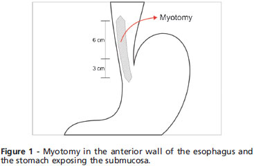
The surgical technique used was Heller myotomy to an extent of around 9 cm, approximately 6 cm of the esophagus and 3 cm of the stomach, after which a valve was fashioned in three posterior-anterior layers as described by Pinotti et al.,11 wrapping the exposed area of submucosa along its entire length. This procedure was performed using either median xifoumbilical laparotomy or videolaparoscopy (Figures 1-4).


Evaluation of dysphagia
The most important symptom analyzed was the grade of dysphagia, based on the classification defined by Saeed et al.21 as shown in Table 1.
Ethics
The study protocol was evaluated and approved by the ethics committee of the School of Medicine, UNICAMP, prior to initiation.
Statistical analysis
Data collected were analyzed using the Epi-Info software program, version 6.04, following prior construction of a database into which all the variables selected for analysis were entered. Frequency tables were constructed for the categorical variables containing values of absolute frequency (n) and percentages (%), while continuous variables were described using measurements of position and dispersion (means, standard deviations, minimum and maximum values and medians). For the categorical variables, the chi-square test or Fisher's exact test was used. In the case of numerical variables, the Mann-Whitney test was used. The Wilcoxon signed rank test for related samples was used for continuous variables, whereas McNemar's test was used to compare the categorical variables between different moments. A significance level of 5% was adopted throughout the statistical analysis (p<0.05).
RESULTS
Of the 67 patients studied, 37 were male (55.22%). Mean age was 42.46 years (SD 13.95; median 42; range 14 - 73 years). Thirty-four patients (50.74%) were between 40 and 59 years of age.
Sixty-one patients (91.04%) were found to have non-advanced megaesophagus grade II. Based on the classification of dysphagia as defined by Saeed et al.,21 50% of the patients were found to have dysphagia grade 1; 42.42% grade 2; and 7.58% grade 3.
The duration of follow-up ranged from 5 to 17 years, with a mean of 8 years. The mean duration of the symptoms of dysphagia was 3.97 years (range 1.13-9.07 years). Fifty-one patients (76.12%) had an epidemiological history of Chagas disease, confirmed or not by serology, whereas the remaining 16 patients (23.88%) were classified as having idiopathic achalasia. Twenty-nine of the patients with chagasic mega-esophagus (56.86%) had concomitant systemic diseases, Chagas cardiopathy in 13 and Chagas megacolon in 16 patients.
Other symptoms reported prior to surgery were weight loss (44.78%), regurgitation (41.79%), epigastric pain (40.3%), vomiting (28.36%), sensation of fullness (25.37%), pyrosis (20.9%) and odynophagia (13.43%). Less common symptoms consisted of eructation, choking and sialorrhea. Surgery was performed by laparotomy in 41 patients (61.19%) and by laparoscopy in 26 patients (38.81%). The most common intraoperative complication was esophageal mucosal perforation occurring during myotomy in 5 cases (7.46%), 4 during laparotomy (9.75%) and 1 during videolaparoscopy (3.84%) (p>0.05), treated by simple suturing followed by oral fasting for 72 h in all cases. Other minor complications occurred in 5 patients: three subcutaneous emphysema and two superficial lesions of the splenic capsule.
The overall mean duration of hospitalization for this sample was 3.01±1.26 days, median 3.00 days. In the laparotomy group, the mean time of hospitalization was 3.32 days, whereas in the videolaparoscopy group, the mean was 2.54 days. This difference was statistically significant (p<0.05).
Overall, a significant improvement in dysphagia was achieved following surgery (p<0.001 compared with presurgical values), with 27 patients (40.3%) having no further symptoms (grade 5). Of the 40 patients who reported some degree of dysphagia following surgery, 3% were classified as grade 1 and 23.88% as grade 2. In the laparotomy group, there was an improvement in dysphagia in 76.17% of cases, whereas in the videolaparoscopy group, improvement occurred in 76.08% of patients. The difference between the presence of symptoms prior to and following surgery was statistically significant in both groups.
With respect to the other symptoms reported by patients, a marked reduction was recorded in the occurrence of odynophagia (1.49%), regurgitation (7.46%), vomiting (2.99%), sensation of fullness (13.43%) and epigastric pain (17.91%). The only symptom that intensified following surgery was pyrosis, which was present in 26.87% of patients.
The dilatation of the cardia was performed in the early postoperative period in 17 patients (25.37%), 7 (17.07%) in the laparotomy group and 10 (38.46%) in the videolaparoscopy group. This difference between the two groups was statistically significant (p<0.05). Surgery had to be repeated in 5 patients (7.46%), 3 in the laparotomy group (7.31%) and 2 in the videolaparoscopy group (7.69%) (p>0.05). Tables 2 and 3 summarize these data.
Figure 5 shows the grade of dysphagia, according to the classification of Saeed et al.21 , before and after surgery performed by laparotomy or laparoscopy using the Heller - Pinotti technique. No statistically significant differences were found between the two groups with respect to the improvement obtained following surgery or the duration of this effect.
DISCUSSION
Chagasic megaesophagus corresponds to at least 80% of cases submitted to surgical treatment in Brazil,16,22,23 unlike countries in the developed world where cases consist almost entirely of idiopathic achalasia24 and affect 1 in every 100,000 individuals.25
The myotomy, as described by Heller, was first performed by laparotomy in 191326 as an alternative to the other therapeutic techniques used up to that time. Despite the numerous modifications that have been introduced to Heller's original technique, the generic denomination of Heller myotomy is still used to describe any of its variations.9 In 1991, myotomy began to be performed by videolaparoscopy and, since then, many surgeons have adopted this option.24
The demographic data of the patients enrolled in the present study (gender, age and the etiology of the disease (chagasic or idiopathic)) are similar to those of patients in other studies reported in the literature.16,23
The majority of Brazilian surgeons prefer conservative surgery (Heller myotomy) for the treatment of achalasia in cases of non-advanced megaesophagus and esophagectomy for advanced cases, whereas dilatation of the cardia is reserved for special circumstances.10
The surgical treatment of non-advanced megaesophagus consists of the myotomy of around 6-7 cm of the abdominal esophagus and of the cardia, around 2-3 cm into the stomach, via laparotomy. This technique has been used since the 1960s; however, in the 1970s, it was associated with a valve fashioned in three posterior-anterior layers as described by Pinotti et al.11 This technique is known in Brazil as Heller-Pinotti surgery.
The introduction of video-assisted surgical techniques permitted myotomy and fundoplication to be performed by laparoscopy. Indeed, Abir et al.27 conducted an extensive literature review and concluded that the majority of authors recommend surgery as the treatment of choice and that the current preference is for videolaparoscopy.
Intraoperative complication rates are low with both types of surgery, mucosal perforation being found in 15% of cases, bleeding in 0.5%, visceral perforation in 0.5%, pneumothorax in 1% and conversion to open surgery in 2-6% of cases.14,28-32 These complications do not, however, alter the final outcome of surgery.
The authors have reported mucosal perforation in 4.1% of surgeries performed by laparotomy11 and in 3.5% of surgeries carried out by videolaparoscopy.24 No cases have been reported of any lesion to the spleen or liver requiring major intervention.
In the present study, intraoperative complication rates were low. Mucosal perforation was the most common complication, occurring in 3.84% of surgeries carried out by videolaparoscopy and in 9.75% of surgeries performed by laparotomy. In all cases, lesions were sutured primarily and no complications occurred.
There were no cases of conversion to open surgery; however, rates reported in the literature range from 1.5% to 22% of laparoscopies.33-35 Rossetti et al.33 reported a conversation rate to open surgery of 1.5% and a morbidity rate of 2.1%. Deb et al.31 reported a rate of conversion to open surgery of 2% and mucosal perforation in 15.16% of cases, suggesting that there is a learning curve and that the rate of intraoperative complications will consequently decrease as the surgeon gains experience.36
In the early postoperative period, 17 patients required dilatation of the cardia. Savary-Gilliard dilators were used because they offer greater security. The reason for larger number of patients undergoing laparoscopic surgery require dilation can be explained by a learning curve early in the series. The size of myotomy was similar in both groups, as well as the technique of fundoplication, covering its entire length. A short myotomy is not recommended.
The Heller-Pinotti surgical technique is the method of choice for cases of non-advanced megaesophagus. For this reason, the present study compared the results of surgery carried out by laparotomy with those obtained following videolaparoscopy for the relief of dysphagia with the objective of evaluating any difference in long-term results between the two different surgical techniques, as described by Katilius and Velanovich.37
Patients included in this study had been followed up for a minimum of 5 years following surgery, a time considered sufficient to evaluate improvements in dysphagia in patients with megaesophagus. Costantini et al.38 reported that the recurrence of symptoms of dysphagia occurs during the first year following surgery in over 50% of patients.
The present study shows that, at follow-up a mean of 8 years after surgery, the improvement in dysphagia persisted in 73.1% of patients and that 40.3% reported no difficulty at all in swallowing. Comparing the improvement obtained in dysphagia between the groups, it was very evident that the outcome after a period of at least 5 years of follow-up was similar in both. In the group submitted to videolaparoscopy, the incidence of dysphagia grade 1 or 2 prior to surgery was 92.31% compared with 26.92% following surgery. In the group of patients submitted to laparotomy, these rates were 94.5% and 26.83% respectively. There was a statistically significant difference between the pre-and postoperative grades of dysphagia within each group (p<0.05); however, there was no statistically significant difference between the two groups, showing that the approach used in the surgical treatment of this symptom does not affect the outcome even after a mean follow-up time of 8 years.
Youssef et al.39 evaluated 110 patients submitted to Heller myotomy by videolaparoscopy, who answered an objective questionnaire on dysphagia as a symptom, the psychologi cal aspects of this disease and their general health status. A marked improvement compared with presurgical condi tions was found for all the items evaluated. These authors concluded that surgery greatly improved the symptom of dysphagia and the patient's quality of life and resulted in greater patient satisfaction.
The mean time of hospitalization in the present study was 2.54 days in the group of patients operated on by videolaparoscopy and 3.32 days in the case of open surgery, with this difference being statistically significant (p<0.05). A shorter hospital stay is beneficial in the surgical treatment of mega-esophagus and was not found to affect the final outcome, which was an improvement in the symptom of dysphagia. Other studies have reported a mean duration of hospitalization ranging from 5 to 12 days for surgery by laparoto my 24,30 and 2 to 7 days for videolaparoscopy.20,30-33,40
According to Herbella et al.,16 the rate of repeat surgery as a result of failure of the original operation was 8.4% in cases of laparotomy, but only 2.4% in the case of intense symptoms of dysphagia postoperatively after a follow-up of over 40 months. Bonatti et al.41 reported a rate of repeat surgery following videolaparoscopy of 2.5% in 75 patients followed up for a mean of 5 years.
Andreollo and Earlam42 reported a rate of repeat surgery of 2.8% in an analysis of over 5,000 cases reported in the literature on Heller myotomy without fundoplication, performed by either abdominal or open chest surgery.
The percentages of repeat surgery reported in the present study (7.31% with open surgery vs. 7.69% with videolaparoscopy) are in agreement with those reported in the literature, although rates vary greatly in different sample populations.
Finally, this is a disease in which the functional alteration is permanent and progressive. Surgical treatment indicated in non-advanced cases for the relief of achalasia does not correct the functional disorder, and recurrence of the principal symptom may occur over the long term.
CONCLUSIONS
The results show that the Heller-Pinotti technique is effective and safe for both methods and has a low rate of intraoperative and postoperative complications. The period of hospitalization was significantly lower in patients operated on by laparoscopic surgery. Relief of dysphagia was excellent or good in 75% of the two groups with a mean follow-up of 8 years. The rate of reoperation was 7% in both groups.
The authors conclude that the two methods were similar in achieving the final result of the relief of dysphagia, and either approach can be chosen, depending on the surgeon's experience.
ACKNOWLEDGMENTS
The statistical analysis was performed by the statistical team of UNICAMP's Research Department.
Received for publication on July 13, 2010; First review completed on September 2, 2010; Accepted for publication on October 4, 2010
- 1. Diamantis T, Pikoulis E, Felekouras E, et al. Laparoscopic esophagomyotomy for achalasia without a complementary antireflux procedure. J Laparoendosc Adv Surg Tech A. 2006;16:345-9., doi: 10.1089/lap.2006.16.345
- 2. Koberle F. Patogenia do megaesôfago brasileiro e europeu. Tese de Livre Docência. Ribeirão Preto, SP: Universidade de São Paulo, 1962.
- 3. Pinotti HW, Betarello A. Megaesophagus: general aspects and surgical treatment. Dis Esophagus. 1990;3:21-6.
- 4. Goldblum JR, White RI, Orringer MB, Appelman HD. Achalasia. A morphologic study of forty-two resected specimens. Am J Surg Pathol. 1994;18:327-37, doi: 10.1097/00000478-199404000-00001.
- 5. de Oliveira RB, Rezende Filho J, Dantas RO, Iazigi N. The spectrum of esophageal motor disorders in Chagas' disease. Am J Gastroenterol. 1995;90:1119-24.
- 6. Mason RJ, Bremner CG. Esophageal length in achalasia. Dis Esophagus. 1995;8:119-24.
- 7. Andreollo NA, Brandalise NA, Leonardi LS. Megaesôfago incipiente: dilatação ou cirurgia? Rev Assoc Med Bras. 1984;30:4-6.
- 8. Felix VN, Cecconello I, Zilberstein B, Moraes-Filho JP, Pinotti HW, Carvalho E. Achalasia: a prospective study comparing the results of dilatation and myotomy. Hepatogastroenterology. 1998;45:97-108.
- 9. Rezende JM. Caminhos da Medicina. História da cirurgia da acalasia do esôfago e do megaesôfago chagásico. Available at: htpp://usuarios.cultura.com.br/jmrezende/CIRACALÁSIA.htm Accessed on Mar 20, 2010.
- 10. Herbella FA, Aquino JL, Stefani-Nakano S, et al. Treatment of achalasia: lessons learned with Chagas' diseases. Dis Esophagus. 2008;21:461-7, doi: 10.1111/j.1442-2050.2008.00811.x.
- 11. Pinotti HW, Gama-Rodrigues JJ, Ellenbogen G, Raia A. Nova técnica no tratamento cirúrgico do megaesôfago. Esofagocardiomiotomia associada com esofagofundogastropexia. Rev Goiana Med. 1974;20:1-6.
- 12. Cecconello I, Sallum RA, Rocha JR, Zilberstein B, Pinotti HW. Tratamento cirúrgico do megaesôfago. In: De Paula AL, editor. Cirurgia Videolaparoscópica. Goiânia: Hospital Samaritano; 1994. p. 123.
- 13. Sallum RA, Rocha JR, Cecconello I, et al. Cardiomiectomia com fundoplicatura no tratamento do megaesôfago. Seguimento tardio. ABCD, Arq. Bras. Cir. Dig. 1994;9:35-41.
- 14. Del Grande JC, Herbella FA, Lourenço LG, Mansur N, Salomão H, Chibly M. Resultados imediatos da cardiomiotomia com fundoplicatura no tratamento do megaesôfago: análise de 104 casos. GED Gastroenterol Endosc Dig. 1996;15:156-60.
- 15. Pinotti HW, Sakai P, Ishioka S. Cardiomyotomy and fundoplication for esophageal achalasia. Jpn J Surg. 1983;13:399-403, doi: 10.1007/ BF02469725.
- 16. Herbella FA, Del Grande JC, Lourenço LG, Mansur NS, Haddad CM. Late results of Heller operation and fundoplication for the treatment of the megaesophagus - analysis of 83 cases. Rev Assoc Med Bras. 1999;45:317-22, doi: 10.1590/S0104-42301999000400006.
- 17. Pinotti HW. First development of cardiomyotomy by videolaparoscopy: a new perspective in the achalasia treatment. ISDE News. 1991;10:8.
- 18. Shimi S, Nathanson LK, Cuschieri A. Laparoscopic cardiomyotomy for achalasia. J R Coll Surg Edinb. 1991;36:152-4.
- 19. Pellegrini C, Wetter LA, Patti M, Leichter R, Mussan G, Mori T, et al. Thoracoscopic esophagomyotomy. Initial experience with a new approach for the treatment of achalasia. Ann Surg. 1992;216:291-6, doi: 10.1097/00000658-199209000-00008.
- 20. Vara-Thorbeck C, Herrainz R. Esophageal achalasia: laparoscopic Heller cardiomyotomy. Int Surg. 1995;80:376-9.
- 21. Saeed ZA, Winchester CB, Ferro PS, Michaletz PA, Schwartz JT, Graham DY. Prospective randomized comparison of polyvinyl bougies and thorough-the-scope balloons for dilation of peptic strictures of the esophagus. Gastrointest Endosc. 1995;41:189-95, doi: 10.1016/S0016 5107(95)70336-5.
- 22. Rezende JM, Moreira H. Megaesôfago e megacolon chagásicos. Revisão histórica e conceitos atuais. Arq Gastroenterol. 1988;25:32-43.
- 23. Oliveira GC, Lopes LR, Andreollo NA, Coelho Neto JS. Surgically treated megaesophagus: epidemiological profile of patients operated in the Clinical Hospital of the State University of Campinas between 1989 and 2005. Rev Soc Bras Med Trop. 2008;41:183-8.
- 24. Desai KM, Soper NJ. Laparoscopic management of idiopathic esophageal achalasia. Rev Gastroenterol Mex. 2004;69:7-13.
- 25. Cacchione RN, Tran DN, Rhoden DH. Laparoscopic Heller myotomy for achalasia. Am J Surg. 2005;190:191-5, doi: 10.1016/j.amjsurg.2005.05.010.
- 26. Heller E. (1913) Apud Heller E. Extramukose Kardioplastik beim chronischem Kardiospasmus mit dilatation des Oesophagus. Mitt Grenzgeb Med Chir. 1914;27:141-3.
- 27. Abir F, Modlin I, Kidd M, Bell R. Surgical treatment of achalasia: current status and controversies. Dig Surg. 2004;21:165-76, doi: 10.1159/000079341.
- 28. Pinotti HW, Cecconello I, Zilberstein B. Megaesôfago. In: Pinotti HW, editor. Tratado de Clínica Cirúrgica do Aparelho Digestivo. São Paulo: Atheneu; 1994. p. 316.
- 29. Sharp KW, Khaitan L, Scholz S, Holzman MD, Richards WO. 100 consecutive minimally invasive Heller myotomies: lessons learned. Ann Surg. 2002;235:631-8, doi: 10.1097/00000658-200205000-00004.
- 30. Douard R, Gaudric M, Chaussade S, Couturier D, Houssin D, Dousset B. Functional results after laparoscopic Heller myotomy for achalasia: A comparative study to open surgery. Surgery. 2004;136:16-24, doi: 10.1016/j.surg.2004.01.011.
- 31. Deb S, Deschamps C, Allen MS, Nichols FC 3rd, Cassivi SD, Crownhart BS, et al. Laparoscopic esophageal myotomy for achalasia: factors affecting functional results. Ann Thorac Surg. 2005;80:1191-4, doi: 10.1016/j.athoracsur.2005.04.008.
- 32. Domene CE, Santo MA, Onari P, Volpe P, Pinotti HW. Cardiomiectomia com fundoplicatura parcial videolaparoscópica no tratamento do megaesôfago não avançado: estudo de 50 casos. Rev Col Bras Cirurg. 1998;35:229-34.
- 33. Rossetti G, Brusciano L, Maffettone V, Napolitano V, Russo G, Amato G, et al. A total fundoplication is not an obstacle to esophageal emptying after Heller myotomy for achalasia: results of a long-term follow up. Ann Surg. 2005;241:614-21, doi: 10.1097/01.sla.0000157271.69192.96.
- 34. Bessell JR, Lally CJ, Schloithe A, Jamieson GG, Devitt PG, Watson DI. Laparoscopic cardiomyotomy for achalasia: long-term outcomes. Aust NZ J Surg. 2006;76:558-62, doi: 10.1111/j.1445-2197.2006.03784.x.
- 35. Dang Y, Mercer D. Treatment of esophageal achalasia with Heller myotomy: retrospective evaluation of patient satisfaction and disease-specific quality of life. Can J Surg. 2006;49:267-71.
- 36. Bloomston M, Serafini F, Boyce HW, Rosemurgy AS. The ''learning curve'' in videoscopic Heller myotomy. JSLS. 2002;6:41-7.
- 37. Katilius M, Velanovich V. Heller myotomy for achalasia: quality of life comparison of laparoscopic and open approaches. JSLS. 2001;5:227-31.
- 38. Costantini M, Zaninotto G, Guirroli E, Rizzetto C, Portale G, Ruol A, et al. The laparoscopic Heller-Dor operation remains an effective treatment for esophageal achalasia at a minimum 6-year follow-up. Surg Endosc. 2005;19:345-51, doi: 10.1007/s00464-004-8941-7.
- 39. Youssef Y, Richards WO, Sharp K, Holzman M, Sekhar N, Kaiser J, et al. Relief of dysphagia after laparoscopic Heller myotomy improves long-term quality of life. J Gastrointest Surg. 2007;11:309-13, doi: 10.1007/s11605-006-0050-6.
- 40. Richter JE. Modern management of achalasia. Curr Treat Options Gastroenterol. 2005;8:275-83, doi: 10.1007/s11938-005-0020-1.
- 41. Bonatti H, Hinder RA, Klocker J, Neuhauser B, Klaus A, Achem SR, et al. Long-term results of laparoscopic Heller myotomy with partial fundoplication for the treatment of achalasia. Am J Surg. 2005;190 874-8, doi: 10.1016/j.amjsurg.2005.08.012.
- 42. Andreollo NA, Earlam RJ. Heller's myotomy for achalasia: is an added antireflux procedure necessary? Br J Surg. 1987;74:765-9, doi: 10.1002/bjs.1800740903.
Publication Dates
-
Publication in this collection
14 Mar 2011 -
Date of issue
2011
History
-
Received
13 July 2010 -
Accepted
04 Oct 2010 -
Reviewed
02 Sept 2010