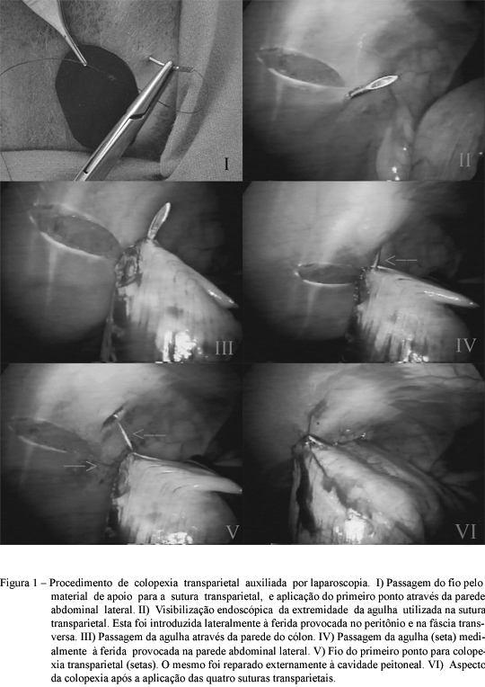
Two groups of dogs, GA (n=8) and GL (n=16) were used to compare the conventional and the transparietal laparsocopic assisted technique for colopexy and to compare with the conventional surgery. In the GA group, the colopexy was proceded by incisional technique trough celiotomy. In the GL group, the colopexy was performed using the transparietal laparoscopic assisted technique. The GL dogs were separated in four subgroups (S1, S2, S3 and S4) with four dogs each. In each subgroup a different stent was used, capton of infusion tube (S1); plastic disks made with NaCl solutions bottles (S2); disks made with rubber (S3) and silicone disks (S4). The time for complete surgery was statically higher in the GL group than GA. Seven GA dogs maintained the colopexy and all of these presented adherences of the omentum in the suture zone. In three GL animals, all of the S4 subgroup the colopexies were not maintained. In all dogs of this subgroup dermatitis and/or cellulites were observed. Best results were obtained in the S3 subgroup dogs. The main histological observations in the 14d-after surgery biopsies in GL animals were related to a higher connective tissue deposition at the colon adherence, and infiltration of this type of tissue into the associated musculature. In the 28d biopsies, no difference was found between groups. In both groups, the collagen fibers presented mature aspect. Concluding, the transparietal laparoscopic assisted technique is viable, however it’s associated with tissue lesions in the regions in contact with the material used to support the suture. The rubber disk presented better results.
endoscopic surgery; laparoscopic assisted surgery; canine


