 Characterization of canine bone marrow mononuclear cells
ABSTRACT:
Bone marrow mononuclear cells (BM-MNC) have been used to treat various diseases. These cells are chemo-attracted to sites of injury and may produce cytokines and trophic factors that act in tissue regeneration. Nevertheless, a challenge associated with this procedure is the lack of studies on the characterization of canine BM-MNC. This study contributed with BM-MNC characterization data. Bone marrow mononuclear cells from four donors were labeled with anti-CD8a, CD9, CD14, CD29, CD34, CD44, CD45, and CD90 for phenotypic characterization. In this study, was obtained strong expression of CD9 (79.48% ± 7.71%), CD29 (96.10% ± 1.71%), CD44 (89.60% ± 3.14%) and CD45 (94.33% ± 4.09%). Therefore, these markers should be considered in the definition of panel for characterization of canine BM-MNC.
Key words:
cells characterization; phenotypic characterization; canine CD markers
RESUMO:
Células mononucleares da medula óssea (CMN-MO) têm sido utilizadas para tratar diversas doenças. Essas células são atraídas quimicamente para locais de lesão e podem produzir citocinas e fatores tróficos que atuam na regeneração tecidual. No entanto, um desafio associado a esse procedimento é a falta de estudos sobre a caracterização de CMN-MO caninas. Nesse sentido, o objetivo deste estudo foi contribuir com dados de caracterização de CMN-MO. Para isso, as células mononucleares da medula óssea de quatro doadores foram marcadas com anticorpos anti-CD8a, CD9, CD14, CD29, CD34, CD44, CD45 e CD90 para caracterização fenotípica. Os resultados obtidos foram uma forte expressão de CD9 (79,48% ±7,71%), CD29 (96,10% ±1,71%), CD44 (89,60% ±3,14%) e CD45 (94,33% ±4,09%). Portanto, esses marcadores devem ser considerados na definição de painel para a caracterização de CMN-MO canina.
Palavras-chave:
caracterização celular; caracterização fenotípica; marcadores CD-canina
INTRODUCTION
Bone marrow mononuclear cells (BM-MNC) contain a subset of hematopoietic progenitor cells and several cells of non-hematopoietic lineage (KAMIYA et al., 2008; GIRALDI-GUIMARÃES et al., 2009). Several clinical studies have focused on the use of the entire BM-MNC fraction, assuming that functional effects depend on a correct balance among multiple cell types and stem cell precursors (MATHIEU et al., 2009). These cells may be easily obtained by a low-cost method and isolated in a short time just before transplantation, minimizing the risks of contamination. Besides, the isolation of BM-MNC does not require a laboratory with a more complex structure, which makes it a more accessible procedure in clinical trials with animals (KAMIYA et al., 2008; GIRALDI-GUIMARÃES et al., 2009; KAMIYA et al., 2014; TAMURA & MAETA, 2020).
Despite the fact that BM-MNC have been used as regenerative strategies for tissue injuries, there are many challenges associated with this therapeutic approach. One of them is the characterization of canine BM-MNC that is poorly defined compared to human cells. The limited knowledge of phenotype of the canine BM-MNC is partly due to the lack of species-specific monoclonal antibodies for many of the cellular markers used for the characterization of human cells (SCREVEN et al., 2014; TAMURA & MAETA, 2020). Conversely, some canine monoclonal antibodies are commercially available and some cross-reacting antibodies have been compared to those of the target species (COBBOLD & METCALFE, 1994; ALLDINGER et al., 2000; SCHUBERTH et al., 2007; TAMURA & MAETA, 2020), but there are no studies in the literature on the characterization of BM-MNC. Therefore, the use of specific or validated antibodies for canine cells characterization may contribute to the definition of a panel of cellular markers to identify canine BM-MNC.
MATERIALS AND METHODS
Bone marrow harvesting and BM-MNC isolation
Four healthy mixed-breed dogs, two males and two females, weighing between 12 and 20 kg, aged between 18 and 36 months were used as bone marrow donors. The routine pretreatment evaluations included a complete medical history, physical examination and hematology and biochemical profiles (serum proteinogram, urea, creatinine, alkaline phosphatase and alanine aminotransferase) to assess the health status of donors.
Animals received general anesthesia with propofol (Propovan® Cristalia) and meperidine (Dolosal® Cristalia) for bone marrow aspiration, which was performed by puncturing the iliac crest with disposable hypodermal needles (16 G) and disposable 10 mL syringes containing 1.0 mL heparin (5,000 IU/mL) anticoagulant solution. All bone marrow was diluted in Dulbecco’s modified Eagle medium (DMEM, Gibco) at a ratio 1:3. Bone marrow was carefully loaded onto Ficoll-Hypaque (Histopaque, Sigma Chemical) density gradient (density 1,077 g/cm3) to isolate BM-MNC. Mononuclear cells were isolated by centrifugation at 400g for 30 minutes, at room temperature (BÖYUM, 1968). After isolation, the BM-MNC were washed twice with DMEM to remove excess Ficoll-Hypaque and finally resuspended in DMEM. Cell counting and cell viability were determined using Trypan blue (Sigma Chemical) exclusion test in a Neubauer chamber (STROBER, 1997).
Phenotypic characterization of BM-MNC
For phenotypic characterization, the samples of BM-MNC were incubated with commercial monoclonal antibodies to analyze canine cell-surface expression of typical marker proteins: anti-CD8a (PerCP-conjugated, clone YCATE55.9; eBioscience), anti-CD9 (RPE-conjugated, clone MM2/57; AbD Serotec), anti-CD14 (APC-conjugated, clone M5E2; BD Pharmingen), anti-CD29 (PE-conjugated, clone HL1255; ABCAM), anti-CD34 (PE-conjugated, clone 1H6; eBioscience), anti-CD44 (Alexa fluor 488-conjugated, clone YKIX337.8.7; ABD Serotec), anti-CD45 (FITC-conjugated, clone YKIX716.13; eBioscience), anti-CD90 (PE-conjugated, clone 5E10; BD Pharmigen). For labeling, aliquots at 1 x 10⁶ mononuclear cells were resuspended in 1.0 mL of PBS. Samples were centrifuged at 400 g for 5 minutes and then incubated in the dark for 30 min with fluorochrome-conjugated specific antibodies, according to the manufacturers’ instructions. Apoptosis and cell viability were assessed using Annexin (PE conjugated; BD Pharmigen) and 7-amino-actinomycin D (7-AAD; BD Pharmigen). The cells were washed with PBS and resuspended in 500 µL of a solution containing 1% formaldehyde. Isotypic IgG1 antibodies were used as controls. Approximately 1 × 10⁵ cells were acquired on a BD FACSCalibur Flow Cytometer (BD Bioscience, San Jose, USA) and were analyzed using FlowJo v8.0.2 software (Tree Star, Ashland, USA).
RESULTS AND DISCUSSION
The bone marrow harvest volume varied from 18 to 36 mL. The average number of BM-MNC cells obtained per mL was 1.14 x 10⁷ (?0.32 x 10⁷), and the cells viability was estimated by trypan blue as 91.7% (± 5.6%).
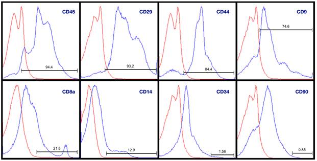
BM-MNC cell-surface antigen expression was evaluated by flow cytometry in four samples (Figure 1). The average cell viability by 7AAD staining and flow cytometry was 83.36% (±5.92%). The results were expressed as the intensity of expression of these markers in cells. With few exceptions, all four sources displayed similar immunophenotypes for the markers analyzed (Table 1).
Immune phenotype by flow cytometry. The bone marrow mononuclear cells were labeled with antibodies against the indicated antigens, and analyzed by flow cytometry. Representative histograms of sample 4 are displayed. On the y-axis is the % of Max (the cell count in each bin divided by the cell count in the bin that contains the largest number of cells) and the x-axis is the fluorescence intensity in a log (100 - 104) scale. Isotype control is shown as a thick red line histogram.
Expression of surface proteins of mononuclear cells derived from four samples of canine bone marrow analyzed by flow cytometry. Cell viability and apoptosis were assessed using 7-amino-actinomycin D (7-AAD) and Annexin.
The number of BM-MNC isolated from canine bone marrow in this study (1.14 x 10⁷ BM-MNC/mL ± 0.32 x 10⁷) was consistent with those reported by other authors (KAMISHINA et al., 2008; SPENCER et al., 2012; TAMURA & MAETA, 2020). It was also evaluated the expression of some cell surface molecules, some of which are commonly used for mononuclear cells and mesenchymal stem cells characterization as CD8a, CD9, CD29, CD44 and CD90 (JUNG et al., 2009; TAKEMITSU et al., 2012; KANG & PARK, 2014; QUINTANILHA et al., 2014; SCREVEN et al., 2014; HUMENIK et al., 2019; TAMURA & MAETA, 2020; KREŠIĆ et al., 2021) as well as CD14, CD34 and CD45, which are negative markers for mesenchymal cells, but that are used for characterization of mononuclear cells, including progenithor endothelial cells (HENRICH et al., 2015; TAMURA & MAETA, 2020; KREŠIĆ et al., 2021).
The results of the expression of CD9, CD29, CD34 and CD44 were consistent with the results obtained by other authors (ALVES et al., 2014; JUNG et al., 2009; SCREVEN et al., 2014) for characterization of mesenchymal stem cells harvested from dogs. However, compared to the values found by TAMURA & MAETA (2020) for the canine-BM-MNC characterization, we found similar labeling values for CD14, but lower values for CD34.
In relation to markers with strongly positive result, it was observed that the expression of CD29 ranged between 93.20 and 97.40% in this study, consistent with the results obtained by TAKEMITSU et al. (2012), ALVES et al. (2014) and HUMENIK et al. (2019) that showed high expression of CD29 (96.00% ± 3.00%, 98.90% ± 0.80% and 98.41% ± 0.53% respectivelly) in canine bone marrow MSC, while KREŠIĆ et al. (2021) showed expression of CD29 in more than 95% of canine adipose-derived MSC in culture. Nonetheless, SCREVEN et al. (2014) showed that CD29+ cells ranged between 2.06 and 2.96% also in canine bone marrow MSC, while TAMURA & MAETA (2020) obtained expression of CD 29 ranged between 10.1-16.6% in canine-BM-MNC.
The expression of CD45 obtained in this study ranged from 87.70 to 98.50%. CD45 is a pan leukocyte marker, expressed on all hematopoietic cells and is also in endothelial progenitor cells and hematopoietic stem cells, but is not expressed in platelets and MSC-precursor; therefore, this marker is used as a negative marker for MSC (BRODERSEN et al., 1998; HENRICH et al., 2015; KREŠIĆ et al., 2021). TAKEMITSU et al. (2012), ALVES et al. (2014) and HUMENIK et al. (2019) obtained expression of CD45 in 1.45% (± 0.60%), 2.00% (± 1.20%) and 0.24% (± 0.07%) of canine bone marrow MSC, respectively. Although, HENRICH et al. (2015) have used CD45 expression to characterize human BM-MNC, these authors showed only concomitant expression of this marker with CD34 or with CD34 and CD133.
The result of CD9 expression obtained in this study (69.60 to 88.80%) is consistent with those reported by JUNG et al. (2009), who used this marker to characterize canine bone marrow MSC. CD9 is expressed in young B cells, platelets, eosinophils, basophils and activated T lymphocytes (BOUCHEIX et al., 1991; ANTON et al., 1995), but is not expressed by hematopoietic progenitors or non-activated lymphocytes (BOUCHEIX et al., 1991).
In this study, a strong expression of CD44 (84.40 to 92.70%) was also observed in canine BM-MNC. This result is consistent with those reported by TAKEMITSU et al. (2012) that showed expression of CD44 in 98.90% (0.25%) of canine bone marrow MSC. However, SCREVEN et al. (2014) obtained CD44+ cells that ranged between 19.90 and 34.20% in canine bone marrow MSC. CD44 is a multifunctional cell adhesion molecule expressed on the surface endothelial cells, epithelial cells, fibroblasts, keratinocytes, leukocytes, MSC and others cells with transendothelial migratory capacity. CD44 antigen functions include cell-cell and cell-substrate adhesion (LEWINSOHN et al., 1990; KREŠIĆ et al., 2021).
In this study, a moderate expression of CD8a (16.90 to 21.50%) and CD14 (12.90 to 38.90%) was also observed. CD8a is a surface glycoprotein expressed in NK cells and thymocytes that facilitates specific antigen recognition. In addition, CD8a antibody binds to major histocompatibility complex class I (MHC-I) and plays a role in mature T cell activation (MOORE et al., 1992; LIN et al., 2010). SCREVEN et al. (2014) showed great variation on expression of MHC-I (4.07 to 12.80%) in canine bone marrow-derived MSC.
In relation to markers with weak positive result, it was observed that the expression of CD34 ranged from 0.50 to 1.58%. The result of CD34 expression is consistent with the result obtained by HENRICH et al. (2015) that showed expression of this marker around 1-2% in human BM-MNC. SCREVEN et al. (2014) obtained expression of CD34 between 1.11 and 2.65% in canine bone marrow-derived MSC. However, TAMURA & MAETA (2020) showed expression of CD34 ranged from 2.6 to 9.1% in phenotypic characterization of canine-BM-MNC. CD34 is expressed in hematopoietic stem cells and endothelial progenitor cells, and is considered as a negative marker for MSC (BARA et al., 2015; HENRICH et al., 2015).
The result of CD90 expression obtained in this study (0.40-1.83%) was significantly lower than those obtained by ALVES et al. (2014) that showed high expression of CD90 (80.04% 1.96) and SCREVEN et al. (2014), that showed CD90+ cells ranging from 17.1 to 27.8%, both in canine bone marrow-derived MSC. Our result to CD90 was also significantly lower than those obtained by TAMURA & MAETA (2020) that showed labeling values for CD90 ranged from 19.6 to 39.3 in canine-BM-MNC. This marker is expressed in MSC, T lymphocytes and monocytes, with weak expression in granulocytes (COBBOLD & METCALFE, 1994; SCREVEN et al., 2014). HENRICH et al. (2015) have not used this marker to characterize human BM-MNC. Therefore, the results of this study are consistent with those reported by HENRICH et al. (2015) that BM-MNC are a heterogenous mixture of diverse cell types. Nevertheless, these results should be confirmed by other researchers, given the limited number of studies in the literature about canine BM-MNC characterization.
The main limitations of this study included the small number of samples and the impossibility of simultaneous labeling with the antibodies used.
CONCLUSION
This study showed the characterization of cell surface molecule that may contribute to defining a panel of cellular markers to identify canine BM-MNC, with the use of CD9, CD29, CD44 and CD45 markers being recommended, due to their strong expression in this study, as well as CD8a and CD14 with moderate expression.
ACKNOWLEDGMENTS
This research was financially supported by the Brazilian Federal Agency: Coordenação de Aperfeiçoamento de Pessoal de Nível Superior (CAPES), Brazil - Financial Code 001.
REFERENCES
-
ALLDINGER, S. et al. Up-regulation of the hyaluronate receptor CD44 in canine distemper demyelinated plaques. Acta neuropathologica, v.99, p.138-146, 2000. Available from: <Available from: https://link.springer.com/article/10.1007/PL00007417 >. Accessed: Sept. 03, 2024. doi: 10.1007/PL00007417.
» https://doi.org/10.1007/PL00007417.» https://link.springer.com/article/10.1007/PL00007417 -
ALVES, E. G. L. et al. Comparison of the osteogenic potential of mesenchymal stem cells from the bone marrow and adipose tissue of young dogs. BMC Veterinary Research, v.10, n.190, p.1-9, 2014. Available from: <Available from: https://link.springer.com/content/pdf/10.1186/s12917-014-0190-y.pdf >. Accessed: Jan. 05, 2024. doi: 10.1186/s12917-014-0190-y.
» https://doi.org/10.1186/s12917-014-0190-y.» https://link.springer.com/content/pdf/10.1186/s12917-014-0190-y.pdf -
ANTON, E. S. et al. CD9 plays a role in Schwann cell migration in vitro. The Journal of Neuroscience, v.15, n.1, p.584-595, 1995. Available from: <Available from: https://www.jneurosci.org/content/jneuro/15/1/584.full.pdf >. Accessed: Jan. 05, 2024. doi: 10.1523/JNEUROSCI.15-01-00584.1995.
» https://doi.org/10.1523/JNEUROSCI.15-01-00584.1995.» https://www.jneurosci.org/content/jneuro/15/1/584.full.pdf -
BARA, J. J. et al. Three-dimensional culture and characterization of mononuclear cells from human bone marrow. Cytotherapy, v.17 n.4, p.458-472, 2015. Available from: <Available from: https://www.sciencedirect.com/science/article/abs/pii/S1465324915000079 >. Accessed: Feb. 15, 2024. doi: 10.1016/j.jcyt.2014.12.011.
» https://doi.org/10.1016/j.jcyt.2014.12.011.» https://www.sciencedirect.com/science/article/abs/pii/S1465324915000079 -
BOUCHEIX, C. et al. Molecular cloning of the CD9 antigen. A new family of cell surface proteins. Journal of Biological Chemistry, v.266, n.1, p.117-122, 1991. Available from: <Available from: https://www.sciencedirect.com/science/article/pii/S0021925818524108 >. Accessed: Sept. 10, 2024. doi: 10.1016/S0021-9258(18)52410-8.
» https://doi.org/10.1016/S0021-9258(18)52410-8.» https://www.sciencedirect.com/science/article/pii/S0021925818524108 -
BÖYUM, A. Isolation of mononuclear cells and granulocytes from human blood. Isolation of monuclear cells by one centrifugation, and of granulocytes by combining centrifugation and sedimentation at 1 g. Scandinavian Journal of Clinical and Laboratory Investigation, v.97, p.77-89. 1968. Available from: <Available from: https://pubmed.ncbi.nlm.nih.gov/4179068/ >. Accessed: Sept. 03, 2023.
» https://pubmed.ncbi.nlm.nih.gov/4179068/ -
BRODERSEN, R. et al. Analysis of the immunological cross reactivities of 213 well characterized monoclonal antibodies with specificities against various leucocyte surface antigens of human and 11 animal species. Veterinary immunology and immunopathology, v.64, n.1, p.1-13, jun. 1998. Available from: <Available from: https://doi.org/10.1016/S0165-2427(98)00117-2 >. Accessed: Sept. 10, 2024. doi: 10.1016/s0165-2427(98)00117-2.
» https://doi.org/10.1016/s0165-2427(98)00117-2.» https://doi.org/10.1016/S0165-2427(98)00117-2 -
COBBOLD, S.; METCALFE, S. Monoclonal antibodies that define canine homologues of human CD antigens: summary of the First International Canine Leukocyte Antigen Workshop (CLAW). Tissue Antigens, v.43, n.3, p.137-154, 1994. Available from: <Available from: https://onlinelibrary.wiley.com/doi/epdf/10.1111/j.1399-0039.1994.tb02315.x >. Accessed: Sept. 10, 2024. doi: 10.1111/j.1399-0039.1994.tb02315.x.
» https://doi.org/10.1111/j.1399-0039.1994.tb02315.x.» https://onlinelibrary.wiley.com/doi/epdf/10.1111/j.1399-0039.1994.tb02315.x -
GIRALDI-GUIMARÃES, A. et al. Treatment with bone marrow mononuclear cells induces functional recovery and decreases neurodegeneration after sensorimotor cortical ischemia in rats. Brain Research, v.1266, p.108-120, 2009. Available from: <Available from: https://www.sciencedirect.com/science/article/pii/S0006899309002571 >. Accessed: Sept. 10, 2024. doi: 10.1016/j.brainres.2009.01.062.
» https://doi.org/10.1016/j.brainres.2009.01.062.» https://www.sciencedirect.com/science/article/pii/S0006899309002571 -
HENRICH, D. et al. Characterization of bone marrow mononuclear cells on biomaterials for bone tissue engineering in vitro. BioMed research international, v.2015, n.1, p.762407, 2015. Available from: <Available from: https://onlinelibrary.wiley.com/doi/pdf/10.1155/2015/762407 >. Accessed: Jan. 05, 2024. doi: 10.1155/2015/762407.
» https://doi.org/10.1155/2015/762407.» https://onlinelibrary.wiley.com/doi/pdf/10.1155/2015/762407 -
HUMENIK, F. et al. Canine bone marrow-derived mesenchymal stem cells: genomics, proteomics and functional analyses of paracrine factors. Molecular & Cellular Proteomics, v.18, n. 9, p.1824-1835, 2019. Available from: <Available from: https://www.sciencedirect.com/science/article/pii/S1535947620317813 >. Accessed: Sept. 03, 2024. doi: 10.1074/mcp.RA119.001507.
» https://doi.org/10.1074/mcp.RA119.001507.» https://www.sciencedirect.com/science/article/pii/S1535947620317813 -
JUNG, D. I. et al. A comparison of autologous and allogenic bone marrow-derived mesenchymal stem cell transplantation in canine spinal cord injury. Journal of the neurological sciences, v.285, n.1-2, p.67-77, 2009. Available from: <Available from: https://www.sciencedirect.com/science/article/abs/pii/S0022510X09006315 >. Accessed: Jan. 05, 2024. doi: 10.1016/j.jns.2009.05.027.
» https://doi.org/10.1016/j.jns.2009.05.027.» https://www.sciencedirect.com/science/article/abs/pii/S0022510X09006315 -
KAMISHINA, H. et al. The frequency, growth kinetics, and osteogenic/adipogenic differentiation properties of canine bone marrow stromal cells. In Vitro Cellular & Developmental Biology-Animal, v.44, p.472-479, 2008. Available from: <Available from: https://link.springer.com/article/10.1007/s11626-008-9137-6 >. Accessed: Jan. 07, 2024. doi: 10.1007/s11626-008-9137-6.
» https://doi.org/10.1007/s11626-008-9137-6.» https://link.springer.com/article/10.1007/s11626-008-9137-6 -
KAMIYA, N. et al. Intra-arterial transplantation of bone marrow mononuclear cells immediately after reperfusion decreases brain injury after focal ischemia in rats. Life sciences, v.83, n.11-12, p.433-437, 2008. Available from: <Available from: https://doi.org/10.1016/j.lfs.2008.07.018 >. Accessed: Sept. 10, 2024. doi: 10.1016/j.lfs.2008.07.018.
» https://doi.org/10.1016/j.lfs.2008.07.018.» https://doi.org/10.1016/j.lfs.2008.07.018 -
KAMIYA, F. et al. Effect of repeated allogeneic bone marrow mononuclear cell transplantation on brain injury following transient focal cerebral ischemia in rats. Life sciences, v.95, n.1, p.22-28, 2014. Available from: <Available from: https://doi.org/10.1016/j.lfs.2013.12.016 >. Accessed: Jan. 07, 2024. doi: 10.1016/j.lfs.2013.12.016.
» https://doi.org/10.1016/j.lfs.2013.12.016.» https://doi.org/10.1016/j.lfs.2013.12.016 -
KANG, M. H.; PARK, H. M. Evaluation of adverse reactions in dogs following intravenous mesenchymal stem cell transplantation. Acta Veterinaria Scandinavica, v.56, p.1-8, 2014. Available from: <Available from: http://www.actavetscand.com/content/56/1/16 >. Accessed: Sept. 11, 2024. doi: 10.1186/1751-0147-56-16.
» https://doi.org/10.1186/1751-0147-56-16.» http://www.actavetscand.com/content/56/1/16 -
KREŠIĆ, N. et al. The expression pattern of surface markers in canine adipose-derived mesenchymal stem cells. International journal of molecular sciences, v.22, n.14, p.7476, 2021. Available from: Available from: <https://www.mdpi.com/1422-0067/22/14/7476 >. Accessed: Sept. 11, 2024. doi: 10.3390/ijms22147476.
» https://doi.org/10.3390/ijms22147476.» <https://www.mdpi.com/1422-0067/22/14/7476 -
LEWINSOHN, D. M. et al. Hematopoietic progenitor cell expression of the H-CAM (CD44) homing-associated adhesion molecule. Blood, v.75, n.3, p.589-595, 1990. Available from: <Available from: https://doi.org/10.1182/blood.V75.3.589.589 >. Accessed: Sept. 03, 2024. doi: 10.1182/blood.V75.3.589.589.
» https://doi.org/10.1182/blood.V75.3.589.589.» https://doi.org/10.1182/blood.V75.3.589.589 -
LIN, Y. C. et al. Canine CD8 T cells showing NK cytotoxic activity express mRNAs for NK cell-associated surface molecules. Veterinary immunology and immunopathology, v.133, n.2-4, p.144-153, 2010. Available from: <Available from: https://doi.org/10.1016/j.vetimm.2009.07.013 >. Accessed: Sept. 03, 2024. doi: 10.1016/j.vetimm.2009.07.013.
» https://doi.org/10.1016/j.vetimm.2009.07.013.» https://doi.org/10.1016/j.vetimm.2009.07.013 -
MATHIEU, M. et al. Cell therapy with autologous bone marrow mononuclear stem cells is associated with superior cardiac recovery compared with use of nonmodified mesenchymal stem cells in a canine model of chronic myocardial infarction. The Journal of thoracic and cardiovascular surgery, v.138, n.3, p.646-653, 2009. Available from: <Available from: https://www.sciencedirect.com/science/article/pii/S0022522309000154 >. Accessed: Sept. 03, 2024. doi: 10.1016/j.jtcvs.2008.12.031.
» https://doi.org/10.1016/j.jtcvs.2008.12.031.» https://www.sciencedirect.com/science/article/pii/S0022522309000154 -
MOORE, P. F. et al. Monoclonal antibodies specific for canine CD4 and CD8 define functional T-lymphocyte subsets and high-density expression of CD4 by canine neutrophils. Tissue Antigens, v.40, n.2, p.75-85, aug. 1992. Available from: <Available from: https://doi.org/10.1111/j.1399-0039.1992.tb01963.x >. Accessed: Sept. 10, 2024. doi: 10.1111/j.1399-0039.1992.tb01963.x.
» https://doi.org/10.1111/j.1399-0039.1992.tb01963.x.» https://doi.org/10.1111/j.1399-0039.1992.tb01963.x -
QUINTANILHA, L. F. et al. Canine mesenchymal stem cells show antioxidant properties against thioacetamide-induced liver injury in vitro and in vivo Hepatology Research, v.44, n.10, p.E206-E217, 2014. Available from: <Available from: https://doi.org/10.1111/hepr.12204 >. Accessed: Sept. 11, 2024. doi: 10.1111/hepr.12204.
» https://doi.org/10.1111/hepr.12204.» https://doi.org/10.1111/hepr.12204 -
SCHUBERTH, H. J. et al. Reactivity of cross-reacting monoclonal antibodies with canine leukocytes, platelets and erythrocytes. Veterinary immunology and immunopathology, v.119, n.1-2, p.47-55, 2007. Available from: <Available from: https://doi.org/10.1016/j.vetimm.2007.06.013 >. Accessed: Sept. 03, 2024. doi: 10.1016/j.vetimm.2007.06.013.
» https://doi.org/10.1016/j.vetimm.2007.06.013.» https://doi.org/10.1016/j.vetimm.2007.06.013 -
SCREVEN, R. et al. Immunophenotype and gene expression profile of mesenchymal stem cells derived from canine adipose tissue and bone marrow. Veterinary immunology and immunopathology, v.161, n.1-2, p.21-31, 2014. Available from: <Available from: https://www.sciencedirect.com/science/article/abs/pii/S0165242714001421 >. Accessed: Sept. 03, 2024. doi: 10.1016/j.vetimm.2014.06.002.
» https://doi.org/10.1016/j.vetimm.2014.06.002.» https://www.sciencedirect.com/science/article/abs/pii/S0165242714001421 -
SPENCER, N. D. et al. In vitro expansion and differentiation of fresh and revitalized adult canine bone marrow-derived and adipose tissue-derived stromal cells. The Veterinary Journal, v.191, n.2, p.231-239, 2012. Available from: <Available from: https://doi.org/10.1016/j.tvjl.2010.12.030 >. Accessed: Sept. 10, 2024. doi: 10.1016/j.tvjl.2010.12.030.
» https://doi.org/10.1016/j.tvjl.2010.12.030.» https://doi.org/10.1016/j.tvjl.2010.12.030 -
STROBER, W. Trypan blue exclusion test of cell viability. Current protocols in immunology, v.21, n.1, p.A.3B.1-A.3B.2, 1997. Available from: <Available from: https://doi.org/10.1002/0471142735.ima03bs21 >. Accessed: Sept. 25, 2025. doi: 10.1002/0471142735.ima03bs21.
» https://doi.org/10.1002/0471142735.ima03bs21.» https://doi.org/10.1002/0471142735.ima03bs21 -
TAKEMITSU, H. et al. Comparison of bone marrow and adipose tissue-derived canine mesenchymal stem cells. BMC veterinary research, v.8, p.1-9, 2012. Available from: <Available from: https://core.ac.uk/download/pdf/81266153.pdf >. Accessed: Sept. 10, 2024. doi: 10.1186/1746-6148-8-150.
» https://doi.org/10.1186/1746-6148-8-150.» https://core.ac.uk/download/pdf/81266153.pdf -
TAMURA, K.; MAETA, N. Efficacy of autologous bone marrow mononuclear cell transplantation in dogs with chronic spinal cord injury. Open Veterinary Journal, v.10, n.2, p.206-215, 2020. Available from: <Available from: https://www.openveterinaryjournal.com/?mno=72682 >. Accessed: Sept. 03, 2024. doi: 10.4314/ovj.v10i2.10.
» https://doi.org/10.4314/ovj.v10i2.10.» https://www.openveterinaryjournal.com/?mno=72682
-
CR-2025-0295.R1
-
DECLARATION OF CONFLICT OF INTEREST
We have no conflict of interest to declare.
-
BIOETHICS AND BIOSECURITY COMMITTEE APPROVAL
This research was performed according to protocol approved by the Local Animal Use Ethics Committee (CEUA-SCA/UFPR number 030/2011).
-
DATA AVAILABILITY STATEMENT
Research data is only available upon request.
-
DECLARATION OF USE OF ARTIFICIAL INTELLIGENCE
The authors declare that no Gen AI was used in the creation of this manuscript.
Edited by
-
ASSOCIATE EDITOR:
Rudi Weiblen (0000-0002-1737-9817)
-
SCIENTIFIC EDITOR:
Felisbina Queiroga (0000-0001-6130-8381)
Research data is only available upon request.