
 Inactivation of Severe Acute Respiratory Syndrome Coronavirus 2, Herpes Simplex Virus Type 1, Vaccinia Virus, and Human Adenovirus Type II in Water using Ultraviolet Light-Emitting Diodes at 265 nm and 275 nm
ABSTRACT
Waterborne viruses, such as adenovirus, herpesvirus, severe acute respiratory syndrome coronavirus 2, and vaccinia virus, pose a health risk due to their persistence and structural variability. Ultraviolet C light-emitting diodes have emerged as a promising alternative to conventional mercury lamps, due to their energy efficiency, durability, and absence of mercury. This study evaluated the inactivation of these four viruses in water by exposure to light-emitting diodes with wavelengths of 265 nm and 275 nm. Ultraviolet light doses were quantified by chemical actinometry; inactivation kinetics were adjusted to log-linear and Weibull models, and statistical differences between treatments were evaluated by analysis of variance (p < 0.05). Results indicated a reduction ≥ 4 logs for all viruses, with doses ranging from 19.0 to 40.9 mJ/cm², depending on the virus, lower than those found in the literature for conventional systems. The Weibull model showed a better fit, suggesting resistant viral subpopulations. No statistically significant differences were observed between wavelengths in all viruses. It is concluded that ultraviolet C light-emitting diodes are highly effective for viral disinfection in water, offering a sustainable and energy-efficient alternative for water disinfection applications.
Keywords:
UV-LED; virus; inactivation; HAdV-II; SARS-CoV-2; HSV; VACV; water disinfection
INTRODUCTION
Microbiological contamination of water by pathogens such as bacteria, viruses, and protozoa — usually associated with the discharge of untreated domestic, industrial, and agricultural waste — poses a direct threat to public health. In regions with inefficient or non-existent treatment systems, access to safe water is particularly compromised. According to the World Health Organization (WHO, 2023), around 2 billion people still relied on unsafe water sources in 2020, including raw water from rivers, lakes, or unprotected springs, exposing themselves to a range of waterborne diseases.
The ingestion of contaminated water can lead to illnesses such as gastroenteritis, encephalitis, and hepatitis, caused by enteric viruses and fecal contamination indicators, such as E. coli and enterococci (Panizzolo et al., 2023). In addition to the routine pollution of water sources, exceptional events, like pandemics or biological accidents, further exacerbate these risks. During such situations, viral transmission tends to intensify due to high transmissibility, as observed in the COVID-19 pandemic (Bui et al., 2022).
Martino et al. (2021) emphasize that population growth and increased global mobility favor the dissemination of pathogens through water, food, and human contact, as is the case with adenoviruses, noroviruses, and coronaviruses. In this context, viral monitoring becomes essential to protect vulnerable populations. Viruses such as adenoviruses, caliciviruses, and enteroviruses are listed in the Contaminant Candidate List 5 of the United States Environmental Protection Agency (USEPA, 2022a), even though they are not yet regulated, due to their persistence potential in public water systems.
Human adenoviruses (HAdV), highly abundant in sewage, stand out for their resistance and prolonged infectivity in aquatic environments (Verani et al., 2019). Their double-stranded DNA structure, lacking a lipid envelope, contributes to this resilience (Dhingra et al., 2019). All serotypes can be excreted in high concentrations in feces (Silva; García-Zapata and Anunciação, 2011), increasing the risk of contamination. Viral replication depends on the host cell machinery, and the virus can remain latent after infection (Bui et al., 2022).
Given the importance of these microorganisms, ultraviolet (UV) radiation disinfection has been widely used, particularly due to its ability to inactivate pathogens resistant to chlorination, such as Cryptosporidium and Giardia (Hijnen and Medema, 2005). The UV-C range (200–280 nm), with peak DNA absorption around 260 nm, is the most effective (USEPA, 2006). Traditional sources like low-pressure (LP) and medium-pressure (MP) mercury lamps have operational and environmental limitations (Chevremont et al., 2012; Chen; Loeb and Kim, 2017). In contrast, ultraviolet C light-emitting diodes (UV-C LEDs) have emerged as a viable alternative, offering energy efficiency, longer service life, and mercury-free operation (Tran et al., 2021; Würtele et al., 2011), with applications in various matrices and full-scale systems (Oguma and Rattanakul, 2021).
The germicidal action of UV radiation is primarily associated with photochemical damage to nucleic acids. UV photons induce the formation of cyclobutane pyrimidine dimers (CPDs) and other photoproducts that interfere with viral genome replication. However, viral susceptibility to UV radiation may vary depending on genome type, genome size, presence of an envelope, capsid structure, and aggregation phenomena in the irradiated medium.
Despite the increasing use of UV-LED systems for microbial disinfection, experimental data comparing the response of different viral structures under controlled UV-LED irradiation conditions remain limited.
Given the growing interest in UV-C LED technology for microbial disinfection in water treatment systems, this study aims to investigate the inactivation efficiency of HAdV type II (HAdV-II), Herpes simplex virus type 1 (HSV-1), severe acute respiratory syndrome coronavirus 2 (SARS-CoV-2), and vaccinia virus (VACV), using LEDs operating at specific wavelengths (265 nm and 275 nm). Furthermore, the study seeks to contribute to the current and future body of literature by presenting experimental data on viral inactivation using UV LEDs. These findings are essential for developing more effective inactivation strategies and advancing the understanding of UV radiation as a viral infection control measure.
METHODOLOGY
Viruses and Cell Lines
SARS-CoV-2 infectivity assays were conducted in a high-containment biosafety level 4 (BSL-4) facility at the Federal Laboratory for Agricultural Defense (LFDA-MG), Pedro Leopoldo, MG, Brazil. VACV assays were carried out at the BSL-3 Laboratory of the Institute of Biological Sciences of UFMG. The assays for HAdV-II and HSV-1 were also conducted in a BSL-3 laboratory at UFMG.
The SARS-CoV-2 strain SP02/BRA (SARS.CoV2/SP02.2020.HIAE.Br) was kindly provided by Dr. Edison Luiz Durigon (USP), and the VACV-F13-GFP strain by Dr. Bernard Moss (NIH, USA). The HSV-1 strain was provided by Dr. Jonatas Abrahão (UFMG), and the HAdV-II strain by Dr. Fernando Rosado Spilki (Feevale University).
Virus Culture and Quantification
All viruses were cultured and quantified using Vero CCL-81 cells (ATCC® CCL-81™). The cells were seeded in 24- or 96-well plates, depending on the virus, and maintained in Dulbecco's Modified Eagle Medium (DMEM) supplemented with 2% fetal bovine serum (FBS) and antibiotics, incubated at 37°C with 5% CO2.
For SARS-CoV-2 and VACV, viral stocks were prepared by infecting Vero CCL-81 cells and storing the harvested supernatant at −80°C (Case et al., 2020). Viral titers were determined using plaque assays in 24-well plates seeded with 1 × 10⁶ cells/well. Serial dilutions (100 μL) of the viral suspension were adsorbed for 1 hour, overlaid with DMEM-2% FBS containing 1.2% (w/v) carboxymethylcellulose (CMC), and incubated for 96 hours at 37°C and 5% CO2. The monolayers were fixed with 10% formalin and stained with 0.5% crystal violet, and the plaques were manually counted.
For HAdV-II and HSV-1, Vero cells were cultured in 96-well plates (approximately 2 × 10⁷ cells/well). Ten-fold serial dilutions of the virus were inoculated in eight replicates (100 μL/well), with uninfected control cells. After 1 hour of adsorption with intermittent agitation, the inoculum was removed and replaced with DMEM supplemented with 1% FBS. Cytopathic effect (CPE) was evaluated to be 96 hours post-infection by light microscopy, followed by fixation with 10% formalin and 0.5% crystal violet staining. Viral titers were calculated using the 50% Tissue Culture Infectious Dose (TCID50) method via the Spearman-Karber approach (Mahy and Kangro, 1996).
UV-C LED Equipment and Dose Determination
UV-C LEDs (Shenzhen Jiu Zhou Xingh He Technology Co., Ltd., China) with peak emissions at 265 nm and 275 nm were used. Each setup consisted of a 4×2 cm printed circuit board containing three LED chips (3.5 × 3.5 mm each), connected in series and emitting light through a quartz lens (1.8 mm diameter). The system included a dimmable LED driver, USB power source, and a universal support stand, positioned 2 cm from the sample surface.
Key specifications included an electric current of 40 mA, voltages of 7.0 V (265 nm) and 6.2 V (275 nm), output power of 4.0 mW (265 nm) and 6.8 mW (275 nm), emission angle of 120°, peak wavelengths at 266.6 nm and 276.3 nm, and full width at half maximum (FWHM) of 10.2 nm and 11.9 nm, respectively. A 9-cm diameter Petri dish with a 1-cm liquid depth and a magnetic stir bar was used as the reactor.
Irradiance was quantified using the chemical actinometry method with ferrioxalate, prepared according to Hatchard and Parker (1956), assuming a quantum yield of 40% (Bolton et al., 2011). Iron concentration was determined using the o-phenanthroline method (APHA; AWWA; WEF, 2017). The maximum irradiation time was 120 seconds.
The LEDs were preheated for 30 seconds before each test for thermal stabilization. Samples were maintained at room temperature and low ambient light to ensure controlled conditions. All experiments were conducted in triplicate.
Viral Inactivation Procedure
Inactivation tests were performed using autoclaved water as the experimental matrix in order to isolate the effect of UV radiation on viral inactivation without interference from suspended solids, natural organic matter, or other constituents that may absorb UV radiation, with 9 mL per Petri dish, exposed to UV-C LED radiation for up to 300 seconds, with samples collected every 15 seconds. During irradiation, the matrix temperature was monitored and showed no significant variation.
After exposure, 50 μL of each sample was diluted in 450 μL of DMEM-2% FBS for viral titration.
For SARS-CoV-2 and VACV, titration was performed by plaque assay (Plaque Forming Units - PFU/mL). Only well exhibiting non-confluent plaques were considered in the counts, using Equation 1:
Where ci is the number of non-confluent PFUs per dilution, ni is the number of replicates, vi is the dilution factor among replicates, and d is the initial dilution factor (Mahy and Kangro, 1996).
For HAdV-II and HSV-1, inactivation was assessed using the TCID50 method with the Spearman-Karber equation (Equation 2):
Where m is the log viral titer, xk is the log of the lowest positive dilution, d is the log of the dilution factor, and pi is the proportion of positive wells at dilution i.
Inactivation Modeling and Statistical Analysis
Inactivation data were fitted to log-linear and Weibull-tail models using GInaFiT software (Geeraerd; Valdramidis and Van Impe, 2005) to describe dose-response relationships.
The log-linear model is represented by Equation 3:
Where No and N are the initial and remaining microorganisms, H is the UV dose (mJ/cm²), and k is the inactivation constant based on UV dose (cm²/mJ).
The Weibull-tail model is given by Equation 4:
Where No and N are the initial and remaining microorganisms, H is the UV dose (mJ/cm²), β is the dose required for first log reduction, p is the shape parameter (p = 1 for linear; p < 1 concave; p > 1 convex), and Nres is the residual density.
Model fits were evaluated using the coefficient of determination (R²) and mean square error (MSE). The dose required for a 4-log reduction (D4) and inactivation constants were calculated for comparison.
Statistical differences among UV wavelengths and tested viruses were evaluated using analysis of variance (ANOVA), followed by Tukey's test (p < 0.05).
It should be noted that the use of autoclaved water represents idealized experimental conditions. In natural waters or wastewater matrices, the presence of organic matter and suspended particles may reduce UV transmission and consequently increase the required inactivation dose. All experiments were conducted in triplicate, and mean values ± standard deviation were used for model fitting and statistical comparisons.
RESULTS AND DISCUSSION
The viruses used in this study were selected based on their sanitary relevance and structural diversity. HAdV-II, known for its high resistance to UV radiation, is widely used as an indicator of fecal contamination and disinfection efficacy. HSV-1, a DNA-enveloped virus, broadens the analysis by including viruses with lipid envelopes in the UV inactivation assessment. SARS-CoV-2, due to its public health relevance and documented presence in sanitary effluents, represents an enveloped respiratory RNA virus of concern. VACV was selected as a safe surrogate for emerging zoonotic viruses, enabling evaluation of viral inactivation efficacy in aquatic environments.
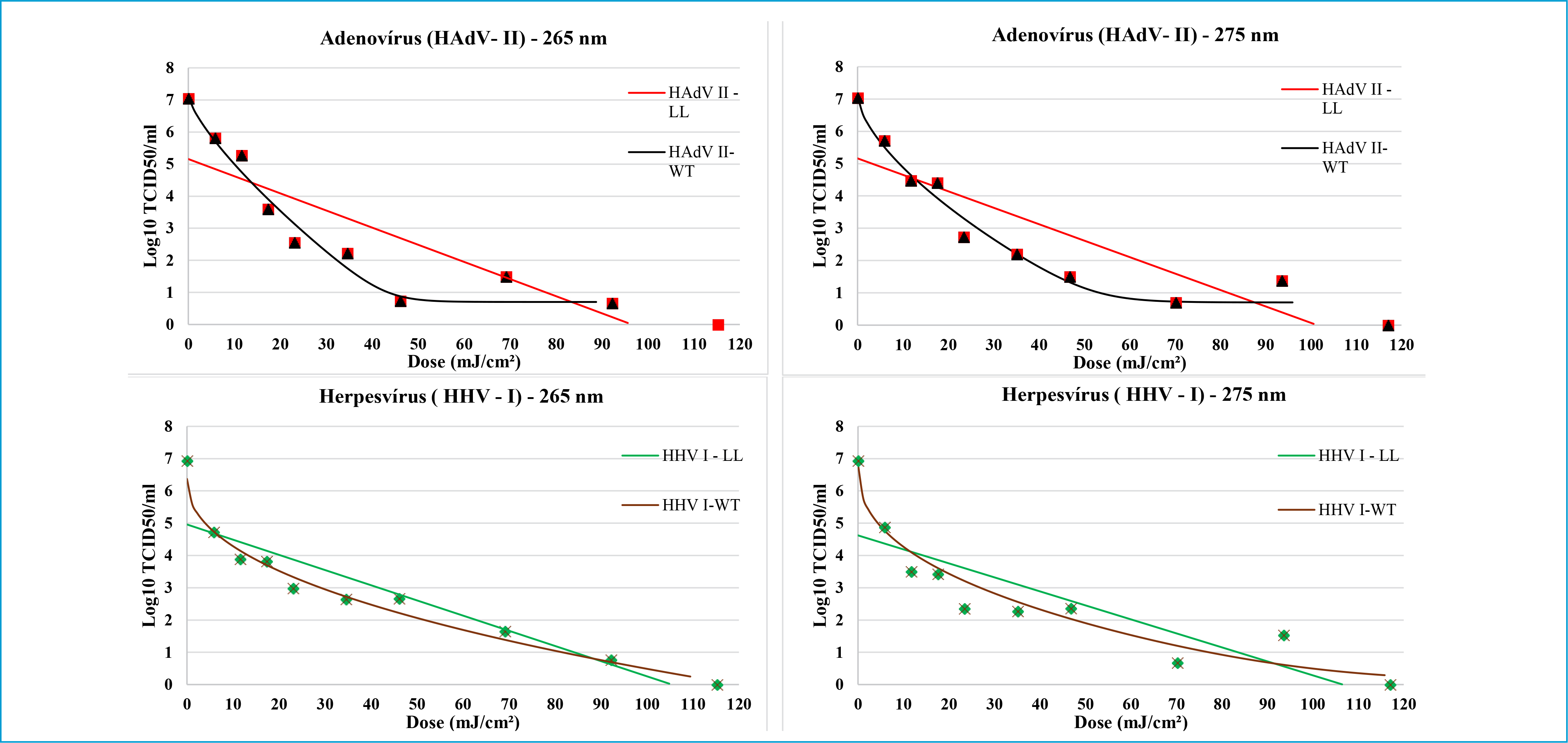
Figures 1 and 2 show the inactivation curves, and the corresponding kinetic parameters obtained from the log-linear and Weibull models are summarized in Table 1. Nonlinear survival curves described by the Weibull model are frequently associated with heterogeneous viral populations or protective mechanisms such as viral aggregation, which may reduce the effective UV exposure of part of the viral population. An initial sharp reduction was followed by the persistence of a more resistant viral fraction. This pattern, typical of heterogeneous populations, contrasts with the conventional log-linear model commonly applied to bacteria. The relatively low coefficients of determination obtained for the log-linear model, particularly for SARS-CoV-2 (R2 = 0.60–0.65), further indicate that the inactivation curves deviate from first-order kinetics and are better represented by the Weibull model.

Viral inactivation curves for HAdV-II and HSV-1 under UV-LED irradiation at 265 nm and 275 nm. Curves represent the fitted log-linear (LL) and Weibull-tail (WT) models used to describe the inactivation kinetics.

Viral inactivation curves for severe acute respiratory syndrome coronavirus 2 and vaccinia virus under ultraviolet light-emitting diodes at 265 nm and 275 nm. Curves represent the fitted log-linear (LL) and Weibull-tail (WT) models used to describe the inactivation kinetics..
Summary of kinetic parameters obtained from log-linear and Weibull models for viral inactivation under UV-LED irradiation at 265 and 275 nm.
The dose required for a 4-log reduction was 24.19 mJ/cm2 (265 nm) and 25.74 mJ/cm2 (275 nm), significantly lower than that estimated by the log-linear model: 76.03 and 79.56 mJ/cm2, respectively. These findings are consistent with those of Keshavarzfathy et al. (2021), who reported 74.4 mJ/cm2 for a 1.6-log reduction using 265 nm LEDs.
No statistically significant difference (p > 0.05) was observed between the 265 and 275 nm LEDs, as also reported by Beck et al. (2017). The nonlinear response may suggest the presence of resistant viral subpopulations, although reductions exceeding 4 logs were achieved at doses far below those required in mercury lamp systems (600–1000 mJ/cm2 – Saguti et al., 2022). It is worth noting that HAdV-II is one of the most UV-sensitive serotypes. HAdV-41, for instance, requires more than twice the dose for a similar level of inactivation (Augsburger et al., 2021). The absence of statistically significant differences between wavelengths suggests comparable inactivation performance under the tested conditions, although additional experiments with more replicates could further strengthen the statistical power of the comparison.
Studies have shown that HAdV exhibit elevated resistance to UV radiation, particularly serotype 40, followed by serotypes 41, 5, 2, and 1 (Oguma and Rattanakul, 2021). This intraspecies variability reinforces the importance of considering more resistant serotypes when establishing microbiological safety criteria, as doses optimized for sensitive strains may be insufficient. Literature indicates that to achieve a 4-log inactivation, required doses range from 138 mJ/cm2 for HAdV-I, 200 mJ/cm2 for HAdV-V, and up to 235.3 mJ/cm2 for HAdV-40 (Baxter et al., 2007; Beck et al., 2017; Nwachuku et al., 2005; Oguma and Rattanakul, 2021).
However, in the present study, the evaluated serotypes exhibited significantly lower required doses when irradiated with LEDs at 265 and 275 nm, demonstrating the higher efficacy of UVC-LED technology. The results support the feasibility of its application in disinfection systems, offering advantages such as reduced energy consumption and lower exposure requirements. Nevertheless, adjustments using the log-linear model indicated that HAdV-II still required higher doses, highlighting the heterogeneity in viral UV response.
For HSV, data fitted to the log-linear model indicated required doses of 31 mJ/cm2 at 265 nm and 29 mJ/cm2 at 275 nm. Although the peak absorption of viral DNA lies between 270 and 280 nm, no statistically significant difference was observed between the two wavelengths. Bui et al. (2022) reported higher resistance in their study using LEDs at 260 and 280 nm, requiring 50 mJ/cm2 and 90 mJ/cm2, respectively, to achieve a 4-log reduction. The authors also noted that the formation of byproducts such as CPDs and pyrimidine-(6-4)-pyrimidone photoproducts (6-PPs) was similar at both wavelengths, reinforcing the potential of UVC-LEDs for HSV inactivation.
The emergence and reemergence of viral diseases, such as mpox (monkeypox), have driven the use of surrogate viral models like VACV in disinfection studies. Although there are no confirmed reports of VACV transmission via water, its environmental persistence is notable: Mahnel, Ottis and Herlyn (1977) demonstrated its viability for over 200 days in aquatic environments. The use of VACV as an experimental surrogate for monkeypox virus is promising, especially given recent detection of monkeypox DNA in wastewater and uncertainties regarding its environmental stability (Atoui et al., 2023; Tiwari et al., 2023).
The relatively higher resistance observed for VACV may be associated with its large double-stranded DNA genome and complex virion structure. Poxviruses possess large genomes and multilayered viral particles, which may require greater cumulative photochemical damage to prevent viral replication. Doses of 38.02 mJ/cm2 and 40.95 mJ/cm2 were required at 265 and 275 nm, respectively, to achieve a 4-log inactivation. Although these values are higher than the 0.3 mJ/cm2 reported by McDevitt et al. (2007) using LP lamps, the discrepancy is attributed to the greater power output of traditional systems. Nevertheless, the results align with those by Koutras and Wade (2022), who reported greater than 4-log inactivation within seven minutes using LP lamps. In our study, the same level of inactivation was achieved in approximately 120 seconds, with comparable performance between the two wavelengths, demonstrating the effectiveness of LEDs against VACV.
SARS-CoV-2 was also evaluated, given its epidemiological relevance. A 4-log reduction was achieved with doses of 23 mJ/cm2 (265 nm) and 19 mJ/cm2 (275 nm). The inactivation curve did not follow a log-linear model, suggesting possible resistance from subpopulations or the formation of aggregates. Nonetheless, the results confirm the high sensitivity of the virus to UV-C radiation. Inagaki et al. (2020) reported a 4-log reduction using 37.5 mJ/cm2 with 280 nm LEDs. Other studies, such as those by Shimoda et al. (2021), Ma et al. (2021a), Minamikawa et al. (2021), and Biasin et al. (2022), demonstrated effectiveness with doses ranging from 1.8 mJ/cm2 to 8.2 mJ/cm2 for a 3-log reduction, using LEDs between 265 and 282 nm
These findings further support the feasibility of using UV-C LEDs at 265 nm and 275 nm as an efficient and safe tool for disinfecting environments and surfaces contaminated with SARS-CoV-2. The combination of high efficacy, short exposure time, and low energy demand positions this technology as a promising alternative to traditional UV sources. Although many previous studies evaluated SARS-CoV-2 inactivation on surfaces or aerosols, the results obtained here contribute specifically to understanding its behavior in aqueous environments.
Among known human coronaviruses, SARS-CoV-2, SARS-CoV, and Middle East respiratory syndrome coronavirus (MERS-CoV) are associated with more severe clinical outcomes, while human coronavirus OC43 (HCoV-OC43), human coronavirus HKU1 (HKU1), human coronavirus NL63 (HCoV-NL63), and human coronavirus 229E (HCoV-229E) typically cause mild infections (Nishioka, 2021). Due to their lower pathogenicity, the latter two are frequently used as experimental surrogates for SARS-CoV-2. Boegel et al. (2021) reported doses of 2.2 mJ/cm2 (HCoV-229E) and 6.8 mJ/cm2 (HCoV-OC43) for a 4-log reduction using 254 nm UV. These values were lower than those obtained by Ma et al. (2021b) — 11.5 mJ/cm2 (270 nm) and 14.8 mJ/cm2 (282 nm) — and comparable to Gerchman et al. (2020), who reported up to 16.4 mJ/cm2, indicating greater UV sensitivity in these surrogate models.
ANOVA with Tukey's test showed that SARS-CoV-2 exhibited higher UV sensitivity than VACV. While HSV-1 exhibited higher resistance than HAdV-II at both wavelengths, SARS-CoV-2 exhibited the highest sensitivity to UV irradiation. It is important to note that HAdV-II and HSV-1 were quantified in Log10 TCID50/mL, while SARS-CoV-2 and VACV were expressed in PFU/mL. This methodological difference may limit direct quantitative comparison of viruses. Therefore, the relative sensitivity among viruses should be interpreted with caution.
Based on the D4 (dose required to achieve a 4-log inactivation) values calculated under the experimental conditions of this study, the relative resistance of the viruses evaluated follows this order: VACV > HSV-1 > HAdV-II > SARS-CoV-2. This trend is consistent with the structural characteristics of the viruses, since large double-stranded DNA viruses tend to exhibit greater resistance to UV irradiation than smaller RNA viruses. In addition, the presence of complex capsid or envelope structures may influence the penetration of UV photons and the extent of nucleic acid damage.
Nevertheless, these observations should be interpreted cautiously, since factors such as viral aggregation, culture conditions, and differences in quantification methods may influence the apparent resistance of the viruses under study.
Variations can be attributed to factors such as genome type and conformation, presence of an envelope, and viral morphology. As shown in Table 2, greater resistance was expected for HAdV-II, as it is non-enveloped and possesses a double-stranded DNA (dsDNA) genome, a feature commonly associated with high resistance to UV radiation (Augsburger et al., 2021).
Maquart and Marlet (2022) reported that on plastic surfaces, doses between 3.2 and 44.7 mJ/cm2 of 265 nm LED irradiation resulted in a 4-log reduction of SARS-CoV-2. On steel, fabric, paper, and cardboard, doses ranged from 22.35 to 89.4 mJ/cm2, achieving reductions of 3.8 ± 0; 4.2 ± 0.9; 2.9 ± 1.6; and 3.3 ± 1.4 logs, respectively. Using LP lamps, doses ranged from 7.14 to 48.8 mJ/cm2 (plastic) and from 46.43 to 185.7 mJ/cm2 (other surfaces), with LEDs showing comparable performance and no statistically significant differences. These results are consistent with the findings of the present study, in which SARS-CoV-2 showed high susceptibility to UVC-LED irradiation at both wavelengths tested.
Differences between studies reflect variables such as viral species, intrinsic sensitivity, surface type, LED model, and quantification method (Maquart and Marlet, 2022). Among human coronaviruses, HCoV-229E and HCoV-OC43 are often used as SARS-CoV-2 surrogates due to their lower pathogenicity (Nishioka, 2021). For 4-log inactivation under 254 nm UV, Boegel et al. (2021) reported 2.2 mJ/cm2 (HCoV-229E) and 6.8 mJ/cm2 (HCoV-OC43), values lower than those obtained by Ma et al. (2021b) with LEDs at 270–282 nm, and consistent with Gerchman et al. (2020).
SARS-CoV-2 showed higher sensitivity to UV radiation than VACV, while HSV-1 was more resistant than HAdV-II. HAdV-II and HSV-1 were quantified by TCID50/mL, and SARS-CoV-2 and VACV by PFU/mL, which limits direct comparisons. ANOVA with Tukey's test revealed a statistically significant difference (p ≤ 0.05) only between the inactivation rates of VACV and SARS-CoV-2. The observed variations reflect factors such as genome type, morphology, and viral envelope (Augsburger et al., 2021; Oguma and Rattanakul, 2021), as well as possible viral aggregation in VACV, which may confer greater UV resistance.
From a practical perspective, the results of this study indicate that UV-C LEDs operating at wavelengths of 265 and 275 nm may represent a viable alternative for viral inactivation in water treatment systems. The relatively low UV doses required for achieving a 4-log reduction suggest that LED-based reactors could operate with reduced energy demand compared to conventional mercury lamp systems. However, further studies considering natural water matrices containing organic matter and suspended particles are necessary to evaluate the performance of this technology under real treatment conditions.
Overall, the results demonstrate that UV-C LED irradiation can effectively inactivate viruses with diverse structural characteristics, including enveloped and non-enveloped viruses as well as RNA and DNA genomes. The absence of statistically significant differences between the wavelengths tested indicates that both 265 nm and 275 nm LEDs can achieve comparable disinfection performance. These findings reinforce the growing potential of UV-C LED technology as a sustainable and flexible approach for viral control in water treatment applications.
CONCLUSION
The results of this study demonstrate that LED-UV systems operating at 265 nm and 275 nm wavelengths effectively inactivated various viruses of interest in water, including SARS-CoV-2, HSV-1, VACV, and HAdV-II. LED performance varied according to viral type, highlighting the importance of morphological and genomic characteristics in the response to UV radiation. SARS-CoV-2 showed the highest sensitivity to UV radiation, while VACV had the highest resistance among the viruses evaluated. However, these results should be interpreted considering the controlled laboratory conditions adopted in this study.
Variations in efficacy may also be associated with factors such as genome type (RNA or DNA), presence or absence of an envelope, viral conformation, and the quantification methods used (PFU or TCID50). The appropriate choice of wavelength and radiation dose should consider these variables to ensure disinfection efficiency, especially in water treatment applications.
These results reinforce the potential of LED-UV technology as a viable alternative to conventional mercury lamps, particularly in scenarios requiring sustainable, safe, and environmentally friendly solutions for viral inactivation. Future studies evaluating different water matrices, reactor configurations, and hydraulic conditions will be important to support the implementation of UV-LED technology in full-scale water treatment systems.
Such studies will be essential for evaluating the scalability and operational feasibility of UV-LED systems in full-scale water treatment plants. These findings contribute to the growing body of evidence supporting UV-LED technology as a promising approach for advanced viral disinfection in water treatment systems.
DATA AVAILABILITY STATEMENT
The datasets generated and/or analyzed during the current study are available from the corresponding author upon reasonable request.
ACKNOWLEDGEMENTS
To the Federal Center for Technological Education of Minas Gerais (CEFET/MG) for their logistical and structural support in conducting this work. To the National Agricultural Laboratory (LANAGRO) of Minas Gerais from the Ministry of Agriculture, Livestock, and Supply (Mapa) for their structural support in the SARS-CoV analyses. To the National Institute of Science and Technology (INCT). To the National Council for Scientific and Technological Development (CNPq). To the Foundation for Research Support of the State of Minas Gerais (FAPEMIG).
REFERENCES
- AMERICAN PUBLIC HEALTH ASSOCIATION – APHA; AMERICAN WATER WORKS ASSOCIATION – AWWA; WATER ENVIRONMENT FEDERATION – WEF. Standard Methods for the Examination of Water and Wastewater 23. ed. Washington: APHA, AWWA, WEF, 2017.
-
ATOUI, Ali; JOURDAIN, Frédéric; MOULY, Damien; CORDEVANT, Christophe; CHESNOT, Thierry; GASSILLOUD, Benoît. A review on mpox (monkeypox) virus shedding in wastewater and its persistence evaluation in environmental samples. Case Studies in Chemical and Environmental Engineering, v. 7, 100315, 2023. https://doi.org/10.1016/j.cscee.2023.100315
» https://doi.org/10.1016/j.cscee.2023.100315 -
AUGSBURGER, Nicolas; RACHMADI, Andri Taruna; ZAOURI, Noor; LEE, Yunho; HONG, Pei Ying. Recent Update on UV Disinfection to Fulfill the Disinfection Credit Value for Enteric Viruses in Water. Environmental Science and Technology, v. 55, n. 24, p. 16283-16298, 2021. https://doi.org/10.1021/acs.est.1c03092
» https://doi.org/10.1021/acs.est.1c03092 -
BAXTER, Carole; HOFMANN, Ron; TEMPLETON, Michael; BROWN, Martha; ANDREWS, Robert. Inactivation of Adenovirus Types 2, 5, and 41 in Drinking Water by UV Light, Free Chlorine, and Monochloramine. Journal of Environmental Engineering, v. 133, n. 1, p. 95-103, 2007. https://doi.org/10.1061/(ASCE)0733-9372(2007)133:1(95)
» https://doi.org/10.1061/(ASCE)0733-9372(2007)133:1(95) -
BECK, Sara; RODRIGUEZ, Roberto; LINDEN, Karl; HARGY, Thomas; LARASON, Thomas; WRIGHT, Harold. Wavelength dependent UV inactivation and DNA damage of adenovirus as measured by cell culture infectivity and long-range quantitative PCR. Environmental Science and Technology, v. 48, n. 1, p. 591-598, 2014. https://doi.org/10.1021/es403850b
» https://doi.org/10.1021/es403850b -
BECK, Sara; RYU, Hodon; BOCZEK, Laura; CASHDOLLAR, Jennifer; JEANIS, Kaitlyn; ROSENBLUM, James; LAWAL, Oliver; LINDEN, Karl. Evaluating UV-C LED disinfection performance and investigating potential dual-wavelength synergy. Water Research, v. 109, p. 207-216, 2017. https://doi.org/10.1016/j.watres.2016.11.024
» https://doi.org/10.1016/j.watres.2016.11.024 -
BIASIN, Mara; STRIZZI, Sergio; BIANCO, Andrea; MACCHI, Alberto; UTYRO, Olga; PARESCHI, Giovanni; LOFFREDA, Alessia; CAVALLERI, Adalberto; LUALDI, Manuela; TRABATTONI, Daria; TACCHETTI, Carlo; MAZZA, Davide; CLERICI, Mario. UV and violet light can neutralize SARS-CoV-2 Infectivity. Journal of Photochemistry and Photobiology, v. 10, 100107, 2022. https://doi.org/10.1016/j.jpap.2021.100107
» https://doi.org/10.1016/j.jpap.2021.100107 -
BOEGEL, S. J.; GABRIEL, Michelle; SASGES, Michael; PETRI, Brian; D’AGOSTINO, M. R.; ZHANG, A.; ANG, J. C.; MILLER, M. S.; MEUNIER, Sarah; AUCOIN, Marc. Robust Evaluation of Ultraviolet-C Sensitivity for SARS-CoV-2 and Surrogate Coronaviruses. Microbiology Spectrum, v. 9, n. 2, e0053721, 2021. https://doi.org/10.1128/spectrum.00537-21
» https://doi.org/10.1128/spectrum.00537-21 -
BOLTON, James; STEFAN, Mihaela; SHAW, Ping Shine; LYKKE, Keith. Determination of the quantum yields of the potassium ferrioxalate and potassium iodide-iodate actinometers and a method for the calibration of radiometer detectors. Journal of Photochemistry and Photobiology A: Chemistry, v. 222, n. 1, p. 166-169, 2011. https://doi.org/10.1016/j.jphotochem.2011.05.017
» https://doi.org/10.1016/j.jphotochem.2011.05.017 -
BUI, Thi Kim Ngan; MAWATARI, Kazuaki; EMOTO, Takahiro; FUKUSHIMA, Shiho; SHIMOHATA, Takaaki; UEBANSO, Takashi; AKUTAGAWA, Masatake; KINOUCHI, Yohsuke; TAKAHASHI, Akira. UV-LED irradiation reduces the infectivity of herpes simplex virus type 1 by targeting different viral components depending on the peak wavelength. Journal of Photochemistry and Photobiology B: Biology, v. 228, 112410, 2022. https://doi.org/10.1016/j.jphotobiol.2022.112410
» https://doi.org/10.1016/j.jphotobiol.2022.112410 -
CASE, James Brett; BAILEY, Adam; DIAMOND, Michael; KIM, Arthur; CHEN, Rita. Growth, detection, quantification, and inactivation of SARS-CoV-2. Virology, v. 548, p. 39-48, 2020. https://doi.org/10.1016/j.virol.2020.05.015
» https://doi.org/10.1016/j.virol.2020.05.015 -
CHEN, Jian; LOEB, Stephanie; KIM, Jae Hong. LED revolution: Fundamentals and prospects for UV disinfection applications. Environmental Science: Water Research and Technology, v. 3, n. 2, p. 188-202, 2017. https://doi.org/10.1039/C6EW00241B
» https://doi.org/10.1039/C6EW00241B -
CHEVREMONT, Anne-Céline; FARNET, Anne Marie; SERGENT, Michelle; COULOMB, Bruno; BOUDENNE, Jean Luc. Multivariate optimization of fecal bioindicator inactivation by coupling UV-A and UV-C LEDs. Desalination, v. 285, p. 219-225, 2012. https://doi.org/10.1016/j.desal.2011.10.006
» https://doi.org/10.1016/j.desal.2011.10.006 -
DHINGRA, Akshay; HAGE, Elias; GANZENMUELLER, Tina; BÖTTCHER, Sindy; HOFMANN, Jörg; HAMPRECHT, Klaus; OBERMEIER, Patrick; RATH, Barbara; HAUSMANN, Fabian; DOBNER, Thomas; HEIM, Albert. Molecular Evolution of Human Adenovirus (HAdV) Species C. Scientific Reports, v. 9, n. 1, p. 1-13, 2019. https://doi.org/10.1038/s41598-018-37249-4
» https://doi.org/10.1038/s41598-018-37249-4 -
GEERAERD, Annemie; VALDRAMIDIS, Vasilis; VAN IMPE, Jan Frans. GInaFiT, a freeware tool to assess non-log-linear microbial survivor curves. International Journal of Food Microbiology, v. 102, n. 1, p. 95-105, 2005. https://doi.org/10.1016/j.ijfoodmicro.2004.11.038
» https://doi.org/10.1016/j.ijfoodmicro.2004.11.038 -
GERCHMAN, Yoram; MAMANE, Hadas; FRIEDMAN, Nehemya; MANDELBOIM, Michal. UV-LED disinfection of Coronavirus: Wavelength effect. Journal of Photochemistry and Photobiology B: Biology, v. 212, 112044, 2020. https://doi.org/10.1016/j.jphotobiol.2020.112044
» https://doi.org/10.1016/j.jphotobiol.2020.112044 - HATCHARD, C. G.; PARKER, C. A. A New Sensitive Chemical Actinometer. II. Potassium Ferrioxalate as a Standard Chemical Actinometer. Proceedings of the Royal Society of London. Series A: Mathematical and Physical, v. 235, n. 1203, p. 518-536, 1956.
-
HIJNEN, Wim; MEDEMA, Gertjan. Inactivation of viruses, bacteria, spores and protozoa by ultraviolet irradiation in drinking water practice: A review. Water Science and Technology: Water Supply, v. 5, n. 5, p. 93-99, 2005. https://doi.org/10.2166/ws.2005.0043
» https://doi.org/10.2166/ws.2005.0043 -
HULO, Chantal; DE CASTRO, Edouard; MASSON, Patrick; BOUGUELERET, Lydie; BAIROCH, Amos; XENARIOS, Ioannis; LE MERCIER, Philippe. ViralZone: A knowledge resource to understand virus diversity. Nucleic Acids Research, v. 39, p. 576-582, 2011. https://doi.org/10.1093/nar/gkq901
» https://doi.org/10.1093/nar/gkq901 -
INAGAKI, Hiroko; SAITO, Akatsuki; SUGIYAMA, Hironobu; OKABAYASHI, Tamaki; FUJIMOTO, Shouichi. Rapid inactivation of SARS-CoV-2 with Deep-UV LED irradiation. Emerging Microbes and Infections, v. 9, n. 1, p. 1744-1747, 2020. https://doi.org/10.1080/22221751.2020.1796529
» https://doi.org/10.1080/22221751.2020.1796529 -
KESHAVARZFATHY, Majid; HOSOI, Yamato; OGUMA, Kumiko; TAGHIPOUR, Fariborz. Experimental and computational evaluation of a flow-through UV-LED reactor for MS2 and adenovirus inactivation. Chemical Engineering Journal, v. 407, 127058, 2021. https://doi.org/10.1016/j.cej.2020.127058
» https://doi.org/10.1016/j.cej.2020.127058 -
KOUTRAS, Carolina; WADE, Richard. Rapid inactivation of vaccinia, a surrogate virus for monkeypox and smallpox, using ultraviolet-C disinfection. bioRxiv, p. 48-51, 2022. https://doi.org/10.1101/2022.10.04.510918
» https://doi.org/10.1101/2022.10.04.510918 -
MA, Ben; GUNDY, Patricia M.; GERBA, Charles; SOBSEY, Mark; LINDEN, Karl. UV Inactivation of SARS-CoV-2 across the UVC Spectrum: KrCl* Excimer, Mercury-Vapor, and Light-Emitting-Diode (LED) Sources. Applied and Environmental Microbiology, v. 87, n. 22, e0153221, 2021a. https://doi.org/10.1128/aem.01532-21
» https://doi.org/10.1128/aem.01532-21 -
MA, Ben; LINDEN, Yarrow; GUNDY, Patricia; GERBA, Charles; SOBSEY, Mark; LINDEN, Karl. Inactivation of coronaviruses and phage Phi6 from irradiation across UVC wavelengths. Environmental Science and Technology Letters, v. 8, n. 5, p. 425-430, 2021b. https://doi.org/10.1021/acs.estlett.1c00178
» https://doi.org/10.1021/acs.estlett.1c00178 - MAHNEL, H; OTTIS, K; HERLYN, M. Stability in drinking and surface water of nine virus species from different genera. Zentralblatt fur Bakteriologie, Parasitenkunde, Infektionskrankheiten und Hygiene, v. 164, n. 1-2, p. 64-84, 1977.
- MAHY, Brian; KANGRO, Hillar. Virology Methods Manual San Diego: Academic Press, 1996.
-
MAQUART, Marianne; MARLET, Julien. Rapid SARS-CoV-2 inactivation by mercury and LED UV-C lamps on different surfaces. Photochemical and Photobiological Sciences, v. 21, n. 12, p. 2243-2247, 2022. https://doi.org/10.1007/s43630-022-00292-2
» https://doi.org/10.1007/s43630-022-00292-2 -
MARTINO, Vincent; OCHSNER, Kyra; PETERS, Paige; ZITOMER, Daniel; MAYER, Brooke K. Virus and Bacteria Inactivation Using Ultraviolet Light-Emitting Diodes. Environmental Engineering Science, v. 38, n. 6, p. 458-468, 2021. https://doi.org/10.1089/ees.2020.0092
» https://doi.org/10.1089/ees.2020.0092 -
MCDEVITT, James; KA, Man Lai; RUDNICK, Stephen; HOUSEMAN, E. Andres; FIRST, Melvin; MILTON, Donald. Characterization of UVC light sensitivity of vaccinia virus. Applied and Environmental Microbiology, v. 73, n. 18, p. 5760-5766, 2007. https://doi.org/10.1128/AEM.00110-07
» https://doi.org/10.1128/AEM.00110-07 -
MINAMIKAWA, Takeo; KOMA, Takaaki; SUZUKI, Akihiro; MIZUNO, Takahiko; NAGAMATSU, Kentaro; ARIMOCHI, Hideki; TSUCHIYA, Koichiro; MATSUOKA, Kaoru; YASUI, Takeshi; YASUTOMO, Koji; NOMAGUCHI, Masako. Quantitative evaluation of SARS-CoV-2 inactivation using a deep ultraviolet light-emitting diode. Scientific Reports, v. 11, n. 1, 5070, 2021. https://doi.org/10.1038/s41598-021-84592-0
» https://doi.org/10.1038/s41598-021-84592-0 -
NISHIOKA, Sérgio Andrade. Sete coronavírus causam doenças em humanos. UNASUS, 2021. Available at: https://www.unasus.gov.br/especial/covid19/markdown/111 Accessed in: Oct. 30, 2025.
» https://www.unasus.gov.br/especial/covid19/markdown/111 -
NWACHUKU, Nena; GERBA, Charles; OSWALD, Amy; MASHADI, Faezeh. Comparative inactivation of adenovirus serotypes by UV light disinfection. Applied and Environmental Microbiology, v. 71, n. 9, p. 5633-5636, 2005. https://doi.org/10.1128/AEM.71.9.5633-5636.2005
» https://doi.org/10.1128/AEM.71.9.5633-5636.2005 -
OGUMA, Kumiko; RATTANAKUL, Surapong. UV inactivation of viruses in water: Its potential to mitigate current and future threats of viral infectious diseases. Japanese Journal of Applied Physics, v. 60, n. 11, 110502, 2021. https://doi.org/10.35848/1347-4065/ac2b4f
» https://doi.org/10.35848/1347-4065/ac2b4f -
PANIZZOLO, Marco; GEA, Marta; CARRARO, Elisabetta; GILLI, Giorgio; BONETTA, Silvia; PIGNATA, Cristina. Occurrence of human pathogenic viruses in drinking water and in its sources: A review. Journal of Environmental Sciences, v. 132, p. 145-161, 2023. https://doi.org/10.1016/j.jes.2022.07.035
» https://doi.org/10.1016/j.jes.2022.07.035 -
SAGUTI, Fredy; CHURQUI, Marianela Patzi; KJELLBERG, Inger; WANG, Hao; OTTOSON, Jakob; PAUL, Catherine; BERGSTEDT, Olof; NORDER, Heléne; NYSTRÖM, Kristina. The UV Dose Used for Disinfection of Drinking Water in Sweden Inadequately Inactivates Enteric Virus with Double-Stranded Genomes. International Journal of Environmental Research and Public Health, v. 19, n. 14, 8669, 2022. https://doi.org/10.3390/ijerph19148669
» https://doi.org/10.3390/ijerph19148669 -
SHIMODA, Hiroshi; MATSUDA, Junji; IWASAKI, Tatsuyuki; HAYASAKA, Daisuke. Efficacy of 265-nm ultraviolet light in inactivating infectious SARS-CoV-2. Journal of Photochemistry and Photobiology, v. 7, 100050, 2021. https://doi.org/10.1016/j.jpap.2021.100050
» https://doi.org/10.1016/j.jpap.2021.100050 -
SILVA, Hugo; GARCÍA-ZAPATA, Marco; ANUNCIAÇÃO, Carlos. Why the Use of Adenoviruses as Water Quality Virologic Marker? Food and Environmental Virology, v. 3, n. 3-4, p. 138-140, 2011. https://doi.org/10.1007/s12560-011-9069-2
» https://doi.org/10.1007/s12560-011-9069-2 -
TIWARI, Ananda; ADHIKARI, Sangeet; KAYA, Devrim; ISLAM, Md Aminul; MALLA, Bikash; SHERCHAN, Samendra; AL-MUSTAPHA, Ahmad; KUMAR, Manish; AGGARWAL, Srijan; BHATTACHARYA, Prosun; BIBBY, Kyle; HALDEN, Rolf; BIVINS, Aaron; HARAMOTO, Eiji; OIKARINEN, Sami; HEIKINHEIMO, Annamari; PITKÄNEN, Tarja. Monkeypox outbreak: Wastewater and environmental surveillance perspective. Science of the Total Environment, v. 856, 159166, 2023. https://doi.org/10.1016/j.scitotenv.2022.159166
» https://doi.org/10.1016/j.scitotenv.2022.159166 -
TRAN, Hai Nguyen; LE, Giang Truong; NGUYEN, Dong Thanh; JUANG, Ruey-Shin; RINKLEBE, Jörg; BHATNAGAR, Amit; LIMA, Eder; IQBAL, Hafiz; SARMAH, Ajit; CHAO, Huan-Ping. SARS-CoV-2 coronavirus in water and wastewater: A critical review about presence and concern. Environmental Research, v. 193, 1110265, 2021. https://doi.org/10.1016/j.envres.2020.110265
» https://doi.org/10.1016/j.envres.2020.110265 - UNITED STATES ENVIRONMENTAL PROTECTION AGENCY – USEPA. Ultraviolet disinfection guidance manual for the final long term 2 enhanced surface water treatment rule Washington: USEPA, 2006. 436 p.
-
UNITED STATES ENVIRONMENTAL PROTECTION AGENCY – USEPA. Contaminant Candidate List 5 - CCL5. USEPA, 2022a. Available at: https://www.epa.gov/ccl/contaminant-candidate-list-5-ccl-5 Accessed in: Oct. 30, 2025.
» https://www.epa.gov/ccl/contaminant-candidate-list-5-ccl-5 -
UNITED STATES ENVIRONMENTAL PROTECTION AGENCY – USEPA. Coronavirus and Drinking Water and Wastewater. USEPA, 2022b. Available at: https://19january2021snapshot.epa.gov/coronavirus/coronavirus-and-drinking-water-and-wastewater_.html Accessed in: Oct. 30, 2025.
» https://19january2021snapshot.epa.gov/coronavirus/coronavirus-and-drinking-water-and-wastewater_.html -
VERANI, Marco; FEDERIGI, Ileana; DONZELLI, Gabriele; CIONI, Lorenzo; CARDUCCI, Annalaura. Human adenoviruses as waterborne index pathogens and their use for Quantitative Microbial Risk Assessment. Science of the Total Environment, v. 651, p. 1469-1475, 2019. https://doi.org/10.1016/j.scitotenv.2018.09.295
» https://doi.org/10.1016/j.scitotenv.2018.09.295 -
WORLD HEALTH ORGANIZATION – WHO. Drinking water. WHO, 2023. Available at: https://www.who.int/news-room/fact-sheets/detail/drinking-water Accessed in: Oct. 30, 2025.
» https://www.who.int/news-room/fact-sheets/detail/drinking-water -
WÜRTELE, Marlene; KOLBE, Tobias; LIPSZ, Martin; KÜLBERG, Alexander; WEYERS, Markus; KNEISSL, Michael; JEKEL, Martin. Application of GaN-based ultraviolet-C light emitting diodes - UV LEDs - for water disinfection. Water Research, v. 45, n. 3, p. 1481-1489, 2011. https://doi.org/10.1016/j.watres.2010.11.015
» https://doi.org/10.1016/j.watres.2010.11.015
Edited by
-
Editor:
Rodrigo Moruzzi, Universidade Estadual Paulista, São Paulo/SP. https://orcid.org/0000-0002-1573-3747

 Source: Authors
 Source: Authors