
 Platelet Distribution Width as a Marker for Severity and Extent of Coronary Artery Disease in Acute Coronary Syndrome
Abstract
Background Previous studies have demonstrated an association between larger platelet volume and coronary thrombus formation in acute coronary syndrome (ACS).
Objectives We aimed to investigate the association of platelet distribution width (PDW) levels with both the clinical severity and the extent of coronary artery disease (CAD) in patients diagnosed with ACS.
Methods A retrospective evaluation was conducted on 1043 consecutive patients diagnosed with ACS. Patients were stratified into two groups based on their PDW values: group 1 (PDW > 11.8 fL) and group 2 (PDW ≤ 11.8 fL). For each patient, Global Registry of Acute Coronary Events (GRACE) score, SYNTAX score, Thrombolysis in Myocardial Infarction (TIMI) flow grade, thrombus grades of culprit lesions, and neutrophil–lymphocyte ratio (NLR) were calculated, and their associations with PDW were examined.
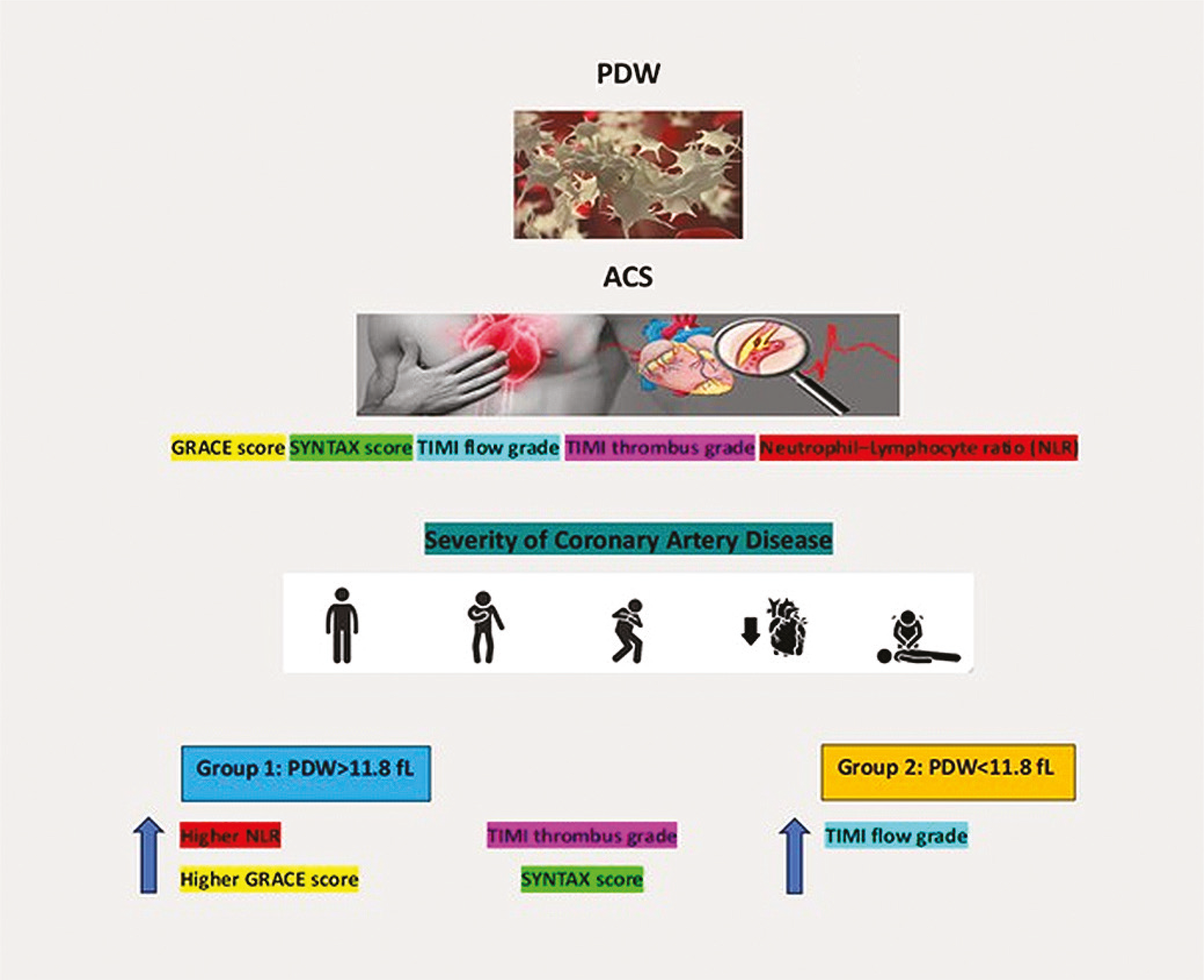
Results The mean age of the patients was 60.1 ± 12.4 years, with the majority being male (78.5%). The mean PDW was 11.8 fL (range: 8.9 to 22.0). Patients in group 1 exhibited higher NLR (3.21 versus 2.13, p < 0.05) and higher GRACE scores (139 versus 126, p < 0.05) compared to group 2. Group 1 patients also showed lower TIMI flow grades (0 and 1). A positive correlation was observed between PDW and GRACE score (R: 0.304, p = 0.01), but not with the SYNTAX score.
Conclusion Elevated PDW levels in patients with ACS were associated with more severe disease, as indicated by higher GRACE scores and reduced TIMI flow grades.
Blood Platelets; Coronary Artery Disease; Acute Coronary Syndrome
Introduction
Acute coronary syndrome (ACS) encompasses a diverse array of clinical presentations resulting from either partial or complete acute obstruction of a coronary vessel. Its well-established pathophysiology is rooted in the rupture of a vulnerable atherosclerotic plaque and subsequent thrombus formation. The clinical spectrum of ACS ranges from unstable angina pectoris to sudden cardiac death.1 Platelets play a pivotal role in the pathogenesis of ACS, participating not only in thrombus formation but also contributing to inflammation and modulation of the immune system.2 Upon activation, platelets undergo enlargement, and two laboratory parameters, namely, mean platelet volume and platelet distribution width (PDW), serve as indicators of platelet size or, equivalently, their activity in circulation.3
Previous investigations have established a link between coronary thrombus formation and larger platelet volume in cases of ACS.4 Additionally, studies have demonstrated an association between PDW and major adverse cardiac events, further underscoring the clinical relevance of platelet parameters in ACS.5
The primary objective of our study is to explore the association between PDW levels and both the clinical severity and extent of coronary artery disease (CAD) in patients diagnosed with ACS. Furthermore, we endeavor to elucidate the relationship between PDW levels and intracoronary thrombus grade, as well as the Thrombolysis in Myocardial Infarction (TIMI) flow rate. Through these investigations, we aim to contribute valuable insights into the role of platelet indices in characterizing the severity and extent of ACS, as well as their potential utility as prognostic markers in this clinical context.
Methods
This retrospective study involved a comprehensive analysis of clinical, angiographic, and laboratory data obtained from a consecutive cohort comprising 1043 patients diagnosed with ACS. The patient population comprised a heterogeneous mix, consisting of 119 individuals presenting unstable angina pectoris, 252 cases of non-ST-segment elevation myocardial infarction, and 672 instances of ST-segment elevation myocardial infarction. To ensure the integrity of calculations concerning CAD complexity, patients with a history of prior coronary artery bypass grafting surgery were meticulously excluded from the study cohort.
Patients were categorized into two distinct groups based on their PDW values: group 1 (comprising patients with PDW > 11.8 fL) and group 2 (encompassing patients with PDW ≤ 11.8 fL). The Global Registry of Acute Coronary Events (GRACE) score, serving as a reflection of pertinent clinical and laboratory parameters, was meticulously calculated for each patient.6 To ensure an objective assessment, an independent interventional cardiologist, blinded to patients’ clinical and demographic data, thoroughly evaluated all angiographic records. The SYNergy between percutaneous coronary intervention with TAXus and cardiac surgery (SYNTAX) score, serving as an indicator of CAD complexity, alongside the TIMI flow grade and thrombus grade of the culprit coronary arteries were diligently determined for each participant.7-10
The SYNTAX scoring system, encompassing a set of 12 main questions, facilitated the assignment of individual scores to each lesion, culminating in the derivation of a comprehensive total SYNTAX score.7-10 Regarding TIMI flow grades, meticulous categorization was employed, encompassing TIMI 0 flow (denoting the absence of contrast penetration beyond stenosis or occlusion), TIMI 1 flow (indicating contrast penetration beyond stenosis with no perfusion of the distal vessel), TIMI 2 flow (reflecting contrast reaching the distal vessel with a decreased filling rate or clearance compared to other coronary arteries), and TIMI 3 flow (where contrast reaches the distal bed and clears at a rate comparable to other coronary arteries).8 On the other hand, TIMI thrombus grades spanned from grade 0 (characterized by the absence of thrombus) to grade 5 (indicative of complete thrombotic occlusion of a vessel).9 Notably, instances where TIMI thrombus grade 5 was initially diagnosed underwent meticulous reclassification via angiographic reassessment subsequent to the removal of total occlusion facilitated by guidewire advancement during balloon angioplasty.11 Furthermore, the neutrophil–lymphocyte ratio (NLR) was calculated for each patient.
Statistical analyses were conducted employing SPSS 17.0 software. Spearman’s rank correlation test facilitated the exploration of associations between PDW, GRACE, and SYNTAX scores. Additionally, the Kruskal-Wallis test was employed to examine differences in PDW values among TIMI flow grades and thrombus grade subclasses. Continuous variable differences between the two PDW groups were evaluated by means of t test and Mann-Whitney tests, while chi-square tests explored differences in categorical variables. Ethical approval for the study was obtained from the local ethics committee, ensuring adherence to the principles outlined in the Helsinki Declaration of 1975, as revised in 2008, concerning ethical standards for human experimentation.
Results
The study cohort was predominantly constituted by male patients, comprising 78.5% of the total participants. The age distribution of the majority fell within the sixth decade, with a mean age of 60.1 ± 12.4 years. Among the diverse clinical presentations, ST-segment elevation myocardial infarction emerged as the most prevalent, representing 64.5% of cases. Table 1 displays a comprehensive delineation of the demographic, clinical, laboratory, and angiographic characteristics.
Upon comparative analysis of the demographic, clinical, laboratory, and angiographic features between the two groups, significant distinctions were observed, as elucidated in Table 2. Specifically, group 1 patients exhibited an elevated mean age, with a notable predominance of females within the subset displaying increased PDW values. Further scrutiny revealed that patients in group 1 manifested heightened NLR and GRACE scores, suggestive of an elevated risk profile. Moreover, a distinct association emerged between TIMI flow grades and PDW values, with higher PDW values correlating with lower TIMI flow grades (0 and 1). However, no significant disparities in TIMI thrombus grades were identified between the two groups.
A correlation analysis provided insightful findings, demonstrating a positive relationship between PDW and GRACE score (R: 0.34, p = 0.01), thereby highlighting a potential linkage between PDW and the GRACE scoring system. Interestingly, no such correlation was discerned when scrutinizing the SYNTAX score.
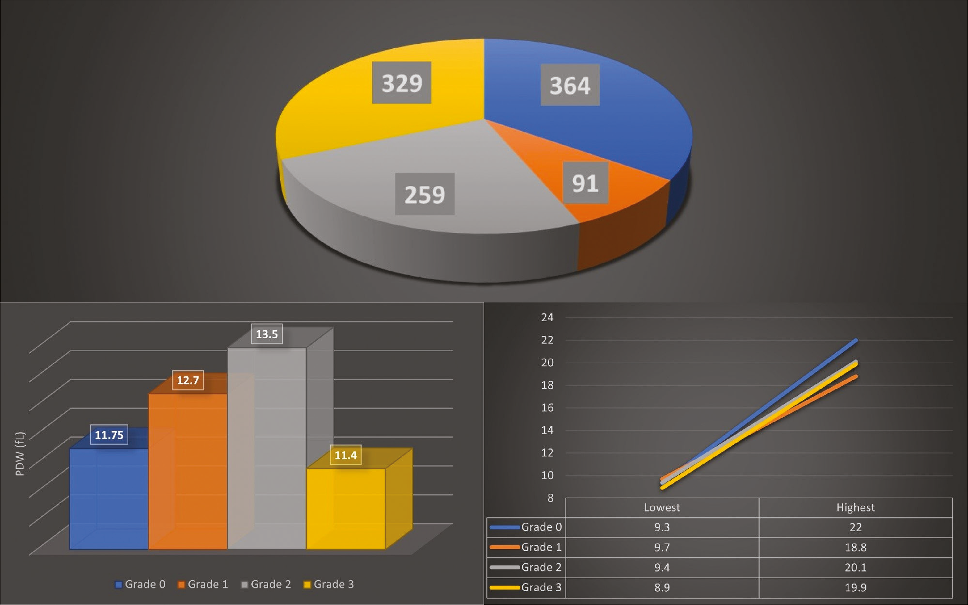
To further elucidate angiographic characteristics, patients were stratified into two distinct groups based on the size of the angiographic thrombus. Group A comprised patients devoid of apparent angiographic thrombus (TIMI thrombus grade 0) and those with a suspected angiographic thrombus (TIMI thrombus grade 1). Conversely, group B encompassed patients presenting with an apparent thrombus (TIMI thrombus grades 2, 3, and 4). Intriguingly, PDW values exhibited no significant disparities between these two groups (11.6 versus 11.9, p = 0.40). Nonetheless, when evaluating PDW values in relation to angiographic TIMI flow grades, a noteworthy observation emerged; patients with TIMI flow grade 3 consistently displayed lower PDW values compared to counterparts with different TIMI flow grades (Figure 1).
Discussion
Beyond their established role in thrombus formation, platelets play crucial roles in inflammation and the immune system. During acute myocardial infarction, platelets assume an active role in leukocyte activation by releasing inflammatory mediators and forming aggregates with leukocytes.12,13 Platelets exhibit heterogeneity in size, reflecting their reactivity, with a decrease in volume during circulation.14-16 Under stress conditions, platelet production is stimulated, leading to the release of larger and more active “stress platelets” into circulation.17 These stress platelets are notably increased during ACS, potentially indicating heightened thrombopoietic activity in the bone marrow.18 The observed enlargement of platelets in ACS may be indicative of increased thrombopoietic activity driven by proinflammatory cytokines, particularly interleukin-6.19 Numerous reports have associated the presence of large platelets with adverse outcomes in patients with ACS, and the increased megakaryocyte heterogeneity resulting from heightened thrombotic activity in the bone marrow contributes to elevated PDW in peripheral blood counts.20,21 Consequently, elevated PDW in patients with ACS may serve as a marker for increased thrombopoietic activity driven by heightened cytokine levels.
In the context of our study, the observed relationship between the GRACE risk score and PDW is postulated to stem from increased thrombopoietic activity influenced by heightened cytokine levels, particularly in response to the physical stress associated with ACS. This finding aligns with previous studies reporting correlations between GRACE score and inflammatory biomarkers.22,23 Notably, we did not observe any association between the extent of CAD and PDW levels, a deviation from prior research.24,25 This discrepancy may be attributed to the chronic and progressive nature of CAD, with our results reflecting a broader clinical spectrum in our patient cohort. Additionally, the use of the Gensini scoring system by Bekler and coauthors in their analysis of CAD extent may offer a more nuanced evaluation of the functional severity of coronary lesions, potentially contributing to the disparate results observed.
In our investigation of the association between intracoronary thrombus load and PDW, a weak inverse correlation was found between TIMI flow grade and PDW, rather than with TIMI thrombus grades. This result may reflect a connection between lower TIMI flow grades and the severity of ACS. The release of “stress platelets” from the bone marrow under stress conditions, mediated by proinflammatory cytokines, may provide an explanation for this observation.17
Notably, patients with elevated PDW values in our study exhibited older age, higher GRACE scores, elevated NLR, and lower TIMI flow grades. We posit that these findings underscore the association of these large stress platelets with the severity of ACS. Furthermore, the preponderance of female patients in group 1 aligns with established knowledge that female patients with ACS tend to be older and exhibit a greater number of major coronary risk factors than their male counterparts.26 The observed higher cytokine levels in these female patients, presenting with more severe ACS, may contribute to an increased release of stress platelets into circulation from the bone marrow, potentially influencing prognosis.
While our study provides valuable insights into the association between PDW and various parameters in patients with ACS, several limitations must be acknowledged. Firstly, the retrospective nature of the study introduces inherent biases and limits the establishment of causal relationships. The exclusion of patients with a history of prior coronary artery bypass grafting surgery aimed to ensure the accuracy of calculations related to CAD complexity; however, it may have introduced a selection bias, thus limiting the generalizability of our findings. Additionally, the study’s reliance on a single-center cohort may impact the external validity, and broader multicenter studies are warranted to validate our observations across diverse populations. Furthermore, the cross-sectional design hinders the exploration of temporal relationships, and prospective studies are needed to elucidate the dynamic changes in platelet indices over time. The use of the GRACE score as a representation of clinical severity may not capture the full spectrum of individual patient characteristics. Despite these limitations, our study offers a foundation for future investigations to delve deeper into the intricate interplay between platelet dynamics and ACS pathophysiology.
Conclusion
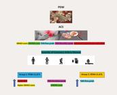
In conclusion, our comprehensive investigation into the association between PDW levels and various clinical, angiographic, and laboratory parameters in patients diagnosed with ACS has revealed intricate connections with the severity and extent of this cardiovascular condition. The study elucidates the potential role of PDW as a valuable marker for heightened thrombopoietic activity, influenced by proinflammatory cytokines, particularly in response to the physical stress associated with ACS. The observed correlations with GRACE score, NLR, and TIMI flow grade underscore the multifaceted involvement of platelets in the pathophysiology of ACS (Central Illustration). Interestingly, our findings did not establish a significant association between PDW levels and the extent of CAD, deviating from previous research, possibly due to the chronic and progressive nature of CAD. This study contributes novel insights into the intricate interplay between platelet indices and the clinical manifestations of ACS, emphasizing the potential of PDW as a prognostic marker and supporting further research to enhance our understanding of platelet dynamics in cardiovascular diseases.
References
-
1 Nardin M, Verdoia M, Laera N, Cao D, De Luca G. New Insights into Pathophysiology and New Risk Factors for ACS. J Clin Med. 2023;12(8):2883. doi: 10.3390/jcm12082883.
» https://doi.org/10.3390/jcm12082883 -
2 Rondina MT, Weyrich AS, Zimmerman GA. Platelets as Cellular Effectors of Inflammation in Vascular Diseases. Circ Res. 2013;112(11):1506-19. doi: 10.1161/CIRCRESAHA.113.300512.
» https://doi.org/10.1161/CIRCRESAHA.113.300512 -
3 Vizioli L, Muscari S, Muscari A. The Relationship of Mean Platelet Volume with the Risk and Prognosis of Cardiovascular Diseases. Int J Clin Pract. 2009;63(10):1509-15. doi: 10.1111/j.1742-1241.2009.02070.x.
» https://doi.org/10.1111/j.1742-1241.2009.02070.x -
4 Huczek Z, Filipiak KJ, Kochman J, Michalak M, Roik M, Piatkowski R, et al. Baseline Platelet Size is Increased in Patients with Acute Coronary Syndromes Developing Early Stent Thrombosis and Predicts Future Residual Platelet Reactivity. A Case-Control Study. Thromb Res. 2010;125(5):406-12. doi: 10.1016/j.thromres.2009.09.003.
» https://doi.org/10.1016/j.thromres.2009.09.003 -
5 Ulucan S, Keser A, Kaya Z, Katlandur H, Özdil H, Bilgi M, et al. Association between PDW and Long Term Major Adverse Cardiac Events in Patients with Acute Coronary Syndrome. Heart Lung Circ. 2016;25(1):29-34. doi: 10.1016/j.hlc.2015.05.017.
» https://doi.org/10.1016/j.hlc.2015.05.017 -
6 Szabó GT, Ágoston A, Csató G, Rácz I, Bárány T, Uzonyi G, et al. Predictors of Hospital Mortality in Patients with Acute Coronary Syndrome Complicated by Cardiogenic Shock. Sensors. 2021;21(3):969. doi: 10.3390/s21030969.
» https://doi.org/10.3390/s21030969 -
7 Farooq V, Girasis C, Magro M, Onuma Y, Morel MA, Heo JH, et al. The CABG SYNTAX Score - An Angiographic Tool to Grade the Complexity of Coronary Disease Following Coronary Artery Bypass Graft Surgery: From the SYNTAX Left Main Angiographic (SYNTAX-LE MANS) Substudy. EuroIntervention. 2013;8(11):1277-85. doi: 10.4244/EIJV8I11A196.
» https://doi.org/10.4244/EIJV8I11A196 -
8 Kern MJ, Moore JA, Aguirre FV, Bach RG, Caracciolo EA, Wolford T, et al. Determination of Angiographic (TIMI Grade) Blood Flow by Intracoronary Doppler flow Velocity During Acute Myocardial Infarction. Circulation. 1996;94(7):1545-52. doi: 10.1161/01.cir.94.7.1545.
» https://doi.org/10.1161/01.cir.94.7.1545 -
9 Gibson CM, Lemos JA, Murphy SA, Marble SJ, McCabe CH, Cannon CP, et al. Combination Therapy with Abciximab Reduces Angiographically Evident Thrombus in Acute Myocardial Infarction: A TIMI 14 Substudy. Circulation. 2001;103(21):2550-4. doi: 10.1161/01.cir.103.21.2550.
» https://doi.org/10.1161/01.cir.103.21.2550 -
10 Ong AT, Serruys PW, Mohr FW, Morice MC, Kappetein AP, Holmes DR Jr, et al. The SYNergy between Percutaneous Coronary Intervention with TAXus and Cardiac Surgery (SYNTAX) Study: Design, Rationale, and Run-in Phase. Am Heart J. 2006;151(6):1194-204. doi: 10.1016/j.ahj.2005.07.017.
» https://doi.org/10.1016/j.ahj.2005.07.017 -
11 Sianos G, Papafaklis MI, Daemen J, Vaina S, van Mieghem CA, van Domburg RT, et al. Angiographic Stent Thrombosis after Routine Use of Drug-Eluting Stents in ST-Segment Elevation Myocardial Infarction: The Importance of Thrombus Burden. J Am Coll Cardiol. 2007;50(7):573-83. doi: 10.1016/j.jacc.2007.04.059.
» https://doi.org/10.1016/j.jacc.2007.04.059 -
12 Metwalli O, Hashem E, Ajabnoor MA Sr, Alama N, Banjar ZM. Study of Some Inflammatory Mediators in the Serum of Patients with Atherosclerosis and Acute Myocardial Infarction. Cureus. 2021;13(10):e18450. doi: 10.7759/cureus.18450.
» https://doi.org/10.7759/cureus.18450 -
13 Furman MI, Barnard MR, Krueger LA, Fox ML, Shilale EA, Lessard DM, et al. Circulating Monocyte-Platelet Aggregates are an Early Marker of Acute Myocardial Infarction. J Am Coll Cardiol. 2001;38(4):1002-6. doi: 10.1016/s0735-1097(01)01485-1.
» https://doi.org/10.1016/s0735-1097(01)01485-1 -
14 Vilella-Figuerola A, Cordero A, Mirabet S, Muñoz-García N, Suades R, Padró T, et al. Platelet-Released Extracellular Vesicle Characteristics Differ in Chronic and in Acute Heart Disease. Thromb Haemost. 2023;123(9):892-903. doi: 10.1055/s-0043-57017.
» https://doi.org/10.1055/s-0043-57017 -
15 Castrichini M, Luzum JA, Pereira N. Pharmacogenetics of Antiplatelet Therapy. Annu Rev Pharmacol Toxicol. 2023;63:211-29. doi: 10.1146/annurev-pharmtox-051921-092701.
» https://doi.org/10.1146/annurev-pharmtox-051921-092701 -
16 Kraemer BF, Martinovic I, Lindemann S. Human Platelets Release TGFBIp in Acute Myocardial Infarction. Heart Vessels. 2022;37(11):1962-70. doi: 10.1007/s00380-022-02086-z.
» https://doi.org/10.1007/s00380-022-02086-z -
17 Tong M, Seth P, Penington DG. Proplatelets and Stress Platelets. Blood. 1987;69(2):522-8. doi: 10.1182/blood.V69.2.522.522.
» https://doi.org/10.1182/blood.V69.2.522.522 -
18 van der Loo B, Martin JF. A Role for Changes in Platelet Production in the Cause of Acute Coronary Syndromes. Arterioscler Thromb Vasc Biol. 1999;19(3):672-9. doi: 10.1161/01.atv.19.3.672.
» https://doi.org/10.1161/01.atv.19.3.672 -
19 Biasucci LM, Vitelli A, Liuzzo G, Altamura S, Caligiuri G, Monaco C, et al. Elevated Levels of Interleukin-6 in Unstable Angina. Circulation. 1996;94(5):874-7. doi: 10.1161/01.cir.94.5.874.
» https://doi.org/10.1161/01.cir.94.5.874 -
20 Heptinstall S, Mulley GP, Taylor PM, Mitchell JR. Platelet-Release Reaction in Myocardial Infarction. Br Med J. 1980;280(6207):80-1. doi: 10.1136/bmj.280.6207.80.
» https://doi.org/10.1136/bmj.280.6207.80 -
21 Bessman JD, Williams LJ, Gilmer PR Jr. Platelet Size in Health and Hematologic Disease. Am J Clin Pathol. 1982;78(2):150-3. doi: 10.1093/ajcp/78.2.150.
» https://doi.org/10.1093/ajcp/78.2.150 -
22 Beygui F, Silvain J, Pena A, Bellemain-Appaix A, Collet JP, Drexler H, et al. Usefulness of Biomarker Strategy to Improve GRACE Score's Prediction Performance in Patients with Non-St-Segment Elevation Acute Coronary Syndrome and Low Event Rates. Am J Cardiol. 2010;106(5):650-8. doi: 10.1016/j.amjcard.2010.04.019.
» https://doi.org/10.1016/j.amjcard.2010.04.019 -
23 Correia LC, Andrade BB, Borges VM, Clarêncio J, Bittencourt AP, Freitas R, et al. Prognostic Value of Cytokines and Chemokines in Addition to the GRACE Score in Non-ST-Elevation Acute Coronary Syndromes. Clin Chim Acta. 2010;411(7-8):540-5. doi: 10.1016/j.cca.2010.01.011.
» https://doi.org/10.1016/j.cca.2010.01.011 -
24 Bekler A, Ozkan MT, Tenekecioglu E, Gazi E, Yener AU, Temiz A, et al. Increased Platelet Distribution Width is Associated with Severity of Coronary Artery Disease in Patients with Acute Coronary Syndrome. Angiology. 2015;66(7):638-43. doi: 10.1177/0003319714545779.
» https://doi.org/10.1177/0003319714545779 -
25 Akin F, Ayca B, Kose N, Altun I, Avsar M, Celik O, et al. Relation of Platelet Indices to Severity of Coronary Artery Disease in Patients Undergoing Primary Percutaneous Coronary Intervention. Perfusion. 2016;31(3):216-22. doi: 10.1177/0267659115594231.
» https://doi.org/10.1177/0267659115594231 - 26 Song XT, Chen YD, Pan WQ, Lü SZ; CRACE investigators. Gender Based Differences in Patients with Acute Coronary Syndrome: Findings from Chinese Registry of Acute Coronary Events (CRACE). Chin Med J. 2007;120(12):1063-7.
-
Study Association
This study is not associated with any graduation program.
-
Ethics Approval and Consent to Participate
This study was approved by the Ethics Committee of the Yeni Yüzyıl University under the protocol number 693906709-300-00-1001-1. All the procedures in this study were in accordance with the 1975 Helsinki Declaration, updated in 2013. Informed consent was obtained from all participants included in the study.
-
Sources of Funding
There were no external funding sources for this study.

 PDW: platelet distribution width