NMR-Based Urine Metabolic Profiling of Cri-du-Chat Syndrome
Abstract
Cri-du-chat syndrome (CDCS) is a rare genetic disorder caused by a partial deletion of chromosome 5. Common features include intellectual disability, developmental delays, microcephaly, and a characteristic high-pitched cry. Although CDCS has been extensively studied from genetic and developmental perspectives, its metabolic implications remain poorly understood. This study aimed to investigate alterations in metabolic pathways involved in the pathological mechanisms of CDCS through urine metabolomic profiling using nuclear magnetic resonance (NMR) spectroscopy. Sixty-three participants were grouped into children, adolescents, and adults. For the first time, an NMR-based metabolomic signature for CDCS was reported, revealing 33 urinary metabolites and providing new insights into syndrome-related biochemical alterations. Statistical analyses revealed significant metabolic alterations compared to control subjects, particularly involving branched-chain and aromatic amino acids, as well as organic acids associated with energy production. Enrichment analysis further highlighted age-specific pathways impacted by the syndrome. In children, pathways related to phenylalanine, tyrosine, and tryptophan biosynthesis and metabolism, as well as butanoate and glyoxylate metabolism, were affected. Adolescents showed prominent alterations in phenylalanine biosynthesis and metabolism, while adults exhibited disruptions in taurine, hypotaurine, beta-alanine, and histidine metabolism. Altogether, these findings demonstrate pervasive impairments in amino-acid metabolism and energy homeostasis, elucidating key biochemical mechanisms that underlie the physiological manifestations of CDCS and highlighting potential metabolic pathways for future therapeutic exploration.
Keywords:
NMR; metabolomics; metabolic syndromes; Cri-du-Chat syndrome; urine
Introduction
The genetic syndrome known as Cri-du-chat syndrome (CDCS) is a rare chromosomal condition characterized by the total or partial deletion of the “short arm” region on chromosome 5 (also referred as 5p-syndrome).1 Firstly reported by Lejeune et al.2 in the early 60s, the occurrence of this condition varies between 1:15,000 and 1:50,000 of living newborns worldwide.3 Due to its rarity, most studies on CDCS are limited to individual case reports or small case series, which restrict the number of subjects available for broader clinical and epidemiological investigations.4-6
The syndrome presents a wide range of symptoms that vary significantly among individuals, including microcephaly, typical high-pitched cry, distinctive facial dysmorphism, pronounced intellectual disability and developmental delays. Developmentally, these challenges manifest as significant delays in basic motor skills, frequently requiring additional support for everyday activities.7,8 Phenotypic severity directly correlates with deletion size and position, with larger deletions typically resulting in more profound effects.9,10
Although the CDCS has been extensively studied from genetic and developmental perspectives, the metabolic impacts have not yet been sufficiently explored. A comprehensive understanding of the systemic effects of the syndrome requires an integrated approach that combines molecular and genetic perspectives.11,12 In this context, metabolomics is particularly relevant for understanding CDCS, as it provides a comprehensive view of the biochemical pathways affected by the genetic aspects of the disorder, helping to identify potential biomarkers and therapeutic targets for improving the overall well-being and functional capabilities of individuals with the syndrome.13,14
Nuclear magnetic resonance (NMR) spectroscopy represents a valuable tool for metabolomic applications due to its reproducibility, quantitative accuracy and non-destructive nature. NMR spectroscopy of biofluids, such as urine and plasma provide useful insights into endogenous metabolic processes, providing a comprehensive understanding of metabolic disturbances.15,16 The urine represents a valuable biofluid due to its abundance of metabolic breakdown products. For this study, a non-invasive collection and straightforward sample preparation made it useful for analyzing the sensitive cohorts.17,18
To our knowledge, this is the first study to characterize the urinary profile of individuals with CDCS through an NMR-based metabolomics approach. Thus, this study aimed to generate metabolomic signatures from the urine of CDCS individuals and identify key metabolic pathways affected by the syndrome.
Experimental
Ethical statement
This study was conducted using procedures approved by the Ethics Committee of the Federal University of São Paulo (Paulist School of Medicine, number: 868.398) and informed consent was obtained from all volunteers.
Sample collection
In total, 63 first-morning (fasting) urine samples were collected at the Cri-du-Chat Advisory and Research Center19 located in São Paulo (Brazil). The participants included children (15 with CDCS, average age 6.13 ± 3.07 years; 6 healthy controls (HC), average age 7.83 ± 2.40 years), adolescents (17 with CDCS, average age 14.64 ± 2.08 years; 11 HC, average age 15.45 ± 1.96 years), and adults (6 with CDCS, average age 25.83 ± 5.49 years; 8 HC, average age 28.65 ± 4.10 years). The collected samples were stored at -80 °C until conducting the NMR analyses. Donors ranged in age from 2 to 35 years and were classified as children (2-10 years), adolescents (11-20 years), and adults (21 35 years). The descriptive characteristics of the samples are presented in Table 1.
Sample preparation
The NMR experimental protocol was optimized based on standardized metabolomics procedures for urine analysis.20 Briefly, 200 µL of each urine sample were mixed with 400 µL of phosphate buffer solution prepared. The buffer solution was prepared by dissolving 0.27 g of NaH2PO4·H2O and 0.54 g of Na2HPO4·2H2O in 100 mL of D2O (pH 7.4 ± 0.4) containing 1 mM sodium-3-trimethylsilylpropionate-d4 (TMSP-d4) as the internal standard and chemical shift calibrant. After homogenization using a vortex-type stirrer (Biomixer® DL-901), the mixture was transferred to a 5 mm NMR tube for spectral acquisition.
NMR experiments and instrumentation
All NMR experiments were performed on a Bruker Avance III 600 MHz spectrometer operating at 14.1 T and equipped with a 5 mm triple resonance carbon inverse (TCI) cryoprobe, under a controlled temperature of 298 K. One-dimensional 1H NMR spectra were acquired using the zgesgp water-suppression pulse sequence (excitation sculpting). A total of 128 scans were collected with 64 k data points, covering a spectral width of 9615 Hz, resulting in an acquisition time of 3.40 s. The 90° pulse was calibrated to 11.010 μs, and the relaxation delay was set to 3.00 s. The transmitter offset frequency (o1p) was adjusted to 4.701 ppm, and the receiver gain was automatically optimized and maintained across all experiments.
Spectra were referenced to TMSP-d4 (d = 0.00 ppm), zero-filled to 64 k points, processed with a spectral size of 32k (SI), and apodized using a 0.3 Hz Lorentzian line-broadening factor. Automatic phase and baseline corrections were applied. All NMR acquisition and processing parameters are reported in Table S1 (Supplementary Information (SI) section). Two-dimensional NMR experiments, including 1H-1H correlated spectroscopy (COSY) and 1H-13C heteronuclear single quantum coherence (HSQC), were acquired to support metabolite identification and structural confirmation, particularly in regions with overlapping resonances.
Data collection and analysis
Processed spectra were imported into Chenomx NMR Suite 8.5 (Chenomx Inc., Edmonton, Canada) for metabolite identification and quantification using the internal reference concentration method. Identification was corroborated by comparing 2D NMR correlations with open-access metabolomics databases, including the Human Metabolome Database (HMDB)21 and Biological Magnetic Resonance Data Bank (BMRB).22 All observed and referenced chemical shifts are listed in Table S2 (SI section). Representative 1H-13C HSQC spectra of the aliphatic and aromatic regions are shown in Figure S1 (SI section), supporting the identification of metabolites.
All statistical analyses and metabolite pathway assessments were performed using MetaboAnalyst 6.0 (Xia Lab, McGill University, Montreal, QC, Canada, 2023).23 To account for urine dilution, all urinary metabolite concentrations were normalized to creatinine prior to statistical analysis. The normalized data were then log10 transformed to improve data distribution and approximate normality before multivariate analysis. Group differences were evaluated using the Mann Whitney U test. Volcano plots were generated to visualize the relationship between fold change and statistical significance. Orthogonal projections to latent structures discriminant analysis (OPLS DA) was applied to model the relationship between metabolic profiles and the predefined classes (CDCS patients vs. control group across different age groups) and to identify metabolites contributing to class discrimination. Variable Importance in Projection (VIP) scores greater than 1.2 were considered significant contributors to group differentiation. Volcano plot analysis further applied a fold-change threshold of 1.5 and p < 0.05, with false discovery rate (FDR) correction to control for multiple testing. Statistical outputs from OPLS-DA and fold-change analysis are provided in Table S2 (SI section).
Results and Discussion
Cri-du-chat syndrome (CDCS) metabolomic profile using 1H NMR spectroscopy
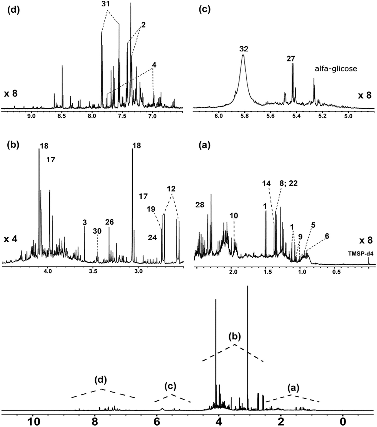
In this study, it is presented the untargeted urinary metabolomic profile of individuals with Cri-du-chat syndrome in contrast with healthy controls using a 1H NMR-based approach. A total of 33 metabolites were identified, including amino acids and their derivatives, organic acids, and methylated amines. A representative 1H NMR spectrum of urine samples from the CDCS group is shown in Figure 1, illustrating the most relevant metabolites detected. The corresponding 1H and 13C chemical shifts for all metabolites are provided in Table S3 (SI section). This work reports, for the first time, the application of NMR based metabolomics to characterize urinary metabolic profiles in individuals with CDCS, addressing the need to understand age-dependent biochemical alterations associated with the syndrome.
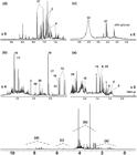
1H NMR spectra (600 MHz, D2O) of urine sample from CDCS individual with region expansions. (a) d 1H = 2.50 to 4.20 and (b) d 1H = 0.80 to 2.50. The numbers indicate the following metabolites: 1: alanine; 2: phenylalanine; 3: glycine; 4: histidine; 5: leucine; 6: isoleucine; 8: threonine; 9: valine; 10: acetate; 12: citrate; 14: 2-hydroxy-isobutyrate; 15: isobutyrate; 17: creatine; 18: creatinine; 19: dimethylamine; 22: lactate; 23: mannitol; 24: methylamine; 26: trimethylamine N-oxide; 27: sucrose; 28: succinate; 32: hippurate; 33: urea.
Age-dependent alterations in amino acid metabolism
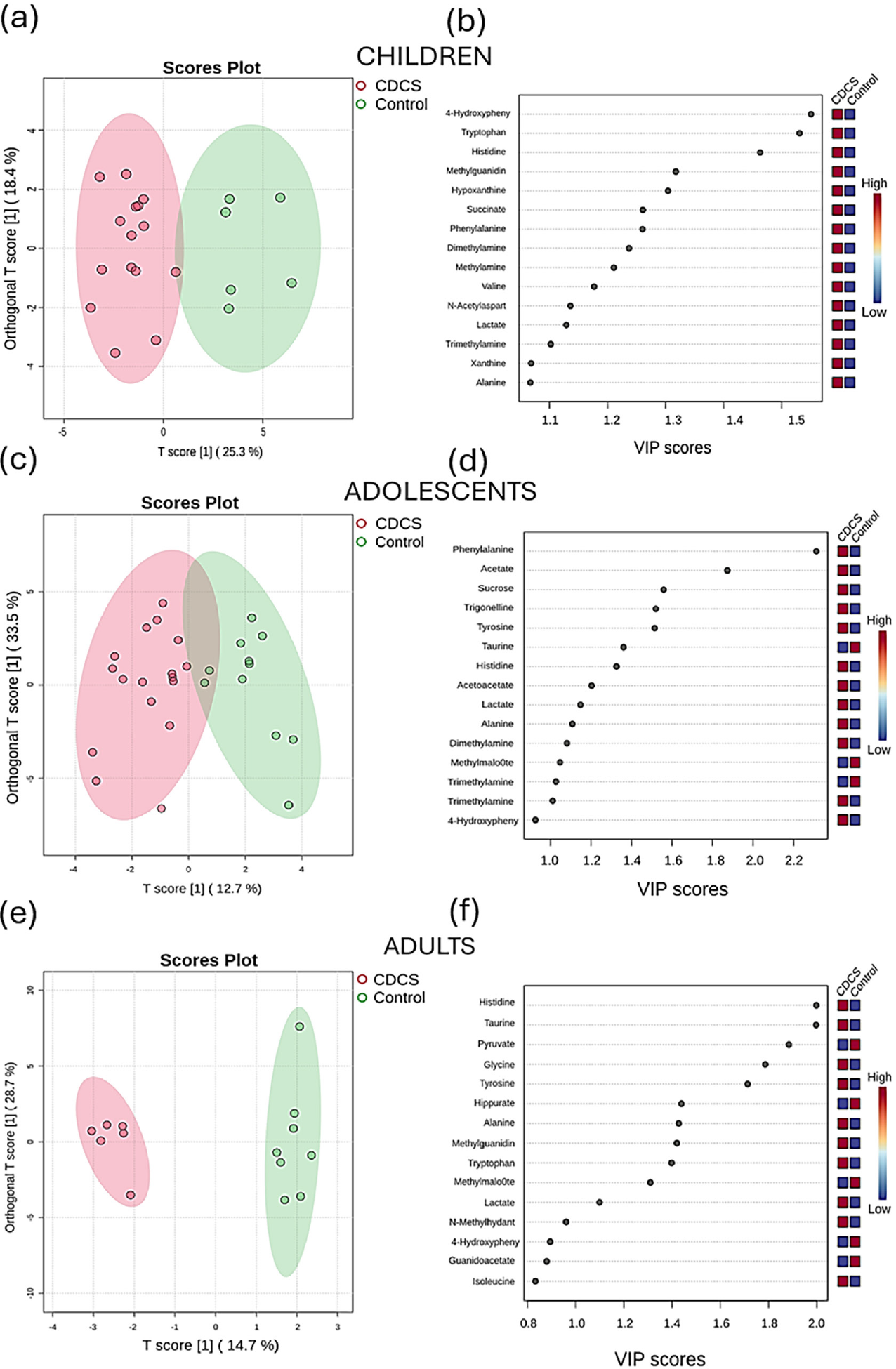
OPLS-DA revealed a clear separation between the CDCS group and the control group, indicating distinct metabolic profiles. The VIP score plot highlights the metabolites that contributed most significantly to this differentiation. In the children and adolescent groups, several metabolites showed a stronger contribution to the separation of the CDCS group, suggesting a potential association with the syndrome phenotype. Complete OPLS DA performance metrics for all age groups are provided in Table S4 (SI section). For the adult cohort, given the small number of adult CDCS patients, permutation testing was performed to assess the risk of overfitting, confirming that both model fit (p = 0.013) and predictive performance (p = 0.002) were significantly better than expected by chance (Table S5 and Figure S2, SI section).
Notably, 4-hydroxyphenylacetate, tryptophan (Trp), histidine (His), phenylalanine (Phe), succinate, dimethylamine, and methylamine were identified as key contributors to group discrimination. These compounds were found at higher levels in the CDCS group and may reflect alterations in amino acid metabolism and energy-related pathways associated with the condition (Figures 2a 2d). In the adult group, taurine and alanine (Ala) also contributed to the separation between control and CDCS groups (Figures 2e-2f).
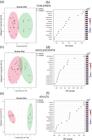
OPLS-DA scores plots and corresponding VIP score plots showing the metabolic differentiation between CDCS and control groups across age categories. (a, b) Children, (c, d) adolescents, and (e, f) adults. The scores plots (left) illustrate the separation between CDCS (red) and control (green) individuals based on their urinary metabolomic profiles. The VIP score plots (right) highlight the metabolites most responsible for the group separation, with higher VIP values indicating greater contribution. Colored heatmaps adjacent to each metabolite represent their relative abundance in each group, from low (blue) to high (red).
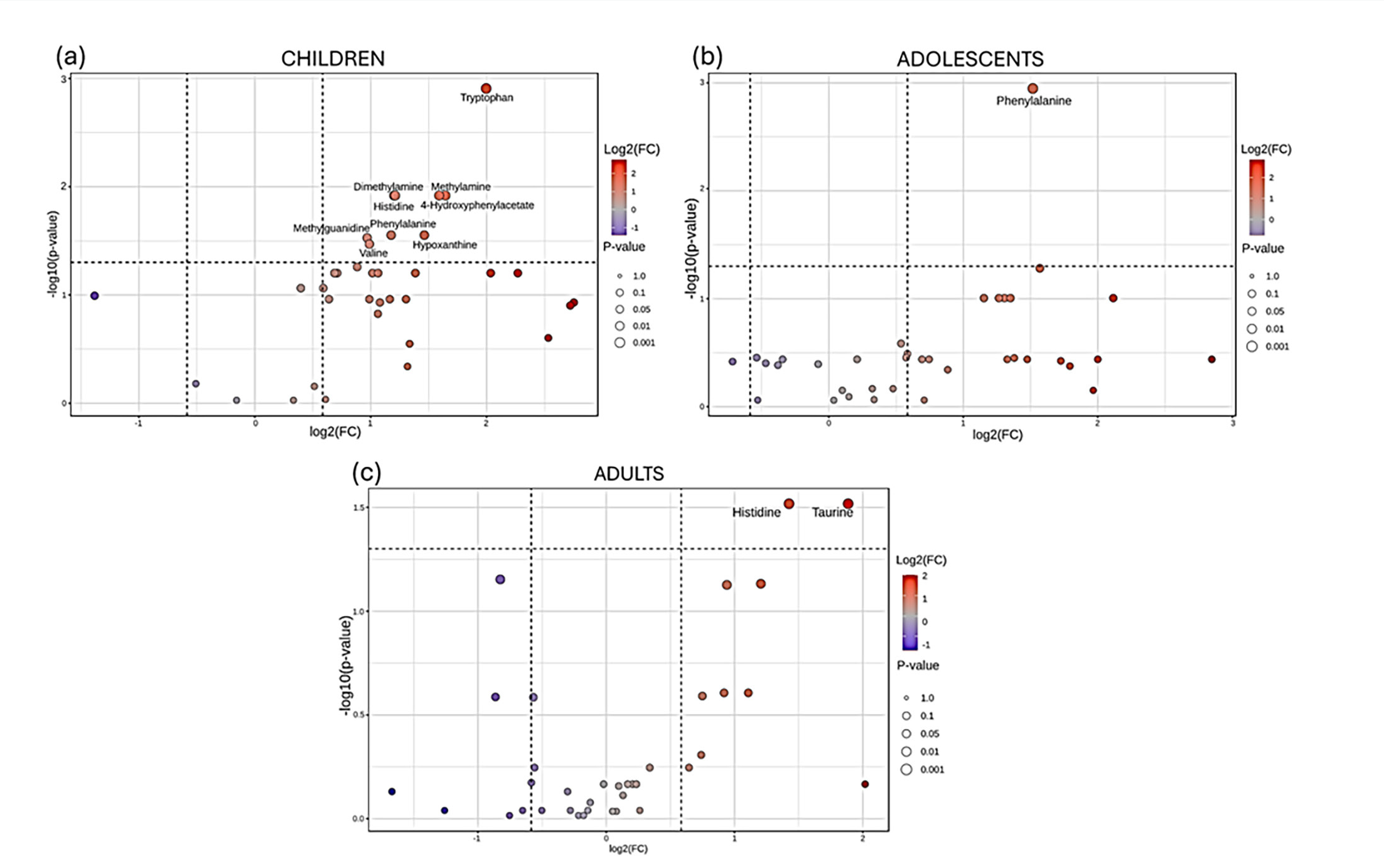
Fold change analysis and volcano plots were employed for identifying metabolites that are both statistically significant and biologically relevant (Figure 3). The fold change quantifies the magnitude of difference in metabolite levels between CDCS and control groups, highlighting those with the most significant variations, while the volcano plots provided a clear visual representation of metabolites that are most affected by the syndrome according within each age category. A summary of the variations is illustrated in Table 2.
Comparison of metabolite levels in urine of CDCS patients and control group from Fold-change analysis
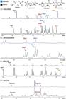
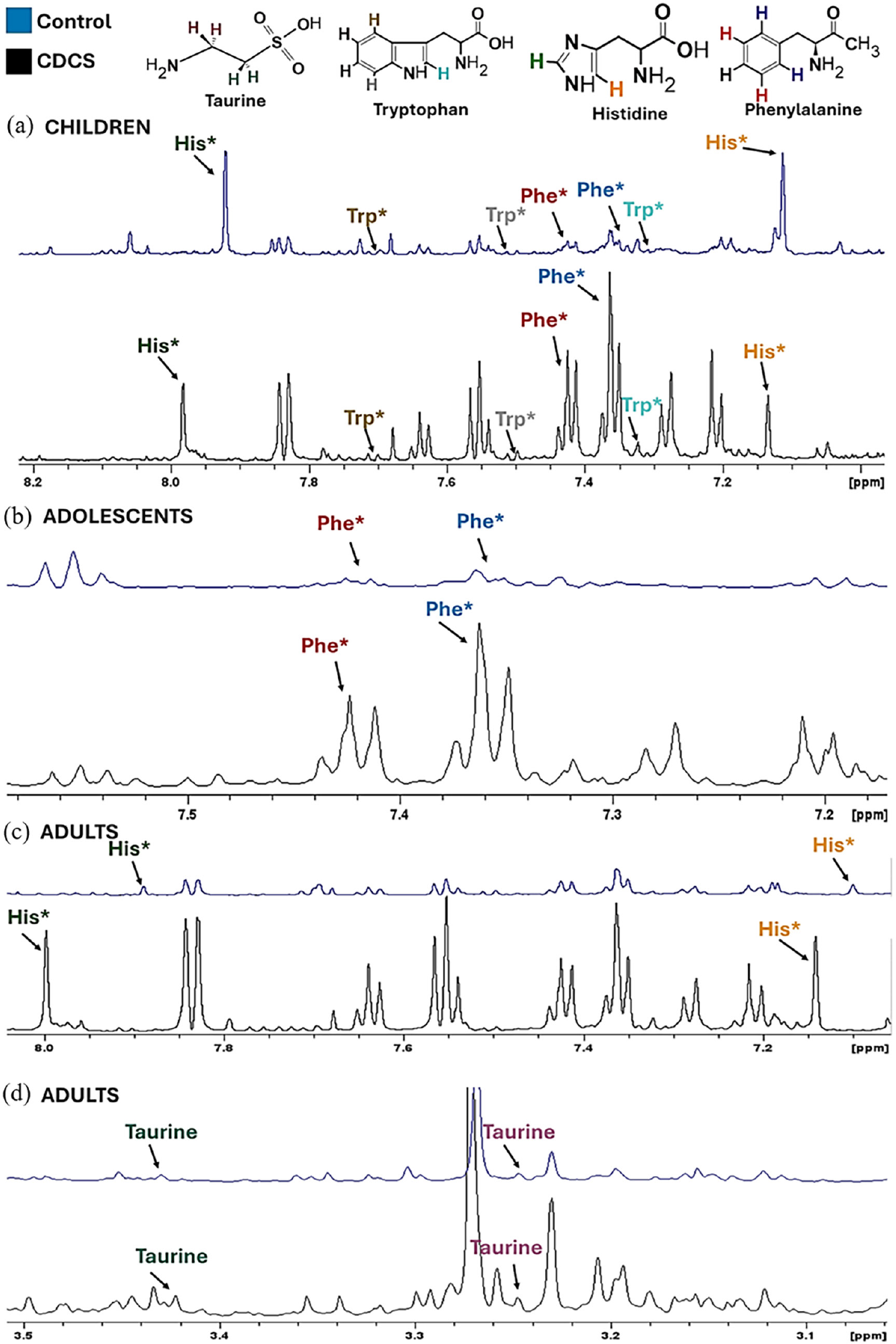
Representative 1H NMR spectra (600 MHz, D2O) of urine from control (blue) and CDCS (black) subjects. (a) Children (expanded aromatic region: 6.8-8.2 ppm), (b) adolescents (expanded aromatic region: 7.0-7.8 ppm), and (c-d) adults (expanded aromatic region: 7.0-8.0 ppm and aliphatic region: 3.0-3.5 ppm). Phe, phenylalanine; His, histidine; Trp, tryptophan.
In the children group, nine metabolites were found to be significantly increased: Trp, His, valine (Val), Phe, 4-hydroxyphenylacetate, methylalanine, dimethylamine, and methylguanidine (Figure 3a). Notably, tryptophan levels were elevated by approximately fourfold, while methylalanine and hypoxanthine showed about a threefold increase. Additionally, His, Val, and Phe were elevated by nearly twofold. In adolescents, only phenylalanine was significantly increased (Figure 3b). In contrast, the adult group exhibited elevated levels of His and taurine (Figure 3c). While taurine did not show significant changes in the other age categories, it was notably increased by nearly fourfold in adults compared to controls. Representative 1H NMR spectra are demonstrated in Figure 3, highlighting the observed variations in phenylalanine, histidine, and taurine in the three groups (children, adolescents and adults).
Key metabolic pathways alterations associated with CDCS
From our findings, elevated levels of His, Phe, Val, and Trp in children and increased Phe in adolescents were shown to significantly affect multiple amino acid metabolic pathways (Figures 4a-4b). Consistent with our results, Araújo et al.24 also reported elevated concentrations of several amino acids in the urine of individuals with CDCS, including aromatic (Tyr, Phe, Trp) and the branched-chain amino acids Val and isoleucine (Ile), among other metabolites. Disruptions in the biosynthesis of these amino acids resemble metabolic patterns observed in Hartnup syndrome, a recessive disorder caused by mutations in SLC6A19, which encodes the B0-type amino acid transporter (B0AT1).25,26 This transporter mediates intestinal absorption and renal reuptake of neutral amino acids, and its dysfunction leads to increased urinary and intestinal loss of Trp, Val and Phe. Notably, SLC6A19 is located on chromosome 5p15.33, a region also affected in CDCS.25 This shared chromosomal locus suggests a mechanistic link between the two conditions, supporting the idea that altered transport and homeostasis of neutral amino acids may contribute to the neurological and psychiatric manifestations characteristic of CDCS.27

Volcano plots showing differential urinary metabolite profiles between CDCS and control groups across age categories. (a) Children, (b) adolescents, and (c) adults. Each point represents a metabolite plotted according to fold change log2FC) on the x-axis and statistical significance (-log10 p-value) on the y-axis. Only metabolites with a fold change greater than 1.5 and p-value lower than 0.05 were considered significantly altered. Color gradient reflects the magnitude of fold change, and circle size indicates p-value.
Together, these observations highlight the importance of the 5p region in amino acid-dependent neurodevelopmental processes and reinforce the role of disrupted amino acid metabolism as a central biochemical feature of the syndrome. Supporting our findings, the excess of amino acids in the blood and urine of CDCS affected individuals has been previously reported in the literature.28,29 In a pioneering study, Kühner et al.28 observed for the first time kidney anomalies, along with increased serum and urine levels of proline and threonine in up to 4 months old infants, while Lejeune et al.30 demonstrated a significant excess of asparagine and aspartate, linked to disorders in purine metabolism.
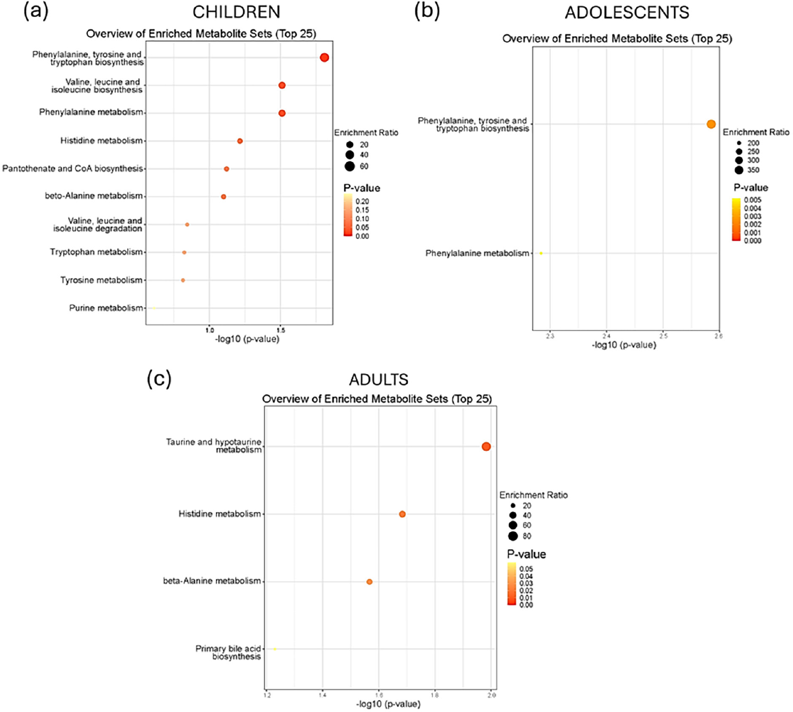
Enrichment analysis highlighted significant metabolic disturbances in pathways related to energy production (butanoate and glyoxylate metabolism) and amino acid metabolism, particularly Phe, tyrosine (Tyr), Trp biosynthesis in children (Figure 5a). In adolescents (Figure 5b), Phe biosynthesis and metabolism were prominently impacted, whereas in adults, disturbances were observed in taurine, hypotaurine, beta-alanine, and His metabolism (Figure 5c). This approach enabled the identification of key metabolites that reflect alterations in multiple molecular pathways associated with the syndrome across different age groups.

Metabolic pathways impacted by the CDCS. (a) Children. The enriched pathways include phenylalanine, tyrosine, and tryptophan biosynthesis, valine, leucine, and isoleucine biosynthesis, as well as histidine metabolism. (b) Adolescents. The most significant pathways are limited to phenylalanine, tyrosine, and tryptophan biosynthesis, and phenylalanine metabolism. (c) Adults. The most enriched pathways include taurine and hypotaurine metabolism, histidine metabolism, beta-alanine metabolism, and primary bile acid biosynthesis.
The literature demonstrates a strong correlation between Phe levels and the prevalence of neurological symptoms, particularly cognitive and behavioral abnormalities, similar to the findings in our investigation of CDCS.31,32 The excess of phenylalanine in blood and urine may be associated with another metabolic disorder, phenylketonuria (PKU).33,34 In PKU, a defect in the enzyme phenylalanine hydroxylase (PAH), responsible for converting Phe into Tyr, impairs phenylalanine metabolism, leading to its toxic accumulation in the brain. If left untreated, this can cause brain damage, developmental delays, and, in severe cases, intellectual disability.35,36 During fetal development, elevated phenylalanine levels, as seen in PKU, can lead to microcephaly, neuronal loss, and hypoplasia, as similarly to reported in CDCS. When exposure continues during early childhood, it may result in progressive neurological damage, severe cognitive impairment, and epilepsy, likely due to disrupted synaptogenesis.31,32
Our results revealed significant alterations in the metabolism of Phe, Tyr, and Trp in individuals with CDCS, indicating potential imbalances in neurotransmitter biosynthesis and amino acid metabolism that may contribute to the neurological manifestations of the syndrome. The metabolism of these amino acids is crucial for the production of essential secondary metabolites, including vitamins, neurotransmitters, and related compounds such as dopamine, serotonin, melatonin, and epinephrine.37,38 Disturbances in these pathways have also been reported in neurological disorders such as schizophrenia and Alzheimer’s disease (AD), where altered Trp metabolism drives neuroinflammation and oxidative stress, processes implicated in neurodegeneration and cognitive decline.39 Collectively, the overlap between our findings in CDCS and those observed in AD and other disorders highlights a possible shared metabolic vulnerability involving dysregulated aromatic amino acid metabolism.
The elevated concentration of Val observed in children with CDCS suggests disturbances in the biosynthesis of branched-chain amino acids (BCAAs), including leucine and isoleucine. This metabolic pattern resembles the alterations reported in maple syrup urine syndrome (MSUD), a rare metabolic syndrome caused by defects in the branched-chain α-ketoacid dehydrogenase complex, leading to the accumulation of Val, Ile, and Leu in plasma and urine.40 As in the CDCS, the characteristic features appear in the neonatal period and include developmental delays, feeding difficulties, metabolic decompensation and irreversible neurological complications.41
Imbalances in β-alanine and histidine metabolism were detected in both children and adults. In agreement with Furtado et al.,29 increased urinary histidine and taurine suggest disruptions in taurine and hypotaurine metabolism as well as purine metabolism, previously linked to CDCS by Lejeune et al.30 Given the essential role of purine metabolism in deoxyribonucleic acid/ribonucleic acid (DNA/RNA) synthesis and neurotransmission, its dysregulation may contribute to neurological symptoms resembling Lesch-Nyhan syndrome, where intellectual and behavioral disturbances are common.42
Furthermore, alterations in Ala, succinate, acetate and formate levels are linked to disturbance in energy production pathways, such as butanoate, glycoxylate and dicarboxylate metabolism. Alanine is a key intermediate in the glucose-alanine cycle, where it is converted to pyruvate for gluconeogenesis in the liver, sustaining energy supply during metabolic stress.39 The glucose-alanine cycle is a key metabolic process where alanine is converted to glucose and then utilized for energy, mainly in muscle glycolysis. This cycle becomes particularly significant during metabolic stress or critical illness, as Ala release increases to support gluconeogenesis, ensuring adequate energy supply including muscle tissues.39 Given that hypotonia is a hallmark of CDCS, disturbances in alanine metabolism may exacerbate energy deficits in muscle tissue, contributing to this phenotype.43
Individuals with CDCS frequently present anomalies affecting multiple organ systems,5,44 including congenital heart defects45,46 and kidney malfunction.28,45,47 In this context, taurine plays a crucial role in various physiological and biological processes within the kidney, often reflected in urinary excretion patterns.48 Moreover, studies suggest a correlation between taurine levels and liver damage.49,50 Elevated urinary taurine levels were observed in adults with CDCS (Figure 3), indicating disruptions in taurine and hypotaurine metabolism (Figure 4), potentially reflecting kidney dysfunction or age-related decline in renal capacity, consistent with previous clinical observations.
Taurine plays a critical role in osmoregulation and bile acid conjugation functions closely linked to renal physiology.51 The increased excretion observed may reflect organ dysfunction, particularly in the kidneys, as supported by previous studies.28,45,47 Notably, these metabolic alterations may also signal an age-related decline in renal function, suggesting that kidney impairment in CDCS may become more pronounced over time.
Collectively, our findings provide novel insights into the metabolic landscape of CDCS, revealing age-dependent alterations across amino acid, energy, and organ-related metabolic pathways. The overlap between these metabolic profiles and those seen in other neurological and metabolic disorders underscores the complex, multisystemic nature of CDCS and highlights potential biochemical targets for future studies.
Conclusions
This study represents the first comprehensive application of NMR-based metabolomic profiling to urine samples from individuals with Cri-du-chat syndrome, revealing distinct, age-dependent metabolic disturbances associated with the condition. By identifying alterations in amino acid metabolism, particularly the elevated excretion of phenylalanine in children, and disruptions in pathways such as the glucose-alanine cycle, our findings contribute important insights into the biochemical mechanisms that may underlie core clinical manifestations of CDCS, including cognitive impairment and persistent hypotonia. These results highlight the value of metabolomic profiling in advancing the understanding of CDCS pathophysiology and point toward potential metabolic biomarkers for monitoring syndrome progression and organ dysfunction. Although the rarity of CDCS constrains sample availability, the trends observed across age groups emphasize the relevance of integrating metabolic data with clinical and genetic evaluations. Future studies with larger cohorts and complementary analytical platforms will help refine these findings and support the development of more targeted, stage-specific therapeutic approaches. Overall, this work provides a foundational metabolic framework for CDCS and underscores the importance of multidisciplinary strategies to improve long-term outcomes and quality of life for affected individuals.
-
This publication is part of the special issue “Omics Sciences”
Supplementary Information
Additional data and materials are provided in the supplementary information file, available free of charge at http://jbcs.sbq.org.br as a PDF file.
Supplementary material 1
Acknowledgments
The authors gratefully acknowledge the financial support provided by CAPES under grant number 88882.332738/2019-1. We also thank the Nuclear Magnetic Resonance Laboratory at the Federal University of São Carlos for providing research infrastructure. Finally, we sincerely thank the Brazilian Association for Research and Support for Families of Cri-du-Chat in Brazil for their valuable contributions.
Data Availability Statement
Data supporting the findings of this study are available from the corresponding author upon reasonable request.
References
-
1 Rodríguez-Caballero, Á.; Torres-Lagares, D.; Rodríguez-Pérez, A.; Serrera-Figallo, M. Á.; Hernández-Guisado, J. M.; Machuca-Portillo, G.; Med. Oral Patol. Oral Cir. Bucal 2010, 15, e473. [Crossref]
» Crossref -
2 Lejeune, J.; Lafourcade, J.; Berger, R.; Vialatte, J.; Boeswillwald, M.; Seringe, P.; Turpin, R.; C. R. Hebd. Seances Acad. Sci. 1963, 257, 3098. [Crossref]
» Crossref -
3 Higurashi, M.; Lijima, K.; Sugimoto, Y.; Ishikawa, N.; Hoshina, H.; Watanabe, N.; Yonwyama, K.; Am. J. Med. Genet. 1980, 6, 189. [Crossref]
» Crossref -
4 Almeida, V. T.; Chehimi, S. N.; Carvalho, G. F. S.; Gasparini, Y.; Nascimento, A. M.; Vieira, L. L.; Wolff, B. M.; Montenegro, M. M.; Kulikowski, L. D.; BMC Res. Notes 2024, 17, 121. [Crossref]
» Crossref -
5 Peng, Y.; Pang, J.; Hu, J.; Jia, Z.; Xi, H.; Ma, N.; Yang, S.; Liu, J.; Huang, X.; Tang, C.; Wang, H.; Mol. Genet. Genomic Med. 2020, 8, e1312. [Crossref]
» Crossref -
6 Almeida, V. T.; Chehimi, S. N.; Gasparini, Y.; Nascimento, A. M.; Carvalho, G. F. S.; Montenegro, M. M.; Zanardo, É. A.; Dias, A. T.; Assunção, N. A.; Kim, C. A.; Kulikowski, L. D.; Mol. Syndromol. 2022, 13, 527. [Crossref]
» Crossref -
7 Teixeira, M. C. T. V.; Emerich, D. R.; Orsati, F. T.; Rimério, R. C.; Gatto, K. R.; Chappaz, I. O.; Kim, C. A.; J. Intellect. Disabil. Res. 2011, 55, 132. [Crossref]
» Crossref -
8 Cerruti Mainardi, P.; Orphanet J. Rare Dis. 2006, 1, 33. [Crossref]
» Crossref -
9 Nguyen, J. M.; Qualmann, K. J.; Okashah, R.; Reilly, A.; Alexeyev, M. F.; Campbell, D. J.; Am. J. Med. Genet., Part C: Semin. Med. Genet. 2015, 169, 224. [Crossref]
» Crossref -
10 Nevado, J.; Bel-Fenellós, C.; Sandoval-Talamantes, A. K.; Hernández, A.; Biencinto-López, C.; Martínez-Fernández, M. L.; Barrúz, P.; Santos-Simarro, F.; Mori-Álvarez, M. Á.; Mansilla, E.; García-Santiago, F. A.; Valcorba, I.; Sáenz-Rico, B.; Martínez-Frías, M. L.; Lapunzina, P.; Front. Genet. 2021, 12, 645595. [Crossref]
» Crossref -
11 Corrêa, T.; Feltes, B. C.; Riegel, M.; Genet. Mol. Biol. 2019, 42, 186. [Crossref]
» Crossref -
12 Holland, P.; Wildhagen, M.; Istre, M.; Reiakvam, O. M.; Dahl, J. A.; Søraas, A.; Clin. Epigenet. 2022, 14, 128. [Crossref]
» Crossref -
13 Pituch, K. A.; Green, V. A.; Didden, R.; Whittle, L.; O’Reilly, M. F.; Lancioni, G. E.; Sigafoos, J.; J. Dev. Phys. Disabil. 2010, 22, 65. [Crossref]
» Crossref -
14 Del Boccio, P.; Rossi, C.; di Ioia, M.; Cicalini, I.; Sacchetta, P.; Pieragostino, D.; Proteomics: Clin. Appl. 2016, 10, 470. [Crossref]
» Crossref -
15 Caracausi, M.; Ghini, V.; Locatelli, C.; Mericio, M.; Piovesan, A.; Antonaros, F.; Pelleri, M. C.; Vitale, L.; Vacca, R. A.; Bedetti, F.; Mimmi, M. C.; Luchinat, C.; Turano, P.; Strippoli, P.; Cocchi, G.; Sci. Rep. 2018, 8, 20834. [Crossref]
» Crossref -
16 Rawat, A.; Misra, G.; Saxena, M.; Tripathi, S.; Dubey, D.; Saxena, S.; Aggarwal, A.; Gupta, V.; Khan, M. Y.; Prakash, A.; Diabetes Metab. Syndr.: Clin. Res. Rev. 2019, 13, 290. [Crossref]
» Crossref -
17 Bouatra, S.; Aziat, F.; Mandal, R.; Guo, A. C.; Wilson, M. R.; Knox, C.; Bjorndahl, T. C.; Krishnamurthy, R.; Saleem, F.; Liu, P.; Dame, Z. T.; Poelzer, J.; Huynh, J.; Yallou, F. S.; Psychogios, N.; Dong, E.; Bogumil, R.; Roehring, C.; Wishart, D. S.; PLoS One 2013, 8, e73076. [Crossref]
» Crossref -
18 Beckonert, O.; Keun, H. C.; Ebbels, T. M. D.; Bundy, J.; Holmes, E.; Lindon, J. C.; Nicholson, J. K.; Nat. Protoc. 2007, 2, 2692. [Crossref]
» Crossref -
19 Cri-du-Chat Advisory and Research Center. [Link] accessed in February 2026
» Link -
20 Emwas, A.-H.; Roy, R.; McKay, R. T.; Ryan, D.; Brennan, L.; Tenori, L.; Luchinat, C.; Gao, X.; Zeri, A. C.; Gowda, G. A. N.; Raftery, D.; Steinbeck, C.; Salek, R. M.; Wishart, D. S.; J. Proteome Res. 2016, 15, 360. [Crossref]
» Crossref -
21 Wishart, D. S.; Tzur, D.; Knox, C.; Eisner, R.; Guo, A. C.; Young, N.; Cheng, D.; Jewell, K.; Arndt, D.; Sawhney, S.; Fung, C.; Nikolai, L.; Lewis, M.; Coutouly, M.-A.; Forsythe, I.; Tang, P.; Shrivastava, S.; Jeroncic, K.; Stothard, P.; Amegbey, G.; Block, D.; Hau, D. D.; Wagner, J.; Miniaci, J.; Clements, M.; Gebremedhin, M.; Guo, N.; Zhang, Y.; Duggan, G. E.; MacInnis, G. D.; Weljie, A. M.; Dowlatabadi, R.; Bamforth, F.; Clive, D.; Greiner, R.; Li, L.; Marrie, T.; Sykes, B. D.; Vogel, H. J.; Querengesser, L.; Nucleic Acids Res. 2007, 35, D521. [Crossref]
» Crossref -
22 Ulrich, E. L.; Akutsu, H.; Doreleijers, J. F.; Harano, Y.; Ioannidis, Y. E.; Lin, J.; Livny, M.; Mading, S.; Maziuk, D.; Miller, Z.; Nakatani, E.; Schulte, C. F.; Tolmie, D. E.; Wenger, R. K.; Yao, H.; Markley, J. L.; Nucleic Acids Res. 2008, 36, D402. [Crossref]
» Crossref -
23 Xia, J.; Psychogios, N.; Young, N.; Wishart, D. S.; Nucleic Acids Res. 2009, 37, W652. [Crossref]
» Crossref -
24 Araújo, B. R.; Furtado, D. Z. S.; de Moura Leite, F. B. V.; de Assunção, N. A.; Carrilho, E.; J. Chromatogr. B 2020, 1153, 122267. [Crossref]
» Crossref -
25 Kravetz, Z.; Schmidt-Kastner, R.; IBRO Neurosci. Rep. 2023, 14, 393. [Crossref]
» Crossref -
26 Bröer, S.; Physiology 2008, 23, 95. [Crossref]
» Crossref -
27 Danthi, S. J.; Liang, B.; Smicker, O.; Coupland, B.; Gregory, J.; Gefteas, E.; Tietz, D.; Klodnitsky, H.; Randall, K.; Belanger, A.; Kuntzweiler, T. A.; SLAS Discovery 2019, 24, 111. [Crossref]
» Crossref -
28 Kühner, U.; Büsse, M.; Buchinger, G.; Z. Kinderheilkd. 1974, 117, 259. [Crossref]
» Crossref -
29 Furtado, D. Z. S.; Leite, F. B. V. M.; Barreto, C. N.; Faria, B.; Jedlicka, L. D. L.; Silva, E. J.; Silva, H. D. T.; Bechara, E. J. H.; Assunção, N. A.; J. Pharm. Biomed. Anal. 2017, 140, 137. [Crossref]
» Crossref -
30 Lejeune, J.; Rethoré, M. O.; Peeters, M.; de Blois, M. C.; Rabier, D.; Parvy, P.; Bardet, J.; Kamoun, P.; Ann. Genet. 1990, 33, 16. [Crossref]
» Crossref -
31 Rovelli, V.; Longo, N.; Mol. Genet. Metab. 2023, 139, 107583. [Crossref]
» Crossref -
32 Bone, A.; Kuehl, A. K.; Angelino, A. F.; Psychosomatics 2012, 53, 517. [Crossref]
» Crossref -
33 Anderson, P. J.; Leuzzi, V.; Mol. Genet. Metab. 2010, 99, S3. [Crossref]
» Crossref -
34 Pilotto, A.; Zipser, C. M.; Leks, E.; Haas, D.; Gramer, G.; Freisinger, P.; Schaeffer, E.; Liepelt-Scarfone, I.; Brockmann, K.; Maetzler, W.; Schulte, C.; Deuschle, C.; Hauser, A. K.; Hoffmann, G. F.; Scheffler, K.; van Spronsen, F. J.; Padovani, A.; Trefz, F.; Berg, D.; Neurology 2021, 96, e399. [Crossref]
» Crossref -
35 Cannet, C.; Bayat, A.; Frauendienst-Egger, G.; Freisinger, P.; Spraul, M.; Himmelreich, N.; Kockaya, M.; Ahring, K.; Godejohann, M.; MacDonald, A.; Trefz, F.; Molecules 2023, 28, 4916. [Crossref]
» Crossref -
36 van Spronsen, F. J.; Blau, N.; Harding, C.; Burlina, A.; Longo, N.; Bosch, A. M.; Nat. Rev. Dis. Prim. 2021, 7, 36. [Crossref]
» Crossref -
37 Almulla, A. F.; Supasitthumrong, T.; Amrapala, A.; Tunvirachaisakul, C.; Abdul Jaleel, A.-K. K.; Oxenkrug, G.; Al-Hakeim, H. K.; Maes, M.; J. Alzheimers Dis. 2022, 88, 1325. [Crossref]
» Crossref -
38 Parthasarathy, A.; Cross, P. J.; Dobson, R. C. J.; Adams, L. E.; Savka, M. A.; Hudson, A. O.; Front. Mol. Biosci. 2018, 5, 19. [Crossref]
» Crossref -
39 Holeček, M.; Int. J. Mol. Sci. 2024, 25, 7037. [Crossref]
» Crossref -
40 Blackburn, P. R.; Gass, J. M.; Pinto e Vairo, F.; Farnham, K. M.; Atwal, H. K.; Macklin, S.; Klee, E. W.; Atwal, P. S.; Appl. Clin. Genet. 2017, 10, 57. [Crossref]
» Crossref -
41 Strauss, K. A.; Wardley, B.; Robinson, D.; Hendrickson, C.; Rider, N. L.; Puffenberger, E. G.; Shelmer, D.; Moser, A. B.; Morton, D. H.; Mol. Genet. Metab. 2010, 99, 333. [Crossref]
» Crossref -
42 Krajewski, O.; Opiełka, M.; Urbanowicz, K.; Chojnowski, K.; Kochany, P.; Pawłowski, K.; Tomaszewska, J.; Peters, G. J.; Smolenski, R. T.; Mazurkiewicz-Bełdzińska, M.; Neurosci. Biobehav. Rev. 2024, 165, 105847. [Crossref]
» Crossref -
43 Felig, P.; Metabolism 1973, 22, 179. [Crossref]
» Crossref -
44 Hills, C.; Moller, J. H.; Finkelstein, M.; Lohr, J.; Schimmenti, L.; Pediatrics 2006, 117, e924. [Crossref]
» Crossref -
45 Honjo, R. S.; Mello, C. B.; Pimenta, L. S. E.; Nuñes-Vaca, E. C.; Benedetto, L. M.; Khoury, R. B. F.; Befi-Lopes, D. M.; Kim, C. A. J.; J. Intellect. Disabil. Res. 2018, 62, 467. [Crossref]
» Crossref -
46 Patrícia, T.; Rosa, R. F. M.; Koshiyama, D. B.; Diehl, Z. T.; Paskulin, G. A.; Zen, P. R. G.; Rev. Paul. Pediatr. 2014, 32, 262. [Crossref]
» Crossref -
47 Egli, F.; Stalder, G.; Humangenetik 1973, 18, 1. [Crossref]
» Crossref -
48 Chesney, R. W.; Han, X.; Patters, A. B.; J. Biomed. Sci. 2010, 17, S4. [Crossref]
» Crossref -
49 Waterfield, C. J.; Turton, J. A.; Scales, M. D. C.; Timbrell, J. A.; Arch. Toxicol. 1991, 65, 548. [Crossref]
» Crossref -
50 Heidari, R.; Jamshidzadeh, A.; Niknahad, H.; Mardani, E.; Ommati, M. M.; Azarpira, N.; Khodaei, F.; Zarei, A.; Ayarzadeh, M.; Mousavi, S.; Abdoli, N.; Shirazi Yeganeh, B.; Saeedi, A.; Najibi, A.; Toxicol. Rep. 2015, 3, 870. [Crossref]
» Crossref -
51 Ripps, H.; Shen, W.; Mol. Vis. 2012, 18, 2673. [Crossref]
» Crossref
Edited by
-
Editor handled this article:
Paulo Wender (Guest)