Analysis of Cocaine, Benzoylecgonine, and Ecgonine Methyl Ester in Hair Strands Via Matrix-Assisted Laser Desorption Ionization Mass Spectrometry (MALDI-MS)
Abstract
Hair can provide chemical information about drug use. This study employed matrix-assisted laser desorption ionization mass spectrometry (MALDI-MS) to detect drugs in hair samples. The technique was used to detect cocaine (COC, molecular weight (Mw) = 303.14 Da) and its main metabolites, benzoylecgonine (BZE, Mw = 289.13 Da), cocaethylene (COCE, Mw = 317.16 Da) and ecgonine methyl ester (EME, Mw = 199.12 Da) in hair strands using 2,5-dihydroxybenzoic acid (DHB, 7.0 mg mL-1) and α-cyano-4-hydroxycinnamic acid (CHCA, at concentrations of 1.5, 3.0, 7.0, and 10 mg mL-1) as the evaluated matrices. Among these, CHCA was selected. Subsequently, the qualitative results obtained by MALDI(+) MS were correlated with the quantitative analyses of gas chromatography-mass spectrometry (GC-MS), demonstrating a significant correlation in responses (70%), with the detection of analytes by MALDI(+) MS even at low concentrations, determined by GC-MS. The concentration ranges obtained by GC MS analysis were from 0.6 to 4.1 ng mg-1 of COC, and from 0.05 to 0.4 ng mg-1 of COCE. The effect of pre-washing on the detection of COC and its metabolites was also evaluated, and the results indicated less influence from the pre-washing stage and confirmed the reduction in COC detection in hair exposed to bleaching.
Keywords:
cocaine; hair; MALDI; TOF MS
Introduction
Recreational drug use has been reported throughout history and has become a recurrent topic in policy and public safety debates.1-4 For example, according to the most recent report by the United Nations Office on Drugs and Crime (UNODC),5 around 284 million people, aged between 15 and 64 years, used drugs in 2020, 26% more than in 2010. Furthermore, greater variety of substances has become available on the illicit market in recent years, along with an expansion in the use of substances of natural origin, such as Cannabis (marijuana), Erythroxylum coca (cocaine), and Papaver somniferum (heroin).5
Significant technological advances and the development of increasingly sensitive and robust methods have allowed for the detection of lower analyte concentrations;6 this is important in toxicological analyses to verify, for example, the use of illicit or licit drugs. In humans, blood or urine matrices are mainly used in toxicological investigations,6 with less common matrices including saliva,7 hair,8,9 sweat,10 meconium,11 and vitreous humor.12,13 With approximate detection times after ingesting a single dose varying from one substance to another, the detection window for drugs is between 3 and 6 months in hair, while the interval for blood and oral fluid is 1 to 2 days and that for urine is 2 to 4 days in comparison.14
The hair strand is a fiber originating from hair follicles located 3 to 4 mm below the epithelial layer.13,15 Hair growth occurs in three stages: anagen, catagen, and telogen. The anagen phase (Figure S1, phases 1 and 4, presented in Supplementary Information, SI section) can last for approximately 3 years and involves the active growth of hair from the bulb; subsequently, growth progresses into the catagen phase, a transitional period, and thereafter the telogen phase, a period of rest, as shown in Figure S1, phases 2 and 3, SI section, respectively. Growth ceases in the catagen phase, with the formation of a keratinized compound within the follicle (Figure S1).16 Compounds can be incorporated into a hair strand through its shaft in several ways: through the bloodstream in the anagen phase, via sweat after hair formation, or from environmental exposure.17
Drugs of abuse, such as cocaine, can be incorporated into the hair shaft through the blood, with the process depending on the physical-chemical properties of each drug and the melanin in the hair.18,19 Cocaine, as a psychomotor stimulant, acts on the central nervous system, leading to euphoria, increased alertness, and increased energy, and the cardiovascular system, resulting in tachycardia, vasoconstriction, and hyperthermia.20,21 It exists in base or salt form and can be administered orally, snorted, smoked, or injected (intravenously).21,22 After administration, cocaine is rapidly distributed throughout the body, with the highest concentrations of the drug present in the brain, spleen, kidneys, lungs, blood, heart, and muscles.23
Cocaine is metabolized in the hepatic tissue, producing the main metabolites benzoylecgonine (BZE, molecular weight (Mw) = 289.13 Da) and ecgonine methyl ester (EME, Mw = 199.12 Da) and minor metabolites such as norcocaine (NCOC, Mw = 289.33 Da), hydroxycocaine (OH-COC, Mw = 319.40 Da), and hydro benzoylecgonine (OH-BE, Mw = 305.32 Da).24-28
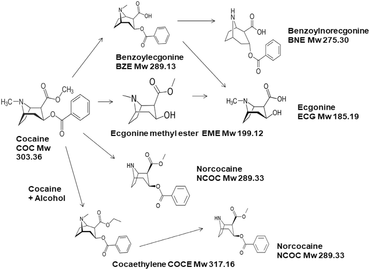
BZE can be formed via chemical hydrolysis at alkaline pH and through the action of esterases, such as carboxylesterase type 1. Further, it can undergo oxidation to form OH-BE. EME is produced via enzymatic hydrolysis with two types of esterases: plasma cholinesterases and hepatic esterases. In addition, BZE and EME can be converted to ecgonine (ECG). Cocaine can undergo n-demethylation to form NCOC, which is metabolized to n-hydroxycocaine and oxidized to cocaine nitroxide.24-28 Furthermore, the administration of cocaine and alcohol leads to the formation of cocaethylene (COCE) via transesterification in the liver,20 with COCE being further transformed into norcocaethylene (NorCE) and ecgonine ethyl ester (EEE). The presence of ethanol also increases cocaine demethylation to NCOC.29Figure 1 shows the main metabolic pathways of cocaine.

Cocaine (COC) metabolic pathways with BZE, BNE, EME, ECG, NCOC, and COCE (adapted from reference 29).
Cocaine, along with its metabolites BZE, EME, and ECG, is excreted in urine in its original, unchanged form over 24 h.21,30 In oral fluids, cocaine is detectable for 5 to 12 h in a single dose and for up to 10 days in the case of chronic use.31 In comparison, as indicated by Garcia Bournissen et al.,32 in 2009, cocaine can be detected in hair strands after 3 to 4 months in former drug users.
Currently, several techniques exist for identifying drugs of abuse, including gas chromatography (GC),32,33 liquid chromatography (LC),34 nuclear magnetic resonance (NMR) spectroscopy,35 attenuated total reflection Fourier transform infrared (ATR FTIR) spectroscopy,36 mass spectrometry (MS),37-45 colorimetric tests (e.g., Scott Test),37,46 and hyphenated techniques (e.g., GC-MS and LC-MS).
A widely adopted technique in the study of drugs in hair is LC-MS, which eliminates the derivatization step of GC MS required in many drug analyses.47 Although GC-MS and LC-MS are widely used in toxicological analyses on hair, they involve laborious sample preparation steps and require large quantities of hair strands.38 In this context, matrix-assisted laser desorption ionization mass spectrometry (MALDI-MS) appears as an analytical method to reduce the preparation time; it is a soft ionization technique, involving the use of pulsed laser beams, such as N2, Nd:YAG, CO2, and Er:YAG, in the ultra violet (UV) and infra-red (IR) regions.48
MALDI has been applied to the study of compounds in hair. For instance, Porta et al.49 observed the spatial distribution of cocaine and its metabolites in hair samples from users by MALDI mass spectrometry imaging (MSI). In addition, Musshoff et al.50 developed a method for analyzing four hair strands from drug users to detect cocaine and its metabolites and cannabinoids using MALDI Fourier transform mass spectrometry (MALDI-FT MS). The analytes were detectable in two samples, providing chronological information about drug consumption as a function of time.
Cuypers et al.51 used MALDI-MSI to observe that cocaine and its metabolites are detectable in hair treated with chemical bleaching. The spatial distribution of the analytes was explored using the time-of-flight (TOF) analyzer, α-cyano-4-hydroxycinnamic acid (CHCA) matrix, and Nd:YAG laser. The authors investigated hair from drug users and hair samples doped with cocaine and also studied the effect of the presence of hydrogen peroxide (H2O2) as a chemical agent. They concluded that chemical treatment reduces the detection of cocaine due to the breaking of the bond between cocaine and melanin in the presence of H2O2.
Therefore, although the MALDI-MS technique is incipient in the study of drugs and their metabolites in hair samples, it serves as a promising tool compared to classical chromatographic techniques (GC and LC) due to its simplified sample preparation, short analysis time, and spatial resolution, enabling the construction of chemical images of the detected components.38,52
Considering this, matrix-assisted laser-desorption and ionization time-of-flight mass spectrometry (MALDI(+)TOF MS) was applied for the rapid analysis of hair strands from drug users, where the sample preparation step was optimized (efficiency of the MALDI CHCA and DHB matrices, and different concentrations of the CHCA matrix, in addition to the hair washing and chemical treatment step). After determining the optimal conditions, 10 hair strands from cocaine users were evaluated, and the results obtained from MALDI(+)TOF MS were compared with those from GC MS analyses.
Experimental
Samples and reagents
Ten natural hair samples (from the leg, armpit, and pubic region) were analyzed, with a standardized size of 2 cm each, and without the addition of chemicals or previous treatments, without identifying gender or age group. The samples were provided by Contraprova Laboratory - Analyses, Teaching and Research LTDA.
In addition, another sample from a donor with no history of drug use was used for subsequent doping. The project was approved by the Research Ethics Council with CAAE number 65316722.3.0000.5542 for use in research with human hair. In addition, the research group has a technical cooperation agreement with the Civil Police of Espírito Santo, process No. 23068.022157/2020-69.
The reagents and solvents used were methanol, ethanol, and acetonitrile, purchased from Vetec® Química Fina Ltda (Rio de Janeiro, Brazil); dichloromethane (JT Baker®, Carnaxide, Portugal); hexane (Merck®, Darmstadt, Germany); acetone (Merck®, Darmstadt, Germany); and hydrochloric acid (Applied Biosystems®, Massachusetts, USA), all with analytical purity greater than 99.5%. The analytical standards; cocaine (COC), with molecular formula (M) of C17H21NO4, Mw = 303.14 Da, and concentration = 1 mg mL-1; benzoylecgonine-D3 (BZE-D3), M = C16D3H19NO4, Mw = 292.13 Da, [BZE-D3] = 100 µg mL-1; and ecgonine methyl ester (EME), M = C10H17NO3, Mw = 199.12 Da, [EME] = 100 µg mL-1, were acquired from Cerilliant (Round Rock, TX, USA).
CHCA and 2,5-dihydroxybenzoic acid (DHB) matrices were purchased from Sigma-Aldrich Chemicals (Oakville, ON, Canada) and were selected based on the literature,19 where 2 μL of the matrix solutions was deposited on hair strand samples with a micropipette.
Sample preparation
To clean the MALDI plate, solvents such as ethanol, water, acetonitrile, and 0.1 mol L-1 hydrochloric acid were used. Briefly, in the first washing step, 50 mL of ethanol, 50 mL of distilled water, and another 50 mL of ethanol were used. Thereafter, the plate was sonicated for 20 min: 10 min in 500 mL of distilled water and then in a solvent mixture of ethanol:water:hydrochloric acid (2:2:1 v/v, 0.1 mol L-1). In the final step, the plate was washed in 50 mL of acetonitrile, as shown in Figure S2 (SI section).
The sample preparation was divided into two steps. The first involved the application of 1 µL of the COC analytical standard (1 mg mL-1), BZE-D3 analytical standard (100 µg mL-1), and EME analytical standard (100 µg mL 1), covered by the DHB and CHCA matrices separately on the MALDI plate (2 µL of 7 mg mL-1 concentration each, Figure S3a, SI section). Four concentrations of the CHCA matrix were then evaluated (from 1 to 10 mg mL-1). In the second step, the optimal matrix and concentration were determined, and the hairs from drug users were glued to the plate with double-sided tape and labeled as samples “A” to “K”. The sample preparation scheme is shown in Figure S3b (SI section).
Initially, two positive samples for COC (A and B) containing three strands of each hair sample were used to evaluate the ionization of analytes with and without the washing step (involving 20 mL each of distilled water, dichloromethane, acetone, and hexane). The objective when washing is to remove impurities such as sweat, and grease, environmental contaminants such as dust, pollution, and traces of shampoos or creams. After determining the efficiency of the sample preparation method in the presence and absence of the washing step, the remaining hair samples (C to K) were analyzed using 2 µL of the 1.5 mg mL-1 CHCA matrix.
MALDI-TOF MS
The MALDI-TOF MS analysis was performed using a Bruker Ultraflex II instrument (Bruker Daltonics, Bremen, Germany), operated in the reflector positive ion mode with a spectral range of m/z 150 to 1000. The laser was focused on the hair segment linearly, with a laser power of 10-20%, laser frequency of 50.0 Hz, and number of shots = 200. The pulsed ion extraction conditions were as follows: IS1 (19.08 kV), IS2 (17.03 kV), and reflector (20.361 kV). The external mass calibration was performed based on the [M + H]+ and [2M + H]+ ions at m/z 190.04 and 379.09 (for CHCA), and m/z 155.02 and 309.02 (for DHB). The data obtained were analyzed using the Data Analysis Daltonics software, USA.
GC-MS
The gas chromatography-tandem mass spectrometry (GC-MS/MS) analysis was performed on a Trace 1300 gas chromatograph coupled with a TSQ 8000 Evo triple quadrupole mass spectrometer (Thermo Fisher Scientific, Waltham, USA). In the analysis, 20 mg of the sample (hair) was washed with dichloromethane for decontamination purposes, and subsequently subjected to grinding using the Fast-Prep® mill. After adding the internal standard (COC-D3), the ground sample was digested with 50 µL of a phosphate buffer (pH 5.4) and under heating for 10 min; the supernatant was partitioned with acetonitrile. The evaporation residue was resuspended with acetonitrile solution in an alkaline medium. After complete evaporation, the residue was resuspended in ethyl acetate for chromatographic analysis using a GC system coupled to a mass spectrometer in tandem mode (GC-MS/MS). The capillary column used for the sample separation had a stationary phase composition including 35% phenyl polysilphenylene-siloxane (DB-35MS) and dimensions of 30 m (length) × 0.25 mm (diameter) × 0.25 μm (film thickness). The method of quantitative analysis of the compound was developed and validated by Contraprova Laboratory - Doping and Toxicology and is accredited by Inmetro according to ABNT NBR ISO/IEC 17025:201753 standard, since 2015.
All quantitative analyses performed by GC-MS/MS were obtained by inserting the result of the relationship between the analyte areas of the sample and its internal standard in a calibration curve produced with analyte-free hair matrices and doped concerning the substance analyzed. In these analyses, the calibration curves produced together with the samples obtained a linear correlation coefficient greater than 0.98. Furthermore, quality controls of low (close to the cut-off value) and high concentrations are processed along with the analytical curve and evaluated for accuracy and precision, before quantifying the analytes in the samples. The limit of quantification (LOQ) of cocaine in this study was 300.8 pg mg-1 and was determined during the analytical method validation process.
Results and Discussion
Evaluation of ionization efficiency of DHB and CHCA matrices
Initially, the COC standard at a concentration of 1 mg mL-1 was analyzed with the DHB and CHCA matrices (both at 7 mg mL-1) to optimize the ionization process for the analyte of interest via MALDI(+) MS. In this analysis (Figure 2), COC was detected in the protonated form, [C17H21NO4 + H]+, m/z 304.08 (CHCA) and m/z 304.16 (DHB). Despite the similarity in the ionization efficiency (total ion current [TIC] = 2 × 10³) of the matrices, when evaluating the TIC values of COC in each mass spectrum, the CHCA matrix was selected for further MALDI(+) analyses because of its lower proton affinity (PA) and pKa: CHCA (PA = 183 kcal mol-1 and pKa = 1.2) versus DHB (PA = 204 kcal mol-1 and pKa = 3.0).54,55

MALDI(+)TOF mass spectra of the COC standard (1 mg mL 1) with the matrices (a) CHCA and (b) DHB, both at a concentration of 7 mg mL-1.
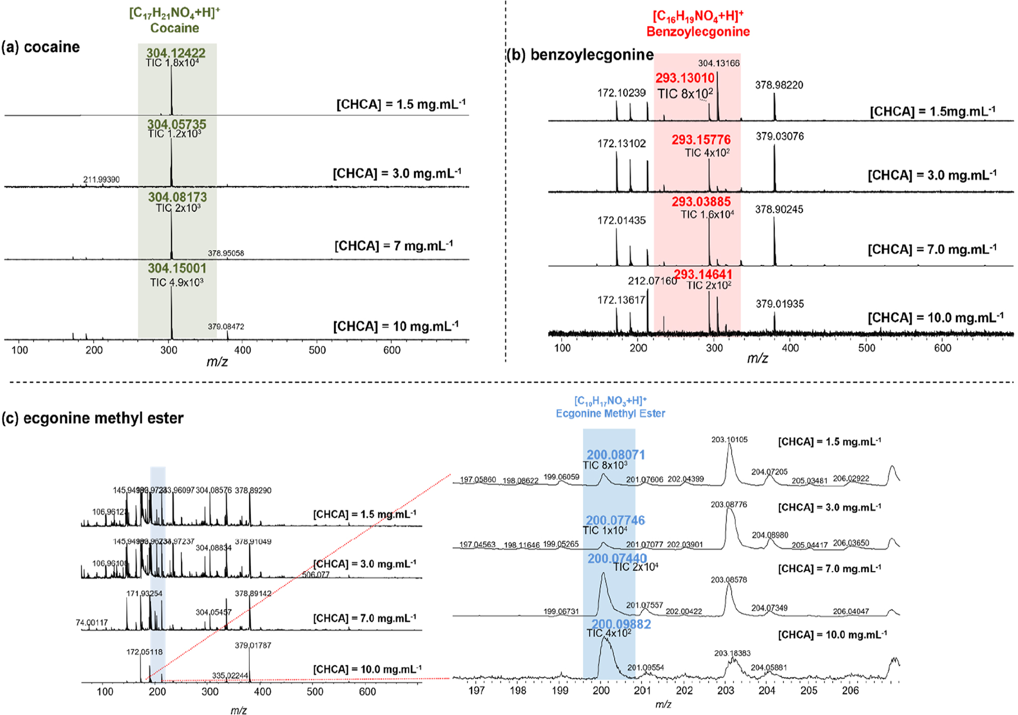
Four concentrations of the CHCA matrix (1.5, 3.0, 7.0, and 10 mg mL-1) were sequentially investigated to identify the optimal working concentration for the ionization of the COC (1 mg mL-1), BZE (100 μg mL-1), and ECG (100 μg mL-1) standards. In the MALDI(+) spectra (Figure 3), the signal for the COC molecule is intense and is the main peak for all tested concentrations of the CHCA matrix. However, this same behavior is not observed for its metabolites. The majority of the BZE and EME standards, ions of m/z 293 and 200, respectively, are not ionized due to their low concentrations (100 μg mL-1). Nevertheless, one may infer that all the tested concentrations of CHCA (1.5, 3.0, 7.0, and 10 mg mL-1) could be used for the detection of at least one monitored target compound. The CHCA concentrations of 1.5 and 7.0 mg mL-1 were optimal in the detection of COC and its metabolites, respectively.

MALDI(+)TOF mass spectra of (a) COC (1 mg mL-1), (b) BZE-D3 (100 µg mL-1), and (c) EME (100 µg mL-1) using 1.5-10 mg mL-1 CHCA.
Table 1 presents the data of the acquired MALDI(+) spectra and further elucidates the results. The lowest tested concentration of the CHCA matrix (i.e., 1.5 mg mL-1) yields the highest sensitivity and efficiency in the ionization of the COC analyte, with a value of TIC(CHCA = 1.5 mg mL-1) = 1.8 × 10⁴, while the remaining matrix concentrations result in TIC values on the order of 103. Therefore, 1.5 mg mL-1 CHCA was selected to further develop the MALDI(+)TOF MS method for the toxicological analysis of hair.
MALDI(+)TOF MS data of the COC standard (1 mg mL-1) and its metabolites, EME and BZE-D3 (100 μg mL-1), for different concentrations (1.5-10 mg mL-1) of the CHCA matrix
Effect of washing the hair strands (pre-analysis)
The hair washing step is used to remove possible impurities such as sweat and grease, as well as environmental contaminants such as dust, pollution, and traces of personal hygiene products, such as cream and shampoo. There is no consensus on a specific washing protocol for hair decontamination in the stages before toxicological analysis.56 However, the Society of Hair Testing (SoHT) Guidelines for Drug Testing in Hair recommends washing with organic products (solvents) and aqueous solutions.52 In this study, the influence of washing was verified in the pre-analysis stage using two samples of COC-positive hair and dichloromethane, water, hexane, and acetone to wash the hair.
Three hairs of two individuals, named “A” and “B” (Figures S4a and S4b, SI section), were analyzed. The acquired MALDI(+) mass spectra show the monitoring of the protonated ion of COC, [C17H21NO4 + H]+, m/z 304, in hair samples “A” and “B”.
In the hair of donor “A”, the lowest TIC value was observed when the hair was washed with water/acetone/hexane (TICcoc = 1.5 × 10³), and the highest TIC value was obtained after washing with dichloromethane (DCM; TICcoc = 7 × 10³), and without the washing step, an intermediate value is observed, TICcoc = 2 × 10³, Table 2. On the other hand, in the analyses of the hair of donor “B”, higher values of TIC can be observed for washing with water:acetone:hexane, TICcoc = 1 × 10⁴, followed by TICcoc = 8 × 10³ (DCM) and TICcoc = 3 × 10³ (no washing). Although the threads belong to the same individual, it is understood that the bioavailability of the compounds can vary, however, the low variation in the sensitivity of the technique in the presence and absence of the washing step is noticeable, therefore, all three methods resulted in the detection of the monitored analyte. Thus, the sequence was standardized without a preliminary step of washing the threads, and consequently optimizing the time of the entire process.
Detection and monitoring of the cocaine molecule in samples “A” and “B”, by MALDI(+) MS, with the CHCA matrix of 1.5 mg mL-1, with and without the hair washing step
Evaluation of hair strand samples from drug users
The MALDI(+)TOF MS analyses allowed for the detection of COC in nine of the ten hair samples, named “A J” (Figures 4a-4j). In addition, COC and EME are markers, among other existing possible ones (Figure 1), that aid in the identification of COC users in toxicological tests.57 Regarding the detection of the COC molecule, a prevalent signal can be observed in nine of the ten samples analyzed; the highest TIC values, 2 × 10⁴ and 1 × 10⁴, correspond to samples “D” and “E”, respectively, while the lowest TIC value (6 × 10²) can be observed for sample “H”.

(a-j, left) MALDI(+) mass spectra of 10 samples of hair strands (named A-J) using the 1.5 mg mL-1 CHCA matrix without washing step, and (right) spectral expansion range (m/z 287 to 307) for the visualization of COC ions.
The results obtained via MALDI(+)TOF MS were compared with those of the classical technique for toxicological studies on hair, namely, GC-MS. The quantitative GC-MS target analyses allowed for the detection of COC in eight of the ten samples analyzed, with COC concentration values ranging from 1.1 ng mg-1 (sample “I”, lowest value) to 4.1 ng mg-1 (sample “E”, highest value). Figure S5 (SI section) depicts the chromatogram of sample “E” (Figure S5a), and its main transition at the m/z 303 → 82 (Figure S5b), and 182 → 82 (Figure S5c). The limit of detection (LOD) and LOQ obtained for COC were 90.2 and 300.8 pg mg-1, respectively, while for BZE, the LOD and LOQ values were 2.0 and 6.6 pg mg-1, respectively.
A similar relationship was observed in 70% of the samples, with the same signals detected by both techniques, MALDI(+) MS and GC-MS (Table 3). On the other hand, 30% showed divergence, which can be justified by the possible variability in the bioavailability of the analytes in the hair, given that they are real samples, in addition to the possible difference between the values of the LOD and LOQ of each technique. Therefore, complementary studies, with replicate analyses are necessary.
Molecular formula, m/z, IR, and TIC of the molecules of interest in the 10 samples of hair strands analyzed by MALDI(+)TOF MS, and the analyte concentrations detected via GC-MS
Additionally, no linear relationship was observed between the concentrations of the analytes quantified via GC-MS and the TIC values obtained from the MALDI(+)TOF MS analyses. However, it is worth noting that the hair sample yielding the lowest concentration value via GC-MS was detected by MALDI(+)TOF MS, thus evidencing the sensitivity and efficiency of the technique. In addition, MALDI(+) involves a shorter analysis time due to the exclusion of the pre-analysis stage (washing). In sample “C”, the analyte was not detected by MALDI(+)TOF MS due to sample loss.
Effect of chemical treatment (bleaching) on hair
The sensitivity of COC detection in hair was verified in the presence and absence of a hair bleaching treatment with H2O2, which was fortified with a COC solution (1 mg mL-1), and analyzed by MALDI(+)TOF MS, with a CHCA matrix concentration of 1.5 mg mL-1. The hair was bleached with a 20% H2O2 solution and then soaked in a COC solution. According to Cuypers et al.,51 the process of bleaching hair with H2O2 can influence and reduce the detection of COC in the strands as the COC-melanin bonds are broken and COC is partially degraded in the process.51
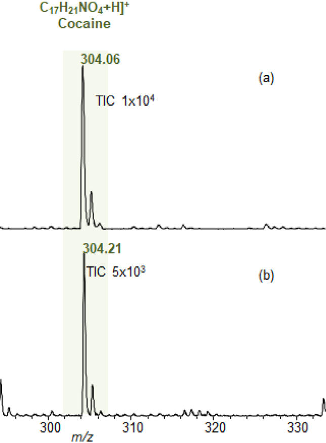
Figures 5a and 5b show the distinct spectral profiles of hair without and with bleaching treatment, respectively. However, from the spectral expansion in the region of m/z 250 to 350, one can observe that the protonated COC is the most abundant ion, [C17H21NO4 + H]+; m/z = 304.06; IR = 100%; TICcoc = 1 × 10⁴, in natural hair (with no bleaching treatment) compared to the same signal (m/z = 304.2197; IR = 14.5%; TICcoc = 5 × 10³) in hair previously subjected to a peroxide lightening treatment (Table S1, SI section). These data agree with the literature51,58-60 and qualitatively reveal the greater abundance of COC in unbleached hair (i.e., hair that does not present a loss of melanin in its composition) since one of the main factors in the incorporation of drugs into hair is their affinity for melanin. However, a greater number of samples and analyses are needed for further confirmation of the results.

MALDI(+) MS analysis of hair samples doped with 1 mg mL-1 of COC in methanol to evaluate the ionization efficiency (a) without and (b) with chemical treatment (1.5 mg mL-1 CHCA was used as the matrix).
Conclusions
In this work, the factors that influence the ionization of COC and its metabolites in hair strands were evaluated via MALDI(+)TOF MS. In addition, the effect of solvent washing was tested in the pre-analysis stage. The qualitative results of hair analyses from users by MALDI(+) MS were related to the quantitative results obtained by the classical GC-MS technique. The quantitative method (GC-MS) yielded data related to 70% of the samples analyzed by MALDI(+) MS, evidencing the sensitivity and precision of the latter method in the analysis of hair samples from COC users, including low concentrations of the monitored analyte (COC = 1.1 ng mg-1). Finally, this study also investigated the influence of hair treatment (bleached hair) on the incorporation of COC into hair, revealing the low efficiency of COC incorporation in treated hair compared to natural hair. Overall, the results support the applicability of the method developed with MALDI(+)TOF MS in the rapid and direct detection of drugs in hair, eliminating the pre-analysis (washing) and chromatographic separation steps, however, complementary studies to investigate the influence of other factors such as the gender and age of users, as well as the stability of metabolites in the hair over time are necessary for more robust and accurate responses.
Supplementary Information
Supplementary information with a summary of the steps of the methodology, illustrations of the sample preparation, spectra resulting from the hair washing test, chromatogram of one of the samples, and a comparative table of the effect of chemical treatment on hair, is available free of charge at http://jbcs.sbq.org.br as a PDF file.
Acknowledgments
The authors thank Coordenação de Aperfeiçoamento de Nível Superior (CAPES, 23038.007083/2014 40); Fundação de Apoio à Pesquisa do Espírito Santo (FAPES) (CNPq/FAPES No. 23/2018 - PRONEM (596/2018 and 597/2018); FAPES/CNPq/Decit-SCTIE-MS/SESA No. 09/2020 - PPSUS (165/2021); FAPES No. 03/2021 - UNIVERSAL (492/2021); FAPES No. 15/2022 - PROFIX 2022 (714/2022 P: 2022-SS849); FAPES No. 019/2022 - Núcleos Capixabas de Excelência em Pesquisa (991/2022 P; 2022-5KMF0); and edital FAPES No. 21/2022 - Apoio à Infraestrutura de Pesquisa (1069/2022 P: 2022-98VRN)); and Conselho Nacional de Desenvolvimento Científico e Tecnológico (CNPq, 310057/2020-5, INCT Forensic) for financial support.
The authors would also like to thank Núcleo de Competência em Química de Petróleo (NCQP)/LabPetro, and Protein Chemistry and Biochemistry Laboratory, Universidade de Brasília (UnB), for the use of their installations; the Civil Police of Espírito Santo and Federal Police of Rio Grande do Sul (INCT-Forense); and Contraprova laboratory, for providing the samples. This study was financed in part by Coordenação de Aperfeiçoamento de Pessoal de Nível Superior (CAPES, finance code 1). W. R. acknowledges Instituto Federal do Espírito Santo (IFES, edital 07/2024 - Prodif) for providing support with regard to English language corrections.
References
- 1 Bruni, A. T.; Velho, J. A.; Oliveira, M. F.; Fundamentos da Química Forense, 2nd ed.; Millennium: Campinas, Brazil, 2019.
-
2 Rodrigues, T. F. C.; Oliveira, R. R.; Decesaro, M. N.; Mathias, T. A.; J. Bras. Psiquiatr. 2019, 68, 73. [Crossref]
» Crossref -
3 Reis, L. M.; Hungaro, A. A.; Oliveira, M. L. F.; Texto & Contexto - Enfermagem 2014, 23, 1050. [Crossref]
» Crossref -
4 Reis, L. M.; Uchimura, T. T.; Oliveira, M. L. F.; Acta Paul. Enfermagem 2013, 26, 276. [Crossref]
» Crossref -
5 United Nations Office on Drugs and Crime (UNODC), World Drug Report 2021, https://www.unodc.org/res/wdr2021/field/WDR21_Booklet_2.pdf, accessed in September 2024.
» https://www.unodc.org/res/wdr2021/field/WDR21_Booklet_2.pdf - 6 Dorta, D. J.; Yonamine, M.; Costa, J. L.; Martinis, B. S.; Toxicologia Forense, 1st ed.; Blucher: São Paulo, Brazil, 2018.
-
7 Gallardo, E.; Queiroz, J. A.; Biomed. Chromatogr. 2008, 22, 795. [Crossref]
» Crossref -
8 Cordero, R.; Paterson, S.; J. Chromatogr. B: Anal. Technol. Biomed. Life Sci. 2007, 850, 423. [Crossref]
» Crossref -
9 Clauwaert, K.; Decaestecker, T.; Mortier, K.; Lambert, W.; Deforce, D.; Van Peteghem, C.; Van Bocxlaer, J.; J. Anal. Toxicol. 2004, 28, 655. [Crossref]
» Crossref -
10 Follador, M.; Yonamine, M.; Moreau, R.; Silva, O.; J. Chromatogr. B: Anal. Technol. Biomed. Life Sci. 2004, 811, 37. [Crossref]
» Crossref -
11 López, P.; Bermejo, A. M.; Tabernero, M. J.; Fernández, P.; Álvarez, I.; J. Appl. Toxicol. 2007, 27, 464. [Crossref]
» Crossref -
12 Fucci, N.; Giovanni, N.; Giorgio, F.; Liddi, R.; Chiarotti, M.; Forensic Sci. Int. 2006, 156, 102. [Crossref]
» Crossref -
13 Musshoff, F.; Rosendahl W.; Madea, B.; Forensic Sci. Int. 2009, 185, 84. [Crossref]
» Crossref -
14 Gjerde, H.; Oiestad, E. L.; Christophersen, A. S.; Nor. Epidemiologi 2011, 21, 5. [Crossref]
» Crossref - 15 Robbins, C. R.; Chemical and Physical Behavior of Human Hair, vol. 1, 4th ed.; Springer-Verlag: New York, USA, 2002.
-
16 Lima, E. C.; Silva, C. L.; Newslab 2007, 14, 156. [Link] accessed in September 2024
» Link -
17 Ueki, R.; Fukusaki, E.; Shimma, S.; J. Biosci. Bioeng. 2022, 133, 89. [Crossref]
» Crossref -
18 Cone, E. J.; Ther. Drug Monit. 1996, 18, 438. [Crossref]
» Crossref -
19 Kintz, P.; Forensic Sci. Int. 2012, 218, 28. [Crossref]
» Crossref -
20 Bortolotti, F.; Gottardo, R.; PascalI, J.; Tagliaro, F.; Curr. Med. Chem. 2012, 19, 5658. [Crossref]
» Crossref - 21 Jenkins, A. J.; Cone, E. J. In Drug Abuse Handbook, 1st ed.; Karch, S. B., ed.; CRC Press: New York, USA, 1998.
-
22 Carrera, M. R. C.; Meijler, M. M.; Janda, K. D.; Bioorg. Med. Chem. 2004, 12, 5019. [Crossref]
» Crossref -
23 Cone, E. J.; J. Anal. Toxicol. 1995, 19, 459. [Crossref]
» Crossref - 24 Levine, B.; Principles of Forensic Toxicology, vol. 1, 3rd ed.; American Association for Clinical Chemistry: New York, USA, 2010.
- 25 Karch, S. B.; Pharmacokinetics and Pharmacodynamics of Abused Drugs, 1st ed.; CRC Press: New York, USA, 2007.
-
26 Kloss, M. W.; Rosen, G. M.; Rauckman, E. J.; Biochem. Pharmacol. 1984, 33, 169. [Crossref]
» Crossref -
27 Kolbrich, E. A.; Barnes, A. J.; Gorelick, D. A.; J. Anal. Toxicol. 2006, 30, 501. [Crossref]
» Crossref -
28 Kovacic, P.; Med. Hypotheses 2005, 64, 350. [Crossref]
» Crossref -
29 European Monitoring Centre for Drugs and Drug Addiction (EMCDDA); Assessing Illicit Drugs in Wastewater - Potential and Limitations of a New Monitoring Approach; EMCDDA: Luxembourg, 2008. [Link] accessed in September 2024
» Link -
30 Baselt, R. C.; Clin. Chem. 2008, 51, 680. [Crossref]
» Crossref -
31 Verstraete, A. G.; Ther. Drug Monit. 2004, 26, 200. [Crossref]
» Crossref -
32 Garcia-Bournissen, F.; Moller, F. M.; Nesterenko, M.; Karaskov, T.; Koren, G.; Forensic Sci. Int. 2009, 189, 24. [Crossref]
» Crossref -
33 Carvalho, T. C.; Tosato, F.; Souza, L. M.; Santos, H.; Merlo, B. B.; Ortiz, R. S.; Vaz, B. G.; Rodrigues, R. R. T.; França, H. S.; Filgueiras, P. R.; Augusti, R.; Romão, W.; Forensic Sci. Int. 2016, 262, 56. [Crossref]
» Crossref -
34 Chen, X.; Zheng, X.; Ding, K.; Zhou, Z.; Zhan, C. G.; Zheng, F.; J. Pharm. Biomed. Anal. 2017, 134, 243. [Crossref]
» Crossref -
35 Rocha, W. W. F.; Leite, J. A.; Correia, R. M.; Tosato, F.; Madeira, N. C. L.; Filgueiras, P. R.; Lacerda Junior, V.; Freitas, J. C. C.; Romão, W.; Neto, A. C.; Anal. Methods 2018, 15, 1685. [Crossref]
» Crossref -
36 Rodrigues, N. V. S.; Cardoso, E. M.; Andrade, M. V. O.; Donnici, C. L.; Sena, M. M.; J. Braz. Chem. Soc. 2013, 24, 507. [Crossref]
» Crossref -
37 Almeida, C. M.; Pinto, F. E.; Santos, N. A.; Souza, L. M.; Merlo, B. B.; Thompson, C. J.; Romão, W.; Microchem. J. 2019, 149, 104002. [Crossref]
» Crossref - 38 Flinders, B.; Bassindale, T.; Heeren, R. M. A. In Emerging Technologies for the Analysis of Forensic Traces: Advanced Sciences and Technologies for Security Applications, 1st ed.; Francese, S., ed.; Springer: Sheffield, England, 2019, ch. 9.
-
39 Pego, A. M. F.; Roveri, F. L.; Kuninari, R. Y.; Leyton, V.; Miziara, I. D.; Yonamine, M.; Forensic Sci. Int. 2017, 274, 83. [Crossref]
» Crossref -
40 Lachenmeier, K.; Musshoff, F.; Madea, B.; Forensic Sci. Int. 2006, 159, 189. [Crossref]
» Crossref -
41 Allochio Filho, J. F.; Santos, N. A.; Borges, K. B.; Lacerda Junior, V.; Pelição, F. S.; Romão, W.; Rapid Commun. Mass Spectrom. 2020, 34, e8747. [Crossref]
» Crossref -
42 Tavares, L. S.; Carvalho, T. C.; Romão, W.; Vaz, B. G.; Chaves, A. R.; J. Am. Soc. Mass Spectrom. 2017, 29, 566. [Crossref]
» Crossref -
43 Sanz, G.; Garcia, L. F.; Yepez, A.; Carvalho, T. C.; Vaz, B. G.; Romão, W.; Barcelos, F. I.; Gil, E. S.; Luque, R.; Electroanalysis 2018, 30, 2094. [Crossref]
» Crossref -
44 Santos, H.; Lima, A. S.; Mazega, A.; Domingos, E.; Thompson, C. J.; Maldaner, A. O.; Romão, W.; Anal. Methods 2017, 9, 3662. [Crossref]
» Crossref -
45 Souza, L. M.; Rodrigues, R. R. T.; Santos, H.; Costa, H. B.; Merlo, B. B.; Filgueiras, P. R.; Romão, W.; Sci. Justice 2016, 56, 73. [Crossref]
» Crossref -
46 Conceição, V. N.; Souza, L. M.; Merlo, B. B.; Filgueiras, P. R.; Poppi, R. J.; Romão, W.; Quim. Nova 2014, 37, 1538. [Crossref]
» Crossref -
47 Miller, E. I.; Wylie, F. M.; Oliver, J. S.; J. Anal. Toxicol. 2008, 32, 457. [Crossref]
» Crossref - 48 Hoffmann, E.; Stroobant, V.; Mass Spectrometry, Principles and Applications, vol. 1, 3rd ed.; Wiley: Chichester, England, 2007.
-
49 Porta, T.; Grivet, C.; Kraemer, T.; Varesio, E.; Hopfgartner, G.; Anal. Chem. 2011, 83, 4266. [Crossref]
» Crossref -
50 Musshoff, F.; Arrey, T.; Strupat, K.; Drug Test. Anal. 2013, 5, 361. [Crossref]
» Crossref -
51 Cuypers, E.; Flinders, B.; Bosman, I. J.; Lusthof, K. J.; Van Asten, A. C.; Tytgat, J.; Heeren, R. M. A.; Forensic Sci. Int. 2014, 242, 103. [Crossref]
» Crossref -
52 Cooper, G. A.; Kronstrand, R.; Kintz, P.; Forensic Sci. Int. 2012, 218, 20. [Crossref]
» Crossref - 53 ABNT NBR ISO/IEC 17025:2017: Requisitos Gerais para a Competência de Laboratórios de Ensaio e Calibração, 2017.
-
54 Santos, N. A.; Almeida, C. M.; Gonçalves, F. F.; Ortiz, R. S.; Kuster, R. M.; Saquetto, D.; Romão, W.; J. Am. Soc. Mass Spectrom. 2021, 32, 946. [Crossref]
» Crossref -
55 Burton, R. D.; Watson, C. H.; Eyler, J. R.; Lang, G. L.; Powell, D. H.; Avery, M. Y.; Rapid Commun. Mass Spectrom. 1997, 11, 443. [Crossref]
» Crossref -
56 Hart, E. D.; Vikingsson, S.; Winecker, R. E.; Evans, A. L.; Cone, E. J.; Mitchell, J. M.; Hayes, E. D.; Flegel, R. R.; J. Anal. Toxicol. 2023, 47, 154. [Crossref]
» Crossref - 57 Chasin, A. A. M.; Silva, E. S.; Carvalho, V. M.; Fundamentals of Toxicology, 4th ed.; Atheneu: São Paulo, Brazil, 2014.
-
58 Tanaka, S.; Lio, R.; Chinaka, S.; Takayama, N.; Hayakawa, K.; Biomed. Chromatogr. 2002, 16, 390. [Crossref]
» Crossref -
59 Cirimele, V.; Kintz, P.; Mangin, P.; J. Anal. Toxicol. 1995, 19, 331. [Crossref]
» Crossref -
60 Nakahara, Y.; KIkura, R.; Arch. Toxicol. 1994, 68, 54. [Crossref]
» Crossref
Edited by
-
Editor handled this article:
Andréa R. Chaves (Associate)