Investigations of Oral Pathogens on Copper(II) Complexes with Thiosemicarbazone: Antimicrobial and Antibiofilm Activities in Addition to in vitro and in vivo Toxicity
Abstract
Oral caries is a major global public health issue involving complex biofilm interactions and contributing to several systemic conditions. Metallic compounds have gained attention as antimicrobial agents due to their notable antibacterial activity. In this work, we investigated the anticariogenic potential of two copper(II) complexes containing Schiff base ligands, namely [CuCl(atc-Me)] (1, atc-Me = 2-acetylpyridine-N(4)-methyl-thiosemicarbazone) and [{Cu(μ atc Me)}2μ-SO4] (2). Minimum inhibitory concentration (MIC), minimum bactericidal concentration (MBC), minimum biofilm inhibitory concentration (MBIC50), and synergistic activity with chlorhexidine were evaluated. The in vivo toxicity of the complexes was further assessed using Caenorhabditis elegans. MBC values indicated a bactericidal effect for both complexes. Synergistic activity with chlorhexidine was observed against Streptococcus mutans and Enterococcus faecalis strains. Cytotoxic evaluation in human fibroblast cultures revealed that complexes 1 and 2 showed half maximal inhibitory concentration (IC50) values of 2.9 and 4.9 µg mL-1, respectively. Toxicity assays in C. elegans showed lethal concentration (LC50) values approximately three times higher than the MIC values for complexes 1 and 2: Molecular docking analyses revealed favorable binding energy values between the copper complexes and bacterial proteins. Overall, this investigation suggests an alternative approach for anticariogenic treatment, highlighting significant antibiofilm activity combined with low toxicity.
Keywords:
copper complexes; thiosemicarbazone; oral pathogens; antibiofilm activity; cytotoxicity
Introduction
Dental caries is the most prevalent oral disease worldwide, affecting approximately 8% of the global population in primary dentition and 35% in permanent dentition.1 According to Listl et al.,2 the direct and indirect costs associated with dental diseases were around $442 billion in the latest global cost survey, corresponding to 4.6% of global health expenditures. The pathogenesis of caries involves complex interactions between tooth structure, bacterial biofilm formation, diet, and duration of contact between bacteria and host.3 The microbial species associated with caries are diverse, including Streptococcus mutans, Streptococcus sobrinus, Prevotella spp., Aggregatibacter actinomycetemcomitans, Enterococcus faecalis, Candida spp., Streptococcus mitis, Streptococcus sanguinis, Streptococcus salivarius, Lactobacillus paracasei.4-6 These microorganisms interact synergistically within complex biofilms, enhancing acid production and contributing to the demineralization of dental tissues.
Currently, prevention is considered the most effective strategy for public health intervention in dental caries. Regular tooth brushing with fluoride toothpaste, flossing, and the use of mouthwashes help reduce dental plaque accumulation and control cariogenic biofilms.7,8 Nevertheless, given the limitations of conventional preventive measures, alternative strategies are required to further inhibit biofilm formation, particularly through the incorporation of antimicrobial agents into oral care products such as toothpaste and mouthwashes.9 Chlorhexidine has been widely used to treat oral infections; however, adverse effects such as tooth staining, bitter taste, taste alteration, hives, and even rare cases of anaphylaxis10 can limit its long-term use. Thus, alternative antibacterial compounds must be incorporated into oral care products to prevent disease progression.
Even with the development of new antibacterial drugs, bacteria continue to evolve resistance mechanisms to evade antibiotic action. Therefore, a potential solution is the discovery of compounds capable of targeting multiple cellular processes simultaneously, since bacteria would need to accumulate several mutations to develop resistance.11 Several metallic compounds, such as silver, palladium, zinc, and copper, have emerged as promising antibacterial agents because they can reach multiple targets at once.12-16 In addition to the metal center, synthetic organic ligands can enhance or potentiate biological activity when coordinated to metals. Thiosemicarbazone ligands, in particular, are attractive due to their well-known antitumor, antibacterial, antifungal and antiviral activities, as well as their versatile coordination chemistry.17-19
According to Nakahata et al.,20 the combination of an organic compound with a metal ion may represent a promising approach for the development of new antimicrobial drugs.21-23 This trend aligns with the growing interest in modifying classical antibiotics through metal coordination, giving rise to so-called “metalloantibiotics.21,22 Copper(II) was selected as the metal center because it is an essential element and typically exhibits lower toxicity than other metals, in addition to its documented antimicrobial activity.20 Furthermore, copper(II) combined with thiosemicarbazone ligands can generate compounds with improved biological properties and potential advantages in their mechanism of action, possibly reducing the risk of bacterial resistance, advantages that are difficult to achieve using purely organic molecules.14,24-26
Considering our previous findings on the antibacterial activity of copper(II) complexes, the present study investigates the anticariogenic potential of copper(II) complexes with thiosemicarbazone ligands by performing antibacterial, antibiofilm, and synergistic assays with chlorhexidine against representative cariogenic bacteria. This is also the first study to assess the in vivo toxicity of these molecules. Additionally, docking simulations were carried out to obtain further insights into the binding modes of these compounds with bacterial proteins and to clarify the differences in their molecular interactions.
Experimental
Synthesis of copper(II) complexes
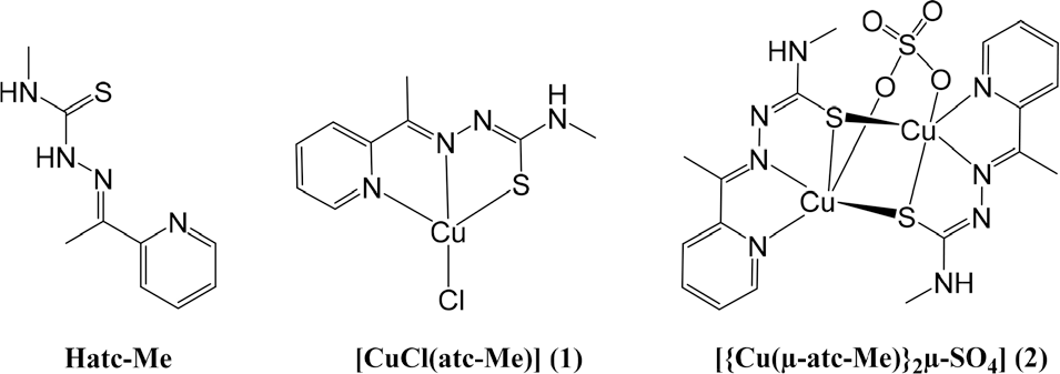
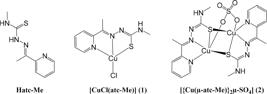
The compounds studied here were previously synthesized and characterized as described by Souza et al.14 Briefly, the complexes were prepared by refluxing CuCl2·5H2O (4) and thiosemicarbazone (Hatc-Me), represented as ligand Hatc Me in a 1:1 molar ratio to obtain complex 1. Complex 2 was synthesized by mixing CuSO4·5H2O (3) and Hatc-Me (1:1), both in methanolic solution (Figure 1). The products were characterized, and their purities evaluated as previously reported.14
Attenuated total reflectance-Fourier transform infrared (ATR FTIR) analysis of copper complexes stability and bacterial interaction
To evaluate potential interactions between the copper complexes and bacterial components, attenuated total reflectance-Fourier transform infrared (ATR FTIR) spectroscopy was performed using an Agilent Cary 630 FTIR spectrometer (Agilent Technologies, Santa Clara, CA, USA). The spectra of the complexes 1 and 2 were recorded to assess their stability both in powder form (pure compound) and as 200 µg mL-1 aqueous solutions over 0 and 24 h. The spectral region from 4000 to 650 cm-1 was analyzed. For pure samples, a small amount of each powdered complex was directly applied onto the ATR crystal. For the diluted condition (200 µg mL-1), each complex (200 µg) was dissolved in 1 mL of Milli-Q water and applied similarly onto the ATR crystal. Spectra were recorded with a resolution of 2 cm-1 and 64 scans. Two replicates per sample were analyzed. Spectral data were preprocessed using Gaussian smoothing, baseline correction, vector normalization, and Minimum Noise Fraction (MNF) denoising.
Bacterial strains
A panel of representative caries-associated bacteria was obtained from the “American Type Culture Collection’ (ATCC): S. mutans (ATCC 25175), S. salivarius (ATCC 25975), S. sanguinis (ATCC 10556), S. mitis (ATCC 49456), S. sobrinus (ATCC 33478), E. faecalis (ATCC 4082) and L. paracasei (ATCC 11578). These microorganisms are part of the culture collection of the Laboratory of Antimicrobial Testing (LEA) at the Federal University of Uberlândia, and are kept in cryopreservation at -20 °C.
Minimal inhibitory concentration (MIC) and minimal bactericidal concentration (MBC)
The MIC was defined as the lowest concentration of the antimicrobial agent capable of inhibiting bacterial growth. The microdilution method, recommended by the Clinical and Laboratory Standards Institute (CLSI),27 was used with adaptations incorporating resazurin as a growth indicator. MIC determinations were performed in triplicate for each microorganism.
Compounds 1-4 and Hatc-Me were dissolved in dimethyl sulfoxide (DMSO; Sigma-Aldrich St. Louis, MO, USA), and Brain Heart Infusion (BHI; Difco, Sparks, MD, USA) was added. The tested concentration range was 0.195-400 μg mL-1.
Cultures of E. faecalis, L. paracasei, S. mutans, S. salivarius, S. mitis, S. sobrinus, and S. sanguinis grown on BHI agar supplemented with 5% defibrinated sheep blood were transferred into BHI broth to achieve turbidity equivalent to 0.5 on the McFarland scale, checked using a densitometer (Densimat®, Biomérieux). The final inoculum concentration in each well was 5 × 10⁵ colony-forming units (CFU) mL-1.
Controls for culture viability, broth sterility, complex sterility, and DMSO effects were included. Chlorhexidine was used as positive control within the concentration range 0.115-59 μg mL-1. Microplates were incubated in a microaerophilic chamber at 37 °C with 10% CO2 for 24 h, except for E. faecalis and S. salivarius, which were incubated under aerobic conditions.
After incubation, 30 μL of resazurin (0.02% aqueous solution) were added to each well. Plates were incubated for an additional 30 min. A blue color indicated growth inhibition, while a pink color indicated bacterial growth.28
For MBC determination,29 10 μL from wells showing inhibited growth were transferred to fresh blood agar prior to resazurin addition. The plates were incubated under the same atmospheric conditions for 24 h. Growth indicated bacteriostatic activity; absence of growth indicated bactericidal activity. MBC values were determined in triplicate.
Minimum biofilm inhibitory concentration 50% (MBIC50)
Selection of the best inoculum concentration and incubation time for the antibiofilm activity assay was accomplished by standardizing biofilm formation (data not shown). Only one microorganism representative of each MIC value was chosen (L. paracasei, E. faecalis, S. salivarius and S. mutans). Complexes 1 and 2 were selected due to their better MIC results.
The antibiofilm activity was evaluated through the determination of the minimum biofilm inhibitory concentration (MBIC50).30 The method used was the microplate dilution, recommended by the Clinical and Laboratory Standards Institute,27 with modifications. The experiments were performed in triplicate and the results were presented graphically.
Two plates for each complex were used: one for optical density reading and the other for counting bacteria (CFU mL-1). Serial dilutions of complexes 1 and 2 were made, with the final concentration ranging from 0.195 400 µg mL-1. Chlorhexidine at concentrations between 0.115 59 µg mL-1 was assessed as positive control. Each strain was added at a concentration of 1 × 10⁶ CFU mL-1. Bacterial strains grown with BHI broth were used as positive control; controls for the sterility of the broth, sample, and the solvent DMSO (Sigma-Aldrich; 1%) were carried out. The microplates were incubated at 37 °C for 24 h with appropriate atmospheres (aerobiosis or microaerophilia).
Each well was washed three times with sterile Milli-Q water and fixed with methanol. Antibiofilm activity was measured by MBIC50 determined by optical density (OD) and by counting the colony-forming units (CFU mL-1). Based on the procedures described by Sandberg et al.,31 OD was quantified in the biofilm by adding 200 µL of crystal violet (0.2%) to the microplate wells. After 15 min at room temperature, the excess dye was removed with tap water and the plates were dried in air at room temperature. Next, 33% of acetic acid were added to each well to resolubilize the dye bound to the cells. The OD of the microplates was measured at 595 nm, after 30 min, using a microtiter plate reader (GloMax®, Promega). The percentage of inhibition was calculated by using the equation: (1 - At595/Ac595) ×100, where At595 and Ac595 are the absorbance values of the wells treated with the samples and the control, respectively.30
To count the CFU29 after the incubation period, the entire volume was carefully aspirated from the wells of the microplate and washed with water to completely remove the non-adherent cells. Then, BHI broth was added to each well and the microplate was subjected to sonication in an ultrasound bath at 38 kHz for 15 min to shed the biofilms. Dilutions of 100 to 10-7 were performed for all wells and 50 µL of each dilution were plated in BHI agar supplemented with defibrinated sheep blood (5%). Subsequently, the plates were incubated in a microaerophilic incubator for 24 h at 37 °C with 10% CO2, except for E. faecalis and S. salivarius, which were incubated under aerobic conditions. After incubation, the colonies were counted and the results were expressed in Log10 by CFU mL-1 and shown graphically.
Fractional inhibitory concentration index (FICI)
The same bacteria used in the MBIC50 assays were chosen as a way to assess the in vitro antibacterial effectiveness of the combination of chlorhexidine with complexes 1 and 2, namely S. mutans, S. salivarius, E. faecalis, and L. paracasei. The experimental approach followed the methodology outlined by Chaturvedi et al.32 with specific adaptations to the culture medium and incubation conditions. Synergy tests were performed in triplicate, and the concentrations of the tested samples and chlorhexidine were standardized at 4× MIC against 1 × 10⁶ CFU mL-1 inoculum.
The fractional inhibitory concentration (FIC) index was determined algebraically, with FICA denoting the MIC of drug A in the combination divided by the MIC of drug A alone, and FICB representing the MIC of drug B in the combination divided by the MIC of drug B alone. The cumulative FIC value (ΣFIC) was calculated as ΣFIC index = FICA + FICB.32 Interpretation of fractional inhibitory concentration index values revealed the following classifications: FICI index values ≤ 0.5 indicated synergism, values > 0.5 and < 1.0 signified an additive effect, values ≥ 1.0 and < 4.0 suggested indifference, and values ≥ 4.0 were indicative of antagonism.33
Cytotoxicity assessment
The GM07492A cell line (non-tumor human lung fibroblasts) was used in this study to assess toxicity. Cells were cultured in Ham F-10 + Dulbecco’s Modified Eagle’s medium (1:1; Sigma-Aldrich) supplemented with 10% fetal bovine serum (Nutricell), antibiotics (0.01 mg mL-1 streptomycin and 0.005 mg mL-1 penicillin; Sigma-Aldrich) and 2.38 mg mL-1 HEPES (Sigma-Aldrich), at 37 °C with 5% CO2.
The evaluation of cytotoxicity was carried out through the colorimetric in vitro toxicology assay - Kit XTT® (Roche Diagnostics), according to the guidelines of the manufacturer. For performing the experiments, 1 × 10⁴ cells were seeded in 96-well microplates. The concentrations tested were determined according to the solubility limit. After solubilization in dimethyl sulfoxide (DMSO; Sigma-Aldrich; 1%), complexes 1, 2 and Hatc-Me were tested at concentrations ranging from 3.9 to 500 µg mL-1, and compounds 3, 4 and chlorhexidine at concentrations ranging from 7.8 to 1,000 µg mL-1. Negative (no treatment), solvent (1% DMSO) and positive (25% DMSO) controls were included. The treatment and analysis procedures were conducted as described by Soares et al.34 The experiments were performed in triplicate and the results are expressed as IC50 (half maximal inhibitory concentration).
Toxicity assessment in Caenorhabditis elegans
The strain of C. elegans was AU37, a mutant strain that is sterile at 25 °C, according to Singulani et al.35 The complexes 1 and 2 were evaluated at concentrations of 11.71 to 375 μg mL-1 and the compounds 3, 4 and Hatc-Me at 46.87 to 1,500 μg mL-1. Dimethyl sulfoxide (DMSO) was used as a solvent (final concentration ≤ 1%).
C. elegans AU37 was grown on nematode growth medium (NGM) plates seeded with Escherichia coli OP50 and incubated at 16 °C for three days. After incubation, the supernatant was washed with the bleaching solution (sodium hypochlorite + NaOH) to synchronize the larvae at the L4 stage. The plates containing larvae synchronized in the L4 stage were washed with M9 and the supernatant was placed in 15 mL conical tubes. Subsequently, 20 µL of the larval suspension were added to each well of a 96-well flat-bottom microplate, along with 80 µL of BHI medium supplemented with antibiotics (streptomycin, ampicillin, and kanamycin) and 100 µL of each sample to be tested. The experiment was then incubated for 24 h at 25 °C. Counting live and dead larvae was performed using an Evos® microscope and the percentage of mortality was calculated.
Molecular docking
Molecular docking studies were performed using AutoDock 4.236 with complex 1 (CCDC 2063732) and complex 2 (CCDC 2083951). Chlorhexidine (CCDC 988972) was obtained from The Cambridge Crystallographic Data Centre. Hatc-Me was drawn in GABEDIT37 and optimized with the semi-empirical method PM7 using the MOPAC2016 program.38 The crystal structures of proteins from S. mutans (PDB 5C2O), S. mitis (PDB 3LE0), S. sanguinis (PDB 4N82), L. paracasei (PDB 6CHK) and E. faecalis (PDB 4LRL) bacteria were obtained from the Protein Data Bank (PDB).
In order to validate the molecular docking protocol, the redocking procedure was performed with the protein and itraconazole. The docking procedure was performed using the AutoDockTools 1.5.6 interface along with AutoGrid4 and AutoDock 4.2,36 which uses the Lamarckian Genetic Algorithm (LGA) to recognize the putative binding site and ligand orientation.
As for the prerequisites of LGA, the water molecules were removed from the protein and polar hydrogens were added, followed by the merging of non-polar hydrogens and the addition of Gasteiger charges.39 The ligands were drawn in GABEDIT36 and optimized with a semi-empirical method PM7 using the MOPAC2016 program.38
The LGA in Autodock was used with 20 runs for docking simulation, with an initial population of 150 individuals, maximum energy evaluations of 25,000,000, maximum generations of 27,000, one elite individual maintained per generation, genetic algorithm docking runs (150) and a ranked cluster analysis performed using a root mean square (RMS) tolerance of 2.0 Å for each docking calculation. Interactions between compounds and the active site were generated using the Discovery Studio Visualizer program.40
ATR-FTIR analysis
To evaluate potential interactions between the copper complexes and bacterial components, attenuated total reflectance-Fourier transform infrared (ATR-FTIR) spectroscopy was performed using an Agilent Cary 630 FTIR spectrometer (Agilent Technologies, Santa Clara, CA, USA).
ATR-FTIR spectra of S. mutans and complexes 1 and 2 were recorded using the Agilent Cary 630 FTIR instrument. The diamond unit in the ATR system serves as the internal reflection element to record the infrared signature in the lipid region 2800-3050 cm-1 and the fingerprint region 1800-800 cm-1. A volume of 4 μL of each sample were placed on the ATR crystal. Bacterial suspensions of S. mutans (density: 2.7 × 10⁹ CFU mL-1) were incubated for 24 h with the complexes 1 and 2 at a final concentration of 400 µg mL-1, prepared in Milli-Q water. The spectra were then recorded with 2 cm-1 resolution and 64 scans. Second derivative spectra were generated from the original data using OriginPro 9.8 (OriginLab, Northampton, MA, USA) and processed with a Savitzky-Golay filter (polynomial order 2, 20-point window).41,42
Results
The solution stability of both monomer and binuclear copper(II) complexes was previously evaluated through UV-Vis spectroscopy and by molar conductivity in DMSO for a 24 h period at room temperature.14 The results indicated that the compounds maintained their structures in solution over the evaluated time. To further investigate the stability of the complexes, representative ATR-FTIR spectra of complexes 1 and 2 were collected both as powders (pure samples) (Figures S1 and S3, Supplementary Information (SI) section) and at 200 µg mL-1 in aqueous solution (Figures S2 and S4, SI section) in the range of 3500-650 cm-1. The spectra recorded immediately after sample preparation and after 24 h revealed no significant changes, demonstrating that the complexes preserved their structural integrity under these conditions.
The antibacterial activity of all samples was assessed by MIC and MBC. Complex 1 demonstrated MIC values of 6.25 µg mL-1 for E. faecalis and S. salivarius, 12.5 µg mL-1 for L. paracasei and 1.56 µg mL-1 for S. sobrinus, S. mitis, S. sanguinis and S. mutans. Complex 2 exhibited MIC values of 12.5 µg mL-1 for E. faecalis, 6.25 µg mL-1 for S. salivarius and L. paracasei, and 0.78 µg mL-1 for S. sobrinus, S. mitis, S. sanguinis and S. mutans. Notably, both complexes demonstrated bactericidal effects at the MBC for all tested species. In contrast, compounds 3, 4 and Hatc-Me displayed MIC values ranging from > 400 to 100 µg mL-1 for all tested bacteria. These results are shown in Table 1.
Results of antibacterial activity minimum inhibitory concentration (MIC) and bactericidal minimum inhibitory (MBC) of compounds 1-4, thiosemicarbazone (Hatc-Me), and chlorhexidine against dental caries bacteria
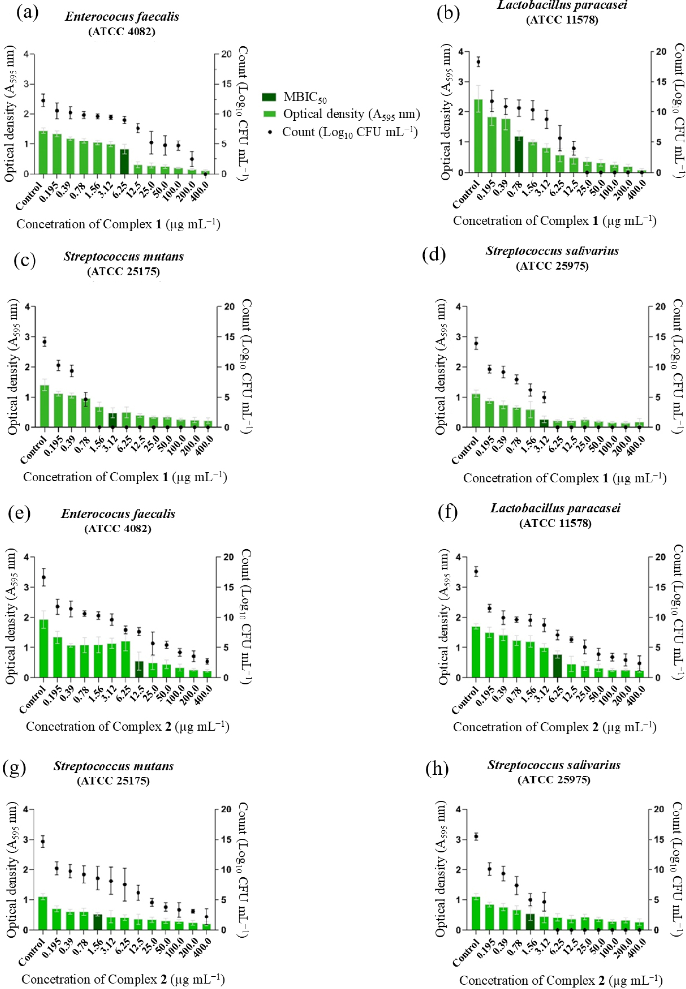
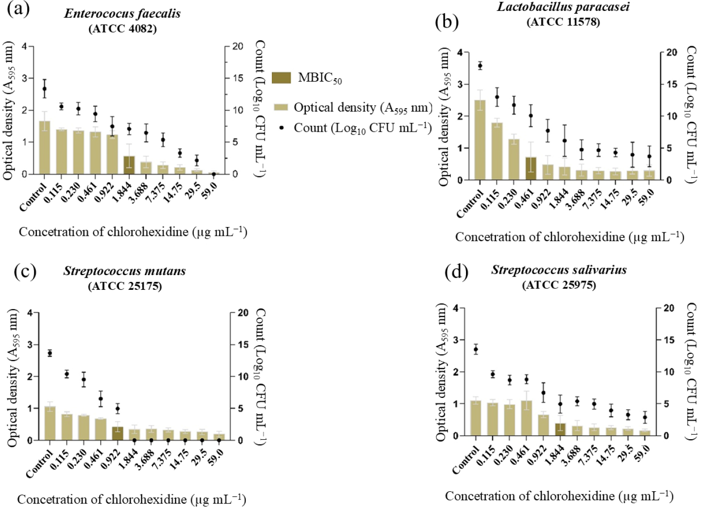
The antibiofilm activity was calculated as MBIC50, with a range of values from 0.78 to 12.5 µg mL-1 (Figure 2). Complex 1 showed MBIC50 values of 0.78 µg mL-1 for L. paracasei, 3.12 µg mL-1 for S. mutans and S. salivarius, and 6.25 µg mL-1 for E. faecalis. Complex 2 presented results of 1.56 µg mL-1 for S. mutans and S. salivarius, 6.25 µg mL-1 for L. paracasei, and 12.5 µg mL-1 for E. faecalis. Chlorhexidine was used as positive control, as shown in Figure 3.
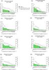
Antibiofilm activity of complexes 1 and 2 against cariogenic bacteria. Optical density and counting of colony forming units (CFU) mL-1 with minimal inhibitory concentration biofilm 50% (MBIC50). (a)-(d) Values from complex 1. (e)-(h): values from complex 2. Values are mean ± standard deviation.
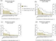
Antibiofilm activity of chlorhexidine (positive control) against cariogenic bacteria. Optical density and counting of colony forming units (CFU) mL-1 with minimal inhibitory concentration biofilm 50% (MBIC50). Values are mean ± standard deviation.
The FICI for complex 1 was 0.74, representing an additive interaction for E. faecalis and 0.24 for S. mutans demonstrating a synergistic interaction. Complex 2 showed FICI values of 0.44 for E. faecalis and 0.45 for S. mutans, both representing synergistic interactions. The complete data are provided in Table 2.
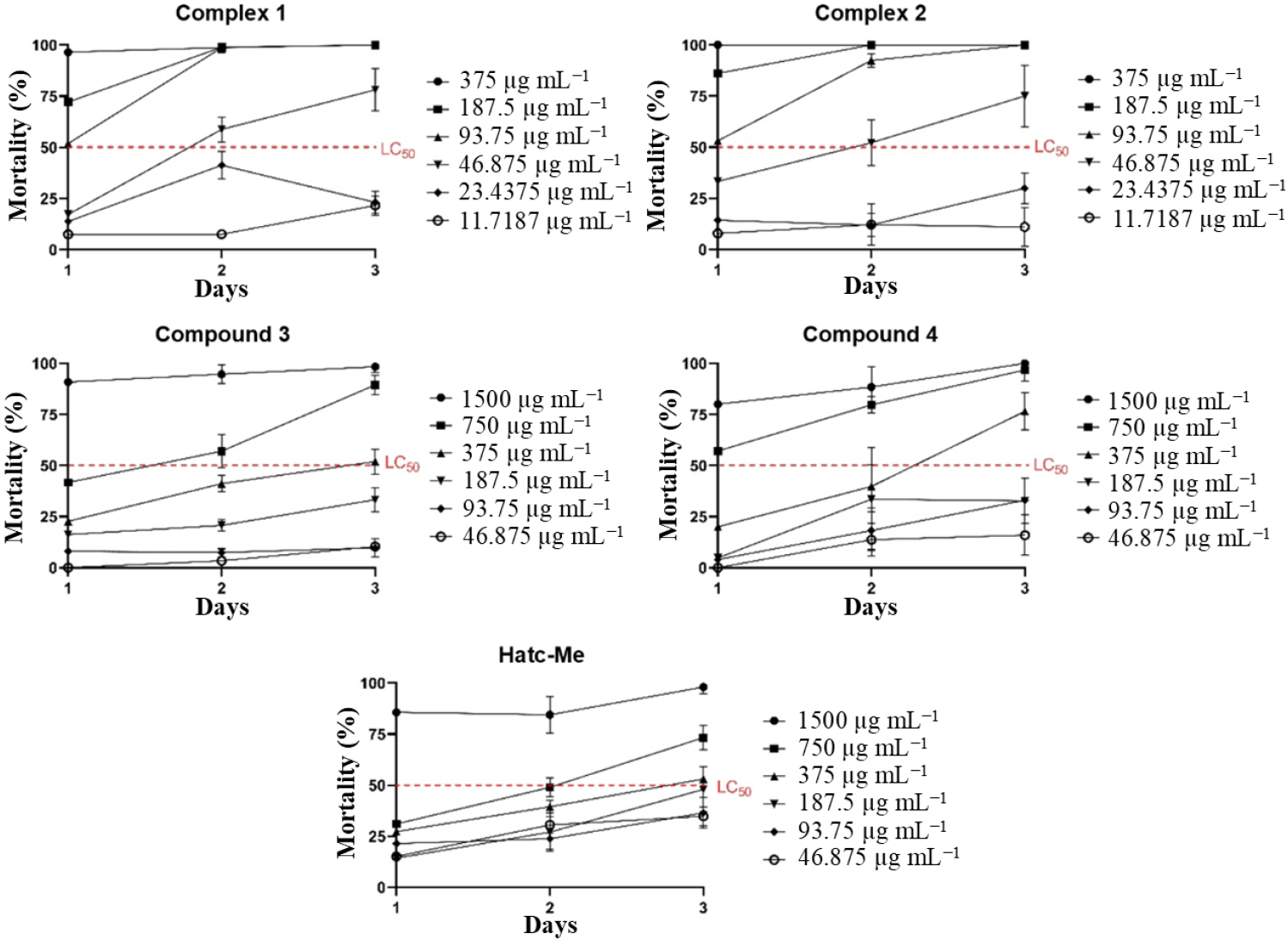
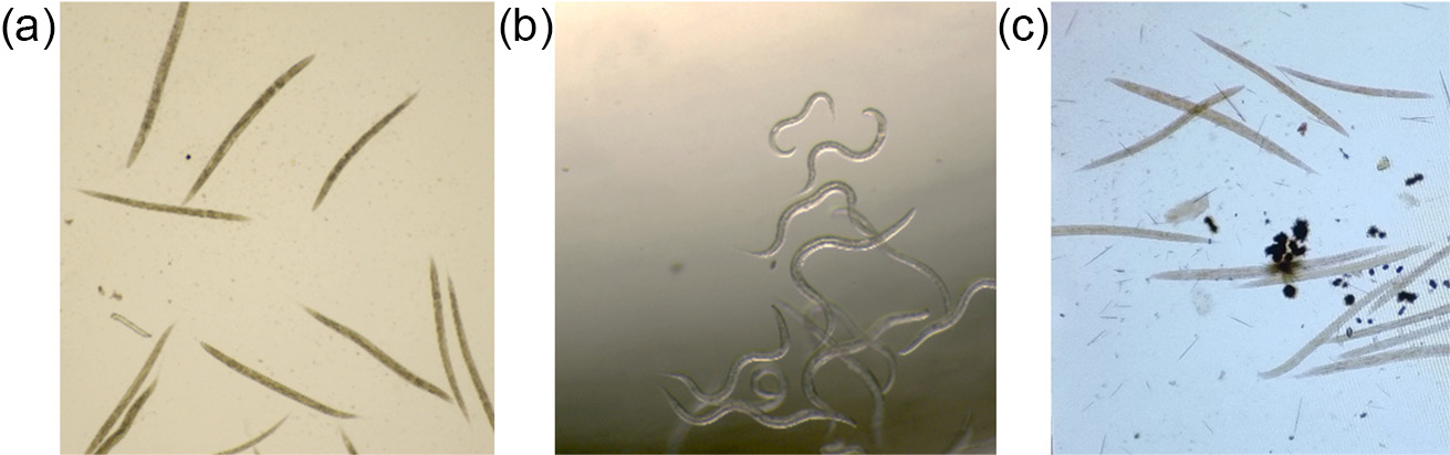
Cytotoxicity results in non-tumor human fibroblasts (GM07492A) are shown in Table 3. After 24 h of treatment, complexes 1 and 2 showed IC50 values of 2.9 and 4.9 µg mL-1, respectively. Toxicity in C. elegans (Figure 4) shows the percentage of larval mortality over three days, with both complexes showing LC50 values of 46.875 µg mL-1 on the second day. Representative transmittance images of live and dead larvae are depicted in Figure 5.
IC50 values obtained against GM07492A cell line after 24 h of treatment with different concentrations of compounds and chlorhexidine
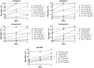
Values of mortality for C. elegans against compounds 1-4 and Hatc-Me during 3 days of counting. LC50 is the lethal concentration that kills 50% of larvae. Values are mean ± standard deviation.
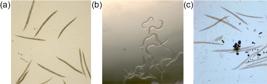
Transmittance images obtained from the EVOS® inverted microscope. (a) C. elegans larvae incubated with complex 1 after 1 day at the concentration of 375 μg mL-1. (b) C. elegans larvae incubated after 1 day, negative control well. (c) C elegans larvae incubated in chlorhexidine, positive control. Needle-shaped larvae were considered dead, and those with a curvilinear shape were considered alive.
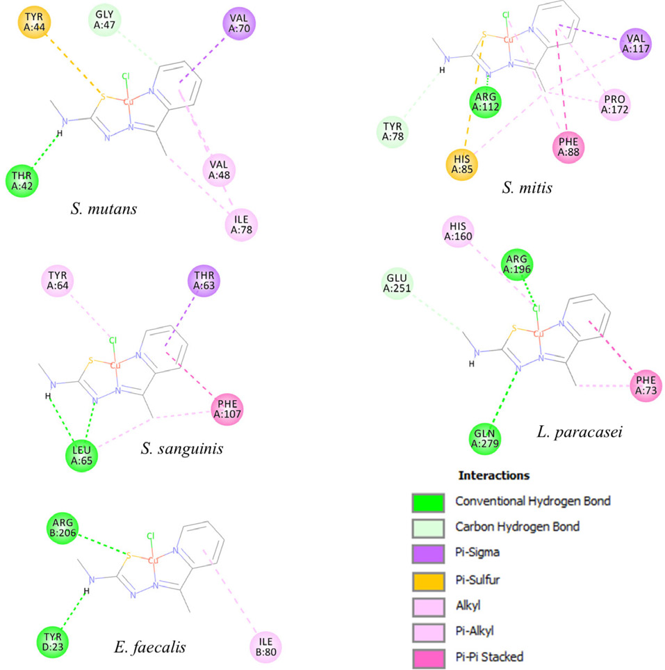
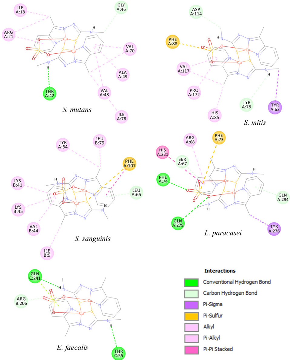
Molecular docking studies were performed to investigate and compare the interactions of complexes 1 and 2 and Hatc-Me with the active sites of proteins from S. mutans, S. mitis, S. sanguinis, L. paracasei and E. faecalis, using chlorhexidine as reference. Redocking was performed with AutoDock4 (Molecular Graphics Laboratory, The Scripps Research Institute, La Jolla, CA, USA, 2009) to validate the structure-activity relationship mechanism. The interaction energy values (∆G) obtained for complexes 1, 2, Hatc-Me and chlorhexidine with the bacterial proteins are presented in Table S2 (SI section). The most favorable conformations for complexes 1 and 2 with each bacterial protein are shown in Figures 6 and 7, respectively.
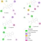
2D representation (DS Visualizer program) of the interactions between complex 1 and the active site of cariogenic bacteria (Discovery Studio Modeling Environment, Release 3.5, Accelrys Software Inc.: San Diego, 2012).
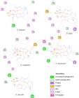
2D representation (DS Visualizer program) of the interactions between complex 2 and the active site of cariogenic bacteria (Discovery Studio Modeling Environment, Release 3.5, Accelrys Software Inc.: San Diego, 2012).
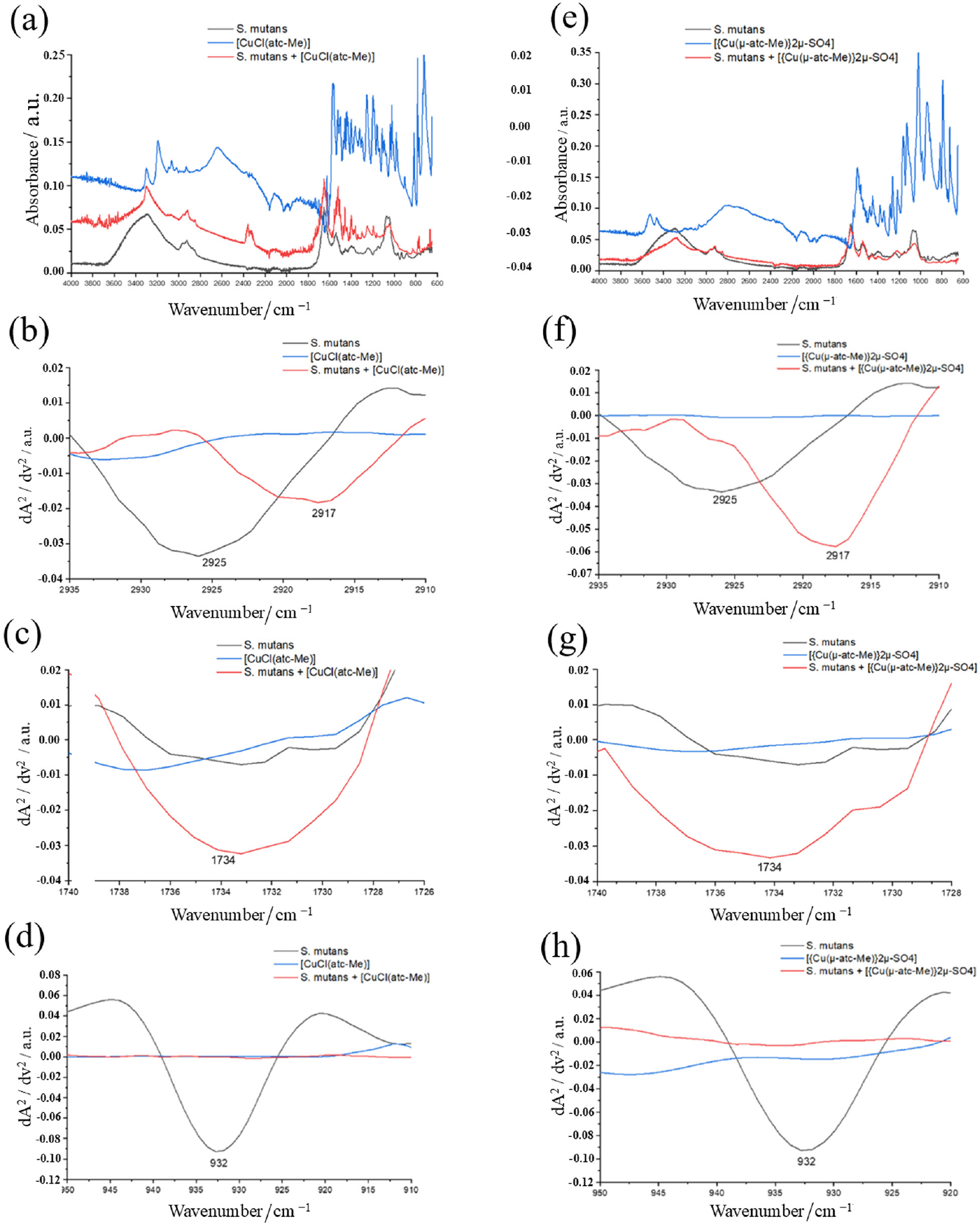
Figure 8 illustrates the ATR-FTIR spectra used to evaluate the molecular interaction between copper complexes 1 and 2 and S. mutans. Characteristic bands associated with lipids, proteins, carbohydrates, and nucleic acids were detected in S. mutans. In the original spectra (Figures 8a and 8e), prominent bands were observed between 1800 and 800 cm-1. Second-derivative spectra (Figures 8b-8d and 8f-8h) enhanced band resolution and allowed the detection of subtle spectral changes related to complex-bacteria interactions.
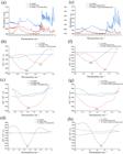
ATR-FTIR of S. mutans alone, complexes 1 and 2 alone, or each complex mixed with S. mutans. (a) and (e) Original spectra; (b), (c), (d), (f), (g) and (h) second-derivative spectra.
The vibrational mode at 2925 cm-1 in S. mutans, attributed to C-H stretching in the lipid region, was absent in the isolated complex 1 (Figure 8b) and complex 2 (Figure 8f). In samples containing S. mutans incubated with complexes 1 and 2, this band shifted to 2917 cm-1 (Figures 8b and 8f), indicating molecular interaction with bacterial lipids.
Another band at 1734 cm-1 was detected only after incubation of S. mutans with the complexes, assigned to fatty acid ester groups, suggesting interaction with membrane lipid components (Figures 8c and 8g). Notably, a band at 932 cm-1, associated with phosphodiester groups of nucleic acids (PMID: PMID: 28494407), was detected only in untreated S. mutans (Figures 8d and 8h) and was absent in all treated samples, suggesting possible interaction or structural modification in the bacterial nucleic acid or polysaccharide regions, as described in Table S1 (SI section).
Discussion
Copper(II) complexes are particularly attractive because copper is an essential micronutrient, which suggests lower toxicity compared to other metal-based agents. Among the various chelating compounds investigated, sulfur-donor ligands have shown potential as antibacterial agents against Mycobacterium tuberculosis, yielding promising results.14 Therefore, copper(II) complexes containing thiosemicarbazone ligands may offer additional biological advantages and could be applied to the treatment of other bacterial diseases.
Complexes 1 and 2 showed strong activity against cariogenic bacteria and biofilms, with toxicity levels lower than their effective inhibitory concentrations. In the present study, MIC values revealed that the complexes (1 and 2) exhibited higher growth-inhibitory activity (0.78 12.5 µg mL-1) compared to compounds 3, 4 and Hatc-Me, which showed MICs between 200 and 400 µg mL-1 (Table 1). Their antibacterial activity at much lower concentrations indicates that metal coordination enhances anticariogenic potential. This is consistent with the findings of Khan et al.,43 who reported that copper(II)-thiosemicarbazone structures showed greater antibacterial activity against both Gram-positive and Gram-negative bacteria compared to the Hatc-Me. MIC values of the complexes 1 and 2 samples with Staphylococcus aureus and S. pyogenes were 32 and 64 µg mL-1, respectively, and 32 µg mL-1 for Salmonella typhimurium and Escherichia coli. For thiosemicarbazone alone, the MIC was 512 µg mL-1 for Gram-positives and 128 µg mL-1 for Gram negatives. Similar results were reported by Azam et al.,44 in which a CuII complex with an azo azomethine ligand was used against S. aureus and E. coli. MIC values for the complex were 128 µg mL-1 (S. aureus) and 64 µg mL-1 (E. coli), while for the Hatc Me, values were 512 and 256 µg mL-1, respectively. Moreover, Abdel-Rahman et al.45 observed that the metal complexes performed better than Hatc-Me in disk diffusion assays using Zn, Cd, Ag, Cu, La and Ni complexes against S. aureus, Candida albicans, E. coli, and Klebsiella pneumoniae. Although different methodologies and bacterial strains were evaluated, these studies collectively reinforce that metal chelation enhances antibacterial potential.
MBC results corroborated the MIC findings. Complexes 1 and 2 showed bactericidal activity for all tested microorganisms at low concentrations, whereas compounds 3, 4 and Hatc-Me were bactericidal or bacteriostatic only at 200-400 µg mL-1. Comparable results were reported by Azam et al.,44 in which Hatc Me was bactericidal at 512 µg mL-1, while its CuII complex showed bactericidal activity at 128 µg mL-1 against S. aureus and bacteriostatic activity against E. coli. These data emphasize the potential of copper(II)-thiosemicarbazone complexes as potent bactericidal agents at low concentrations.
Regarding biofilms, complexes 1 and 2 significantly inhibited both biomass and cell viability. Complex 1 showed the lowest MICB50 value against L. paracasei (0.78 µg mL-1), while complex 2 was most effective against S. mutans and S. salivarius (1.56 µg mL-1). The similarity between MBIC50 and MIC values indicates that higher concentrations are not required to simultaneously kill planktonic cells and reduce biofilm biomass. This behavior aligns with the findings of Li et al.,46 who reported comparable effects using thiosemicarbazone complexes against Candida glabrata. The ability to inhibit biofilms at low concentrations is particularly valuable, as many existing antibiotics show limited antibiofilm activity and higher concentrations may be associated with increased toxicity.47
The FICI revealed synergistic effects between chlorhexidine and complexes 1 and 2 against S. mutans; against E. faecalis, complex 1 showed an additive effect while complex 2 demonstrated synergism. This is the first study to evaluate the synergistic potential of copper(II) thiosemicarbazone complexes combined with chlorhexidine. Previous literature22 suggests that combining new molecules with classic therapeutic agents may enhance treatment efficacy. However, due to the scarcity of studies on thiosemicarbazone synergism, deeper comparisons are limited. The only related study, conducted by Moreira et al.,48 evaluated thiosemicarbazone complexes with meropenem against Enterobacter cloacae and reported concentration-dependent synergistic effects. More investigations are needed to fully understand how thiosemicarbazones interact with antimicrobials.
Cytotoxicity assays showed IC50 values of 2.9 and 4.9 µg mL-1 for complexes 1 and 2, respectively, while compounds 3, 4 and Hatc-Me displayed lower toxicity. These findings indicate that the copper(II) complexes are more cytotoxic than the uncomplexed compounds. Similar results were reported by Kate et al.,49 who observed higher toxicity for the CuII complex compared to the Hatc-Me in HeLa cells. Ng et al.50 also described greater toxicity of copper(II) complexes against HK-2 renal cells and the ovarian cancer line A2780. Although the observed toxicity is not unexpected for metal complexes, further in vivo assays are necessary to establish their relevance and safety under physiological conditions.
The toxicity assessment in C. elegans provides valuable insights obtained from an intact, metabolically active organism with complete digestive, endocrine, sensory, reproductive and neuromuscular systems,51 unlike in vitro assays that present important limitations. Because this is the first study to evaluate the toxicity of copper(II) thiosemicarbazone complexes in C. elegans, an in-depth comparison with previous work is not possible. The lethal concentration (LC50) value for complexes 1 and 2 was 46.875 µg mL-1 after 48 h of exposure, approximately threefold higher than the highest MIC value. This suggests that the antibacterial activity observed is not due to general toxicity but rather to the biological activity of the complexes themselves. Based on these results, the MIC, MBIC50 and synergism concentrations can be considered safe in this model.
Molecular docking studies were carried out using crystal structures of key oral bacterial proteins from S. mutans, S. mitis, S. sanguinis, L. paracasei, and E. faecalis, all of which are relevant to dental biofilm formation and oral infections.52,53 Docking quality was validated using root mean square deviation (RMSD) values, where values between 0 and 2 Å are considered acceptable.54 The docking protocol successfully preserved the native ligand-protein interactions extracted from the PDB, confirming its reliability. Binding energy values (Table S4) indicated that complex 2 displayed more favorable interaction energies (-5.01 to -7.79 kcal mol-1) than complex 1 (-4.78 to -6.53 kcal mol-1) and Hatc Me (-5.07 to -6.85 kcal mol-1). These results mirror the biological assays, where complex 2 generally exhibited the best antibacterial performance, suggesting that it forms stronger and more effective interactions within the active sites of target bacterial proteins.
Figures 6 and 7 illustrate the predicted minimum-energy binding modes of complexes 1 and 2 with their respective bacterial protein targets. For clarity, the non-polar hydrogen atoms of the metal complex are omitted. Dashed green lines indicate hydrogen bonds, dashed purple and pink lines indicate hydrophobic, and dashed orange lines indicate sulfur interactions. Unbound green amino acids indicate van der Waals interactions. Analysis of the most favorable conformation for complex 2 with L. paracasei revealed hydrogen bonds with Gln and Phe, as well as π-π, π-S, and π-alkyl interactions with His and Arg. These interactions help stabilize the complex within the active site of the protein, contributing to the high binding energy observed. Thus, the analysis of binding modes revealed key hydrogen bonds and hydrophobic interactions that stabilize the complexes within the active sites, supporting their possible inhibitory effects on these pathogenic bacteria.
Conclusions
This work reports a promising anticariogenic effect of copper(II) complexes with thiosemicarbazone. In addition, the complexes showed low toxicity toward human cells. The present work describes, for the first time, the synergistic activity of a copper(II) thiosemicarbazone complex with chlorhexidine against cariogenic bacteria, showing encouraging results, as well as in vivo toxicity assessed with C. elegans. The molecular docking results corroborate with the in vitro biological assays, in which complex 2 was demonstrated to be the best candidate when compared with Hatc-Me and complex 1. In conclusion, this complex represents a promising candidate for the development of future anticariogenic agents.
Supplementary Information
Supplementary data is available free of charge at http://jbcs.sbq.org.br as a PDF file.
Supplementary PDF
Acknowledgments
The financial support from CNPq - 307974/2019-7, 406221/2025 1, scientific initiation scholarship and FAPEMIG - APQ-01164-22 are acknowledge.
Data Availability Statement
The data supporting the findings of this study are available within the article and its SI section.
References
-
1 Schulz, S. M.; Yáñez, N. Y.; MOJ Orthop Rheumatol 2022, 14, 176. [Crossref]
» Crossref -
2 Listl, S.; Galloway, J.; Mossey, P. A.; Marcenes, W.; J. Dent. Res 2015, 94, 1355. [Crossref]
» Crossref -
3 Pitts, N. B.; Zero, D. T.; Marsh, P. D.; Ekstrand, K.; Weintraub, J. A.; Ramos-Gomez, F., Tagami, J.; Twetman, S.; Tsakos, G.; Ismail, A.; Nat. Rev. Dis. Primers 2017, 3, 17030. [Crossref]
» Crossref -
4 Jakubovics, N. S.; Goodman, S. D.; Mashburn-Warren, L.; Stafford, G. P.; Cieplik, F.; Periodontol 2000 2021, 86, 32. [Crossref]
» Crossref -
5 Zhang, Y.; Wang, X.; Li, H.; Ni, C.; Du, Z.; Yan, F.; Biomed. Pharmacother 2018, 99, 883. [Crossref]
» Crossref -
6 Arweiler, N. B.; Netuschil, L.; In Microbiota of the Human Body; Schwiertz, A., ed.; Springer, Switzerland, 2016, p. 45. [Crossref]
» Crossref -
7 Jepsen, S.; Blanco, J.; Buchalla, W.; Carvalho, J. C.; Dietrich, T.; Dorfer, C.; Eaton, K. A.; Figuero, E.; Frencken, J. E.; Graziani, F.; Higham, S. M.; Kocher, T.; Maltz, M.; Ortiz-Vigon, A.; Schmoeckel, J.; Sculean, A.; Tenuta, L. M.; van der Veen, M. H.; Machiulskiene, V.; J. Clin. Periodontol 2017, 44, S85. [Crossref]
» Crossref -
8 Nittayananta, W.; Wongwitthayakool, P.; Srichana, T.; Setthanurakkul, C.; Yampuen, P.; Terachinda, P.; Deebunjerd, T.; Tachapiriyakun, J.; BMC Oral Health 2023, 23, 840. [Crossref]
» Crossref -
9 Mathur, V. P.; Dhillon, J. K.; Indian J. Pediatr 2018, 85, 202. [Crossref]
» Crossref -
10 Silvestri, D. L.; McEnery-Stonelake, M.; Dermatitis 2013, 24, 112. [Crossref]
» Crossref -
11 Zhu, M.; Tse, M. W.; Weller, J.; Chen, J.; Blainey, P. C.; Ann. N. Y. Acad. Sci 2021, 1496, 82. [Crossref]
» Crossref -
12 Frei, A.; Zuegg, J.; Elliott, A. G.; Baker, M.; Braese, S.; Brown, C.; Chen F.; Dowson, C. G.; Dujardin, G.; Jung, N.; King, A. P.; Mansour, A. M.; Massi, M.; Moat, J.; Mohamed, H. A.; Renfrew, A. K.; Rutledge, P. J.; Sadler, P. J.; Todd, M. H.; Willans, C. E.; Wilson, J. J.; Cooper, M. A.; Blaskovich, M. A. T.; Chem. Sci 2020, 11, 2627. [Crossref]
» Crossref -
13 Pereira, G. M.; Nunes, J. H. B.; Cruz, Á. B.; Pereira, D. H.; Buglio, K. E.; Ruiz, A. L. T. G.; de Carvalho, J. E.; Frajácomo, S. C. L.; Lustri, W. R.; Bergamini, F. R. G.; Corbi, P. P.; J. Fluor. Chem. 2023, 266, 110096. [Crossref]
» Crossref -
14 Souza, R. A. C.; Costa, W. R. P.; Faria, E. F.; Bessa, M. A. S.; Menezes, R. D.; Martins, C. H. G.; Maia, P. I. S.; Deflon, V. M.; Oliveira, C. G.; J. Inorg. Biochem 2021, 223, 111543. [Crossref]
» Crossref -
15 Al-Fakeh, M. S.; Osman, S. O. M.; Gassoumi, M.; Rabhi, M.; Omer, M.; Nanomaterials 2021, 11, 2666. [Crossref]
» Crossref -
16 Almoudi, M. M.; Hussein, A. S.; Hassan, M. I. A.; Zain, N. M.; Saudi Dent. J 2018, 30, 283. [Crossref]
» Crossref -
17 Singh, V.; Palakkeezhillam, V. N. V.; Manakkadan, V.; Rasin, P.; Valsan, A. K.; Kumar, V. S.; Sreekanth, A.; Polyhedron 2023, 245, 116658. [Crossref]
» Crossref -
18 Oliveira, C. G.; Maia, P. I. S.; Souza, P. C.; Pavan, F. R.; Leite, C. Q.; Viana, R. B.; Batista, A. A.; Nascimento, O. R.; Deflon, V. M.; J. Inorg. Biochem 2014, 132, 21. [Crossref]
» Crossref -
19 Oliveira, C. G.; Romero-Canelon, I.; Silva, M. M.; Coverdale, J. P. C.; Maia, P. I. S.; Batista, A. A.; Castelli, S.; Desideri, A.; Sadler, P. J.; Deflon, V. M.; Dalton Trans 2019, 48, 16509. [Crossref]
» Crossref -
20 Nakahata, D. H.; de Paiva, R. E. F.; Lustri, W. R.; Ribeiro, C. M.; Pavan, F. R.; da Silva, G. G.; Ruiz, A.; de Carvalho, J. E.; Corbi, P. P.; J. Inorg. Biochem 2018, 187, 85. [Crossref]
» Crossref -
21 Chylewska, A.; Biedulska, M.; Sumczynski, P.; Makowski, M.; Curr. Med. Chem 2018, 25, 1729. [Crossref]
» Crossref -
22 Namiecinska, E.; Sobiesiak, M.; Malecka, M.; Guga, P.; Rozalska, B.; Budzisz, E.; Curr. Med. Chem 2019, 26, 664. [Crossref]
» Crossref -
23 Wyszogrodzka, G.; Marszalek, B.; Gil, B.; Dorozynski, P.; Drug Discovery Today 2016, 21, 1009. [Crossref]
» Crossref -
24 Pitucha, M.; Korga-Plewko, A.; Czylkowska, A.; Rogalewicz, B.; Drozd, M.; Iwan, M., Kubik, J.; Humeniuk, E.; Adamczuk, G.; Karczmarzyk, Z.; Fornal, E.; Wysocki, W.; Bartnik, P.; Int. J. Mol. Sci 2021, 22, 3104. [Crossref]
» Crossref -
25 Pahontu, E.; Julea, F.; Rosu, T.; Purcarea, V.; Chumakov, Y.; Petrenco, P.; Gulea, A.; J. Cell. Mol. Med 2015, 19, 865. [Crossref]
» Crossref -
26 Ohui, K.; Afanasenko, E.; Bacher, F.; Ting, R. L. X.; Zafar, A.; Blanco-Cabra, N.; Torrents E., Domotor, O.; May, N. V.; Darvasiova, D.; Enyedy, E. A.; Popovic-Bijelic, A.; Reynisson, J.; Rapta, P.; Babak, M. V.; Pastorin, G.; Arion, V. B.; J. Med. Chem 2019, 62, 512. [Crossref]
» Crossref - 27 Clinical and Laboratory Standards Institute (CLSI); Methods for Dilution Antimicrobial Susceptibility Tests for Bacteria That Grow Aerobically, 11th ed.; CLSI standard M07; CLSI: Wayne, USA, 2018.
-
28 Sarker, S. D.; Nahar, L.; Kumarasamy, Y.; Methods 2007, 42, 321. [Crossref]
» Crossref -
29 Leandro, L. F.; Moraes, T. D. S.; de Oliveira, P. F.; Alves, J. M.; Senedese, J. M.; Ozelin, S. D.; Resende, F. A.; De Grandis, R. A.; Varanda, E. A.; Bastos, J. K.; Tavares, D. C.; Martins, C. H. G.; J. Med. Microbiol. 2016, 65, 937. [Crossref]
» Crossref -
30 Wei, G. X.; Campagna, A. N.; Bobek, L. A.; J. Antimicrob. Chemother 2006, 57, 1100. [Crossref]
» Crossref -
31 Sandberg, M.; Maattanen, A.; Peltonen, J.; Vuorela, P. M.; Fallarero, A.; Int. J. Antimicrob. Agents 2008, 32, 233. [Crossref]
» Crossref -
32 Chaturvedi, V.; Ramani, R.; Ghannoum, M. A.; Killian, S. B.; Holliday, N.; Knapp, Ostrosky-Zeichner, C. L.; Messer, S. A.; Pfaller, M. A.; Iqbal, N. J.; Arthington-Skaggs, B. A.; Vazquez, J. A.; Sein, T.; Rex, J. H.; Walsh, T. J.; Antimicrob. Agents Chemother 2008, 52, 1500. [Crossref]
» Crossref -
33 Lewis, R. E.; Diekema, D. J.; Messer, S. A., Pfaller, M. A.; Klepser, M. E.; J. Antimicrob. Chemother 2002, 49, 345. [Crossref]
» Crossref -
34 Soares, M. H.; Dias, H. J.; Vieira, T. M.; de Souza, M. G. M.; Cruz, A. F. F.; Badoco, F. R.; Nicolella, H. D.; Cunha, W. R.; Groppo, M.; Martins, C. H. G.; Tavares, D. C.; Magalhaes, L. G.; Crotti, A. E. M.; Chem. Biodiversity 2017, 14, e1700149. [Crossref]
» Crossref -
35 Singulani, J. L.; Scorzoni, L.; Gomes, P. C.; Nazare, A. C.; Polaquini, C. R.; Regasini, L. O.; Fusco-Almeida, A. M.; Mendes-Giannini, M. J. S.; Future Med. Chem 2017, 9, 1863. [Crossref]
» Crossref -
36 Morris, G. M.; Huey, R.; Lindstrom, W.; Sanner, M. F.; Belew, R. K.; Goodsell, D. S.; Olson, A. J.; J. Comput. Chem 2009, 30, 2785. [Crossref]
» Crossref -
37 Allouche, A. R.; J. Comput. Chem. 2011, 32, 174. [Crossref]
» Crossref - 38 Stewart, J. J. P.; MOPAC2016, Stewart Computational Chemistry, Colorado Springs, CO, USA, 2016.
- 39 Sanner, M. F.; J. Mol. Graph. Model 1999, 17, 57.
- 40 Biovia, version 17.2.0.16349; Discovery Studio Visualize, CA, USA, 2017.
-
41 Santos, I. A.; Pereira, A.; Guevara-Veja, M.; de Paiva, R. E. F.; Sabino-Silva, R.; Bergamini, F. R. G.; Corbi, P. P.; Jardim, A. C. G.; Acta Trop 2022, 227, 106300. [Crossref]
» Crossref -
42 Grosche, V. R.; Souza, L. P. F.; Ferreira, G. M.; Guevara-Veja, M.; Carvalho, T.; Silva, R.; Calmon, M. F.; Rahal, P.; da Silva, L. C. N.; Andrade, B. S.; Teixeira, C. S.; Sabino-Silva, R.; Jardim, A. C. G.; Viruses 2023, 15, 1886. [Crossref]
» Crossref -
43 Khan, S. A.; Asiri, A. M.; Al-Amry, K.; Malik, M. A.; Sci. World J 2014, 2014, 592375. [Crossref]
» Crossref -
44 Azam, M.; Al-Resayes, S. I.; Wabaidur, S. M.; Altaf, M.; Chaurasia, B.; Alam, M.; Shukla, S. N.; Gaur, P.; Albaqami, N. T. M.; Islam, M. S.; Park, S.; Molecules 2018, 23, 813. [Crossref]
» Crossref -
45 Abdel-Rahman, L. H.; Alzarzah, S. F.; Abdel-Hameed, M.; Shehata, M. R.; El-Saghier, A.; Appl. Organomet. Chem 2024, 38, e7631. [Crossref]
» Crossref -
46 Li, X.; Li, L.; Zhang, H.; Chi, X.; Jiang, Y.; Ni, T.; J. Enzyme Inhib. Med. Chem 2023, 38, 2202362. [Crossref]
» Crossref -
47 Wu, H.; Moser, C.; Wang, H. Z.; Hoiby, N.; Song, Z. J.; Int. J. Oral Sci 2015, 7, 1. [Crossref]
» Crossref -
48 Moreira, J. S.; Galvão, D. S.; Xavier, C. F. C.; Cunha, S.; Pita, S.; Reis, J. N.; Freitas, H. F.; J. Biomol. Struct. Dyn 2022, 40, 14223. [Crossref]
» Crossref -
49 Kate, A. N.; Kumbhar, A. A.; Khan, A. A.; Joshi, P. V.; Puranik, V. G.; Bioconjug. Chem 2014, 25, 102. [Crossref]
» Crossref -
50 Ng, N. S.; Wu, M. J.; Aldrich-Wright, J. R.; J. Inorg. Biochem 2018, 180, 61. [Crossref]
» Crossref -
51 Hunt, P. R.; J. Appl. Toxicol 2017, 37, 50. [Crossref]
» Crossref -
52 Marsh, P. D.; BMC Oral Health 2006, 6, S14. [Crossref]
» Crossref -
53 Kreth, J.; Zhang, Y.; Herzberg, M. C.; J. Bacteriol 2008, 190, 4632. [Crossref]
» Crossref -
54 Thomsen, R.; Christensen, M. H.; J. Med. Chem 2006, 49, 3315. [Crossref]
» Crossref
Edited by
-
Editor handled this article:
Brenno A. D. Neto (Editor-in-Chief)